Embed Size (px)
Citation preview
Clinical StudyUtility of Intraoperative Neuromonitoring during MinimallyInvasive Fusion of the Sacroiliac Joint
Michael Woods,1 Denise Birkholz,2 Regina MacBarb,3
Robyn Capobianco,3 and Adam Woods2
1Missoula Bone and Joint, 2360 Mullan Road, Suite C, Missoula, MT 59808, USA2Pronerve, 7600 East Orchard Road, Suite 200, Greenwood Village, CO 80111, USA3SI-BONE, Inc., 3055 Olin Avenue, Suite 2200, San Jose, CA 95128, USA
Correspondence should be addressed to Robyn Capobianco; [email protected]
Received 30 September 2014; Accepted 18 November 2014; Published 4 December 2014
Academic Editor: Allen L. Carl
Copyright © 2014 Michael Woods et al.This is an open access article distributed under the Creative Commons Attribution License,which permits unrestricted use, distribution, and reproduction in any medium, provided the original work is properly cited.
Study Design. Retrospective case series. Objective. To document the clinical utility of intraoperative neuromonitoring duringminimally invasive surgical sacroiliac joint fusion for patients diagnosed with sacroiliac joint dysfunction (as a direct resultof sacroiliac joint disruptions or degenerative sacroiliitis) and determine stimulated electromyography thresholds reflective offavorable implant position. Summary of Background Data. Intraoperative neuromonitoring is a well-accepted adjunct to minimallyinvasive pedicle screw placement. The utility of intraoperative neuromonitoring during minimally invasive surgical sacroiliac jointfusion using a series of triangular, titanium porous plasma coated implants has not been evaluated.Methods. Amedical chart reviewof consecutive patients treated with minimally invasive surgical sacroiliac joint fusion was undertaken at a single center. Baselinepatient demographics and medical history, intraoperative electromyography thresholds, and perioperative adverse events werecollected after obtaining IRB approval. Results. 111 implants were placed in 37 patients. Sensitivity of EMG was 80% and specificitywas 97%. Intraoperative neuromonitoring potentially avoided neurologic sequelae as a result of improper positioning in 7% ofimplants. Conclusions. The results of this study suggest that intraoperative neuromonitoring may be a useful adjunct to minimallyinvasive surgical sacroiliac joint fusion in avoiding nerve injury during implant placement.
1. Introduction
Minimally invasive (MIS) sacroiliac (SI) joint fusion hasgained popularity as a safe and effective treatment option forpatients with recalcitrant symptoms of SI joint degenerationor disruption (based on joint asymmetry via radiographicimaging or contrast leakage during diagnostic joint block)[1, 2]. A commonmethod uses a series of triangular, titaniumporous plasma spray (TPS) coated implants (iFuse ImplantSystem, SI-BONE, Inc; San Jose, CA) [3].
Similar to MIS pedicle screw procedures, surgery isperformed under indirect visualization using fluoroscopicguidance and the implants are placed in bone adjacent toseveral neural structures. Achieving clear visualization can bedifficult as the trajectory is muchmore anatomically complexthan in lumbar spinal procedures (Figure 1). Bony landmarksare often obscured and can therefore be misinterpreted [4].
Thus, there is a potential risk for neural encroachment dueto improper implant placement with possible neurologicsequelae. Given the consequences of iatrogenic nerve injury,it is advisable to employ neurologic structure localizationtechniques.
Intraoperative neuromonitoring (IOM) is a well-docu-mented technique used to decrease the risk of iatrogenicnerve injury during MIS spinal procedures performed underlimited visualization [5, 6]. This technology is dependentupon the greater electrical resistance of bone compared tothe surrounding fluid and soft tissue. As a result, an implantthat is entirely embedded in bone will be electrically shieldedwithin certain limits from adjacent neural structures [7].Should the implant come within close proximity to a nerve,the implant will no longer be electrically shielded, resultingin neural stimulation upon passage of current through
Hindawi Publishing CorporationAdvances in OrthopedicsVolume 2014, Article ID 154041, 7 pageshttp://dx.doi.org/10.1155/2014/154041
2 Advances in Orthopedics
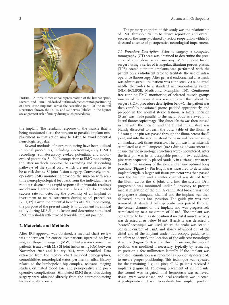
Figure 1: A three-dimensional representation of the lumbar spine,sacrum, and ilium. Red dashed outlines depict common positioningof three iFuse implants across the sacroiliac joint. Of the neuralstructures shown, the L5, S1, and S2 nerves (labeled in the figure)are at greatest risk of injury during such procedures.
the implant. The resultant response of the muscle that isbeing monitored alerts the surgeon to possible implant mis-placement so that action may be taken to avoid potentialneurologic sequelae.
Several methods of neuromonitoring have been utilizedin spinal procedures, including electromyography (EMG)recordings, somatosensory evoked potentials, and motor-evoked potentials [8–10]. In comparison to EMGmonitoring,the latter methods monitor the ascending and descendingpathways of the spinal cord, which are not considered tobe at risk during SI joint fusion surgery. Conversely, intra-operative EMG monitoring provides the surgeon with real-time neurophysiological feedback from the individual nerveroots at risk, enabling a rapid response if unfavorable readingsare obtained. Intraoperative EMG has a high documentedsuccess rate for detecting the proximity of an implant orinstrument to neural structures during spinal procedures[7, 11, 12]. Given the potential benefits of EMG monitoring,the purpose of the present study is to document its clinicalutility during MIS SI joint fusion and determine stimulatedEMG thresholds reflective of favorable implant position.
2. Materials and Methods
After IRB approval was obtained, a medical chart reviewwas undertaken for consecutive patients operated on by asingle orthopedic surgeon (MW). Thirty-seven consecutivepatients, treated with MIS SI joint fusion using IOM betweenNovember 2012 and January 2014, were identified. Dataextracted from the medical chart included demographics,comorbidities, neurological status, pertinent medical historyrelated to the lumbopelvic hip complex, relevant imagingstudies, estimated blood loss, and perioperative and post-operative complications. Stimulated EMG thresholds duringsurgery were obtained directly from the neuromonitoringtechnologist’s records.
The primary endpoint of this study was the relationshipof EMG threshold values to device reposition and overallsuccess of the surgery defined by lack of reoperationwithin 30days and absence of postoperative neurological impairment.
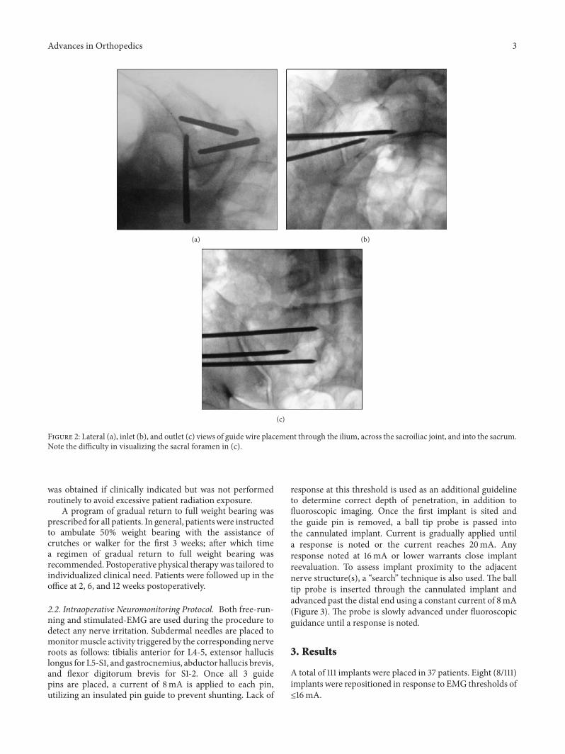
2.1. Procedure Description. Prior to surgery, a computedtomography (CT) scan was obtained to determine the pres-ence of anomalous sacral anatomy. MIS SI joint fusionsurgery using a series of triangular, titanium porous plasma(TPS) coated titanium implants was performed with thepatient on a radiolucent table to facilitate the use of intra-operative fluoroscopy. After general endotracheal anesthesiawas administered, the patient was connected via subdermalneedle electrodes to a standard neuromonitoring system(NIM-ECLIPSE, Medtronic, Memphis, TN). Continuousfree-running EMG monitoring of selected muscle groupsinnervated by nerves at risk was employed throughout thesurgery (IOM procedure description below). The patient wasthen carefully positioned prone, padded appropriately, andprepped in the normal sterile fashion. A lateral incision(3 cm) was made parallel to the sacral body as viewed on alateral fluoroscopic image.The gluteal fascia was then incisedin line with the incision and the gluteal musculature wasbluntly dissected to reach the outer table of the ilium. A3.2mm guide pin was passed through the ilium, across the SIjoint, and into the sacrum lateral to the neural foramen usingan insulated soft tissue retractor. The pin was intermittentlystimulated at 8 milliamperes (mA) during advancement toensure that no neurologic structures were encountered. Afterthe first pin was in an acceptable position, two additionalpins were sequentially placed caudally in a triangular patternto reflect the anatomy of the joint and ensure optimal bonypurchase (Figure 2). Pin length was measured to determineimplant length. A larger soft tissue protector was then passedover the first pin and a center channel was drilled fromthe ilium, across the SI joint, and into the sacrum. Drillprogression was monitored under fluoroscopy to preventmedial migration of the pin. A cannulated broach was usedto prepare a triangular channel and the implant was thendelivered into its final position. The guide pin was thenremoved. A standard ball-tip probe was passed throughthe center channel of the implant and was progressivelystimulated up to a maximum of 20mA. The implant wasconsidered to be in a safe position if no distal muscle activitywas detected at or below 16mA. If activity was detected, a“search” technique was used, where the probe was set to aconstant current of 8mA and slowly advanced out of thedistal end of the implant under fluoroscopic guidance inan effort to identify the location of the adjacent neurologicstructure (Figure 3). Based on this information, the implantposition was modified if necessary, typically by retractingits position a few millimeters laterally. If the implant wasadjusted, stimulation was repeated (as previously described)to ensure proper positioning. This technique was repeatedfor the remaining 2 guide pins; all 37 patients received 3implants (Figure 4). Following placement of all implants,the wound was irrigated, final hemostasis was achieved,tissue layers were closed, and local anesthetic was injected.A postoperative CT scan to evaluate final implant position
Advances in Orthopedics 3
(a) (b)
(c)
Figure 2: Lateral (a), inlet (b), and outlet (c) views of guide wire placement through the ilium, across the sacroiliac joint, and into the sacrum.Note the difficulty in visualizing the sacral foramen in (c).
was obtained if clinically indicated but was not performedroutinely to avoid excessive patient radiation exposure.
A program of gradual return to full weight bearing wasprescribed for all patients. In general, patientswere instructedto ambulate 50% weight bearing with the assistance ofcrutches or walker for the first 3 weeks; after which timea regimen of gradual return to full weight bearing wasrecommended. Postoperative physical therapywas tailored toindividualized clinical need. Patients were followed up in theoffice at 2, 6, and 12 weeks postoperatively.
2.2. Intraoperative Neuromonitoring Protocol. Both free-run-ning and stimulated-EMG are used during the procedure todetect any nerve irritation. Subdermal needles are placed tomonitormuscle activity triggered by the corresponding nerveroots as follows: tibialis anterior for L4-5, extensor hallucislongus for L5-S1, and gastrocnemius, abductor hallucis brevis,and flexor digitorum brevis for S1-2. Once all 3 guidepins are placed, a current of 8mA is applied to each pin,utilizing an insulated pin guide to prevent shunting. Lack of
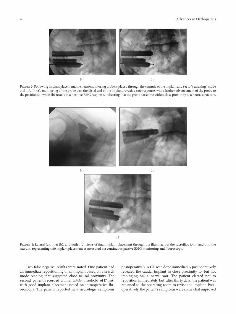
response at this threshold is used as an additional guidelineto determine correct depth of penetration, in addition tofluoroscopic imaging. Once the first implant is sited andthe guide pin is removed, a ball tip probe is passed intothe cannulated implant. Current is gradually applied untila response is noted or the current reaches 20mA. Anyresponse noted at 16mA or lower warrants close implantreevaluation. To assess implant proximity to the adjacentnerve structure(s), a “search” technique is also used. The balltip probe is inserted through the cannulated implant andadvanced past the distal end using a constant current of 8mA(Figure 3). The probe is slowly advanced under fluoroscopicguidance until a response is noted.
3. Results
A total of 111 implants were placed in 37 patients. Eight (8/111)implants were repositioned in response to EMG thresholds of≤16mA.
4 Advances in Orthopedics
(a) (b)
Figure 3: Following implant placement, the neuromonitoring probe is placed through the cannula of the implant and set to “searching”modeat 8mA. In (a), monitoring of the probe past the distal end of the implant reveals a safe response, while further advancement of the probe tothe position shown in (b) results in a positive EMG response, indicating that the probe has come within close proximity to a neural structure.
(a) (b)
(c)
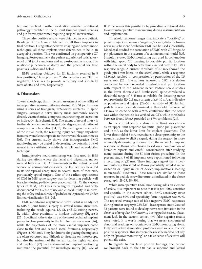
Figure 4: Lateral (a), inlet (b), and outlet (c) views of final implant placement through the ilium, across the sacroiliac joint, and into thesacrum, representing safe implant placement as measured via continuous passive EMGmonitoring and fluoroscopy.
Two false negative results were noted. One patient hadan immediate repositioning of an implant based on a searchmode reading that suggested close neural proximity. Thesecond patient recorded a final EMG threshold of 17mA,with good implant placement noted on intraoperative flu-oroscopy. The patient reported new neurologic symptoms
postoperatively. ACT scan done immediately postoperativelyrevealed the caudal implant in close proximity to, but notimpinging on, a nerve root. The patient elected not toreposition immediately, but, after thirty days, the patient wasreturned to the operating room to revise the implant. Post-operatively, the patient’s symptoms were somewhat improved
Advances in Orthopedics 5
but not resolved. Further evaluation revealed additionalpathology unrelated to the SI joint (lumbar spinal stenosisand piriformis syndrome) requiring surgical intervention.
Three false positive results were obtained in one patient.Readings of 10mA were obtained for all three implants infinal position. Using intraoperative imaging and searchmodetechniques, all three implants were determined to be in anacceptable position.This was confirmed on postoperative CTimaging. Postoperatively, the patient experienced satisfactoryrelief of SI joint symptoms and no postoperative issues. Therelationship between anatomy and the potential for falsepositives is discussed below.
EMG readings obtained for 111 implants resulted in 8true positives, 3 false positives, 2 false negatives, and 98 truenegatives. These results provide sensitivity and specificityrates of 80% and 97%, respectively.
4. Discussion
To our knowledge, this is the first assessment of the utility ofintraoperative neuromonitoring during MIS SI joint fusionusing a series of triangular, TPS-coated implants. In spinesurgery, iatrogenic nerve injury most commonly occursdirectly viamechanical compression, stretching, or lacerationor indirectly via ischemia [13]. The extent of neural injury isfurther dependent on the magnitude, degree, and duration ofcompression or laceration [14, 15]. Depending on the severityof the initial insult, the resulting injury can range anywherefrom recoverable neurapraxia to the irreversible axonotmesis[16]. The current study shows that intraoperative EMGmonitoring may be useful in decreasing the potential risk ofneural injury utilizing a relatively simple and reproducibletechnique.
Intraoperative neuromonitoring was first used in 1898during operations where the facial and trigeminal nerveswere at high risk [17]. Advancements in the technique andscience of neuromonitoring over the last century have ledto its widespread acceptance in several areas of medicine,particularly spinal surgery. One of the earliest applicationsof IOM in MIS spine surgery was for detecting pedicle wallbreaches during pedicle screw placement [18]. Of the varioustypes of IOM, EMG has been highly regarded and well-documented for its ease of use and clinical utility in improv-ing the safety and accuracy of pedicle screw procedures underlimited visualization [18–24].
EMGmonitoringmay likewise prove useful as an adjunctto MIS SI joint fusion surgery as several neural structures,including the cauda equina, L5, S1, and S2 exiting nerves,lie within close proximity to implant trajectory (Figure 1)[25]. Specifically, the trajectory of the most cephalad implantpasses in close proximity to the fifth lumbar nerve root [26],while the trajectories of the more caudal implants comeclose to the first and second sacral foramina, respectively(Figure 1). Not only bony landmarks for placing the implantsare often obscured and difficult to visualize on fluoroscopy,but also the anatomy of the sacrum can be highly variableand dysplastic [27]. Safe instrument and implant positioningminimize the potential for neurologic injury [11]. Use of
IOM decreases this possibility by providing additional datato assist intraoperative maneuvering during instrumentationand implantation.
Threshold response ranges that indicate a “positive,” orpossibly injurious, versus a “negative,” or safe, distance from anervemust be identified before EMGcanbe used successfully.Moed et al. studied the correlation of EMGwith CT for guidepin placement in the sacrum of a canine animal model [13].Stimulus-evoked EMG monitoring was used in conjunctionwith high speed CT imaging to correlate pin tip locationwithin the sacral body to determine a neural proximity EMGresponse range. A current threshold of 6.3mA showed theguide pin 1mm lateral to the sacral canal, while a response≤5.9mA resulted in compression or penetration of the L5nerve root [26]. The authors reported a 0.801 correlationcoefficient between recorded thresholds and pin locationwith respect to the adjacent nerve. Pedicle screw studiesin the lower thoracic and lumbosacral spine correlated athreshold range of 8–15mA as reliable for detecting nerveroot proximity [13, 21] and currents of≤6–10mA as indicativeof possible neural injury [28–30]. A study of 512 lumbarpedicle screw cases determined a threshold response of≥15mA to coincide with a 98% confidence that the screwwas within the pedicle (as verified via CT), while thresholdsbetween 10 and 15mA provided an 87% confidence [21].
In the current study, a stimulus of 8mA was chosenas an upper limit response for initial guide pin placementand 16mA as the lower limit for implant placement. Thelower threshold of 8mA necessitates a closer proximity to theneural structure to elicit a signal, aiding the surgeon in moreaccurately determining distance from the nerve. An implantresponse of 16mA was chosen based on a combination ofliterature reports and careful consideration after studyingmany patients during the SI joint fusion procedure. In thepresent study, 8 of 111 implants were repositioned followinga recording of ≤16mA. These findings suggest that a neu-romonitoring threshold of 16mA potentially avoided nerveirritation or injury in 7% of device implantations, leadingto successful outcomes. These results are similar to thosereported in pedicle screw literature, as indicated in the aboveparagraph [21–23, 28–30].
While intraoperative EMG monitoring adds an elementof safety, it is important to note that it is not 100% sensitiveand specific. In the current cohort, EMG sensitivity (truepositive) was 80% and specificity (true negative) was 97%.The reported average rate of false-negative EMG responsesduring lumbar surgery is 23% [24]. In a separate study, 2 out of32 patients were found to develop nerve root irritation in theabsence of irregular EMGactivity during pedicle screw place-ment [31]. In the current cohort, two false-negative resultswere noted. It is worth noting that we never encounteredabnormal readings on spontaneous EMG monitoring alone.Only with active stimulation protocols were we able to elicitpositive responses.This study emphasizes the need to not relyonly on “passive monitoring” or a false sense of security willpotentially exist.
In regards to our false positive findings, the patientwe brought back to the OR had a superior and lateral
6 Advances in Orthopedics
sacral deficiency, which is not uncommon. This resulted inan unusually wide SI joint superiorly that was traversed bythe implants, especially the superior implant. Our hypothesisis that even though the implants in this case were safelypositioned as confirmed by CT, the superior edge of theimplant was effectively uncovered for a variable distance bythe lack of a bony “roof ” due to the dysplasia and, therefore,not as well insulated.This could have contributed to the lowerthreshold readings.
Such situations demonstrate the technical limitations ofneuromonitoring. Specifically, it is imperative that surgeonsand neurophysiologists are aware that a negative neuromon-itoring reading cannot determine safe implant position with100% certainty. The benefits of neuromonitoring, when usedin conjunction with preoperative CT imaging, fluoroscopy,and meticulous surgical technique, merit its use in MIS SIjoint implantation. This surgical adjunctive technique hasthe potential to optimize positive patient outcomes whenimplanting medical devices in close proximity to neuralstructures. However, there is no substitute for experience andsound surgical judgment in each specific case.
Limitations of this study include small sample size, singlesurgeon experience, and absence of a control group. Thesmall patient size was reflective of the number of patientsavailable in the private practice office. All patients includedin this study were followed postoperatively for a minimumof 3 months. The benefits of evaluating patients from asingle center include a consistent diagnostic and therapeuticapproach. Hopefully, in the near future, other surgeons willadd to this body of knowledge and help validate the outcomesof this limited study.
5. Conclusions
This study provides insight into the potential benefit of IOMfor MIS SI joint fusion. Specifically, results suggest that astimulation threshold of ≤16mA may indicate a potentiallyhazardous implant placement. In addition, the use of an8mA search mode technique for initial guide pin placementand final implant position allows the surgeon to determineproximity to adjacent neural structures. Given the potentialof these thresholds to decrease the risk of iatrogenic nerveinjury, IOM may be a useful adjunct to MIS SI joint fusionsurgery.
Conflict of Interests
This study was performed at Missoula Bone & Joint, LLC andwas sponsored by SI-BONE, Inc. No funds were received byMichael Woods, Denise Birkholz, AdamWoods or MissoulaBone & Joint LL in support of this work. Michael Woods isa paid consultant for SI-BONE, Inc. Robyn Capobianco andRegina MacBarb are employees of SI-BONE, Inc.
Acknowledgments
The authors wish to thank Kristen Munk for her assistancewith chart preparation.
References
[1] A. G. Smith, R. Capobianco, D. Cher et al., “Open versus mini-mally invasive sacroiliac joint fusion: amulti-center comparisonof perioperative measures and clinical outcomes,” Annals ofSurgical Innovation and Research, vol. 7, no. 1, article 14, 2013.
[2] L. Rudolf, “Sacroiliac joint arthrodesis-MIS technique withtitanium implants: report of the first 50 patients and outcomes,”The Open Orthopaedics Journal, vol. 6, no. 1, pp. 495–502, 2012.
[3] M. P. Lorio, D. W. Polly Jr., I. Ninkovic et al., “Utilization ofminimally invasive surgical approach for sacroiliac joint fusionin surgeon population of ISASS and SMISS membership,” TheOpen Orthopaedics Journal, vol. 8, pp. 1–6, 2014.
[4] M. L. C. Routt Jr., P. T. Simonian, S. G. Agnew, and F. A. Mann,“Radiographic recognition of the sacral alar slope for optimalplacement of iliosacral screws: a cadaveric and clinical study,”Journal of Orthopaedic Trauma, vol. 10, no. 3, pp. 171–177, 1996.
[5] M. G. Fehlings, D. S. Brodke, D. C. Norvell, and J. R. Dettori,“The evidence for intraoperative neurophysiological monitor-ing in spine surgery: does it make a difference?” Spine, vol. 35,no. 9S, pp. S37–S46, 2010.
[6] R. R. Lall, J. S. Hauptman, C. Munoz et al., “Intraoperativeneurophysiological monitoring in spine surgery: indications,efficacy, and role of the preoperative checklist,” NeurosurgicalFocus, vol. 33, no. 5, article E10, 2012.
[7] B. Bose, L. R. Wierzbowski, and A. K. Sestokas, “Neurophys-iologic monitoring of spinal nerve root function during instru-mented posterior lumbar spine surgery,” Spine, vol. 27, no. 13,pp. 1444–1450, 2002.
[8] D. S. Dinner, H. Luders, R. P. Lesser, andH.H.Morris, “Invasivemethods of somatosensory evoked potential monitoring,” Jour-nal of Clinical Neurophysiology, vol. 3, no. 2, pp. 113–130, 1986.
[9] D.H. York, R. J. Chabot, and R.W.Gaines, “Response variabilityof somatosensory evoked potentials during scoliosis surgery,”Spine, vol. 12, no. 9, pp. 864–876, 1987.
[10] D. L. Helfet, E. A. Hissa, S. Sergay, and J. W. Mast, “Somatosen-sory evoked potential monitoring in the surgical managementof acute acetabular fractures.,” Journal of orthopaedic trauma,vol. 5, no. 2, pp. 161–166, 1991.
[11] B. R. Moed, B. K. Ahmad, J. G. Craig, G. P. Jacobson, and M. J.Anders, “Intraoperative monitoring with stimulus-evoked elec-tromyography during placement of iliosacral screws: an initialclinical study,” Journal of Bone and Joint Surgery, vol. 80, no. 4,pp. 537–546, 1998.
[12] L. X.Webb,W. de Araujo, P. Donofrio et al., “Electromyographymonitoring for percutaneous placement of iliosacral screws,”Journal of Orthopaedic Trauma, vol. 14, no. 4, pp. 245–254, 2000.
[13] B. R. Moed, J. W. Maxey, and G. J. Minster, “Intraoperativesomatosensory evoked potential monitoring of the sciaticnerve: an animal model,” Journal of Orthopaedic Trauma, vol.6, no. 1, pp. 59–65, 1992.
[14] S. Sunderland, “A classification of peripheral nerve injuriesproducing loss of function,” Brain, vol. 74, no. 4, pp. 491–516,1951.
[15] M. G. Burnett and E. L. Zager, “Pathophysiology of peripheralnerve injury: a brief review,” Neurosurg Focus, vol. 16, no. 5,article E1, 2004.
[16] A. Ahmadian, A. R. Deukmedjian, N. Abel, E. Dakwar, andJ. S. Uribe, “Analysis of lumbar plexopathies and nerve injuryafter lateral retroperitoneal transpsoas approach: diagnosticstandardization,” Journal of Neurosurgery: Spine, vol. 18, no. 3,pp. 289–297, 2013.
Advances in Orthopedics 7
[17] R. E. Minahan and A. S. Mandir, “Neurophysiologic intraop-erative monitoring of trigeminal and facial nerves,” Journal ofClinical Neurophysiology, vol. 28, no. 6, pp. 551–565, 2011.
[18] M. R. Isley, X.-F. Zhang, J. R. Balzer, and R. E. Leppanen,“Current trends in pedicle screw stimulation techniques: lum-bosacral, thoracic, and cervical levels,”Neurodiagnostic Journal,vol. 52, no. 2, pp. 100–175, 2012.
[19] J. C. Rodriguez-Olaverri, N. C. Zimick, A. Merola et al., “Usingtriggered electromyographic threshold in the intercostal mus-cles to evaluate the accuracy of upper thoracic pedicle screwplacement (T3-T6),” Spine, vol. 33, no. 7, pp. E194–E197, 2008.
[20] B. Calancie, N. Lebwohl, P. Madsen, and K. J. Klose, “Intraop-erative evoked EMG monitoring in an animal model: a newtechnique for evaluating pedicle screw placement,” Spine, vol.17, no. 10, pp. 1229–1235, 1992.
[21] S. D. Glassman, J. R. Dimar, R. M. Puno, J. R. Johnson, C. B.Shields, and R. D. Linden, “A prospective analysis of intraoper-ative electromyographicmonitoring of pedicle screw placementwith computed tomographic scan confirmation,” Spine, vol. 20,no. 12, pp. 1375–1379, 1995.
[22] D. H. Clements, D. E. Morledge, W. H. Martin, and R. R.Betz, “Evoked and spontaneous electromyography to evaluatelumbosacral pedicle screw placement,” Spine, vol. 21, no. 5, pp.600–604, 1996.
[23] B. L. Raynor, L. G. Lenke, K. H. Bridwell, B. A. Taylor, and A.M. Padberg, “Correlation between low triggered electromyo-graphic thresholds and lumbar pedicle screw malposition:analysis of 4857 screws,” Spine, vol. 32, no. 24, pp. 2673–2678,2007.
[24] R. M. Beatty, P. McGuire, J. M. Moroney, and F. P. Holladay,“Continuous intraoperative electromyographic recording dur-ing spinal surgery,” Journal of Neurosurgery, vol. 82, no. 3, pp.401–405, 1995.
[25] S. Mirkovic, J. J. Abitbol, J. Steinman et al., “Anatomic consider-ation for sacral screw placement,” Spine, vol. 16, no. 6, pp. S289–S294, 1991.
[26] B. R. Moed, M. J. Hartman, B. K. Ahmad, D. D. Cody, and J. G.Craig, “Evaluation of intraoperative nerve-monitoring duringinsertion of an iliosacral implant in an animal model,” Journalof Bone and Joint Surgery—American Volume, vol. 81, no. 11, pp.1529–1537, 1999.
[27] A. N. Miller and M. L. C. Routt Jr., “Variations in sacral mor-phology and implications for iliosacral screw fixation,” Journalof the American Academy of Orthopaedic Surgeons, vol. 20, no.1, pp. 8–16, 2012.
[28] B. Calancie, P. Madsen, N. Lebwohl, and J. H. Owen, “Stimulus-evoked EMG monitoring during transpedicular lumbosacralspine instrumentation: initial clinical results,” Spine, vol. 19, no.24, pp. 2780–2786, 1994.
[29] L. G. Lenke, A. M. Padberg, M. H. Russo, K. H. Bridwell,and D. E. Gelb, “Triggered electromyographic threshold foraccuracy of pedicle screw placement: an animal model andclinical correlation,” Spine, vol. 20, no. 14, pp. 1585–1591, 1995.
[30] J. Maguire, S. Wallace, R. Madiga, R. Leppanen, and V. Draper,“Evaluation of intrapedicular screw position using intraopera-tive evoked electromyography,” Spine, vol. 20, no. 9, pp. 1068–1074, 1995.
[31] W. C. Welch, R. D. Rose, J. R. Balzer, and G. B. Jacobs, “Eval-uation with evoked and spontaneous electromyography duringlumbar instrumentation: a prospective study,” Journal of Neuro-surgery, vol. 87, no. 3, pp. 397–402, 1997.

Submit your manuscripts athttp://www.hindawi.com
Stem CellsInternational
Hindawi Publishing Corporationhttp://www.hindawi.com Volume 2014
Hindawi Publishing Corporationhttp://www.hindawi.com Volume 2014
MEDIATORSINFLAMMATION
of
Hindawi Publishing Corporationhttp://www.hindawi.com Volume 2014
Behavioural Neurology
EndocrinologyInternational Journal of
Hindawi Publishing Corporationhttp://www.hindawi.com Volume 2014
Hindawi Publishing Corporationhttp://www.hindawi.com Volume 2014
Disease Markers
Hindawi Publishing Corporationhttp://www.hindawi.com Volume 2014
BioMed Research International
OncologyJournal of
Hindawi Publishing Corporationhttp://www.hindawi.com Volume 2014
Hindawi Publishing Corporationhttp://www.hindawi.com Volume 2014
Oxidative Medicine and Cellular Longevity
Hindawi Publishing Corporationhttp://www.hindawi.com Volume 2014
PPAR Research
The Scientific World JournalHindawi Publishing Corporation http://www.hindawi.com Volume 2014
Immunology ResearchHindawi Publishing Corporationhttp://www.hindawi.com Volume 2014
Journal of
ObesityJournal of
Hindawi Publishing Corporationhttp://www.hindawi.com Volume 2014
Hindawi Publishing Corporationhttp://www.hindawi.com Volume 2014
Computational and Mathematical Methods in Medicine
OphthalmologyJournal of
Hindawi Publishing Corporationhttp://www.hindawi.com Volume 2014
Diabetes ResearchJournal of
Hindawi Publishing Corporationhttp://www.hindawi.com Volume 2014
Hindawi Publishing Corporationhttp://www.hindawi.com Volume 2014
Research and TreatmentAIDS
Hindawi Publishing Corporationhttp://www.hindawi.com Volume 2014
Gastroenterology Research and Practice
Hindawi Publishing Corporationhttp://www.hindawi.com Volume 2014
Parkinson’s Disease
Evidence-Based Complementary and Alternative Medicine
Volume 2014Hindawi Publishing Corporationhttp://www.hindawi.com


![ASET 2 0 1 0 media kit - Home - ASET · polysomnography, nerve conduction studies, long term monitoring, intraoperative neuromonitoring and related electroneurodiagnostics [END]](https://img.pdfslide.net/doc/110x75/5edc3a4dad6a402d6666ce80/aset-2-0-1-0-media-kit-home-polysomnography-nerve-conduction-studies-long.jpg)


![Neuromonitoring [PDF, 2.5 MB]](https://img.pdfslide.net/doc/110x75/586f5f371a28abf0508bd912/neuromonitoring-pdf-25-mb.jpg)