Embed Size (px)
Citation preview
Clinical Symptoms of Blood System Affection: Anemia and Polycythemia; Leukopenias and
Leukocytosis; Leukemia; Hemorrhagic Syndrome.
LECTURE IN INTERNAL MEDICINE PROPAEDEUTICS
M. Yabluchansky, L. Bogun, L.Martymianova, O. Bychkova, N. Lysenko, N. Makienko
V.N. Karazin National University Medical School’ Internal Medicine Dept.
Anemia
Definition
Anemia (anaemia) is a decrease in the amount of RBCs or hemoglobin in the blood or a lowered ability of the blood to carry oxygen
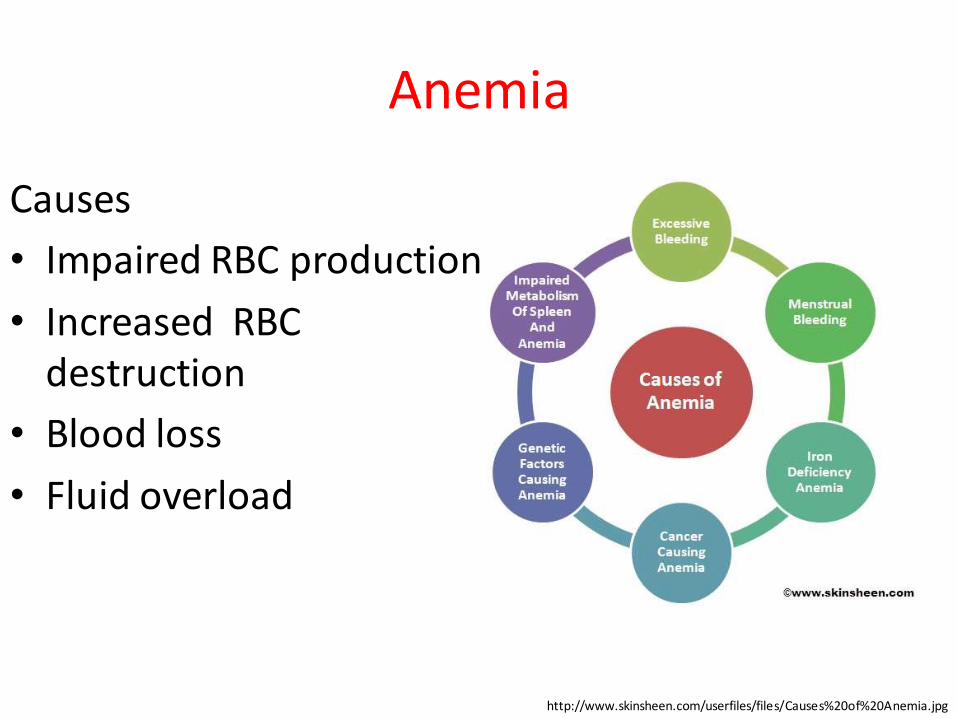
Anemia
Causes
• Impaired RBC production
• Increased RBC destruction
• Blood loss
• Fluid overload
http://www.skinsheen.com/userfiles/files/Causes%20of%20Anemia.jpg
AnemiaCauses: Impaired production 1
• Disturbance of proliferation and differentiation of stem cells (Pure red cell aplasia, Aplastic anemia, Anemia of renal failure, Anemia of endocrine disorders)
• Disturbance of proliferation and maturation of erythroblasts (Pernicious anemia, Anemia of folic acid deficiency, Megaloblastic anemia, Anemia of prematurity, Iron deficiency anemia, Thalassemias, Congenital dyserythropoietic anemias, atc.)
Anemia
Causes: Impaired production 2
• Other mechanisms of impaired RBCs production (Myelophthisic anemia, Myelodysplastic syndrome, Anemia of chronic inflammation)
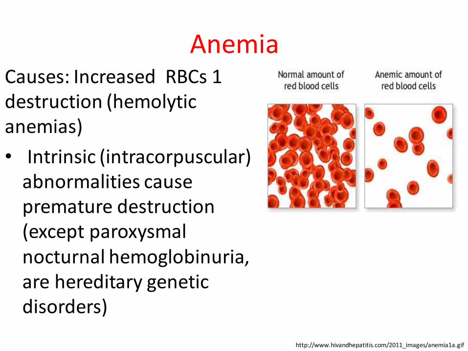
AnemiaCauses: Increased RBCs 1 destruction (hemolytic anemias)
• Intrinsic (intracorpuscular) abnormalities cause premature destruction (except paroxysmal nocturnal hemoglobinuria, are hereditary genetic disorders)
http://www.hivandhepatitis.com/2011_images/anemia1a.gif
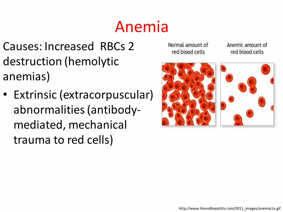
AnemiaCauses: Increased RBCs 2 destruction (hemolytic anemias)
• Extrinsic (extracorpuscular) abnormalities (antibody-mediated, mechanical trauma to red cells)
http://www.hivandhepatitis.com/2011_images/anemia1a.gif
AnemiaCauses: Blood loss
• Anemia of prematurity
• Trauma or surgery, causing acute blood loss
• Gastrointestinal tract lesions (acute bleeds: peptic ulcers, chronic blood loss (angiodysplasia)
• Gynecologic disturbances (chronic blood loss)
• Menstruation, among young women or older women with fibroids
• Infection by intestinal nematodes feeding on blood
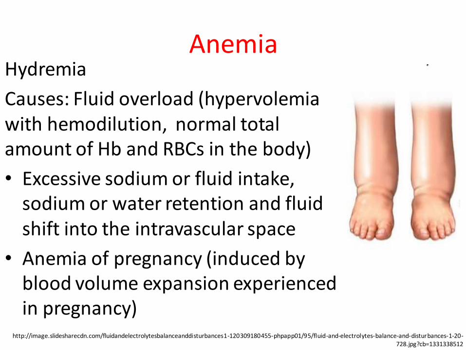
AnemiaHydremia
Causes: Fluid overload (hypervolemia with hemodilution, normal total amount of Hb and RBCs in the body)
• Excessive sodium or fluid intake, sodium or water retention and fluid shift into the intravascular space
• Anemia of pregnancy (induced by blood volume expansion experienced in pregnancy)
http://image.slidesharecdn.com/fluidandelectrolytesbalanceanddisturbances1-120309180455-phpapp01/95/fluid-and-electrolytes-balance-and-disturbances-1-20-728.jpg?cb=1331338512
Anemia
Diagnostic steps
• Clinical manifestations
• Hematological syndromes (blood tests abnormalities)
• Blood biochemistry abnormalities
• Bone marrow abnormalities
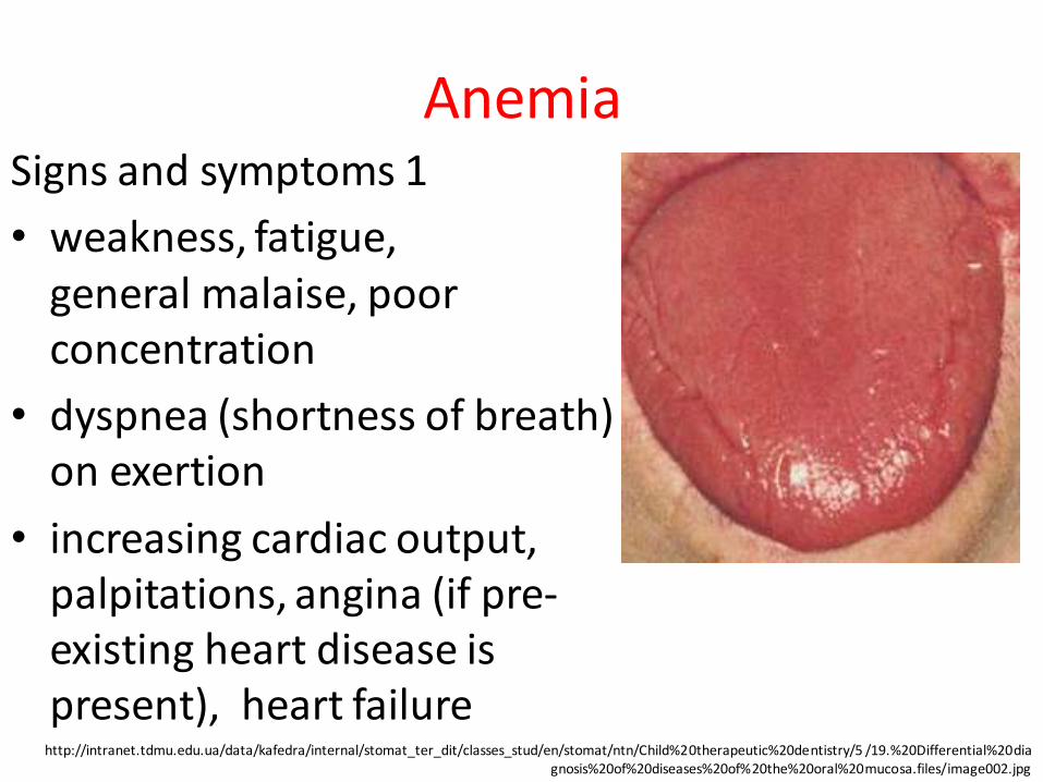
AnemiaSigns and symptoms 1
• weakness, fatigue, general malaise, poor concentration
• dyspnea (shortness of breath) on exertion
• increasing cardiac output, palpitations, angina (if pre-existing heart disease is present), heart failure
http://intranet.tdmu.edu.ua/data/kafedra/internal/stomat_ter_dit/classes_stud/en/stomat/ntn/Child%20therapeutic%20dentistry/5 /19.%20Differential%20diagnosis%20of%20diseases%20of%20the%20oral%20mucosa.files/image002.jpg
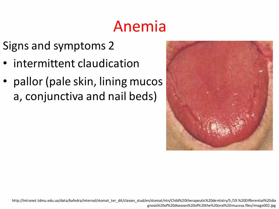
AnemiaSigns and symptoms 2
• intermittent claudication
• pallor (pale skin, lining mucosa, conjunctiva and nail beds)
http://intranet.tdmu.edu.ua/data/kafedra/internal/stomat_ter_dit/classes_stud/en/stomat/ntn/Child%20therapeutic%20dentistry/5 /19.%20Differential%20diagnosis%20of%20diseases%20of%20the%20oral%20mucosa.files/image002.jpg
Anemia
Additional signs of severe anemia
• hyper dynamic circulation (tachycardia, bounding pulse, flow murmurs, cardiac ventricular hypertrophy)
• heart failure
• pica
• restless legs syndrome (iron-deficiency anemia)
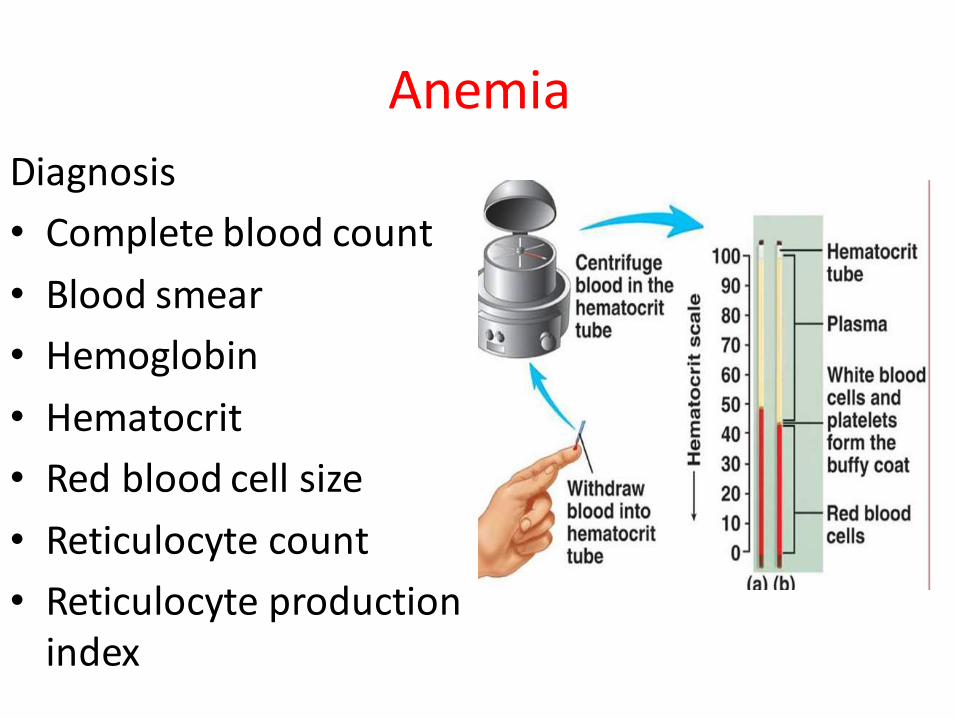
Anemia
Diagnosis
• Complete blood count
• Blood smear
• Hemoglobin
• Hematocrit
• Red blood cell size
• Reticulocyte count
• Reticulocyte production index
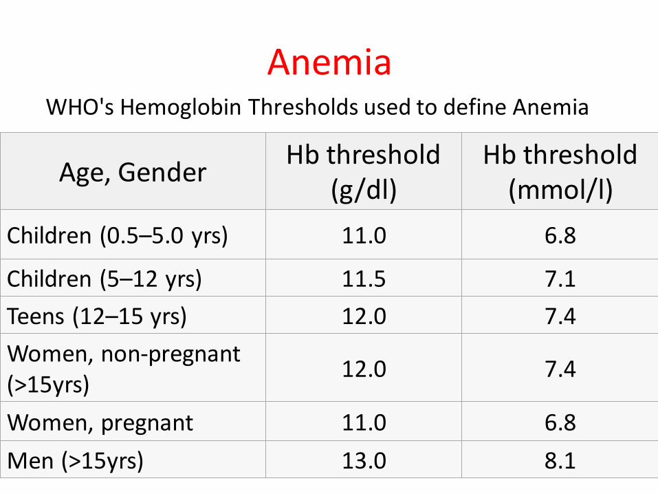
Anemia
Age, Gender Hb threshold
(g/dl)Hb threshold
(mmol/l)
Children (0.5–5.0 yrs) 11.0 6.8
Children (5–12 yrs) 11.5 7.1
Teens (12–15 yrs) 12.0 7.4
Women, non-pregnant (>15yrs)
12.0 7.4
Women, pregnant 11.0 6.8
Men (>15yrs) 13.0 8.1
WHO's Hemoglobin Thresholds used to define Anemia
Anemia
Color index
• Hypochromic (<0,85) – e. g chronic posthemorrhagic, Fe-deficient
• Normochromic (0,85 – 1,05) – e. g acute posthemorrhagic , hemolytic
• Hyperchromic (>1,05) – e.g. B12-deficient, folate-deficient, aplastic
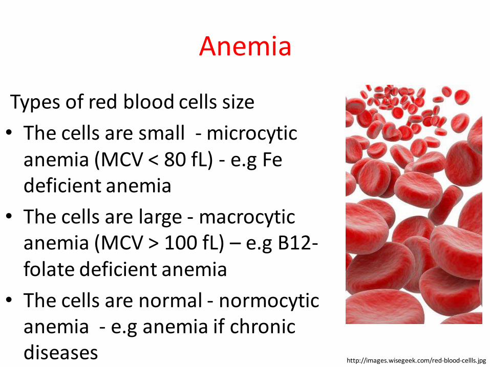
Anemia
Types of red blood cells size
• The cells are small - microcytic anemia (MCV < 80 fL) - e.g Fe deficient anemia
• The cells are large - macrocytic anemia (MCV > 100 fL) – e.g B12-folate deficient anemia
• The cells are normal - normocytic anemia - e.g anemia if chronic diseases http://images.wisegeek.com/red-blood-cellls.jpg
Anemia
Regenerative abilities of bone marrow-reticulocytes, 0/00
• Normoregenerative ( 6 – 12) – anemias due to deficiency (Fe, B12-folate, etc)
• Hyperregenerative (> 12) – hemolytic, acute posthemorhagic
• Hyporegenerative (< 6) - aplastic
Anemia
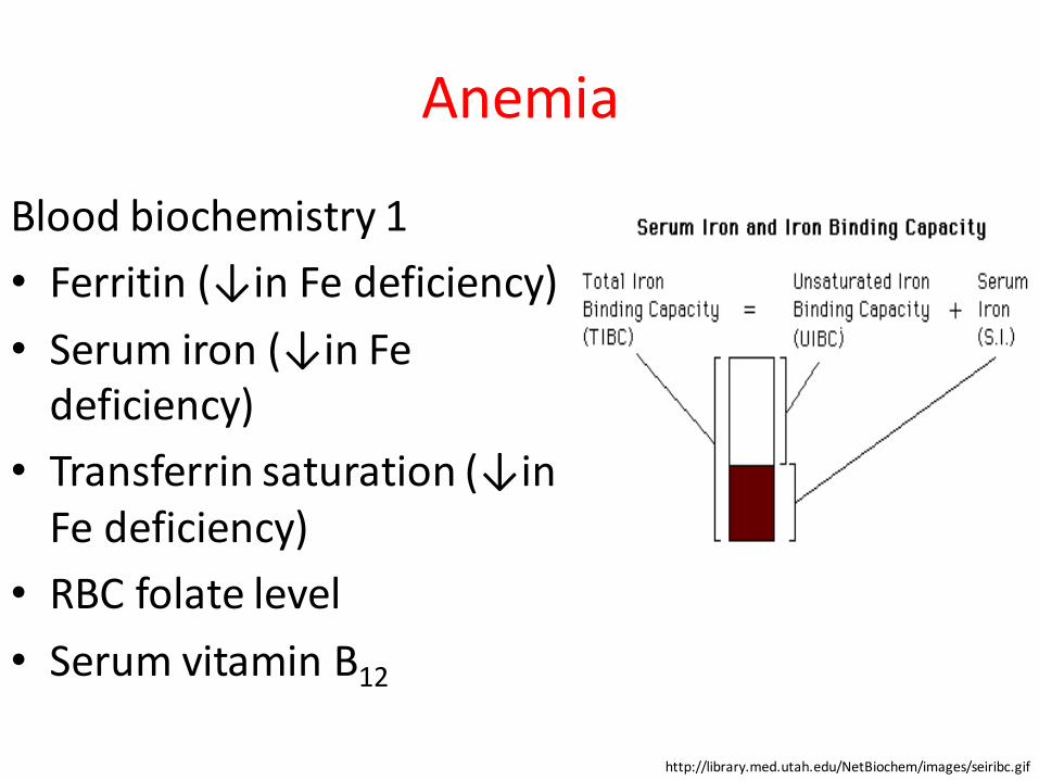
Blood biochemistry 1
• Ferritin (↓in Fe deficiency)
• Serum iron (↓in Fe deficiency)
• Transferrin saturation (↓in Fe deficiency)
• RBC folate level
• Serum vitamin B12
http://library.med.utah.edu/NetBiochem/images/seiribc.gif
Anemia
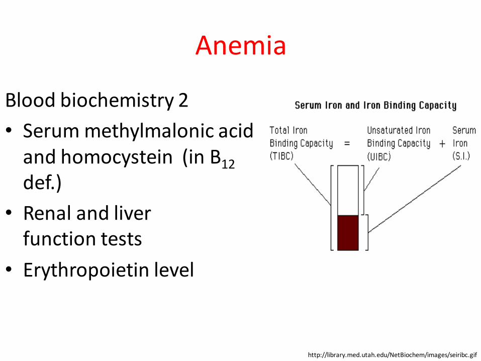
Blood biochemistry 2
• Serum methylmalonic acid and homocystein (in B12
def.)
• Renal and liver function tests
• Erythropoietin level
http://library.med.utah.edu/NetBiochem/images/seiribc.gif
Polycythemia
Definition
Polycythemia is a myeloproliferative condition that results in an increased level of circulating red blood cells in the bloodstream with increase inhematocrit, hemoglobin, or red blood cell count above the normal limits
Polycythemia
Synonyms
Erythremia
Osler-Vaquez disease
Polycythemia rubra vera
Primary polycythemia
Splenomegalic polycythemia
Vaquez-Osler disease
Polyglobulia
Polycythemia
Risk factors:
• Hypoxia from long standing (chronic) lung disease and smoking
• Chronic carbon monoxide (CO) exposure
• People living at high altitudes due to low environmental oxygen levels
• People with genetic mutations and familial types of polycythemia and certain hemoglobin abnormalities
PolycythemiaCauses 1
• Primary (a slow-growing type of blood cancer)
–Polycythemia Vera
–Primary familial and congenital polycythemia
PolycythemiaCauses 2
• Secondary 1
–Physiologically appropriate (adaptation to living at high altitudes, Iatrogenic, etc. )
–Chronic hypoxia (COPD, hypoventilation syndrome, chronic heart diseases, sleep apnea, pulmonary hypertension)
PolycythemiaCauses 2
• Secondary 2
–Erythropoietin secreting tumors (hepatocellular carcinoma, renal cell carcinoma, adenocarcinomas, uterine tumors)
–Relative polycythemia (the underlying cause is reduced blood plasma)
Polycythemia
Signs and symptoms Polycythemia Vera 1
• Trouble breathing when lying down
• Dizziness
• Excess bleeding
• Full feeling in the left upper abdomen (enlarged spleen)
• Headache
Polycythemia
Signs and symptoms Polycythemia Vera 2
• Itchiness, especially after a warm bath
• Red skin coloring, especially of the face
• Shortness of breath
• Phlebitis
Polycythemia
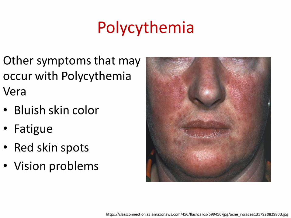
Other symptoms that may occur with Polycythemia Vera
• Bluish skin color
• Fatigue
• Red skin spots
• Vision problems
https://classconnection.s3.amazonaws.com/456/flashcards/599456/jpg/acne_rosacea1317920829803.jpg
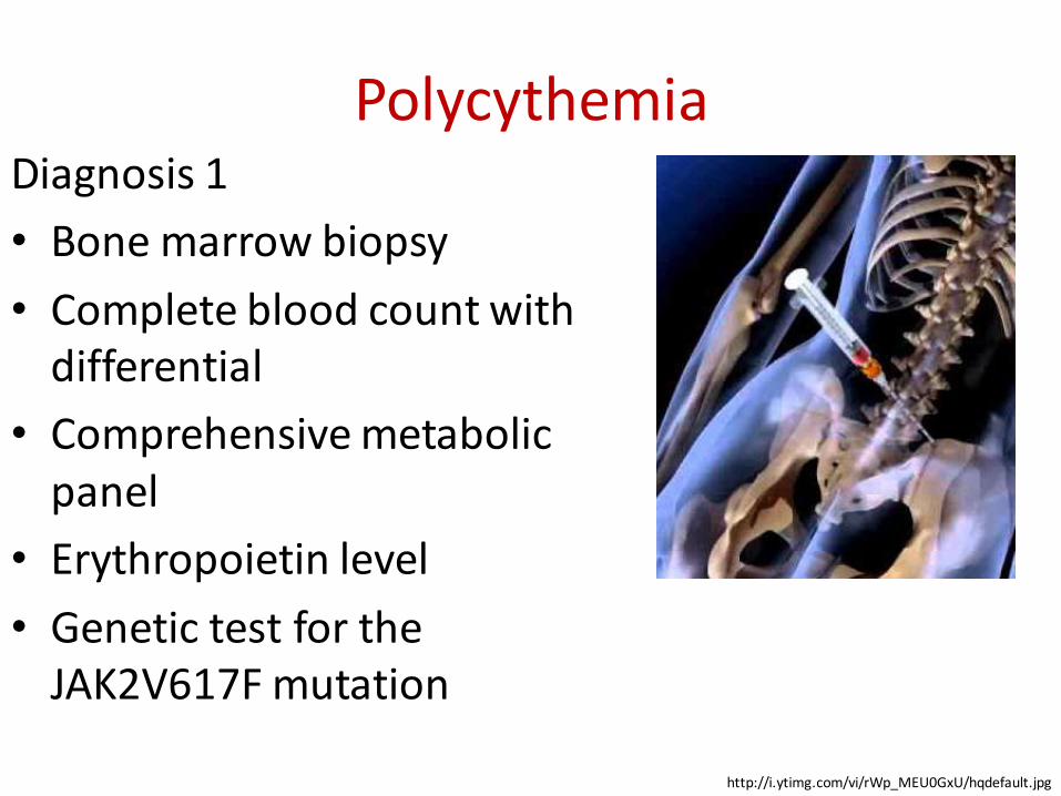
PolycythemiaDiagnosis 1
• Bone marrow biopsy
• Complete blood count with differential
• Comprehensive metabolic panel
• Erythropoietin level
• Genetic test for the JAK2V617F mutation
http://i.ytimg.com/vi/rWp_MEU0GxU/hqdefault.jpg
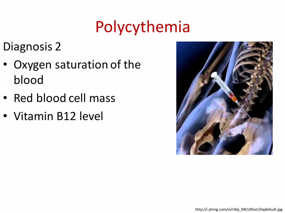
PolycythemiaDiagnosis 2
• Oxygen saturation of the blood
• Red blood cell mass
• Vitamin B12 level
http://i.ytimg.com/vi/rWp_MEU0GxU/hqdefault.jpg
PolycythemiaDiagnosis (WHO criteria ): 1
• Major criteria
–Hemoglobin > 18.5 g/dL in men and > 16.5 g/dLin women, or other evidence of increased red blood cell volume
–Presence of JAK2617V F or other functionally similar mutation, such as JAK2 exon 12 mutation
PolycythemiaDiagnosis (WHO criteria ): 2
• Minor criteria
–Bone marrow biopsy showing hypercellularity for age with trilineage growth (panmyelosis) with prominent erythroid, granulocytic, and megakaryocytic proliferation
– Serum erythropoietin level below the reference range for normal
–Endogenous erythroid colony formation in vitro

PolycythemiaDiagnosis (The Polycythemia Vera Study Group criteria : the diagnosis is established if all three category A criteria are present, or if criteria A1 plus A2 plus any two criteria from category B are present):
• Category A criteria
–Total red blood cell mass ≥ 36 mL/kg in males or ≥ 32 mL/kg in females
–Arterial oxygen saturation ≥ 92%
– Splenomegaly
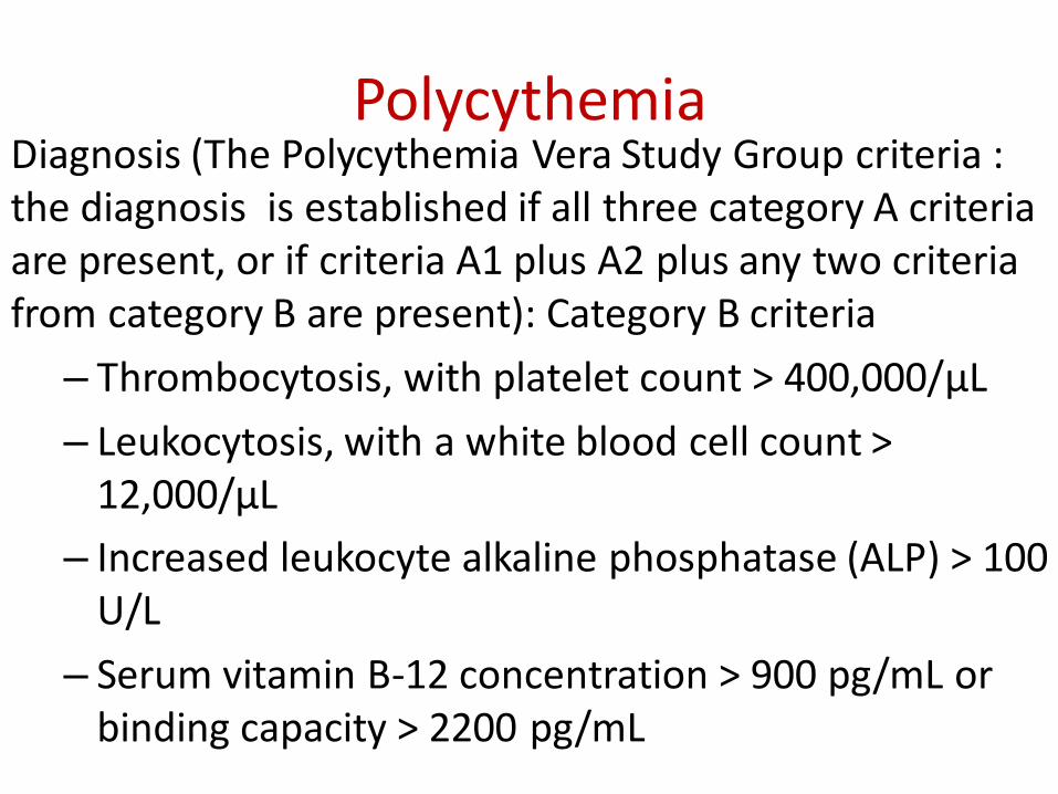
PolycythemiaDiagnosis (The Polycythemia Vera Study Group criteria : the diagnosis is established if all three category A criteria are present, or if criteria A1 plus A2 plus any two criteria from category B are present): Category B criteria
– Thrombocytosis, with platelet count > 400,000/μL
– Leukocytosis, with a white blood cell count > 12,000/μL
– Increased leukocyte alkaline phosphatase (ALP) > 100 U/L
– Serum vitamin B-12 concentration > 900 pg/mL or binding capacity > 2200 pg/mL
Leukopenia
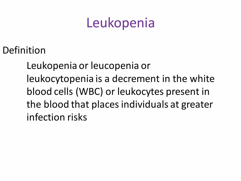
Definition
Leukopenia or leucopenia or leukocytopenia is a decrement in the white blood cells (WBC) or leukocytes present in the blood that places individuals at greater infection risks
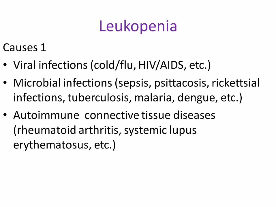
LeukopeniaCauses 1
• Viral infections (cold/flu, HIV/AIDS, etc.)
• Microbial infections (sepsis, psittacosis, rickettsial infections, tuberculosis, malaria, dengue, etc.)
• Autoimmune connective tissue diseases (rheumatoid arthritis, systemic lupus erythematosus, etc.)
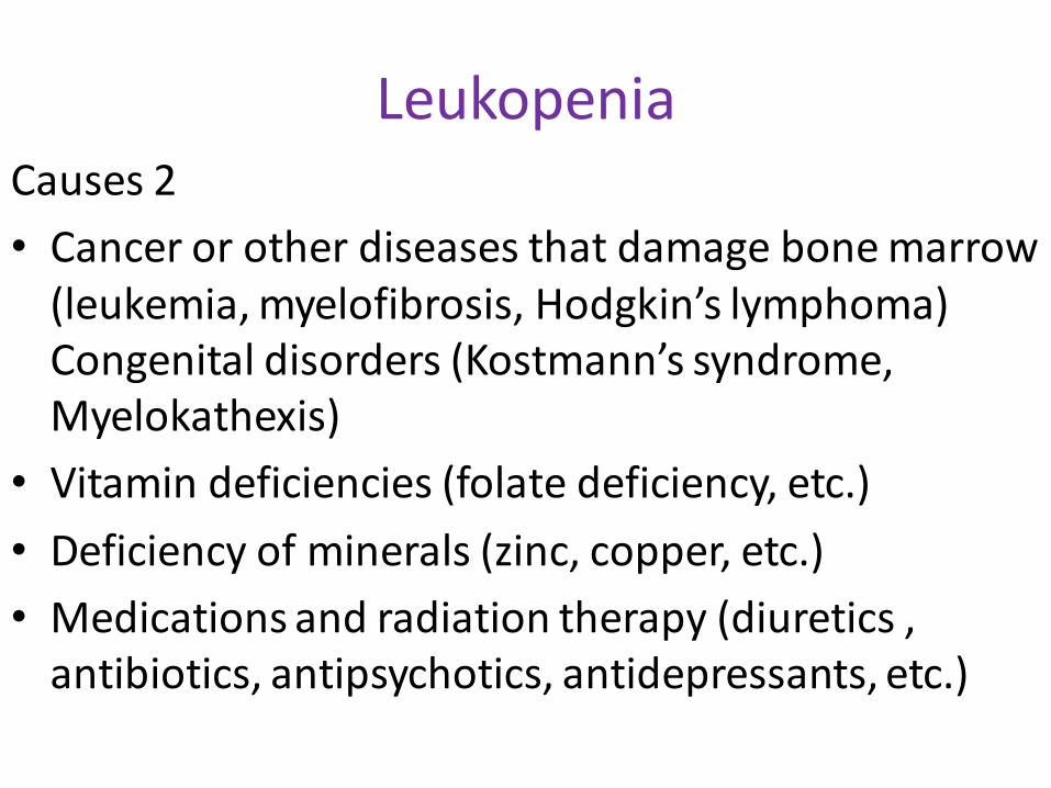
LeukopeniaCauses 2
• Cancer or other diseases that damage bone marrow (leukemia, myelofibrosis, Hodgkin’s lymphoma)Congenital disorders (Kostmann’s syndrome, Myelokathexis)
• Vitamin deficiencies (folate deficiency, etc.)
• Deficiency of minerals (zinc, copper, etc.)
• Medications and radiation therapy (diuretics , antibiotics, antipsychotics, antidepressants, etc.)
Leukopenia
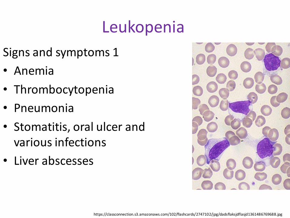
Signs and symptoms 1
• Anemia
• Thrombocytopenia
• Pneumonia
• Stomatitis, oral ulcer and various infections
• Liver abscesses
https://classconnection.s3.amazonaws.com/102/flashcards/2747102/jpg/dadsflaksjdflasjd1361486769688.jpg
Leukopenia
Signs and symptoms 2
• Metrorrhagia, menorrhagia
• Neurasthenia
• Fatigue and hot flashes
• Strong desire to consume hot drinks
https://classconnection.s3.amazonaws.com/102/flashcards/2747102/jpg/dadsflaksjdflasjd1361486769688.jpg
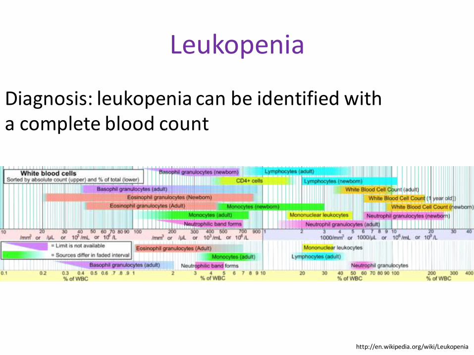
Leukopenia
Diagnosis: leukopenia can be identified with a complete blood count
http://en.wikipedia.org/wiki/Leukopenia
Leukocytosis
Definition
A white blood cell (the leukocyte) count above the normal range (<50 x109/L ) in
the blood
Leukocytosis
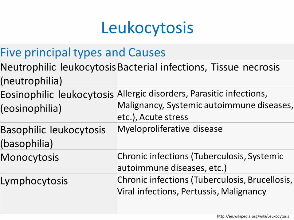
Five principal types and Causes Neutrophilic leukocytosis(neutrophilia)
Bacterial infections, Tissue necrosis
Eosinophilic leukocytosis(eosinophilia)
Allergic disorders, Parasitic infections, Malignancy, Systemic autoimmune diseases, etc.), Acute stress
Basophilic leukocytosis(basophilia)
Myeloproliferative disease
Monocytosis Chronic infections (Tuberculosis, Systemic autoimmune diseases, etc.)
Lymphocytosis Chronic infections (Tuberculosis, Brucellosis, Viral infections, Pertussis, Malignancy
http://en.wikipedia.org/wiki/Leukocytosis
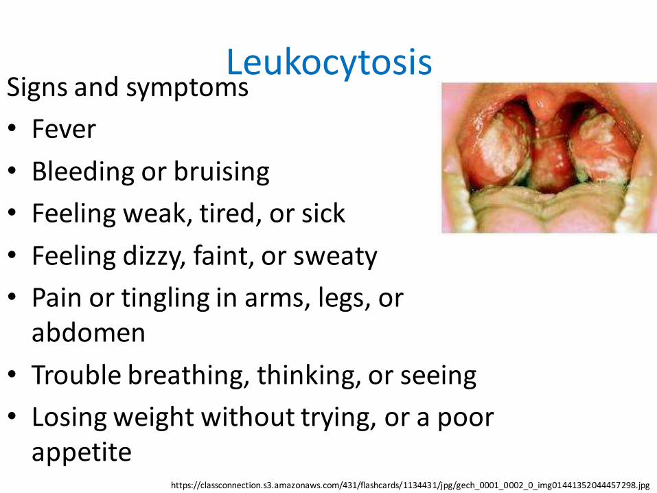
LeukocytosisSigns and symptoms
• Fever
• Bleeding or bruising
• Feeling weak, tired, or sick
• Feeling dizzy, faint, or sweaty
• Pain or tingling in arms, legs, or abdomen
• Trouble breathing, thinking, or seeing
• Losing weight without trying, or a poor appetite
https://classconnection.s3.amazonaws.com/431/flashcards/1134431/jpg/gech_0001_0002_0_img01441352044457298.jpg
Leukocytosis
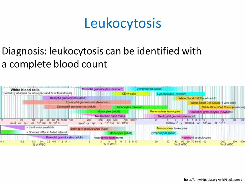
Diagnosis: leukocytosis can be identified with a complete blood count
http://en.wikipedia.org/wiki/Leukopenia
Leukocytosis
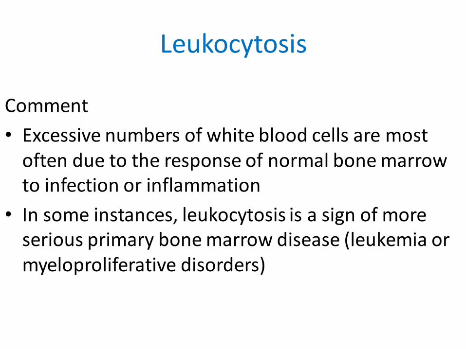
Comment
• Excessive numbers of white blood cells are most often due to the response of normal bone marrow to infection or inflammation
• In some instances, leukocytosis is a sign of more serious primary bone marrow disease (leukemia or myeloproliferative disorders)
Leukemia
Definition
Abnormal proliferation of the blood- forming tissues that usually begins in the bone marrow and results in high numbers of abnormal white blood cells
Leukemia
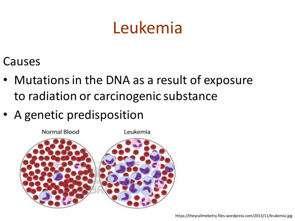
Causes
• Mutations in the DNA as a result of exposure to radiation or carcinogenic substance
• A genetic predisposition
https://theycallmebetty.files.wordpress.com/2013/11/leukemia.jpg
Leukemia
http://en.wikipedia.org/wiki/Leukemia
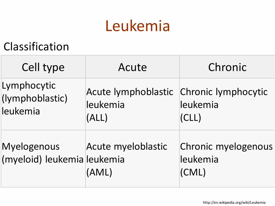
Cell type Acute Chronic
Lymphocytic (lymphoblastic)leukemia
Acute lymphoblastic leukemia(ALL)
Chronic lymphocytic leukemia(CLL)
Myelogenous (myeloid) leukemia
Acute myeloblastic leukemia(AML)
Chronic myelogenous leukemia(CML)
Classification
Leukemia
Signs and symptoms 1
• Fever or chills
• Persistent fatigue, weakness
• Frequent or severe infections
• Losing weight without trying
• Swollen lymph nodes, enlarged liver or spleen
• Easy bleeding or bruising
Leukemia
Signs and symptoms 2
• Recurrent nosebleeds
• Tiny red spots in skin (petechiae)
• Excessive sweating, especially at night
• Bone pain or tenderness
Leukemia
Acute leukemia is characterized by a rapid increase in the number of immature blood cells (blasts)crowding due to such cells makes the bone marrow unable to produce healthy blood cells immediate treatment is required in acute leukemia due to the rapid progression and accumulation of themalignant cells, which then spill over into the bloodstream and spread to other organs of the body
Leukemia
Chronic leukemia is characterized by the excessive buildup of relatively mature, but still abnormal, whiteblood cells typically taking months or years to progress, the cells are produced at a much higher rate than normal, resulting in many abnormal white blood cells mostly occurs in older people, but cantheoretically occur in any age group
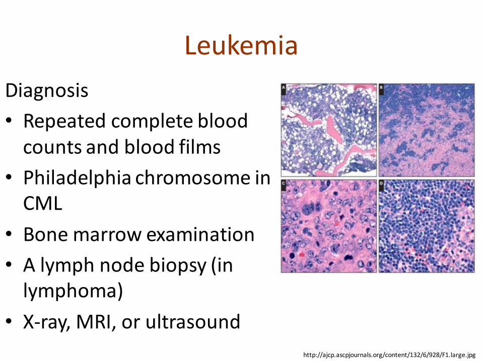
Leukemia
Diagnosis
• Repeated complete blood counts and blood films
• Philadelphia chromosome in CML
• Bone marrow examination
• A lymph node biopsy (in lymphoma)
• X-ray, MRI, or ultrasound
http://ajcp.ascpjournals.org/content/132/6/928/F1.large.jpg
Hemorrhagic Syndrome
Definition
The extravasation of red blood cells from the vasculature into the skin and/or subcutaneous tissue in form of petechiae, purpura, and ecchymosis (collectively referred to as purpura) with purpuric rashes formation
Hemorrhagic Syndrome
Appendix for Definition
The extravasation occur internally, where red blood cells leaks from blood vessels inside the body
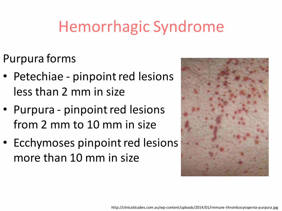
Hemorrhagic Syndrome
Purpura forms
• Petechiae - pinpoint red lesions less than 2 mm in size
• Purpura - pinpoint red lesions from 2 mm to 10 mm in size
• Ecchymoses pinpoint red lesions more than 10 mm in size
http://clinicalstudies.com.au/wp-content/uploads/2014/01/immune-thrombocytopenia-purpura.jpg
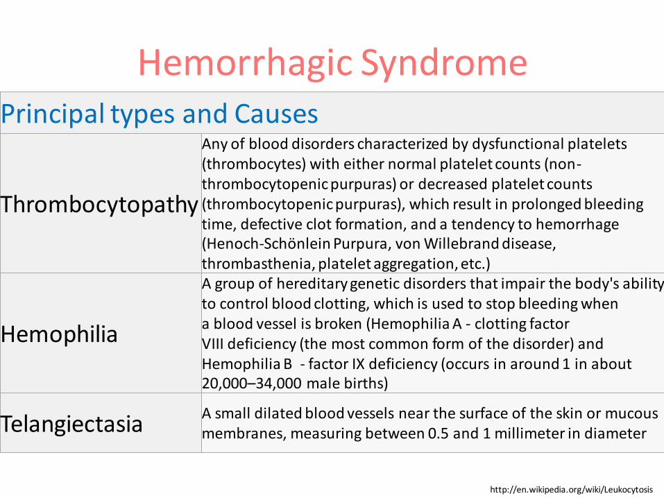
Hemorrhagic Syndrome Principal types and Causes
Thrombocytopathy
Any of blood disorders characterized by dysfunctional platelets(thrombocytes) with either normal platelet counts (non-thrombocytopenic purpuras) or decreased platelet counts (thrombocytopenic purpuras), which result in prolonged bleeding time, defective clot formation, and a tendency to hemorrhage (Henoch-Schönlein Purpura, von Willebrand disease,thrombasthenia, platelet aggregation, etc.)
Hemophilia
A group of hereditary genetic disorders that impair the body's ability to control blood clotting, which is used to stop bleeding when a blood vessel is broken (Hemophilia A - clotting factor VIII deficiency (the most common form of the disorder) and Hemophilia B - factor IX deficiency (occurs in around 1 in about 20,000–34,000 male births)
Telangiectasia A small dilated blood vessels near the surface of the skin or mucous membranes, measuring between 0.5 and 1 millimeter in diameter
http://en.wikipedia.org/wiki/Leukocytosis
Hemorrhagic Syndrome
Signs and symptoms 1
• Purpura, sometimes mucosal bleeding (localization, distribution)
• Arthritis and Arthralgia
• Central Nervous, Gastrointestinal, Cardiovascular, Urethral Systems involvement
• Prolonged, heavy menstrual periods (menorrhagia)
• Unexplained nosebleeds
Hemorrhagic Syndrome
Signs and symptoms 2
• Extended bleeding after minor cuts, blood draws or vaccinations, minor surgery or dental procedures
• Bleeding after aspirin
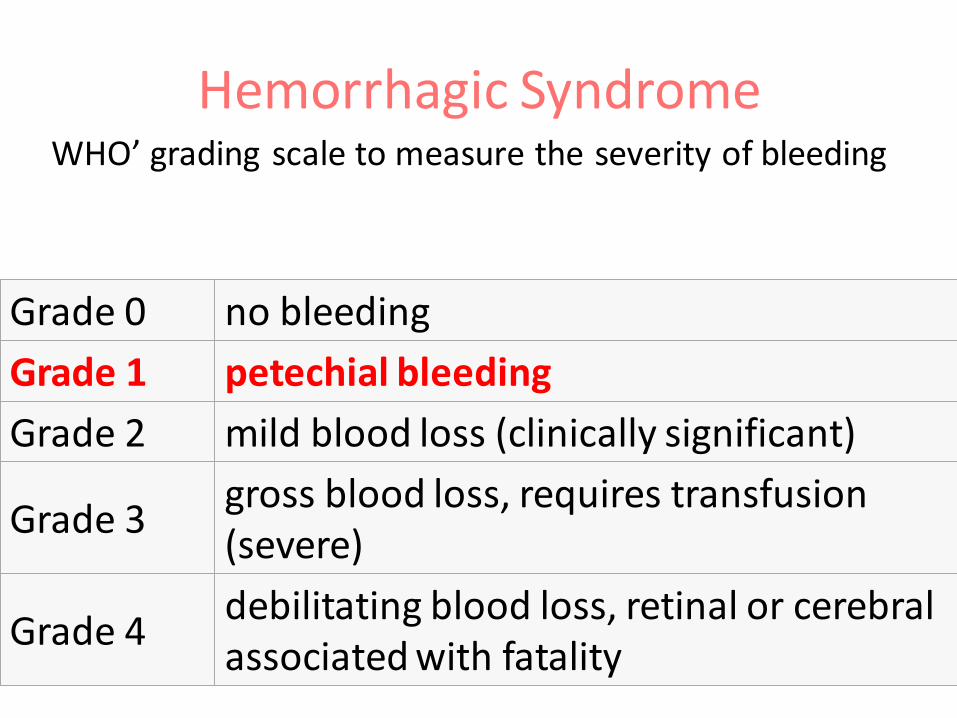
Hemorrhagic Syndrome WHO’ grading scale to measure the severity of bleeding
Grade 0 no bleeding
Grade 1 petechial bleeding
Grade 2 mild blood loss (clinically significant)
Grade 3gross blood loss, requires transfusion (severe)
Grade 4debilitating blood loss, retinal or cerebral associated with fatality
Hemorrhagic Syndrome
Diagnosis
• The platelet count
• The platelet function (bleeding time, platelet aggregation studies, von Willebrand Factor studies, specialized tests)
• A coagulation screen (clotting factor deficiencies)
• If the patient is on warfarin, INR (International Normalized Ratio)
• Autoantibody screen for connective tissue disorders
Glossary of blood disorders pathology’ terms
GLOSSARY of BLOOD RELATED TERMSBLOOD AND HEMATOLOGY DICTIONARY
BLOODBOOK.COM


![[PPT]PEMERIKSAAN LABORATORIUM PADA ANEMIA … · Web viewPEMERIKSAAN LABORATORIUM PADA ANEMIA HEMOLITIK ELLYZA NASRUL Anemia hemolitik - Klasifikasi anemia berdasarkan morfologi anemia](https://img.pdfslide.net/doc/110x75/5c85338309d3f279718c7183/pptpemeriksaan-laboratorium-pada-anemia-web-viewpemeriksaan-laboratorium-pada.jpg)