Embed Size (px)
Citation preview
)259( COPYRIGHT 2017 © BY THE ARCHIVES OF BONE AND JOINT SURGERY
Arch Bone Jt Surg. 2017; 5(4): 259-262. http://abjs.mums.ac.ir
the online version of this article abjs.mums.ac.ir
Lindsay F. Remy, MD; Jacob Azurdia, MD; Ashraf Fansa, MD; Nabil A. Ebraheim, MDT
Research performed at the University of Toledo Medical Center in Toledo, Ohio, USA
Corresponding Author: Lindsay F. Remy, Department of Orthopedic Surgery, University of Toledo Medical Center, Toledo, Ohio, USAEmail: [email protected]
CASE REPORT
Received: 17 February 2016 Accepted: 4 April 2016
Closed Internal Degloving of the Flank
AbstractOriginally described in 1853 by Dr. Morel-Lavellee, closed internal degloving injuries represent an important, although uncommon, source of morbidity in trauma patients. These injuries are typically the result of a shearing or crushing force that traumatically separates the skin and subcutaneous tissue from the underlying fat. This results in disruption of perforating blood vessels and lymphatics, leading to hematoma/seroma formation. We describe two cases in which industrial crush injuries resulted in lumbar transverse process fracture. Both patients developed closed degloving injuries of the flank. To the author’s knowledge, this is the first case series describing the occurrence of closed internal degloving injuries of the flank with transverse process fractures. We advise that a high level of suspicion for these lesions to occur with transverse spinal fractures should be maintained, as they may arise several years after initial injury.
Keywords: Hematoma, Lumbosacral region, Spinal fractures, Subcutaneous tissue, Wounds and injuries
Introduction
Originally described in 1853 by Dr. Morel-Lavellee, closed internal degloving injuries represent an important, although uncommon, source of
morbidity in trauma patients (1). The term Morel-Lavellee lesion is the term used to describe the classic lesion initially described overlying the greater trochanter. Similar closed degloving injuries can occur anywhere on the trunk and extremities. These injuries are typically the result of a shearing or crushing force that traumatically separates the skin and subcutaneous tissue from the underlying fat. This results in disruption of perforating blood vessels and lymphatics, leading to hematoma/seroma formation (1).
Closed internal degloving injuries may be described acutely or chronically. Because a small post-traumatic seroma or hematoma may resolve uneventfully without treatment, the incidence of these lesions is unknown. They have been described in the literature as early as a few hours to as late as 13 years after injury (1, 2).
We describe two cases in which industrial crush injuries resulted in lumbar transverse process fractures. Both patients developed closed degloving injuries of the flank. To the author’s knowledge, this is the first case series describing a possible correlation between transverse
process fractures and closed internal degloving injuries of the flank.
Case presentationThe first patient is an industrial laborer who suffered
a work-related crush injury in 2005 when he was pinned between two semi-trucks. The mechanism of injury involved simultaneous shearing and crushing of the patient’s upper torso in-between both semi-trailers while the patient was standing up. His only known injuries from the event were a T12 compression fracture and transverse process fractures of L2 and L3. These were treated with use of a thoracolumbarsacral orthosis (TLSO) for comfort until healed. He recalls having had a hematoma of the lumbosacral region at the time of the injury that did not receive any intervention.
The patient was seen in clinic in 2007 complaining of discomfort from a lumbosacral “mass”. This was clinically concerning for a chronic closed degloving injury. Magnetic resonance imaging (MRI) was requested from the Bureau of Worker’s Compensation (BWC) for diagnostic confirmation before determining management. Due to his minimal symptoms, he did not follow-through with work-up of the mass.
CLOSED INTERNAL DEGLOVING OF THE FLANKTHE ARCHIVES OF BONE AND JOINT SURGERY. ABJS.MUMS.AC.IRVOLUME 5. NUMBER 4. JULY 2017
)260(
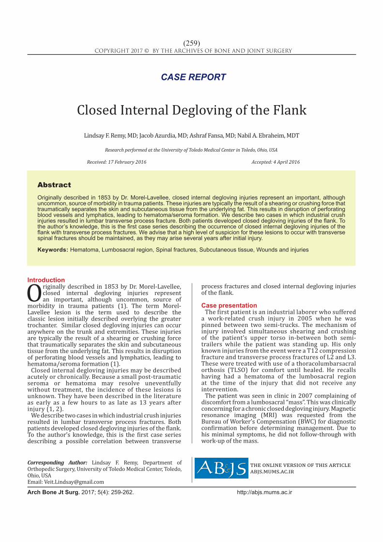
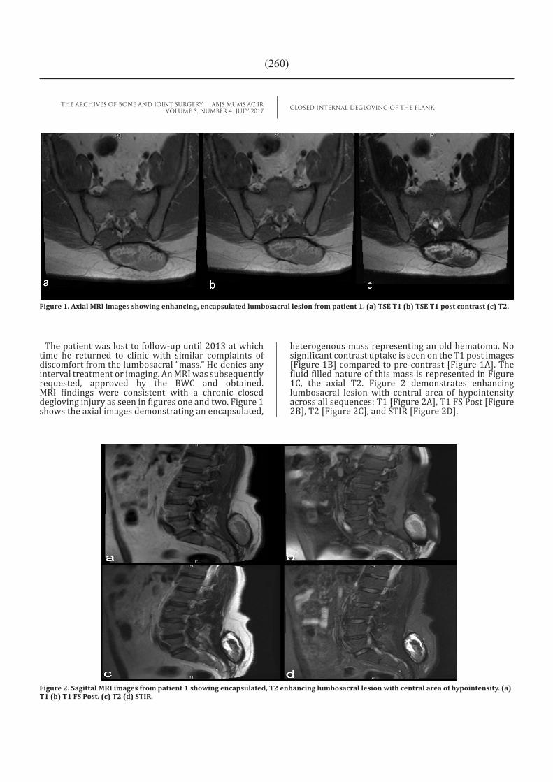
heterogenous mass representing an old hematoma. No significant contrast uptake is seen on the T1 post images [Figure 1B] compared to pre-contrast [Figure 1A]. The fluid filled nature of this mass is represented in Figure 1C, the axial T2. Figure 2 demonstrates enhancing lumbosacral lesion with central area of hypointensity across all sequences: T1 [Figure 2A], T1 FS Post [Figure 2B], T2 [Figure 2C], and STIR [Figure 2D].
The patient was lost to follow-up until 2013 at which time he returned to clinic with similar complaints of discomfort from the lumbosacral “mass.” He denies any interval treatment or imaging. An MRI was subsequently requested, approved by the BWC and obtained. MRI findings were consistent with a chronic closed degloving injury as seen in figures one and two. Figure 1 shows the axial images demonstrating an encapsulated,
Figure 1. Axial MRI images showing enhancing, encapsulated lumbosacral lesion from patient 1. (a) TSE T1 (b) TSE T1 post contrast (c) T2.
Figure 2. Sagittal MRI images from patient 1 showing encapsulated, T2 enhancing lumbosacral lesion with central area of hypointensity. (a) T1 (b) T1 FS Post. (c) T2 (d) STIR.
CLOSED INTERNAL DEGLOVING OF THE FLANKTHE ARCHIVES OF BONE AND JOINT SURGERY. ABJS.MUMS.AC.IRVOLUME 5. NUMBER 4. JULY 2017
)261(
weeks later and found to have persistent subcutaneous fluid collection, but was asymptomatic. Since then, he has been lost to follow-up.
DiscussionClosed degloving injuries such as those described in this
case series can be diagnosed by ultrasound, computed tomography (CT), or magnetic resonance imaging (MRI). These lesions have characteristic imaging findings in each modality. Sonographically, closed degloving injuries are usually hypoechoic and well circumscribed. Appearance can vary by stage of hematoma organization or degradation at time of imaging. Hyperechoic nodules may represent internal fat lobules. Computed tomography of the lesion shows characteristic
fluid-fluid levels resulting from settling of blood products within the collection; a capsule may be visible in chronic cases (1).
Magnetic resonance imaging is the preferred imaging modality in the evaluation of closed degloving injuries due to the degree of detail it provides regarding soft tissue anatomy. Signal variations correspond to internal content and chronicity of the lesions. In the setting of a simple fluid collection, MRI signal findings would be homogeneously hypointense on T1-weighted sequences and hyperintense on T2-weighted sequence. If they contain a high internal concentration of methemoglobin, a product of hemoglobin degradation, lesions may also appear homogeneously bright on both T1- and T2-weighted sequences. A hypointense peripheral ring representing hemosiderin and fibrous tissue capsule may be present. Post-contrast images may show patchy enhancement both internal and peripheral to the lesion, consistent with an organizing hematoma (3, 4).
The thick, well-fixed nature of the thoracolumbar aponeurosis may contribute to the susceptibility of the flank to closed degloving injuries. A similar mechanism is thought to be related to the high frequency of lesions in the proximal thigh area due to the firm attachments
From the patient’s history, it was felt this lesion was likely related to his 2005 accident. He underwent CT guided aspiration of 30ml fluid. At the time of aspiration, a percutaneous drain was placed and then removed the following day with no output. He did well initially and our plan was for elective removal of the cavity wall if symptoms recurred. However, three weeks after aspiration he presented to the emergency department with secondary infection of the lesion. He was taken for excisional debridement and wound VAC application the following day. Intraoperative cultures grew Methicillin-Sensitive Staph Aureus (MSSA) for which he was treated with intravenous ceftriaxone. This was continued per the Infectious Disease recommendations for four months. During this time he returned for repeat excisional debridement on three occasions. Once the wound was nearly closed and without signs of infection, he was transitioned to oral cefazolin. The wound closed by secondary intention over the following five months after final debridement. He is now doing well.
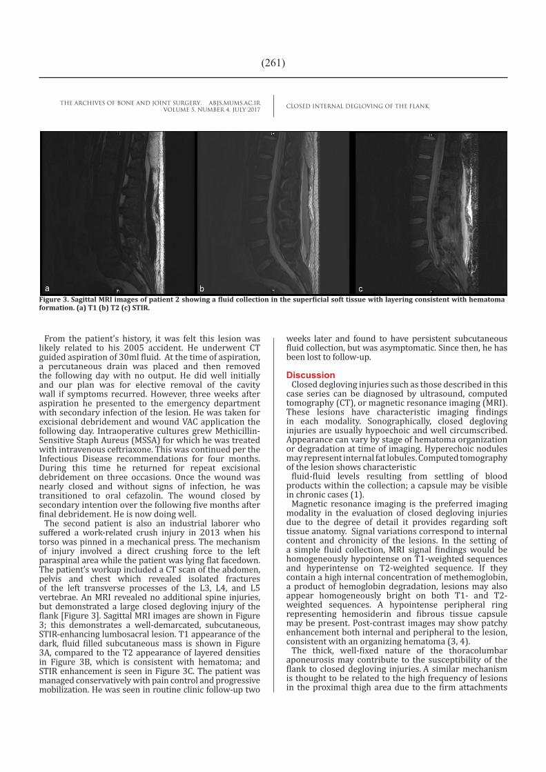
The second patient is also an industrial laborer who suffered a work-related crush injury in 2013 when his torso was pinned in a mechanical press. The mechanism of injury involved a direct crushing force to the left paraspinal area while the patient was lying flat facedown. The patient’s workup included a CT scan of the abdomen, pelvis and chest which revealed isolated fractures of the left transverse processes of the L3, L4, and L5 vertebrae. An MRI revealed no additional spine injuries, but demonstrated a large closed degloving injury of the flank [Figure 3]. Sagittal MRI images are shown in Figure 3; this demonstrates a well-demarcated, subcutaneous, STIR-enhancing lumbosacral lesion. T1 appearance of the dark, fluid filled subcutaneous mass is shown in Figure 3A, compared to the T2 appearance of layered densities in Figure 3B, which is consistent with hematoma; and STIR enhancement is seen in Figure 3C. The patient was managed conservatively with pain control and progressive mobilization. He was seen in routine clinic follow-up two
Figure 3. Sagittal MRI images of patient 2 showing a fluid collection in the superficial soft tissue with layering consistent with hematoma formation. (a) T1 (b) T2 (c) STIR.
CLOSED INTERNAL DEGLOVING OF THE FLANKTHE ARCHIVES OF BONE AND JOINT SURGERY. ABJS.MUMS.AC.IRVOLUME 5. NUMBER 4. JULY 2017
)262(
References
1. Morel-Lavallée AF. Décollements traumatiques de la peau et des couches sous jacentes. Arch Gen Med. 1863; 9(1):20-38.
2. Hak DJ, Olson SA, Matta JM. Diagnosis and management of closed internal degloving injuries associated with pelvic and acetabular fractures: the Morel-Lavallee lesion. J Trauma. 1997; 42(6):1046-51.
3. Mellado JM, Perez del Palomar L, Diz L, Ramos A, Sauri A. Long-standing Morel-Lavallée lesions of
the trochanteric region and proximal thigh: MRI features in five patients. AJR Am J Roentgenol. 2004; 182(5):1289-94.
4. Chokshi FH, Jose J, Clifford PD. Morel-Lavellee lesion. Am J Orthop. 2010; 39(5):252-3.
5. Nickerson TP, Zeilinski MD, Jenkins DH, Schiller HJ. The Mayo Clinic experience with Morel-Lavallee lesions: establishment of a practice management guideline. J Trauma Acute Care Surg. 2014; 76(2):493-7.
of the fascia lata and the iliotibial band with relative mobility of the overlying skin.
In the setting of a patient with transverse process fracture of the lumbar spine from crush mechanism, examination for associated closed degloving injury of the flank is recommended. Treatment will vary by both patient and surgeon preferences. In their retrospective review of 87 of these lesions, 83% of patients in whom percutaneous aspiration yielded more than 50 mL of fluid had recurrence of the lesion. On the other hand only 33% of patients yielding less than 50 mL on aspiration had recurrence. Based on this finding they recommend surgical debridement if aspiration yields more than 50 mL (5). If the skin is not viable or if the lesion is already infected, we recommend open debridement and closure by secondary intention often best achieved with a negative pressure dressing. If the skin is viable, open or minimally invasive debridement can be performed,
Lindsay F. Remy MDJacob Azurdia MDAshraf Fansa MDNabil A. Ebraheim MDTDepartment of Orthopedic Surgery, University of Toledo Medical Center, Toledo, Ohio, USA
typically with postoperative drain placement. Our first case developed a fulminant degloving lesion two years post initial injury. Therefore, we advise that in the setting of transverse process fractures, a high level of suspicion for these lesions should be maintained as they may arise several years post initial injury.