Embed Size (px)
Citation preview
1
Coagulation tests in
children
Katrien Devreese, MD, PhD Coagulation Laboratory
Ghent University Hospitial 25 April 2015

2 2
The young are not just miniature adults Physiology of pediatric hemostasis differs from adults Course of hemostatic disorders may differ
Correct interpretation of coagulation test results
Introduction
3 3
Hemostasis
Introduction
Coagulation Anticoagulation
4 4
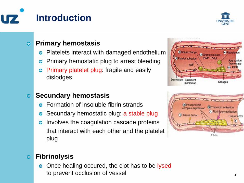
Primary hemostasis Platelets interact with damaged endothelium Primary hemostatic plug to arrest bleeding Primary platelet plug: fragile and easily dislodges
Secundary hemostasis
Formation of insoluble fibrin strands Secundary hemostatic plug: a stable plug Involves the coagulation cascade proteins
that interact with each other and the platelet plug
Fibrinolysis
Once healing occured, the clot has to be lysed to prevent occlusion of vessel
Introduction
5 5
Primary hemostasis Platelet count Platelet morphology Platelet function
Secundary hemostasis
Clotting assays: aPTT, PT, thrombin time Fibrinogen Coagulation factors (FXII, FXI, FIX, FVIII; FII, FV, FVII, FX; FXIII; VWF) Coagulation inhibitors (AT, PC, PS)
Fibrinolysis
Plasminogen, t-PA, α2-antiplasmin, PAI
Introduction
6 6
Testing hemostasis in children Bleeding
Screening tests: complete blood count prothrombin time (PT) activated partial thromboplastin time (aPTT) (Platelet function analysis (PFA))
Further laboratory evaluation: additional tests Thrombosis
Inherited thrombophilia markers (antithrombin, protein C, protein S, FVLeiden, FIIG20210A)
Issues related to coagulation testing in children
Laboratory tests for hemostasis
7 7
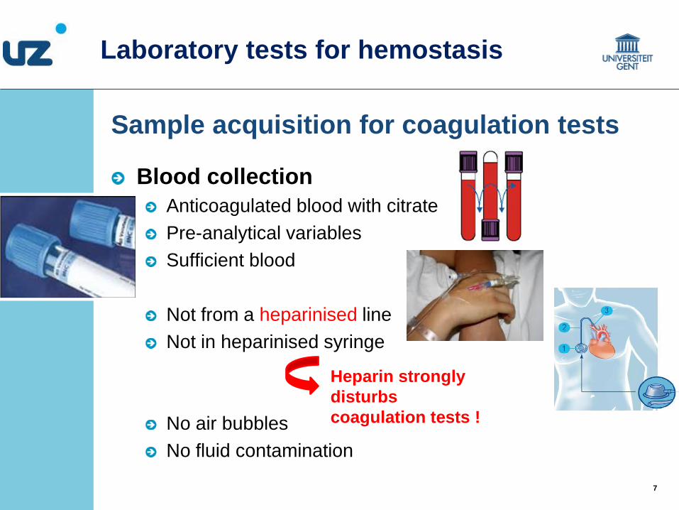
Blood collection Anticoagulated blood with citrate Pre-analytical variables Sufficient blood Not from a heparinised line Not in heparinised syringe
No air bubbles No fluid contamination
Laboratory tests for hemostasis
Sample acquisition for coagulation tests
Heparin strongly disturbs coagulation tests !
8 8
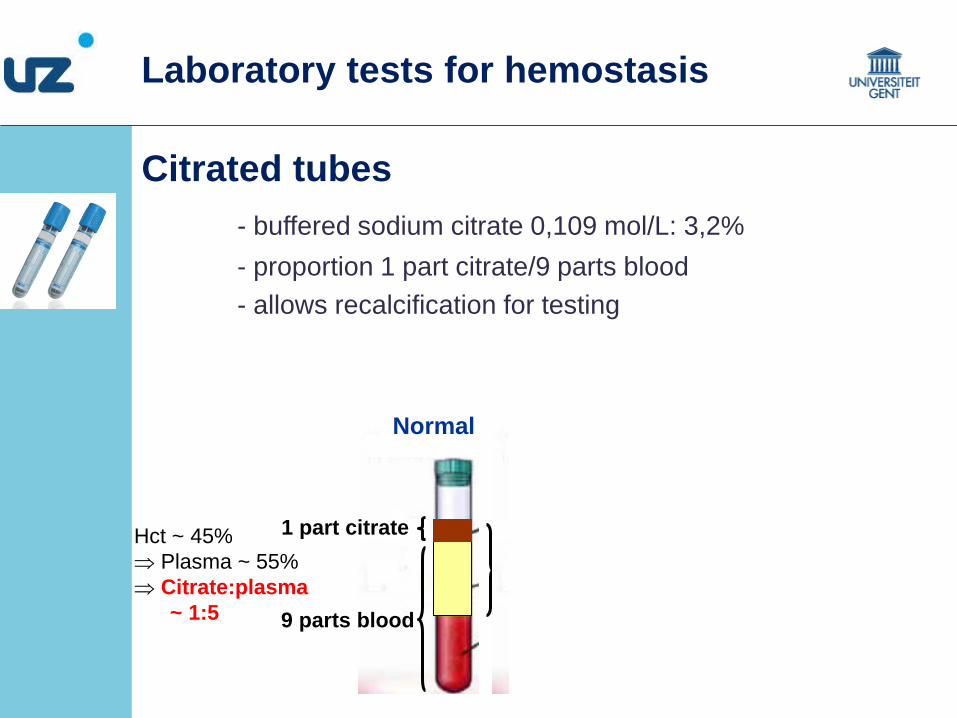
1 part citrate
9 parts blood
Hct ~ 45% ⇒ Plasma ~ 55% ⇒ Citrate:plasma ~ 1:5
Citrate:plasma ~ 1:3 -dilution effect on plasma -excess of citrate that binds to calcium added in reaction mix
Citrated tubes - buffered sodium citrate 0,109 mol/L: 3,2% - proportion 1 part citrate/9 parts blood - allows recalcification for testing
High hematocrit / underfilling of tube Normal
False prolonged aPTT
Laboratory tests for hemostasis
9 9
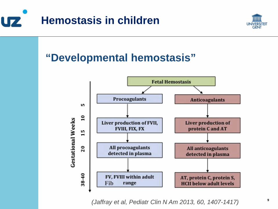
“Developmental hemostasis”
Hemostasis in children
(Jaffray et al, Pediatr Clin N Am 2013, 60, 1407-1417)
Fib
10 10
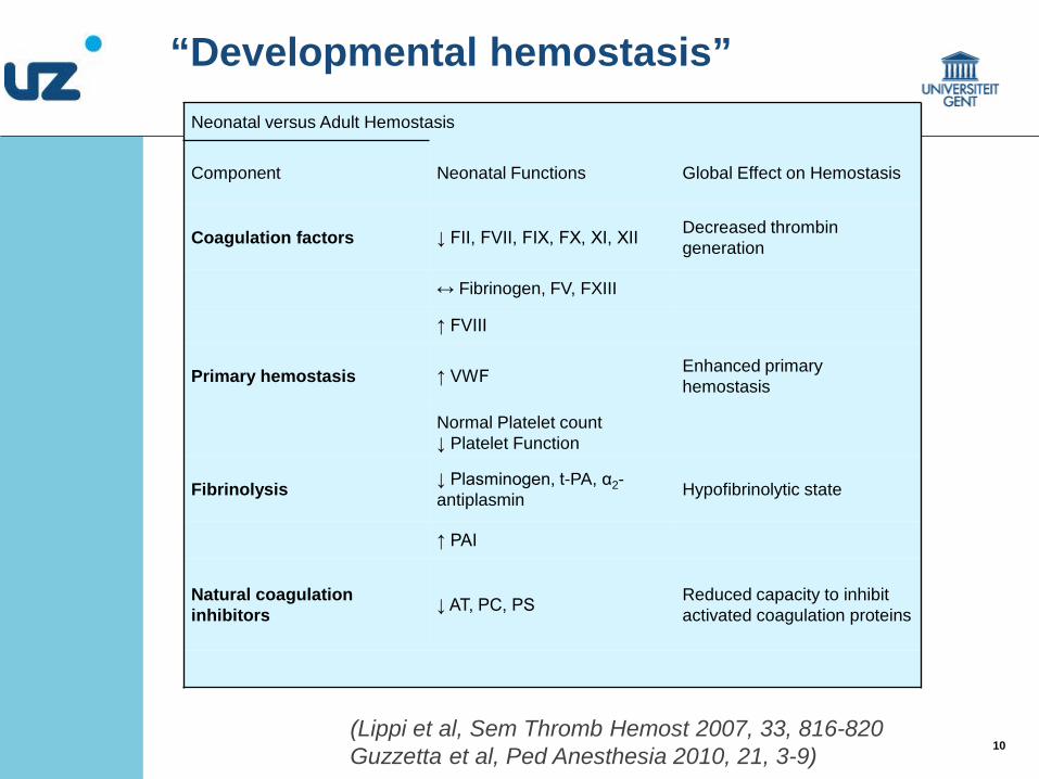
Neonatal versus Adult Hemostasis
Component Neonatal Functions Global Effect on Hemostasis
Coagulation factors ↓ FII, FVII, FIX, FX, XI, XII Decreased thrombin generation
↔ Fibrinogen, FV, FXIII
↑ FVIII
Primary hemostasis ↑ VWF Enhanced primary hemostasis
Normal Platelet count ↓ Platelet Function
Fibrinolysis ↓ Plasminogen, t-PA, α2-antiplasmin Hypofibrinolytic state
↑ PAI
Natural coagulation inhibitors ↓ AT, PC, PS Reduced capacity to inhibit
activated coagulation proteins
(Lippi et al, Sem Thromb Hemost 2007, 33, 816-820 Guzzetta et al, Ped Anesthesia 2010, 21, 3-9)
“Developmental hemostasis”
11 11
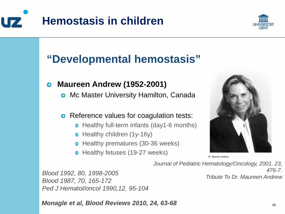
“Developmental hemostasis”
Maureen Andrew (1952-2001) Mc Master University Hamilton, Canada Reference values for coagulation tests:
Healthy full-term infants (day1-6 months) Healthy children (1y-16y) Healthy prematures (30-36 weeks) Healthy fetuses (19-27 weeks)
Hemostasis in children
Journal of Pediatric Hematology/Oncology, 2001, 23, 476-7.
Tribute To Dr. Maureen Andrew Blood 1992, 80, 1998-2005 Blood 1987, 70, 165-172 Ped J Hematol/oncol 1990,12, 95-104
Monagle et al, Blood Reviews 2010, 24, 63-68
12 12
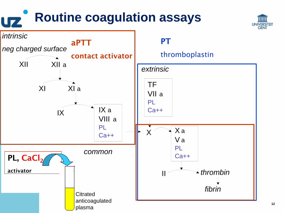
thrombin
XII XII a
XI XI a
IX
II
X
intrinsic
extrinsic
X a
V a PL Ca++
TF VII a PL Ca++ IX a
VIII a PL Ca++
neg charged surface
common
fibrin
aPTT
contact activator
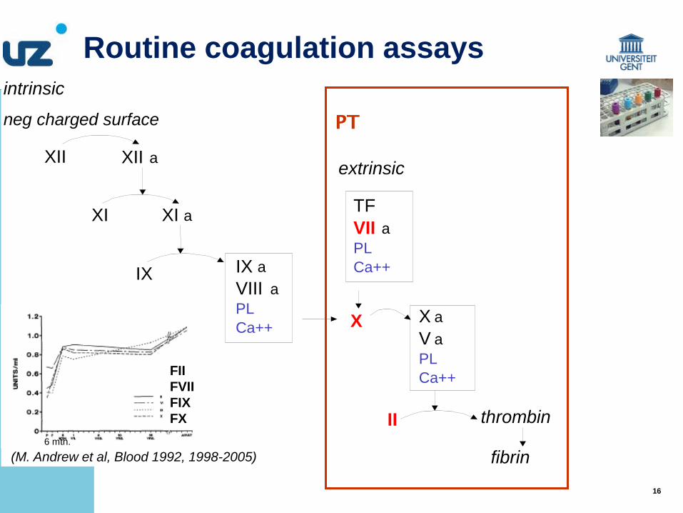
Routine coagulation assays
PT
thromboplastin
PL, CaCl2
activator
Citrated anticoagulated plasma
13 13
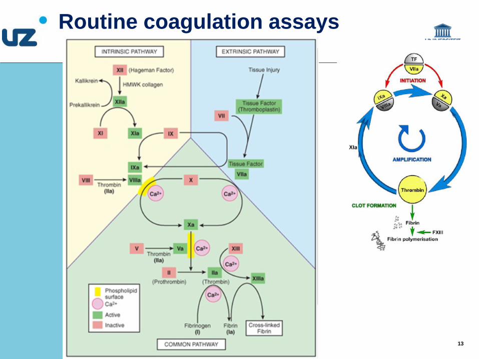
Routine coagulation assays
14 14
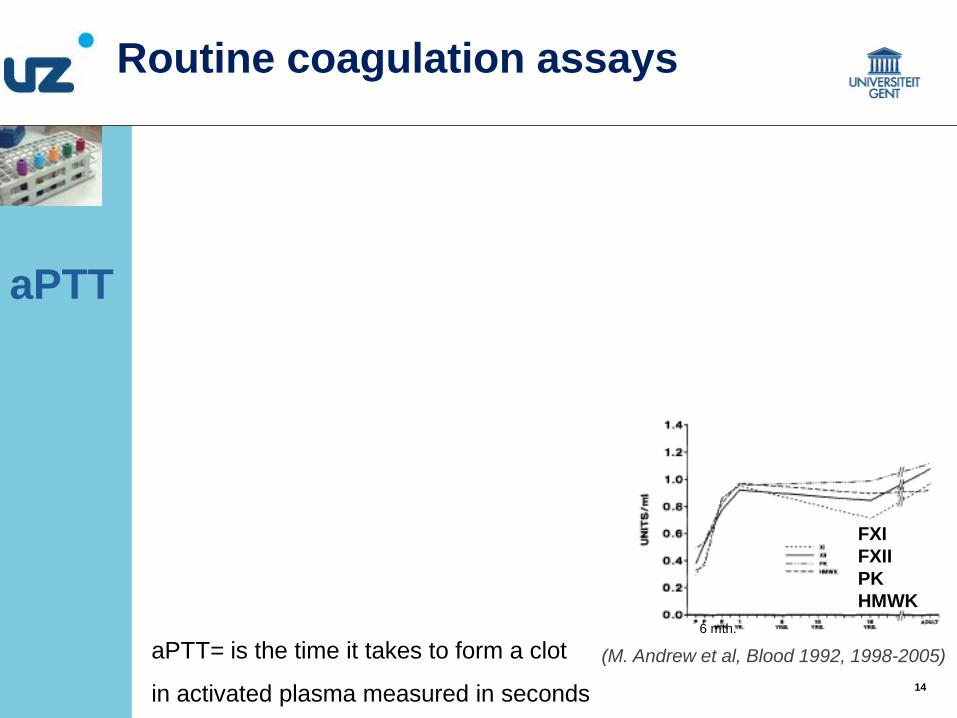
aPTT
FXI FXII PK HMWK
(M. Andrew et al, Blood 1992, 1998-2005) 6 mth.
Routine coagulation assays
aPTT= is the time it takes to form a clot
in activated plasma measured in seconds
15 15
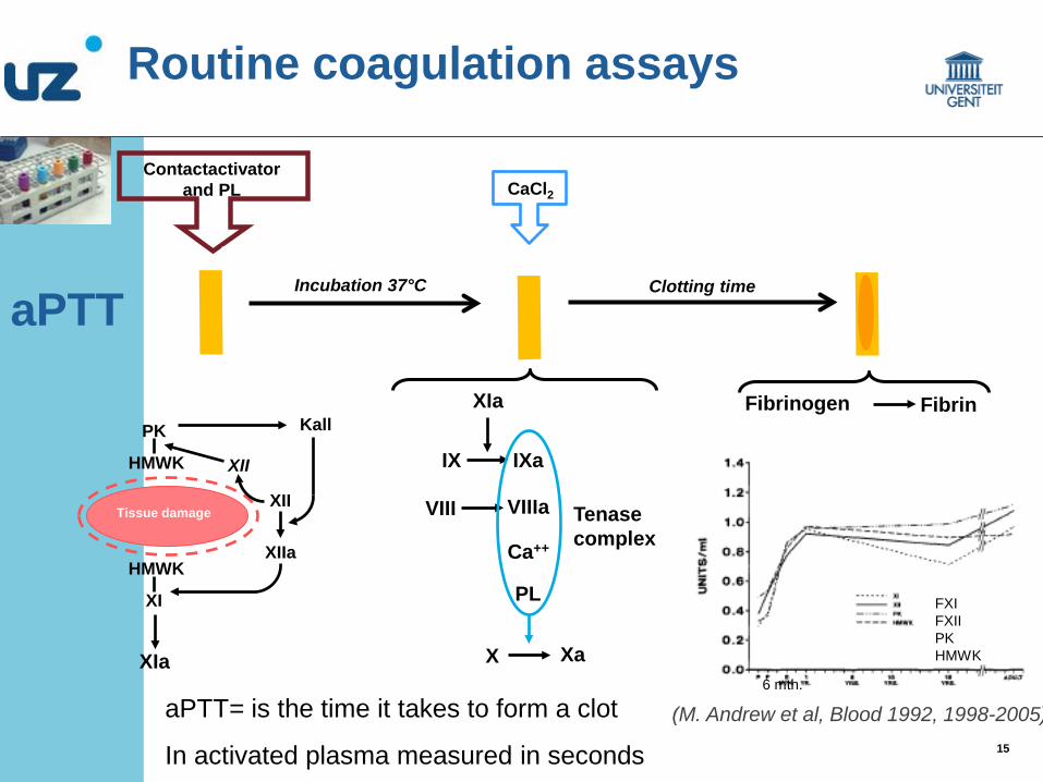
Contactactivator and PL
Incubation 37°C
CaCl2
Clotting time
X Xa
VIII VIIIa
IX IXa
Ca++
PL
XIa Fibrinogen Fibrin
Tenase complex
HMWK
XIa
XII Tissue damage
PK
XII
Kall
XIIa HMWK
XI
aPTT
aPTT= is the time it takes to form a clot
In activated plasma measured in seconds
FXI FXII PK HMWK
(M. Andrew et al, Blood 1992, 1998-2005) 6 mth.
Routine coagulation assays
16 16
thrombin
XII XII a
XI XI a
IX
II
X
intrinsic
extrinsic
X a
V a PL Ca++
TF VII a PL Ca++ IX a
VIII a PL Ca++
neg charged surface
fibrin
PT
Routine coagulation assays
(M. Andrew et al, Blood 1992, 1998-2005) 6 mth.
FII FVII FIX FX
17 17
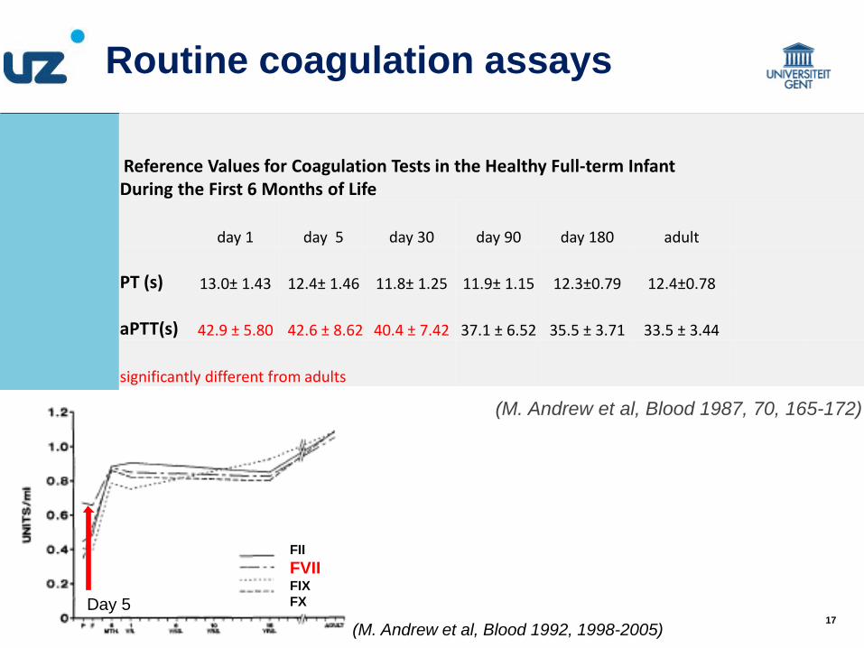
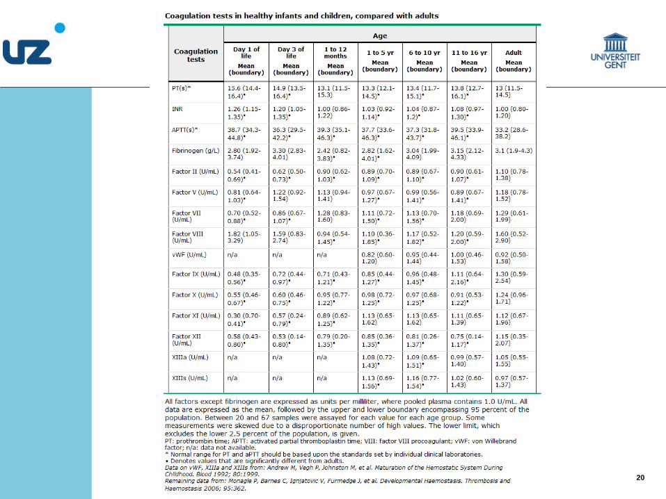
Reference Values for Coagulation Tests in the Healthy Full-term Infant During the First 6 Months of Life
day 1 day 5 day 30 day 90 day 180 adult
PT (s) 13.0± 1.43 12.4± 1.46 11.8± 1.25 11.9± 1.15 12.3±0.79 12.4±0.78
aPTT(s) 42.9 ± 5.80 42.6 ± 8.62 40.4 ± 7.42 37.1 ± 6.52 35.5 ± 3.71 33.5 ± 3.44
significantly different from adults
(M. Andrew et al, Blood 1987, 70, 165-172)
Routine coagulation assays
(M. Andrew et al, Blood 1992, 1998-2005)
FII FVII FIX FX Day 5
18 18
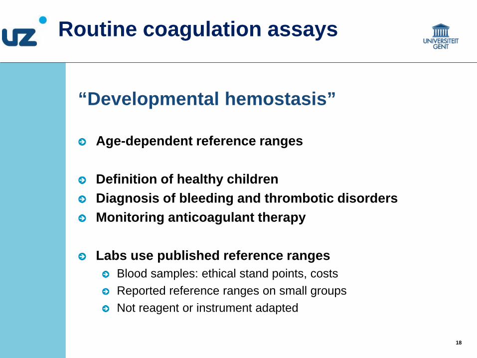
Age-dependent reference ranges
Definition of healthy children Diagnosis of bleeding and thrombotic disorders Monitoring anticoagulant therapy Labs use published reference ranges
Blood samples: ethical stand points, costs Reported reference ranges on small groups Not reagent or instrument adapted
Routine coagulation assays
“Developmental hemostasis”
19 19
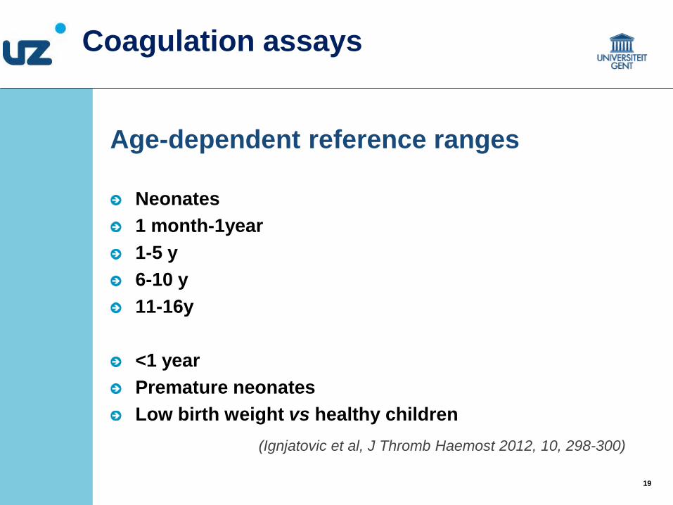
Age-dependent reference ranges
Neonates 1 month-1year 1-5 y 6-10 y 11-16y <1 year Premature neonates Low birth weight vs healthy children
(Ignjatovic et al, J Thromb Haemost 2012, 10, 298-300)
Coagulation assays
20 20
21 21
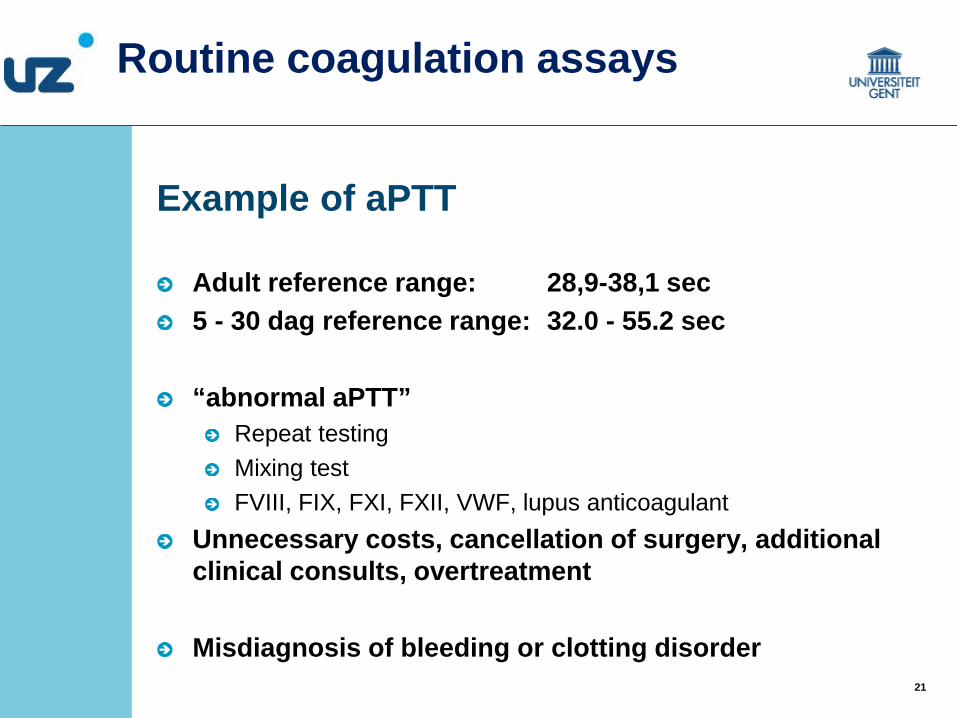
Example of aPTT
Adult reference range: 28,9-38,1 sec 5 - 30 dag reference range: 32.0 - 55.2 sec “abnormal aPTT”
Repeat testing Mixing test FVIII, FIX, FXI, FXII, VWF, lupus anticoagulant
Unnecessary costs, cancellation of surgery, additional clinical consults, overtreatment Misdiagnosis of bleeding or clotting disorder
Routine coagulation assays
22 22
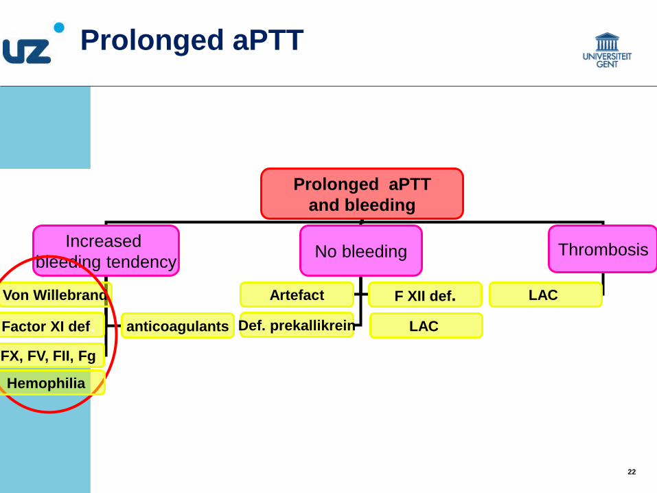
Prolonged aPTT and bleeding
Increased bleeding tendency No bleeding Thrombosis
Von Willebrand
Factor XI def. anticoagulants
FX, FV, FII, Fg
Artefact F XII def. Def. prekallikrein
LAC
Prolonged aPTT
LAC
Hemophilia
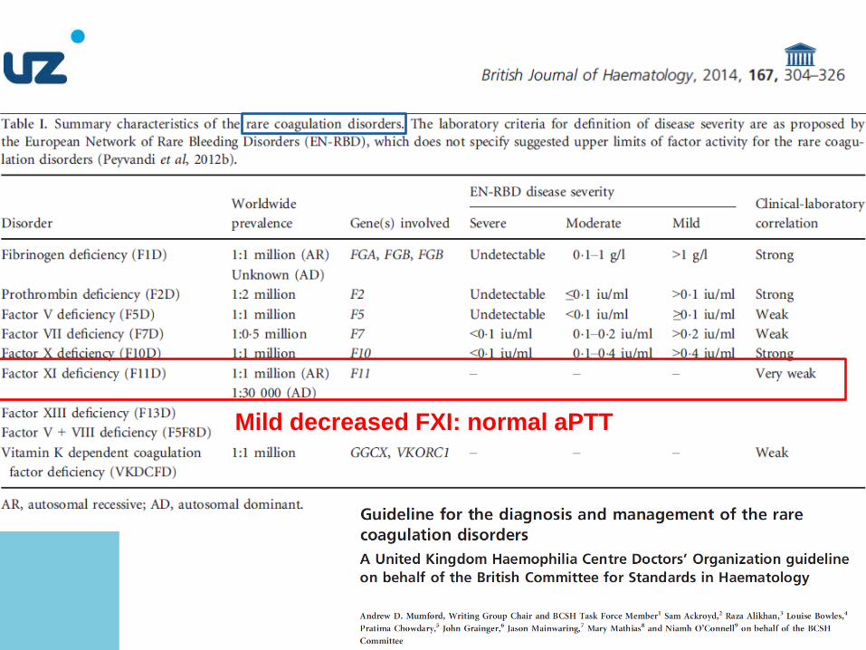
23 23
Mild decreased FXI: normal aPTT
24 24
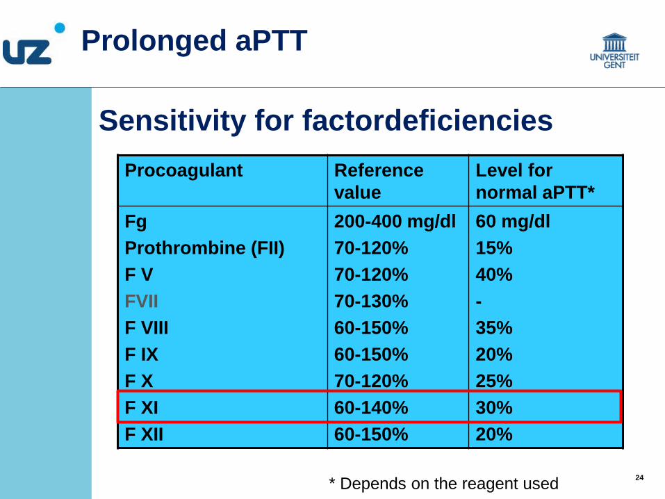
Sensitivity for factordeficiencies Procoagulant Reference
value Level for normal aPTT*
Fg Prothrombine (FII) F V FVII F VIII F IX F X F XI F XII
200-400 mg/dl 70-120% 70-120% 70-130% 60-150% 60-150% 70-120% 60-140% 60-150%
60 mg/dl 15% 40% - 35% 20% 25% 30% 20%
Prolonged aPTT
* Depends on the reagent used
25 25
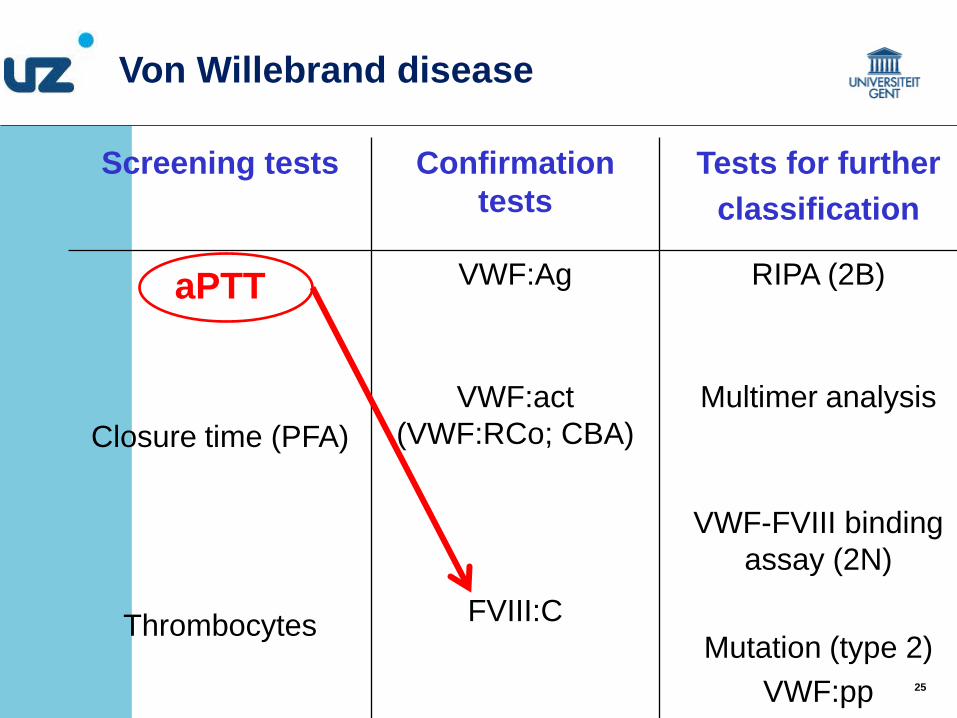
Screening tests Confirmation tests
Tests for further classification
aPTT
VWF:Ag RIPA (2B)
Closure time (PFA) VWF:act
(VWF:RCo; CBA) Multimer analysis
Thrombocytes
FVIII:C
VWF-FVIII binding assay (2N)
Mutation (type 2)
VWF:pp
Von Willebrand disease
26 26
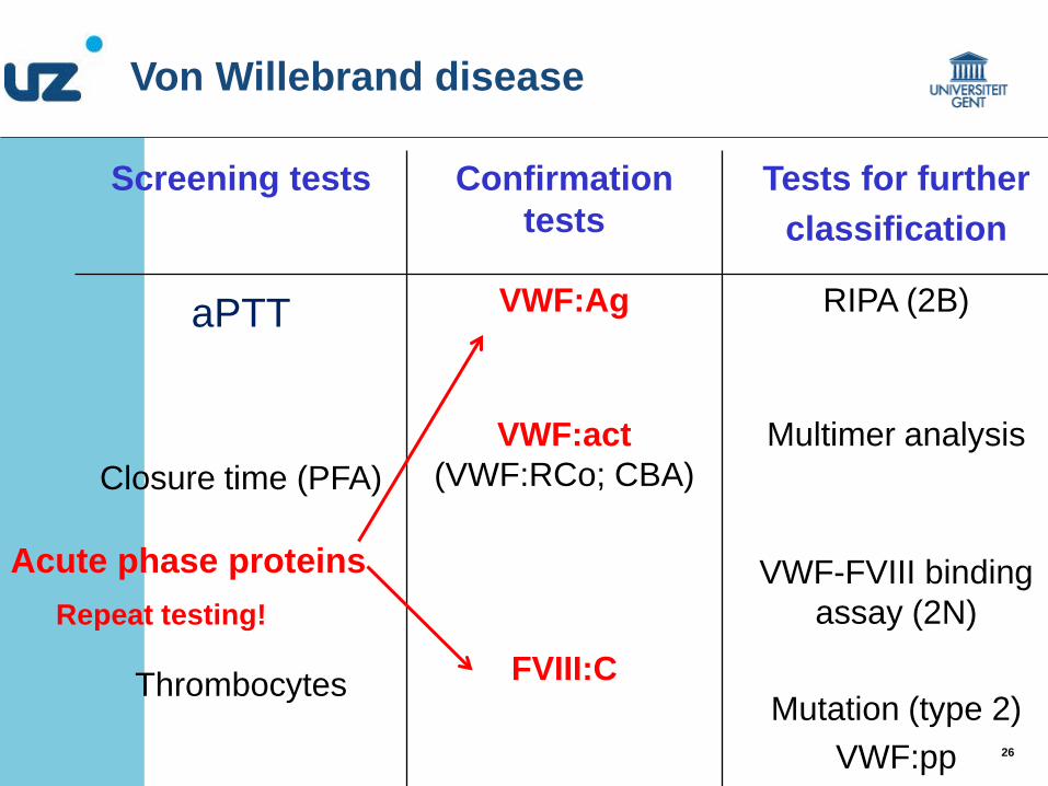
Screening tests Confirmation tests
Tests for further classification
aPTT
VWF:Ag RIPA (2B)
Closure time (PFA) VWF:act
(VWF:RCo; CBA) Multimer analysis
Thrombocytes
FVIII:C
VWF-FVIII binding assay (2N)
Mutation (type 2)
VWF:pp
Von Willebrand disease
Acute phase proteins Repeat testing!
27 27
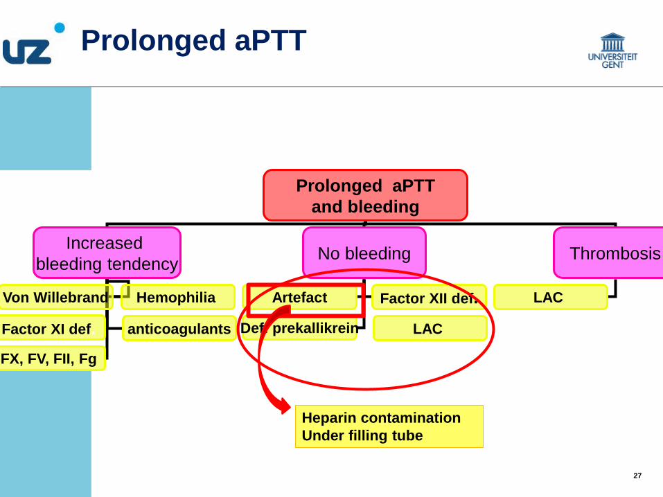
Prolonged aPTT and bleeding
Increased bleeding tendency No bleeding Thrombosis
Von Willebrand Hemophilia
Factor XI def. anticoagulants
FX, FV, FII, Fg
Artefact Factor XII def. Def. prekallikrein
LAC
Prolonged aPTT
LAC
Heparin contamination Under filling tube
28 28
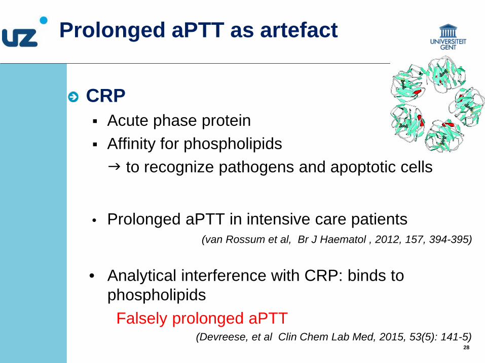
CRP Acute phase protein Affinity for phospholipids to recognize pathogens and apoptotic cells
• Prolonged aPTT in intensive care patients
• Analytical interference with CRP: binds to phospholipids
Falsely prolonged aPTT
Prolonged aPTT as artefact
(van Rossum et al, Br J Haematol , 2012, 157, 394-395)
(Devreese, et al Clin Chem Lab Med, 2015, 53(5): 141-5)
29 29
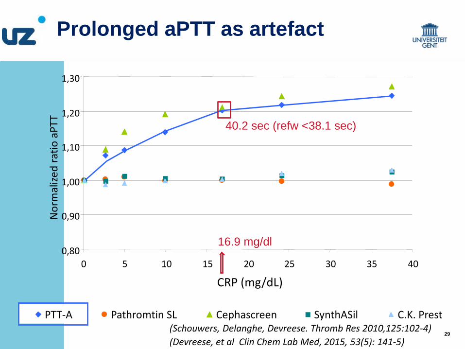
0,80
0,90
1,00
1,10
1,20
1,30
0 5 10 15 20 25 30 35 40
CRP (mg/dL)
Nor
mal
ized
ratio
aPT
T
PTT-A Pathromtin SL Cephascreen SynthASil C.K. Prest
40.2 sec (refw <38.1 sec)
16.9 mg/dl
(Schouwers, Delanghe, Devreese. Thromb Res 2010,125:102-4) (Devreese, et al Clin Chem Lab Med, 2015, 53(5): 141-5)
Prolonged aPTT as artefact
30 30
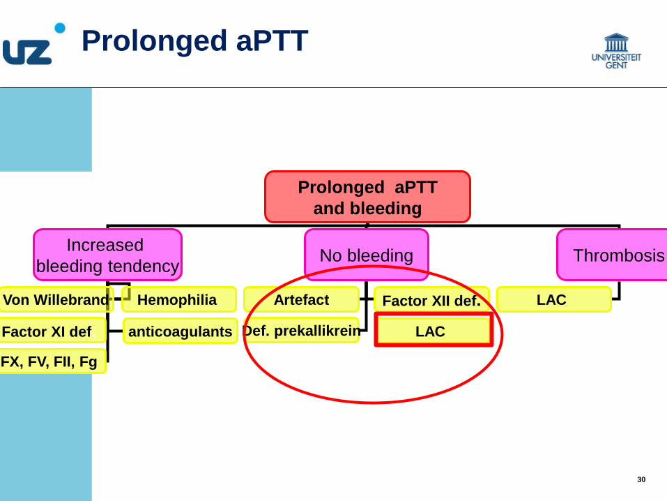
Prolonged aPTT and bleeding
Increased bleeding tendency No bleeding Thrombosis
Von Willebrand Hemophilia
Factor XI def. anticoagulants
FX, FV, FII, Fg
Artefact Factor XII def. Def. prekallikrein
LAC
Prolonged aPTT
LAC
31 31
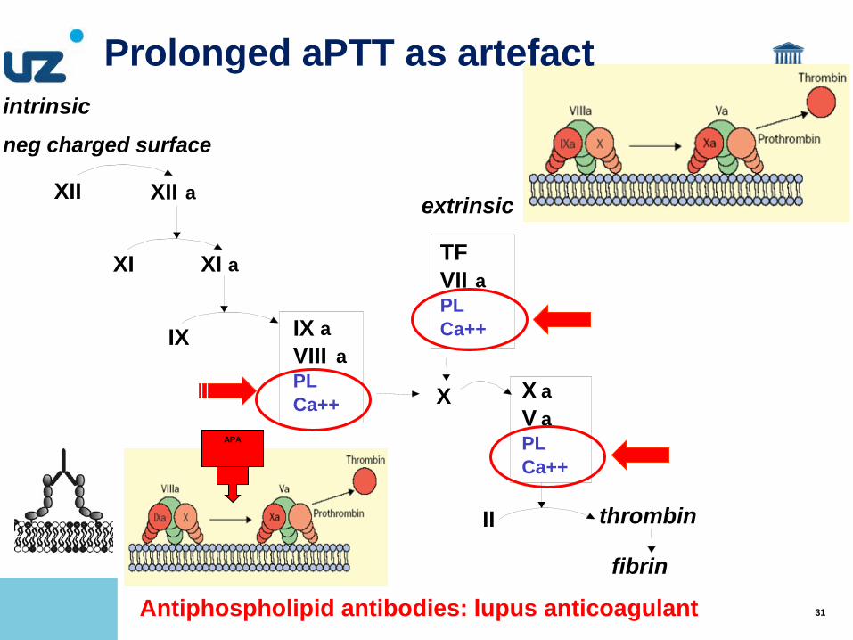
thrombin
XII XII a
XI XI a
IX
II
X
intrinsic
extrinsic
X a
V a PL Ca++
TF VII a PL Ca++ IX a
VIII a PL Ca++
neg charged surface
common
fibrin
APA
Prolonged aPTT as artefact
Antiphospholipid antibodies: lupus anticoagulant
32 32
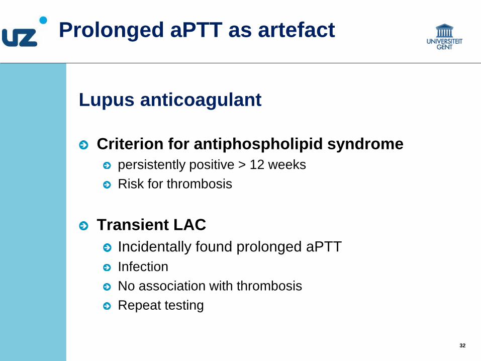
Lupus anticoagulant
Criterion for antiphospholipid syndrome persistently positive > 12 weeks Risk for thrombosis
Transient LAC
Incidentally found prolonged aPTT Infection No association with thrombosis Repeat testing
Prolonged aPTT as artefact
33 33
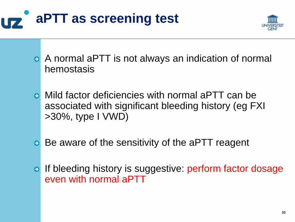
A normal aPTT is not always an indication of normal hemostasis Mild factor deficiencies with normal aPTT can be associated with significant bleeding history (eg FXI >30%, type I VWD) Be aware of the sensitivity of the aPTT reagent If bleeding history is suggestive: perform factor dosage even with normal aPTT
aPTT as screening test
34 34
trombin
XII XII a
XI XI a
IX
II
X
intrinsic
extrinsic
X a
V a PL Ca++
TF VII a PL Ca++ IX a
VIII a PL Ca++
neg charged surface
common
fibrin
aPTT
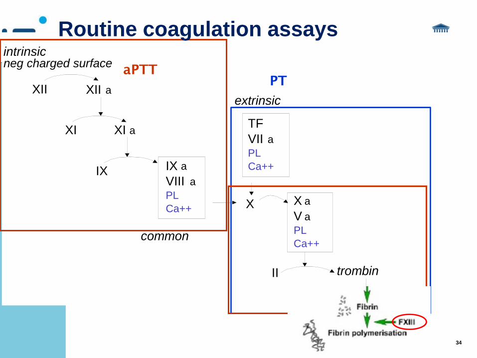
Routine coagulation assays
PT
35 35
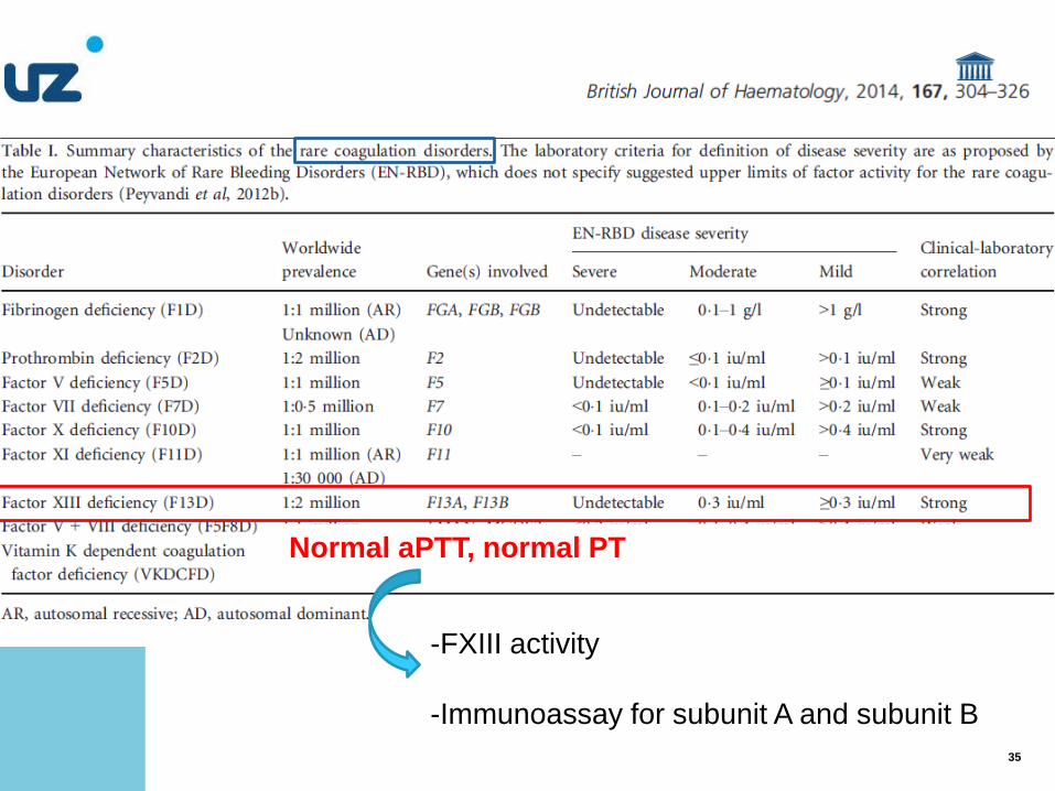
Normal aPTT, normal PT
-FXIII activity -Immunoassay for subunit A and subunit B
36 36
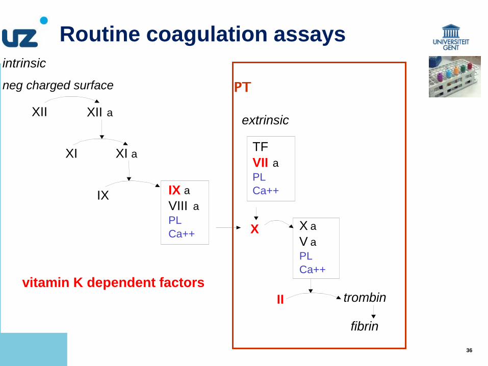
trombin
XII XII a
XI XI a
IX
II
X
intrinsic
extrinsic
X a
V a PL Ca++
TF VII a PL Ca++ IX a
VIII a PL Ca++
neg charged surface
fibrin
PT
Routine coagulation assays
vitamin K dependent factors
37 37
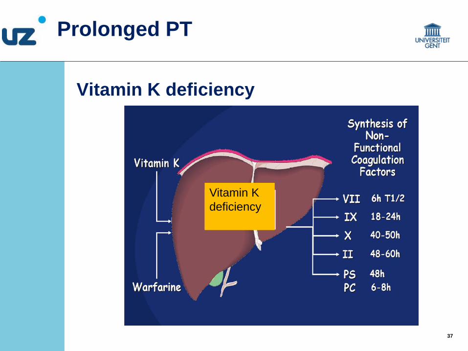
Vitamin K deficiency
Vitamin K deficiency
Prolonged PT
38 38
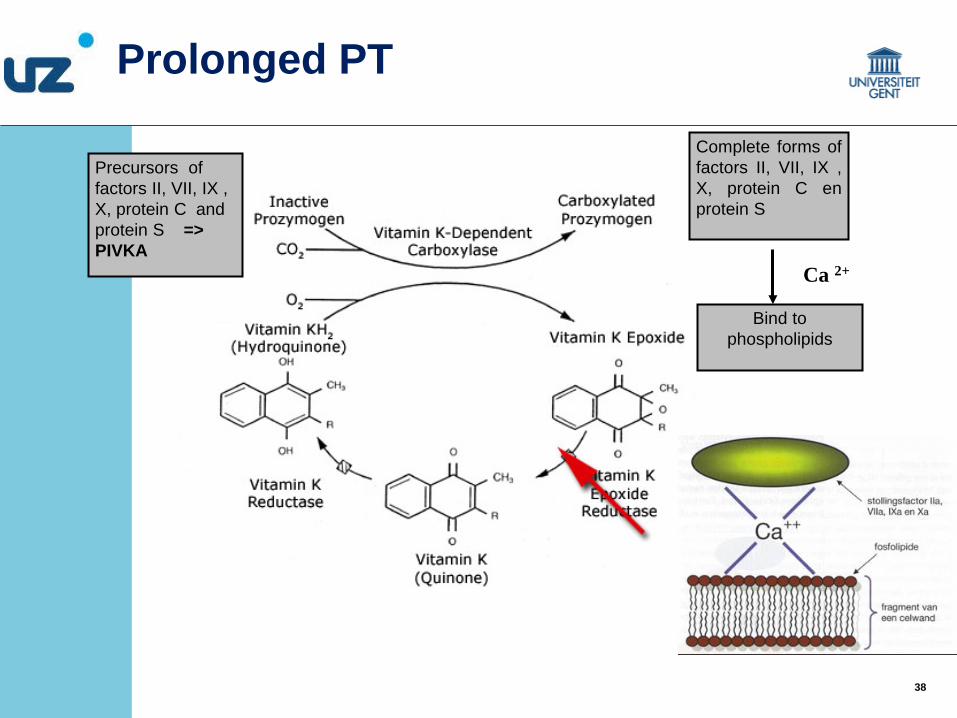
Precursors of factors II, VII, IX , X, protein C and protein S => PIVKA
Complete forms of factors II, VII, IX , X, protein C en protein S
Bind to phospholipids
Ca 2+
Prolonged PT
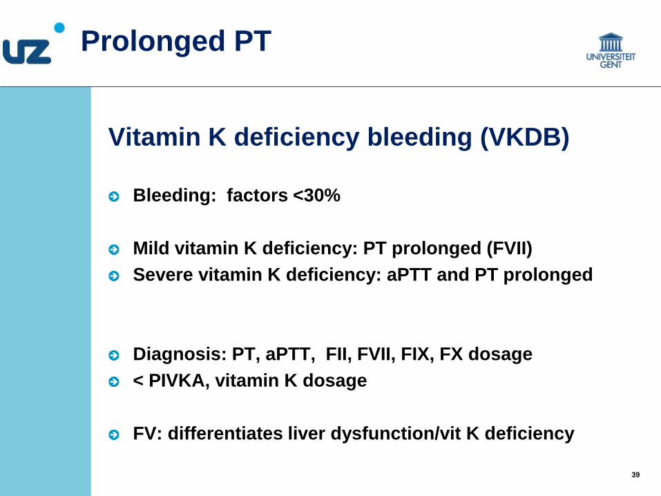
39 39
Bleeding: factors <30% Mild vitamin K deficiency: PT prolonged (FVII) Severe vitamin K deficiency: aPTT and PT prolonged Diagnosis: PT, aPTT, FII, FVII, FIX, FX dosage < PIVKA, vitamin K dosage FV: differentiates liver dysfunction/vit K deficiency
Vitamin K deficiency bleeding (VKDB)
Prolonged PT
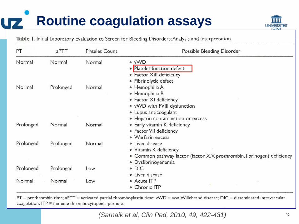
40 40 (Sarnaik et al, Clin Ped, 2010, 49, 422-431)
Routine coagulation assays
41 41
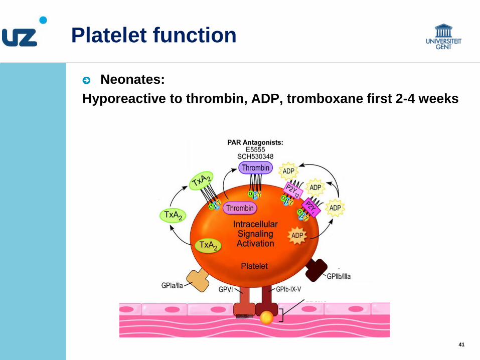
Platelet function
Neonates: Hyporeactive to thrombin, ADP, tromboxane first 2-4 weeks
42 42
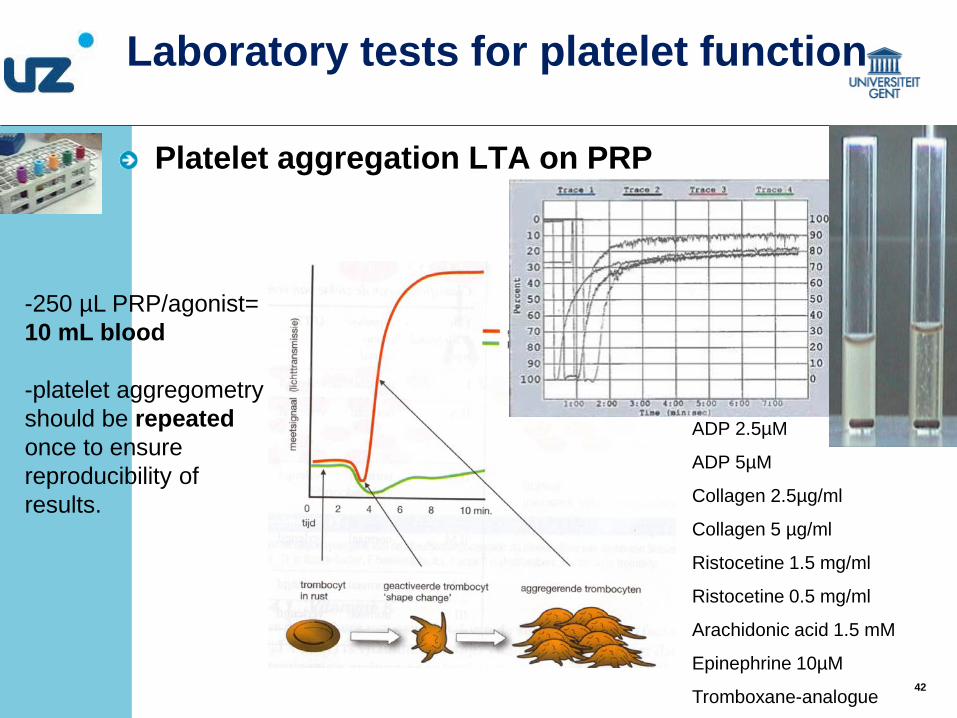
Platelet aggregation LTA on PRP
-250 µL PRP/agonist= 10 mL blood -platelet aggregometry should be repeated once to ensure reproducibility of results.
Laboratory tests for platelet function
ADP 2.5µM
ADP 5µM
Collagen 2.5µg/ml
Collagen 5 µg/ml
Ristocetine 1.5 mg/ml
Ristocetine 0.5 mg/ml
Arachidonic acid 1.5 mM
Epinephrine 10µM
Tromboxane-analogue
43 43
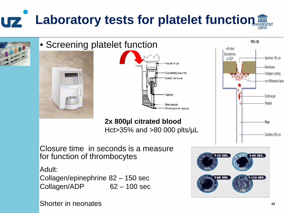
• Screening platelet function
Closure time in seconds is a measure for function of thrombocytes Adult: Collagen/epinephrine 82 – 150 sec Collagen/ADP 62 – 100 sec Shorter in neonates
Laboratory tests for platelet function
2x 800µl citrated blood Hct>35% and >80 000 plts/µL
44 44
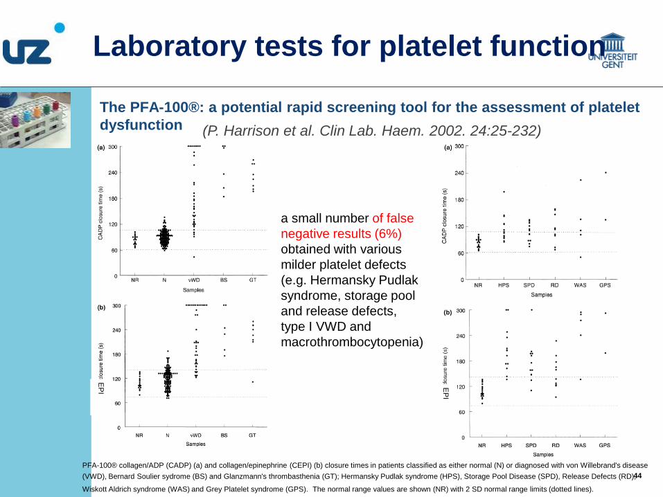
The PFA‐100®: a potential rapid screening tool for the assessment of platelet dysfunction
PFA-100® collagen/ADP (CADP) (a) and collagen/epinephrine (CEPI) (b) closure times in patients classified as either normal (N) or diagnosed with von Willebrand's disease (VWD), Bernard Soulier sydrome (BS) and Glanzmann's thrombasthenia (GT); Hermansky Pudlak syndrome (HPS), Storage Pool Disease (SPD), Release Defects (RD),
Wiskott Aldrich syndrome (WAS) and Grey Platelet syndrome (GPS). The normal range values are shown (NR) with 2 SD normal range limits (dotted lines).
(P. Harrison et al. Clin Lab. Haem. 2002. 24:25-232)
a small number of false negative results (6%) obtained with various milder platelet defects (e.g. Hermansky Pudlak syndrome, storage pool and release defects, type I VWD and macrothrombocytopenia)
EP
I
EP
I
Laboratory tests for platelet function
45 45
Interpretation with caution Diagnosis of bleeding disorder or thrombophilia based on
Clinical symptoms Family history Reproducible laboratory results
Interpretation of coagulation tests
46 46
Immature hemostatic system 3-6 months Differences between adults and children: physiological Age appropriate reference ranges Sample integrity, repeat testing Routine coagulation tests and additional testing An abnormal laboratory test result is not sufficient to define a disease A normal test result does not exclude a disease
Coagulation tests in children: Conclusions
47 47
THANK YOU FOR YOUR ATTENTION