Embed Size (px)
Citation preview

T O P I C 5 | T H E S K E L E T A L S Y S T E M | P a g e 1 | 26
College of Nursing
ANATOMY & PHYSIOLOGY THEORY
Course Module
1stSemester SY 2020 - 2021
Course : MC2
Course Title : Anatomy & Physiology Theory
Course Credits : 3 units (54 hours)
Contact Hours/week : 3 hours/ week
Course Description: This course deals with the basic concepts of Anatomy and Physiology,
introduction of anatomical and medical terms, clinical presentation of common diseases and
disorders. It also includes recognition of system changes that accompany old age health problems to
enhance understanding of students with minimal background in biological sciences pursuing the
nursing career. It also emphasizes a substantial amount of laboratory exposure that involves actual
laboratory work using anatomical models, visual aids, reference charts and prepared slides using
microscope visualization.
TOPIC 6: THE SKELETAL SYSTEM
Although the word skeleton comes from the Greek word meaning “dried-up body,” our internal
framework is beautifully formed and proportioned. Strong yet light, it is perfectly adapted for its
functions of protecting the body and allowing motion. No other animal has such long legs
(compared to the arms or forelimbs) or such a strange foot, and few have grasping hands with
opposable thumbs.
The bones of the skeleton are part of the skeletal system, which also includes joints, cartilages, and
ligaments (fibrous cords that bind the bones together at joints). The skeleton is divided into two parts:
the axial skeleton, the bones that form the longitudinal axis of the body, and the appendicular
skeleton, the bones of the limbs and girdles that attach them to the axial skeleton. Joints give these
parts of the skeleton flexibility and allow movement to occur.
LEARNING OBJECTIVES
Given relevant questions, the student is able to:
Know the structure of a typical bone and its microscopic features.
Develop the skill in identifying major bones of the body.
Determine how bones develop and grow.
Explain how skeletal muscles produce movement at joints and identify types of joint
movements.
Know some bone diseases
Relate changes effects of aging associated with the Skeletal system.
ACTIVATE NEW KNOWLEDGE
WHAT: The skeletal system is the internal frame of the body and includes bones, cartilages, and joints.
HOW: In addition to providing structure, bones articulate, or come together, at joints to allow body
movement.
WHY: The skeleton is essential for protecting organs, producing blood cells, storing essential minerals,
and anchoring skeletal muscles so that their contractions cause body movements.
ACQUIRE NEW KNOWLEDGE
FUNCTIONS OF THE BONES
• Support. Bones, the “steel girders” and “reinforced concrete” of the body, form the internal
framework that supports the body and cradles its soft organs. The bones of the legs act as pillars to
support the body trunk when we stand,and the rib cage supports the thoracic wall.
T O P I C 5 | T H E S K E L E T A L S Y S T E M | P a g e 2 | 26
• Protection. Bones protect soft body organs. For example, the fused bones of the skull provide a
snug enclosure for the brain, allowing someone to head a soccer ball without worrying about injuring
the brain. The vertebrae surround the spinal cord, and the rib cage helps protect the vital organs of
the thorax.
• Allow movement. Skeletal muscles, attached to bones by tendons, use the bones as levers to move
the body and its parts. As a result, we can breathe, walk, swim, and throw a ball. Before continuing,
take a moment to imagine that your bones have turned to putty. What if you were running when this
change took place? Now imagine your bones forming a rigid metal framework inside your body
(without joints). What problems could you envision with this arrangement? These images should help
you understand how well our skeletal system provides support and protection while allowing
movement.
• Storage. Fat is stored in the internal (marrow) cavities of bones. Bone itself serves as a storehouse for
minerals, the most important of which are calcium and phosphorus. Most of the body’s calcium is
deposited in the bones as calcium salts, but a small amount of calcium in its ion form (Ca2+) must be
present in the blood at all times for the nervous system to transmit messages, for muscles to contract,
and for blood to clot. Problems occur not only when there is too little calcium in the blood, but also
when there is too much. Hormones control the movement of calcium to and from the bones and
blood according to the needs of the body. Indeed, “deposits” and “withdrawals” of calcium (and
other minerals) to and from bones go on almost all the time.
• Blood cell formation. Blood cell formation, or hematopoiesis, occurs within the marrow cavities of
certain bones.
CLASSIFICATION OF BONES
The adult skeleton is composed of 206 bones. There are two basic types of osseous, or bone, tissue:
Compact bone is dense and looks smooth and homogeneous, whereas spongy bone has a spiky,
open appearance like a sponge.
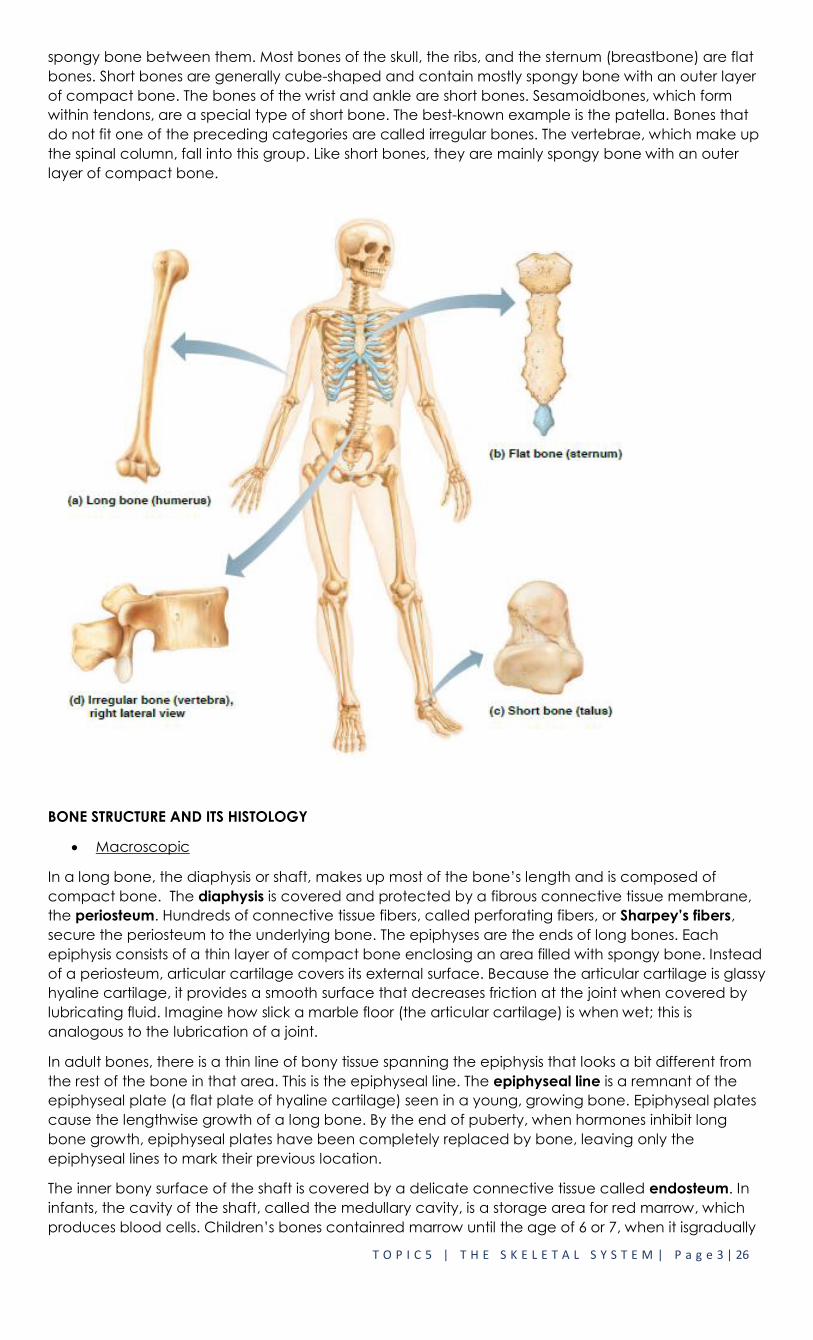
Additionally, bones come in many sizes and shapes. For example, the tiny pisiform bone of the wrist is
the size and shape of a pea, whereas the femur, or thigh bone, is nearly 2 feet long and has a large,
ball-shaped head. The unique shape of each bone fulfills a particular need. Bones are classified
according to shape into four groups: long, short, flat, and irregular. As their name suggests, long
bones are typically longer than they are wide. As a rule, they have a shaft with enlarged ends. Long
bones are mostly compact bone but also contain spongy bone at the ends. All the bones of the
limbs,except the patella (kneecap) and the wrist andankle bones are long bones.Flat bones are thin,
flattened, and usuallycurved. They have two thin layers of compactbone sandwiching a layer of
T O P I C 5 | T H E S K E L E T A L S Y S T E M | P a g e 3 | 26
spongy bone between them. Most bones of the skull, the ribs, and the sternum (breastbone) are flat
bones. Short bones are generally cube-shaped and contain mostly spongy bone with an outer layer
of compact bone. The bones of the wrist and ankle are short bones. Sesamoidbones, which form
within tendons, are a special type of short bone. The best-known example is the patella. Bones that
do not fit one of the preceding categories are called irregular bones. The vertebrae, which make up
the spinal column, fall into this group. Like short bones, they are mainly spongy bone with an outer
layer of compact bone.
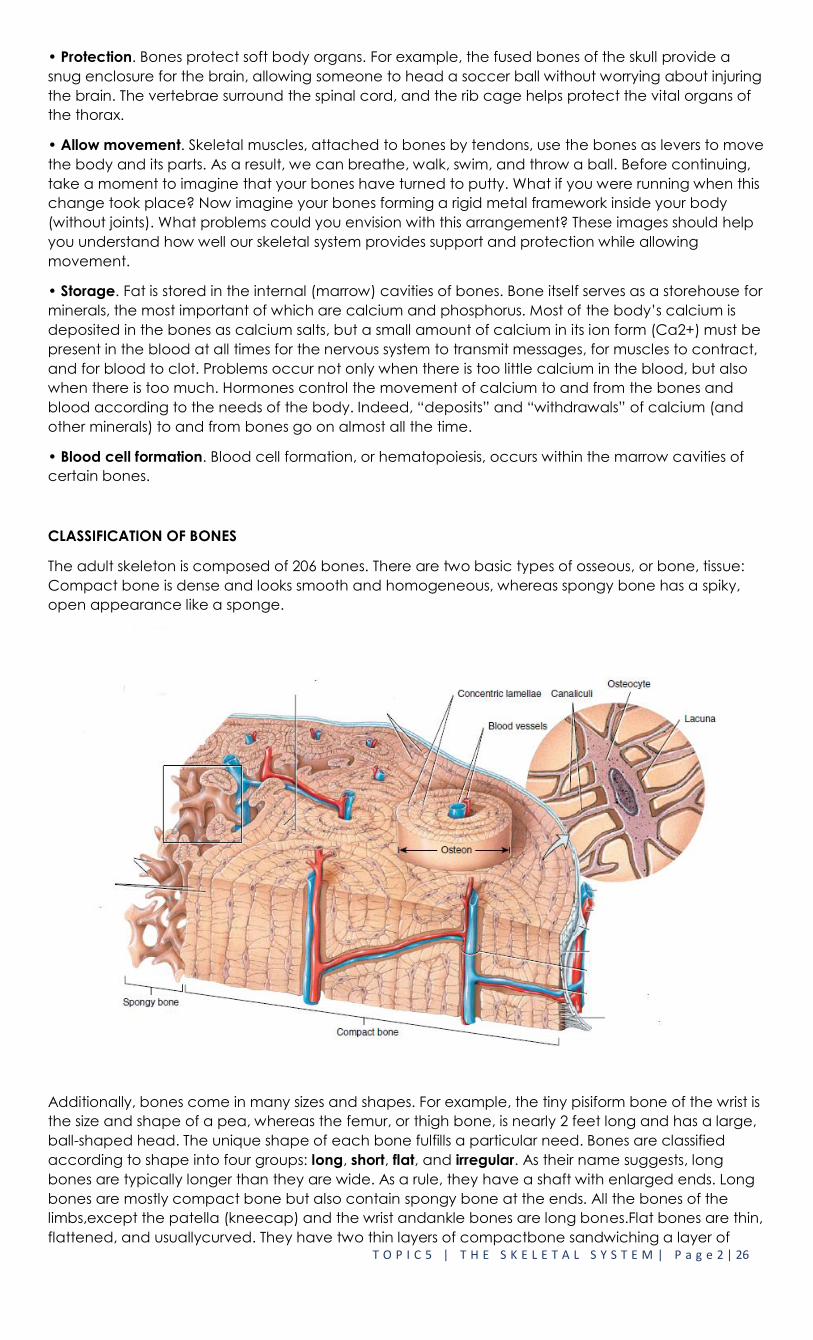
BONE STRUCTURE AND ITS HISTOLOGY
Macroscopic
In a long bone, the diaphysis or shaft, makes up most of the bone’s length and is composed of
compact bone. The diaphysis is covered and protected by a fibrous connective tissue membrane,
the periosteum. Hundreds of connective tissue fibers, called perforating fibers, or Sharpey’s fibers,
secure the periosteum to the underlying bone. The epiphyses are the ends of long bones. Each
epiphysis consists of a thin layer of compact bone enclosing an area filled with spongy bone. Instead
of a periosteum, articular cartilage covers its external surface. Because the articular cartilage is glassy
hyaline cartilage, it provides a smooth surface that decreases friction at the joint when covered by
lubricating fluid. Imagine how slick a marble floor (the articular cartilage) is when wet; this is
analogous to the lubrication of a joint.
In adult bones, there is a thin line of bony tissue spanning the epiphysis that looks a bit different from
the rest of the bone in that area. This is the epiphyseal line. The epiphyseal line is a remnant of the
epiphyseal plate (a flat plate of hyaline cartilage) seen in a young, growing bone. Epiphyseal plates
cause the lengthwise growth of a long bone. By the end of puberty, when hormones inhibit long
bone growth, epiphyseal plates have been completely replaced by bone, leaving only the
epiphyseal lines to mark their previous location.
The inner bony surface of the shaft is covered by a delicate connective tissue called endosteum. In
infants, the cavity of the shaft, called the medullary cavity, is a storage area for red marrow, which
produces blood cells. Children’s bones containred marrow until the age of 6 or 7, when it isgradually

T O P I C 5 | T H E S K E L E T A L S Y S T E M | P a g e 4 | 26
replaced by yellow marrow, which stores adipose (fat) tissue. In adult bones, red marrow is confined
to cavities in the spongy bone of the axial skeleton, the hip bones, and the epiphyses of long bones
such as the humerus and femur.
Microscopic
Under a microscope, you can see that spongy bone is composed of small needlelike pieces of
bone called trabeculae and lots of “open” space filled by marrow, blood vessels and nerves. In
compact bone, the mature bone cells, osteocytes, are found within the bone matrix in tiny
cavities called lacunae. The lacunae are arranged in concentric circles called lamellae around
central canals (also called Haversian canals). Each complex consisting of a central canal and
matrix rings is called an osteon, or Haversian system, and is the structural and functional unit of
compact bone.
Central canalsrun lengthwise through the bony matrix, carrying blood vessels and nerves to all
areas of the bone. Tiny canals, radiate outward from the central canals to all lacunae. The
canaliculi form a transportation system that connects all the bone cells to the nutrient supply and
waste removal services through the hard bone matrix. Because of this elaborate network
ofcanals, bone cells are well nourished in spite of the hardness of the matrix, and bone injuries
heal quickly. The communication pathway from the outside of the bone to its interior (and the
central canals) is completed by perforating canals (also called Volkmann’s canals), which run in
the compact bone at right angles to the shaft (diaphysis) and central canals.

T O P I C 5 | T H E S K E L E T A L S Y S T E M | P a g e 5 | 26
DEVELOPMENT AND GROWTH OF BONE
Ossification is theprocess by which bone forms or osteogenesis.
Bone formation occurs in four principal situations:
o The initial formation of bones in an embryo and fetus (Intramembranous and
Endochondral)
o The growth of bones during infancy, childhood, and adolescence until their adult sizes
are reached,
o The remodeling of bone (replacement of old bone by new bone tissue throughout life),
and
o The repair of fractures (breaks in bones) throughout life.
Intramembranous Ossification
Intramembranous ossification is the simpler of the two methods of bone formation. The flat bones of
the skull, most of the facial bones, mandible (lower jawbone), and the medial part of the clavicle
(collar bone) are formed in this way. Also, the “soft spots” that help the fetal skull pass through the
birth canal later harden as they undergo intramembranous ossification, which occurs as follows.
1. Development of the ossification center. At the site where the bone will develop, specific chemical
messages cause the cells of the mesenchyme to cluster together and differentiate, first into
osteoprogenitor cells and then into osteoblasts. The site of such a cluster is called an ossification
center. Osteoblasts secrete the organic extracellular matrix of bone until they are surrounded by it.
T O P I C 5 | T H E S K E L E T A L S Y S T E M | P a g e 6 | 26
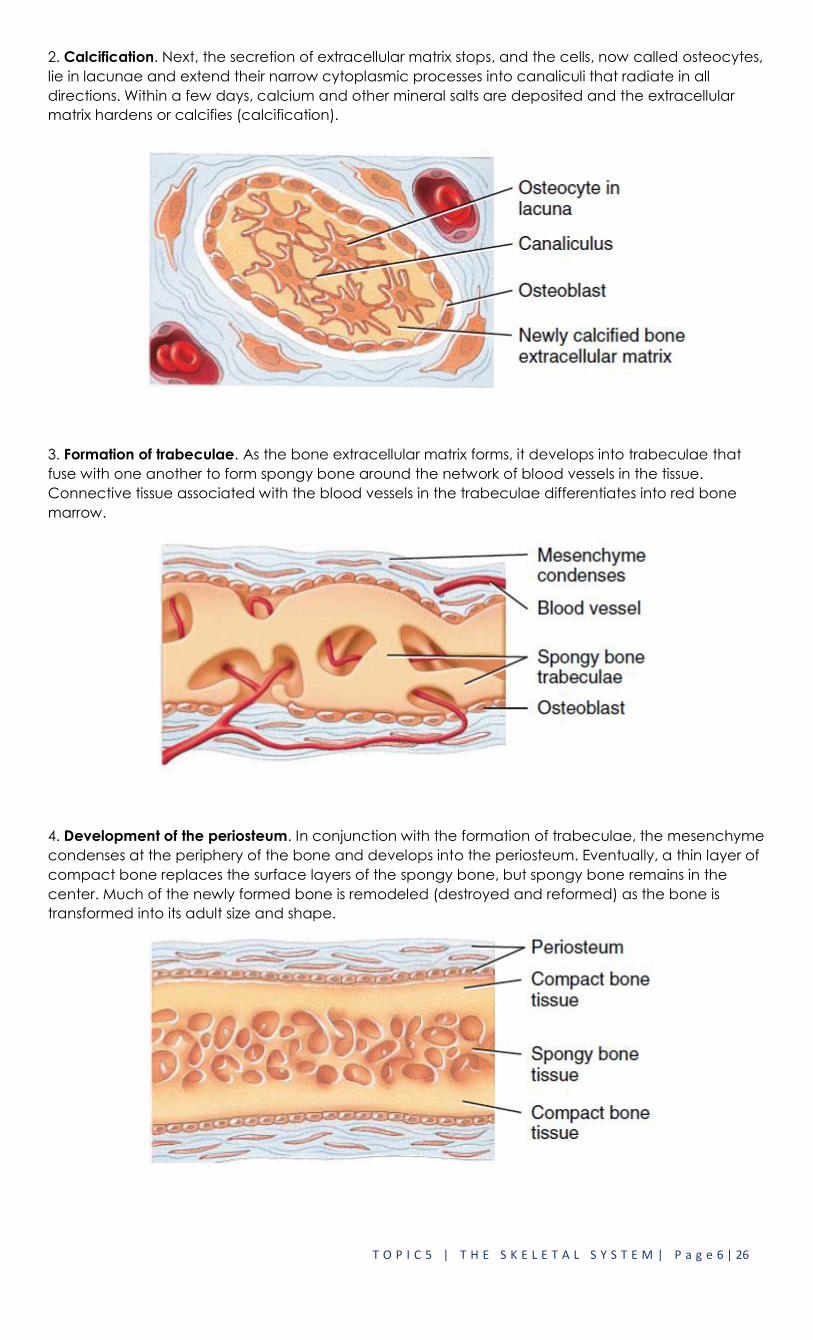
2. Calcification. Next, the secretion of extracellular matrix stops, and the cells, now called osteocytes,
lie in lacunae and extend their narrow cytoplasmic processes into canaliculi that radiate in all
directions. Within a few days, calcium and other mineral salts are deposited and the extracellular
matrix hardens or calcifies (calcification).
3. Formation of trabeculae. As the bone extracellular matrix forms, it develops into trabeculae that
fuse with one another to form spongy bone around the network of blood vessels in the tissue.
Connective tissue associated with the blood vessels in the trabeculae differentiates into red bone
marrow.
4. Development of the periosteum. In conjunction with the formation of trabeculae, the mesenchyme
condenses at the periphery of the bone and develops into the periosteum. Eventually, a thin layer of
compact bone replaces the surface layers of the spongy bone, but spongy bone remains in the
center. Much of the newly formed bone is remodeled (destroyed and reformed) as the bone is
transformed into its adult size and shape.
T O P I C 5 | T H E S K E L E T A L S Y S T E M | P a g e 7 | 26
Endochondral Ossification
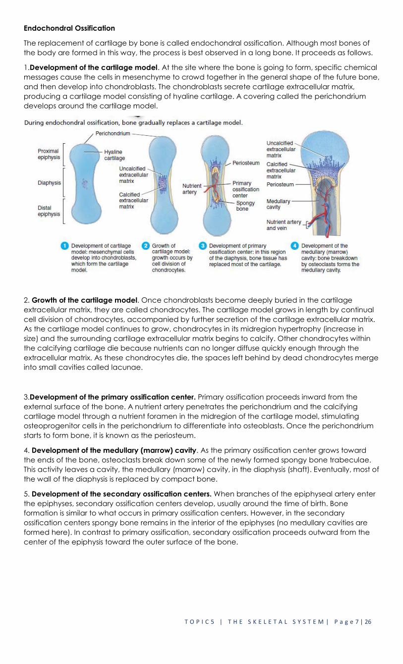
The replacement of cartilage by bone is called endochondral ossification. Although most bones of
the body are formed in this way, the process is best observed in a long bone. It proceeds as follows.
1.Development of the cartilage model. At the site where the bone is going to form, specific chemical
messages cause the cells in mesenchyme to crowd together in the general shape of the future bone,
and then develop into chondroblasts. The chondroblasts secrete cartilage extracellular matrix,
producing a cartilage model consisting of hyaline cartilage. A covering called the perichondrium
develops around the cartilage model.
2. Growth of the cartilage model. Once chondroblasts become deeply buried in the cartilage
extracellular matrix, they are called chondrocytes. The cartilage model grows in length by continual
cell division of chondrocytes, accompanied by further secretion of the cartilage extracellular matrix.
As the cartilage model continues to grow, chondrocytes in its midregion hypertrophy (increase in
size) and the surrounding cartilage extracellular matrix begins to calcify. Other chondrocytes within
the calcifying cartilage die because nutrients can no longer diffuse quickly enough through the
extracellular matrix. As these chondrocytes die, the spaces left behind by dead chondrocytes merge
into small cavities called lacunae.
3.Development of the primary ossification center. Primary ossification proceeds inward from the
external surface of the bone. A nutrient artery penetrates the perichondrium and the calcifying
cartilage model through a nutrient foramen in the midregion of the cartilage model, stimulating
osteoprogenitor cells in the perichondrium to differentiate into osteoblasts. Once the perichondrium
starts to form bone, it is known as the periosteum.
4. Development of the medullary (marrow) cavity. As the primary ossification center grows toward
the ends of the bone, osteoclasts break down some of the newly formed spongy bone trabeculae.
This activity leaves a cavity, the medullary (marrow) cavity, in the diaphysis (shaft). Eventually, most of
the wall of the diaphysis is replaced by compact bone.
5. Development of the secondary ossification centers. When branches of the epiphyseal artery enter
the epiphyses, secondary ossification centers develop, usually around the time of birth. Bone
formation is similar to what occurs in primary ossification centers. However, in the secondary
ossification centers spongy bone remains in the interior of the epiphyses (no medullary cavities are
formed here). In contrast to primary ossification, secondary ossification proceeds outward from the
center of the epiphysis toward the outer surface of the bone.

T O P I C 5 | T H E S K E L E T A L S Y S T E M | P a g e 8 | 26
6. Formation of articular cartilage and the epiphyseal (growth) plate. The hyaline cartilage that
covers the epiphyses becomes the articular cartilage. Prior to adulthood, hyaline cartilage remains
between the diaphysis and epiphysis as the epiphyseal (growth) plate, the region responsible for the
lengthwise growth of long bones that you will learn about next.
ORGANIZATIONAL STRUCTURE
AXIAL SKELETON AND ITS COMPONENTS
The skeleton is divided into two parts, the axial and appendicular skeletons. The axial skeleton forms
the longitudinal axis of the body. It can be divided into three parts- the skull, the vertebral column,
and the thoracic cage.
SKULL
The skull is formed by two sets of bones. The cranium encloses and protects the fragile brain tissue.
The facial bones form a cradle for the eyes that is open to the anterior and allow the facial muscles
to show our feelings through smiles or frowns. All but one of the bones of the skull arejoined together
by sutures, which are interlocking, immovable joints. Only the mandible (jawbone) is attached to the
rest of the skull by a freely movable joint.

T O P I C 5 | T H E S K E L E T A L S Y S T E M | P a g e 9 | 26
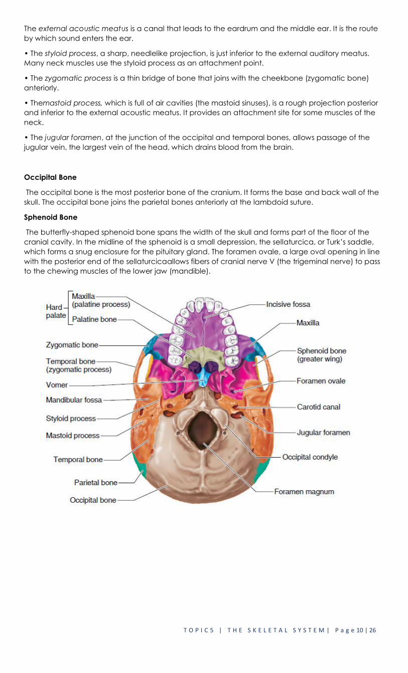
Cranium
The boxlike cranium is composed of eight large flat bones. Except for two sets of paired bones (the
parietal and temporal), they are all single bones.
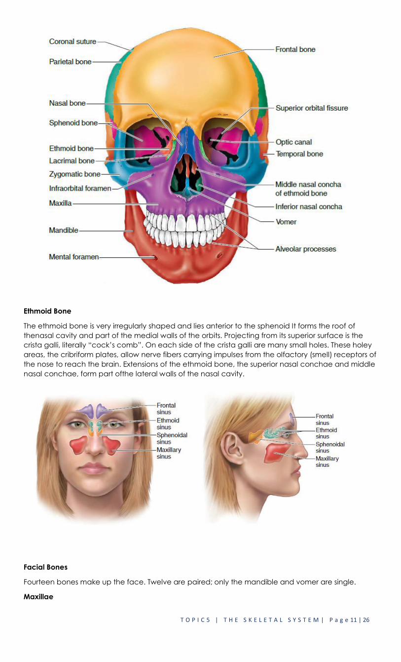
Frontal Bone
The frontal bone forms the forehead, the bony projections under the eyebrows, and the superior part
of each eye’s orbit.
Parietal Bones
The paired parietal bones form most of the superior and lateral walls of the cranium. The sagittal
suture is formed at the midline where the two parietal bones meet, and the coronal suture is formed
where the paired parietal bones meet the frontal bone.
Temporal Bones
The temporal bones lie inferior to the parietal bones and join them at the squamous sutures. Several
important bone markings appear on the temporal bones.
T O P I C 5 | T H E S K E L E T A L S Y S T E M | P a g e 10 | 26
The external acoustic meatus is a canal that leads to the eardrum and the middle ear. It is the route
by which sound enters the ear.
• The styloid process, a sharp, needlelike projection, is just inferior to the external auditory meatus.
Many neck muscles use the styloid process as an attachment point.
• The zygomatic process is a thin bridge of bone that joins with the cheekbone (zygomatic bone)
anteriorly.
• Themastoid process, which is full of air cavities (the mastoid sinuses), is a rough projection posterior
and inferior to the external acoustic meatus. It provides an attachment site for some muscles of the
neck.
• The jugular foramen, at the junction of the occipital and temporal bones, allows passage of the
jugular vein, the largest vein of the head, which drains blood from the brain.
Occipital Bone
The occipital bone is the most posterior bone of the cranium. It forms the base and back wall of the
skull. The occipital bone joins the parietal bones anteriorly at the lambdoid suture.
Sphenoid Bone
The butterfly-shaped sphenoid bone spans the width of the skull and forms part of the floor of the
cranial cavity. In the midline of the sphenoid is a small depression, the sellaturcica, or Turk’s saddle,
which forms a snug enclosure for the pituitary gland. The foramen ovale, a large oval opening in line
with the posterior end of the sellaturcicaallows fibers of cranial nerve V (the trigeminal nerve) to pass
to the chewing muscles of the lower jaw (mandible).
T O P I C 5 | T H E S K E L E T A L S Y S T E M | P a g e 11 | 26
Ethmoid Bone
The ethmoid bone is very irregularly shaped and lies anterior to the sphenoid It forms the roof of
thenasal cavity and part of the medial walls of the orbits. Projecting from its superior surface is the
crista galli, literally “cock’s comb”. On each side of the crista galli are many small holes. These holey
areas, the cribriform plates, allow nerve fibers carrying impulses from the olfactory (smell) receptors of
the nose to reach the brain. Extensions of the ethmoid bone, the superior nasal conchae and middle
nasal conchae, form part ofthe lateral walls of the nasal cavity.
Facial Bones
Fourteen bones make up the face. Twelve are paired; only the mandible and vomer are single.
Maxillae

T O P I C 5 | T H E S K E L E T A L S Y S T E M | P a g e 12 | 26
The two maxillae or maxillary bones,fuse to form the upper jaw. All facial bones except the mandible
join the maxillae; thus they are the main, or “keystone,” bones of the face. The maxillae carry the
upper teeth in the alveolar process.
Palatine Bones
The paired palatine bones lie posterior to the palatine processes of the maxillae.
Zygomatic Bones
The zygomatic bones are commonly referred to as the cheekbones. They also form a good-sized
portion of the lateral walls of the orbits.
Lacrimal Bones
The lacrimal bones are fingernail-sized bones forming part of the medialwall of each orbit. Each
lacrimal bone has a groove that serves as a passageway for tears.
Nasal Bones
The small rectangular bones forming the bridge of the nose are the nasal bones. (The lower part of
the skeleton of the nose is made up of hyaline cartilage.
Vomer Bone
The single bone in the median line of the nasal cavity is the vomer. (Vomer means “plow,” which
refers to the bone’s shape.) The vomer forms the inferior part of the bony nasal septum, which
separates the two nostrils.
Inferior Nasal Conchae
The inferior nasal conchae are thin, curved bones projecting medially from the lateral walls of the
nasal cavity. (As mentioned earlier, the superior and middle conchae are similar but are parts of the
ethmoid bone.)
Mandible
The mandible, or lower jaw, is the largest and strongest bone of the face. It joins the temporal bones
on each side of the face, forming the only freely movable joints in the skull. The horizontal part of the
mandible (the body) forms the chin. Two upright bars of bone (the rami) extend from the body to
connect the mandible with the temporal bone. The lower teeth lie in alveoli (sockets) in the alveolar
process at the superior edge of the mandibular body.
The Hyoid Bone
The hyoid bone is closely related to the mandible and temporal bones. The hyoid bone is unique in
that it is the only bone of the body that does not articulate (form a joint) with any other bone.
Instead, it is suspended in the midneck region about 2 cm (1 inch) above the larynx (voicebox),
where it is anchored by ligaments to the styloid processes of the temporal bones. Horseshoe-shaped,
with a body and two pairs of horns, the hyoid bone serves as a movable base for the tongue and as
an attachment point for neck muscles that raise and lower the larynx when we swallow and speak.
VERTEBRAL COLUMN (SPINE)
Serving as the axial support of the body, the vertebral column, or spine, extends from the skull, which
it supports, to the pelvis, where it transmits the weight of the body to the lower limbs. the spine is
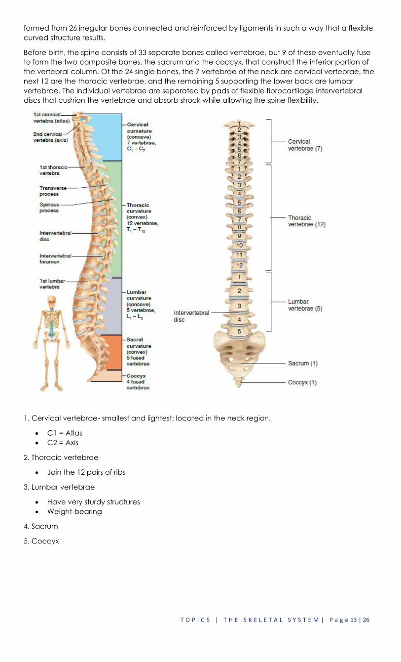
T O P I C 5 | T H E S K E L E T A L S Y S T E M | P a g e 13 | 26
formed from 26 irregular bones connected and reinforced by ligaments in such a way that a flexible,
curved structure results.
Before birth, the spine consists of 33 separate bones called vertebrae, but 9 of these eventually fuse
to form the two composite bones, the sacrum and the coccyx, that construct the inferior portion of
the vertebral column. Of the 24 single bones, the 7 vertebrae of the neck are cervical vertebrae, the
next 12 are the thoracic vertebrae, and the remaining 5 supporting the lower back are lumbar
vertebrae. The individual vertebrae are separated by pads of flexible fibrocartilage intervertebral
discs that cushion the vertebrae and absorb shock while allowing the spine flexibility.
1. Cervical vertebrae- smallest and lightest; located in the neck region.
C1 = Atlas
C2 = Axis
2. Thoracic vertebrae
Join the 12 pairs of ribs
3. Lumbar vertebrae
Have very sturdy structures
Weight-bearing
4. Sacrum
5. Coccyx

T O P I C 5 | T H E S K E L E T A L S Y S T E M | P a g e 14 | 26
Thoracic Vertebrae
The 12 thoracic vertebrae (T1 to T12) are all typical. They are larger than the cervical vertebrae and
are distinguished by the fact that they are the only vertebrae to articulate with the ribs. The body is
somewhat heart-shaped and has two costal facets (articulating surfaces) on each side, which
receive the heads of the ribs.
Lumbar Vertebrae
The five lumbar vertebrae (L1 to L5) have massive, blocklike bodies that are somewhat kidney bean–
shaped. Their short, hatchet-shaped spinous processes make them look like a moose head from the
lateral aspect. Because most of the stress on the vertebral column occurs in the lumbar region, these
are the sturdiest of the vertebrae.
Sacrum
The sacrum is formed by the fusion of five vertebrae. Superiorly it articulates with L5, and inferiorly it
connects with the coccyx.Each winglike ala articulates laterally with the hip bone, forming a
sacroiliac joint. The sacrum forms the posterior wall of the pelvis. Its posterior midline surface is
roughened by the median sacralcrest, the fused spinous processes of the sacralvertebrae. This is

T O P I C 5 | T H E S K E L E T A L S Y S T E M | P a g e 15 | 26
flanked laterally by the posterior sacral foramina. The vertebral canal continues inside the sacrum as
the sacral canal and terminates in a large inferior opening called the sacral hiatus.
Coccyx
The coccyx is formed from the fusion of three to five tiny, irregularly shaped vertebrae. It is the human
“tailbone,” a remnant of the tail that other vertebrate animals have.
THORACIC CAGE
The sternum, ribs, and thoracic vertebrae make up the bony thorax . The bony thorax is routinely
called the thoracic cage because it forms a protective cage of slender bones and cartilages around
the organs of the thoracic cavity (heart, lungs, and major blood vessels).
Sternum
The sternum (breastbone) is a typical flat bone and the result of the fusion of three bones—the
manubrium, body, and xiphoid process. It is attached directly to the first seven pairs of ribs via costal
cartilages. The sternum has three important bony landmarks— the jugular notch, the sternal angle,
and the xiphisternal joint.
• The jugular notch (concave upper border of the manubrium) can be palpated easily; generally it is
at the level of the third thoracicvertebra.
• The sternal angle results where the manubrium and body meet at a slight angle to each other, so
that a transverse ridge is formed at the level of the second ribs.
• The xiphisternal joint, the point where the sternal body and xiphoid process fuse, lies at the level of
the ninth thoracic vertebra.
Ribs
Twelve pairs of ribs form the walls of the bony thorax. All the ribs articulate with the vertebral column
posteriorly and then curve downward and toward the anterior body surface. The true ribs, the first
seven pairs, attach directly to the sternum by costal cartilages. False ribs, the next five pairs, either
attach indirectly to the sternum or are not attached to the sternum at all. The last two pairs of false
ribs lack the sternal attachments, so they are also called floating ribs.
The intercostal spaces (spaces between the ribs) are filled with the intercostal muscles, which aid in
breathing.

T O P I C 5 | T H E S K E L E T A L S Y S T E M | P a g e 16 | 26
APPENDICULAR SKELETON
Bones of the Shoulder Girdle
Each pectoral girdle, or shoulder girdle, consists of two bones—a clavicle and a scapula.Each
pectoral girdle, or shoulder girdle, consists of two bones—a clavicle and a scapula.
The paired clavicles, or collarbones, are slender, doubly curved bones. Each clavicle attaches to the
manubrium of the sternum medially (at its sternal end) and to the scapula laterally, where it helps to
form the shoulder joint.
The paired scapulae, or shoulder blades, are commonly called “wings” because they flare when we
move our arms posteriorly.

T O P I C 5 | T H E S K E L E T A L S Y S T E M | P a g e 17 | 26
Bones of the Upper Limbs
Arm
The arm is formed by a single bone, the humerus, which is a typical long bone. At its proximal end is a
rounded head that fits into the shallow glenoidcavity of the scapula. Immediately inferior to the head
is a slight constriction called the anatomical neck. Anterolateral to the head are two bony
projections separated by the intertubercularsulcus the greater tubercle and lesser tubercle,which
are sites of muscle attachment.
Forearm
Two bones, the radius and the ulna, form the skeleton of the forearm. When the body is in the
anatomical position, the radius is the lateral bone; that is, it is on the thumb side of the forearm.
When the upper limb is in the anatomical position, the ulna is the medial bone (on the little finger
side) of the forearm. On its proximal end are the anterior coronoid process and the posterior
olecranon, which are separated by the trochlear notch.
Hand
The skeleton of the hand consists of the carpals, the metacarpals, and the phalanges. The eight
carpal bones, arranged intwo irregular rows of four bones each, form the part of the hand called the
carpus, or the wrist.
The carpals are bound together by ligaments that restrict movements between them.The palm of the
hand consists of the metacarpals. The metacarpals are numbered 1 to 5 from the thumb side of the
hand toward the littlefinger.
The phalanges are the bones of the fingers. Each hand contains 14 phalanges. There are three in
each finger (proximal, middle, and distal), except in the thumb, which has only two (proximal and
distal).

T O P I C 5 | T H E S K E L E T A L S Y S T E M | P a g e 18 | 26
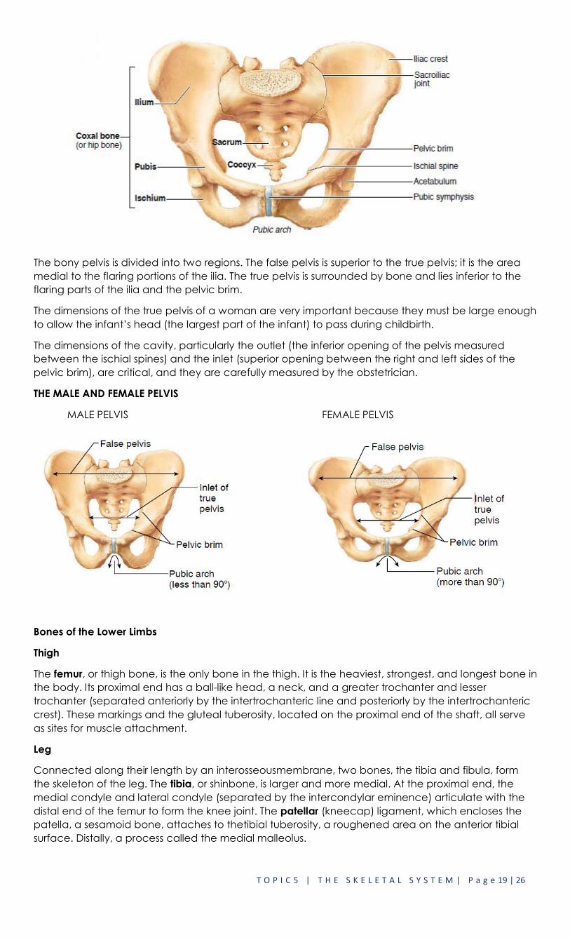
Bones of the Pelvic Girdle
The pelvic girdle is formed by two coxalbones, commonly called hip bones, and the sacrum.
Together with the coccyx, the pelvic girdle forms the pelvis. Note that the terms pelvic girdle (the
coxal bones and the sacrum) and pelvis (the coxal bones, sacrum, and coccyx) have slightly
different meanings.
Each hip bone is formed by the fusion of three bones: the ilium, ischium, and pubis. The ilium, which
connects posteriorly with the sacrum at the sacroiliac joint, is a large, flaring bone that forms most of
the hip bone.
The upper edge of an ala, the iliac crest, is an important anatomical landmark that is always kept in
mind by those who give intramuscular injections. The iliac crest ends anteriorly in the anterior superior
iliac spine and posteriorly in the posterior superior iliac spine.
The ischium is the “sit-down bone,” so called because it forms the most inferior part of the coxal
bone.
The pubis is the most anterior and inferior part of a coxal bone. Fusion of the rami of the pubis
anteriorly and the ischium posteriorly forms a bar of bone enclosing the obturator foramen, an
opening that allows blood vessels and nerves to pass into the anterior part of the thigh. The pubic
bones of each hip bone articulate anteriorly to form a cartilaginous joint, the pubic symphysis.
The ilium, ischium, and pubis fuse at the deep socket called the acetabulumwhich means “vinegar
cup.” The acetabulum receives the head of the thigh bone.
T O P I C 5 | T H E S K E L E T A L S Y S T E M | P a g e 19 | 26
The bony pelvis is divided into two regions. The false pelvis is superior to the true pelvis; it is the area
medial to the flaring portions of the ilia. The true pelvis is surrounded by bone and lies inferior to the
flaring parts of the ilia and the pelvic brim.
The dimensions of the true pelvis of a woman are very important because they must be large enough
to allow the infant’s head (the largest part of the infant) to pass during childbirth.
The dimensions of the cavity, particularly the outlet (the inferior opening of the pelvis measured
between the ischial spines) and the inlet (superior opening between the right and left sides of the
pelvic brim), are critical, and they are carefully measured by the obstetrician.
THE MALE AND FEMALE PELVIS
MALE PELVIS FEMALE PELVIS
Bones of the Lower Limbs
Thigh
The femur, or thigh bone, is the only bone in the thigh. It is the heaviest, strongest, and longest bone in
the body. Its proximal end has a ball-like head, a neck, and a greater trochanter and lesser
trochanter (separated anteriorly by the intertrochanteric line and posteriorly by the intertrochanteric
crest). These markings and the gluteal tuberosity, located on the proximal end of the shaft, all serve
as sites for muscle attachment.
Leg
Connected along their length by an interosseousmembrane, two bones, the tibia and fibula, form
the skeleton of the leg. The tibia, or shinbone, is larger and more medial. At the proximal end, the
medial condyle and lateral condyle (separated by the intercondylar eminence) articulate with the
distal end of the femur to form the knee joint. The patellar (kneecap) ligament, which encloses the
patella, a sesamoid bone, attaches to thetibial tuberosity, a roughened area on the anterior tibial
surface. Distally, a process called the medial malleolus.
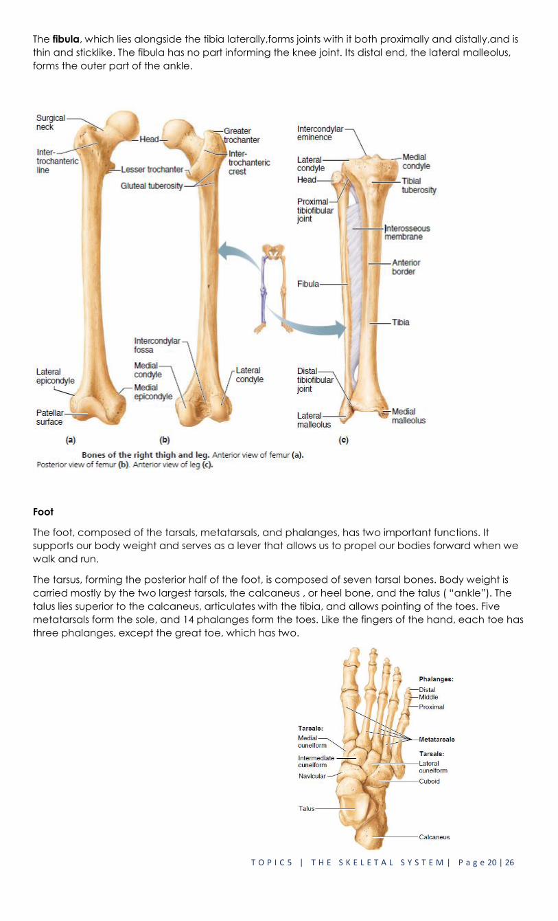
T O P I C 5 | T H E S K E L E T A L S Y S T E M | P a g e 20 | 26
The fibula, which lies alongside the tibia laterally,forms joints with it both proximally and distally,and is
thin and sticklike. The fibula has no part informing the knee joint. Its distal end, the lateral malleolus,
forms the outer part of the ankle.
Foot
The foot, composed of the tarsals, metatarsals, and phalanges, has two important functions. It
supports our body weight and serves as a lever that allows us to propel our bodies forward when we
walk and run.
The tarsus, forming the posterior half of the foot, is composed of seven tarsal bones. Body weight is
carried mostly by the two largest tarsals, the calcaneus , or heel bone, and the talus ( “ankle”). The
talus lies superior to the calcaneus, articulates with the tibia, and allows pointing of the toes. Five
metatarsals form the sole, and 14 phalanges form the toes. Like the fingers of the hand, each toe has
three phalanges, except the great toe, which has two.

T O P I C 5 | T H E S K E L E T A L S Y S T E M | P a g e 21 | 26
ARTICULAR SYSTEM (JOINTS)
Joints, also called articulations, are the sites where two or more bones meet. They have two
functions: They hold the bones together securely but also give the rigid skeleton mobility.
The bone-binding function of joints is just as important as their role in mobility. The immovable joints of
the skull, for instance, form a snug enclosure for the vital brain.
Joints are classified in two ways—functionally and structurally. The functional classification focuses on
the amount of movement the joint allows. On this basis, there are synarthroses, or immovable joints;
amphiarthroses, or slightly movablejoints; and diarthroses , or freely movable joints.
Structurally, there are fibrous, cartilaginous, and synovial joints. These classifications are based on
whether fibrous tissue, cartilage, or a joint cavity separates the bony regions at the joint.
3 Types of Fibrous Joints (Synarthroses)
Syndesmoses Sutures Gomphoses
>fibrous bands or ligaments
connect 2 bones
> Found only in the skull;
teethlike projections
>occur between the root of
tooth and alveolar process of
mandible or maxilla.
Cartilaginous Joints (Amphiarthroses)
1. Synchondroses
2. Symphyses
have hyaline cartilage between
articulating bones.
pad or disk of fibrocartilage connects 2
bones
Synovial joints
Synovial joints are joints in which the articulating bone ends are separated by a joint cavity
containing synovial fluid.

T O P I C 5 | T H E S K E L E T A L S Y S T E M | P a g e 22 | 26
Structures of Synovial Joint
Articular cartilage. Articular (hyaline) cartilagecovers the ends of the bones forming thejoint.
Articular capsule. The joint surfaces are enclosedby a sleeve, or layer, of fibrous
connectivetissue, which is lined with a smooth synovialmembrane (the reason these joints
arecalled synovial joints).
Joint cavity. The articular capsule encloses acavity, called the joint cavity, which
containslubricating synovial fluid secreted by the synovialmembrane.
Reinforcing ligaments. The fibrous layer ofthe capsule is usually reinforced with ligaments.
o Bursae (“purses”) are flattened fibrous sacs lined withsynovial membrane and
containing a thin film ofsynovial fluid. They are common where ligaments,muscles, skin,
tendons, or bones rub together.
Types of Joint Movements
The shapes of the articulating bone surfaces determine what movements are allowed at a joint.
Based on such shapes, our synovial joints can be classified as plane, hinge, pivot, condylar ,saddle, or
ball-and-socket joints.
In a plane joint the articular surfaces are essentially flat, and only short slipping or gliding movements
are allowed. The movements of plane joints are nonaxial; that is, gliding back and forth does not
involve rotation around any axis. The intercarpal joints of the wrist are the best examples of plane
joints.
• In a hinge joint the cylindrical end of one bone fits into a trough-shaped surface on another bone.
Angular movement is allowed in just one plane, like a door hinge. Examples are the elbow joint, ankle
joint, and the joints between the phalanges of the fingers. Hinge joints are classified as uniaxial they
allow movement around one axis only.
T O P I C 5 | T H E S K E L E T A L S Y S T E M | P a g e 23 | 26
• In a pivot jointthe rounded end of one bone fits into a sleeve or ring of bone (and possibly
ligaments). Because the rotating bone can turn only around its long axis,pivot joints are also uniaxial
joints. To understand the motion of this type of joint, think about a basketball player pivoting around
one foot that stays planted in one spot. The proxiproximalradioulnar joint and the joint between the
atlas and the dens of the axis are examples.
•In a condylar joint (“knucklelike”), the eggshapedarticular surface of one bone fits into an oval
concavity in another).Both of these articular surfaces are oval. Condylar joints allow the moving bone
to travel (1) from side to side and (2) back and forth, but the bone cannot rotate around its long axis.
•In saddle joints, each articular surface has both convex and concave areas, like a saddle for a
horse. These biaxial joints allow essentially the same movements as condylar joints. The best examples
of saddle joints are the carpometacarpal joints in the thumb, which are responsible for our
opposable thumbs.
T O P I C 5 | T H E S K E L E T A L S Y S T E M | P a g e 24 | 26
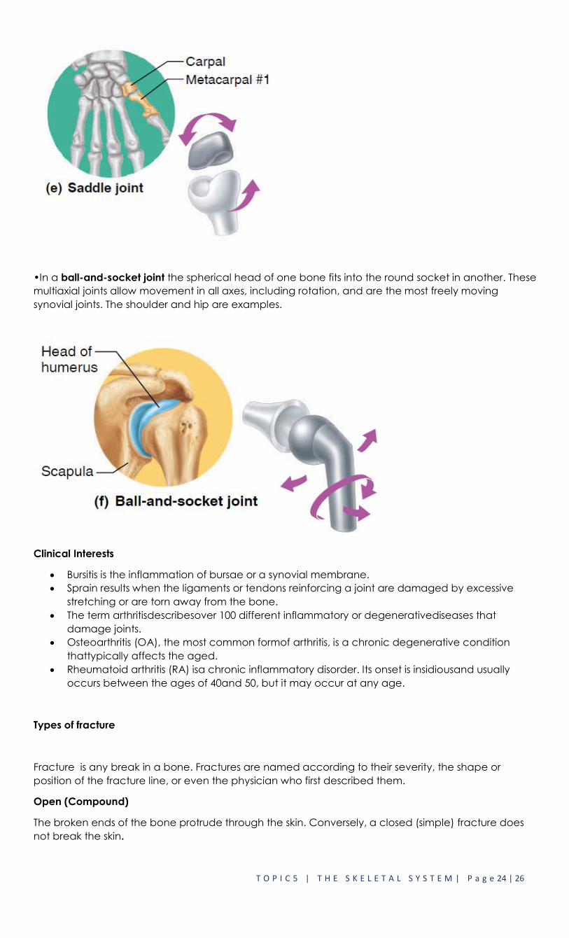
•In a ball-and-socket joint the spherical head of one bone fits into the round socket in another. These
multiaxial joints allow movement in all axes, including rotation, and are the most freely moving
synovial joints. The shoulder and hip are examples.
Clinical Interests
Bursitis is the inflammation of bursae or a synovial membrane.
Sprain results when the ligaments or tendons reinforcing a joint are damaged by excessive
stretching or are torn away from the bone.
The term arthritisdescribesover 100 different inflammatory or degenerativediseases that
damage joints.
Osteoarthritis (OA), the most common formof arthritis, is a chronic degenerative condition
thattypically affects the aged.
Rheumatoid arthritis (RA) isa chronic inflammatory disorder. Its onset is insidiousand usually
occurs between the ages of 40and 50, but it may occur at any age.
Types of fracture
Fracture is any break in a bone. Fractures are named according to their severity, the shape or
position of the fracture line, or even the physician who first described them.
Open (Compound)
The broken ends of the bone protrude through the skin. Conversely, a closed (simple) fracture does
not break the skin.

T O P I C 5 | T H E S K E L E T A L S Y S T E M | P a g e 25 | 26
Comminuted
The bone is splintered, crushed, or broken into pieces at the site of impact, and smaller bone
fragments lie between the two main fragments.
Greenstick
A partial fracture in which one side of the bone is broken and the other side bends; similar to the way
a green twig breaks on one side while the other side stays whole, but bends; occurs only in children,
whose bones are not fully ossified and contain more organic material than inorganic material.
Impacted
One end of the fractured bone is forcefully driven into the interior of the other.

T O P I C 5 | T H E S K E L E T A L S Y S T E M | P a g e 26 | 26
Pott
Fracture of the distal end of the lateral leg bone (fi bula), with serious injury of the distal
tibialarticulation.
Colles
Fracture of the distal end of the lateral forearm bone (radius) in which the distal fragment is
displaced posteriorly.