Embed Size (px)
Citation preview

Common Lesions and Conditions of the Oral Cavity
K. Mark Anderson DDS, MS
University of Tennessee College of Dentistry

Everyday Lumps and Bumps
Case #1
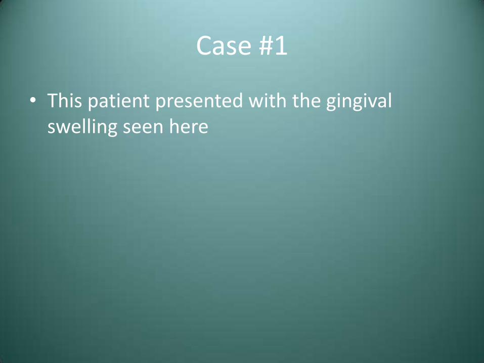
• This patient presented with the gingival swelling seen here
Case #1
Case #2
• A 14 year old female presented with this lesion of the gingiva

Case #2

Cases 1 and 2

Differential Diagnosis – “The 3 P’s”
• Pyogenic Granuloma
• Peripheral Ossifying Fibroma
• Peripheral Giant Cell Granuloma
Pyogenic Granuloma (Pregnancy Tumor)
• Common non-neoplastic proliferation of granulation tissue
• Not a true granuloma
• Response to local irritation or trauma

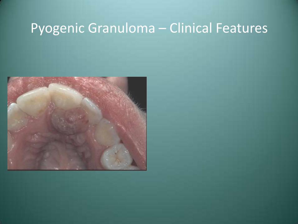
Pyogenic Granuloma – Clinical Features
• F>M, children and young adults
• Common during pregnancy

Pyogenic Granuloma – Clinical Features
• Rapidly growing, smooth or lobulated, ulcerated mass
• Easily bleeds
• Any mucosal surface, with most involving the gingiva

Pyogenic Granuloma – Clinical Features
• Rapidly growing, smooth or lobulated, ulcerated mass
• Easily bleeds
• Any mucosal surface, with most involving the gingiva
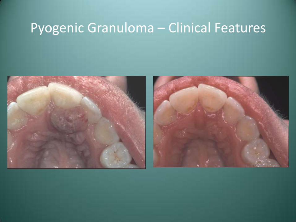
Pyogenic Granuloma – Clinical Features
Pyogenic Granuloma – Clinical Features

Pyogenic Granuloma – Treatment and Prognosis
• Conservative surgical excision with removal of any local factors
• Lesions associated with pregnancy may spontaneously regress postpartum
• Recurrences occur due to remaining local factors (calculus)

Peripheral Ossifying Fibroma
• Relatively common reactive lesion, probably arising from periodontal ligament
• This lesion is unrelated to the central ossifying fibroma

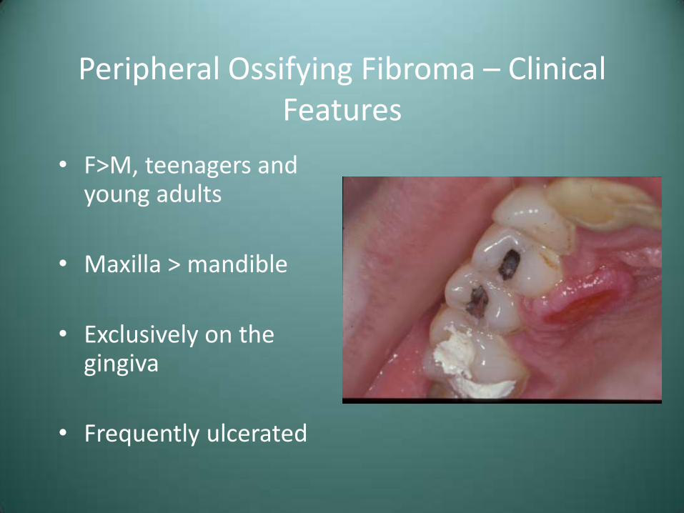
Peripheral Ossifying Fibroma – Clinical Features
• F>M, teenagers and young adults
• Maxilla > mandible
• Exclusively on the gingiva
• Frequently ulcerated

Peripheral Ossifying Fibroma – Clinical Features
• F>M, teenagers and young adults
• Maxilla > mandible
• Exclusively on the gingiva
• Frequently ulcerated
Peripheral Ossifying Fibroma – Clinical Features
• F>M, teenagers and young adults
• Maxilla > mandible
• Exclusively on the gingiva
• Frequently ulcerated

Peripheral Ossifying Fibroma – Treatment and Prognosis
• Local excision down to the periosteum
• Elimination of local factors or irritants
• Approximately 16% recurrence rate

Peripheral Giant Cell Granuloma
• Relatively common reactive lesion of the gingiva
• Histologically identical to the central giant cell granuloma
Peripheral Giant Cell Granuloma – Clinical Features
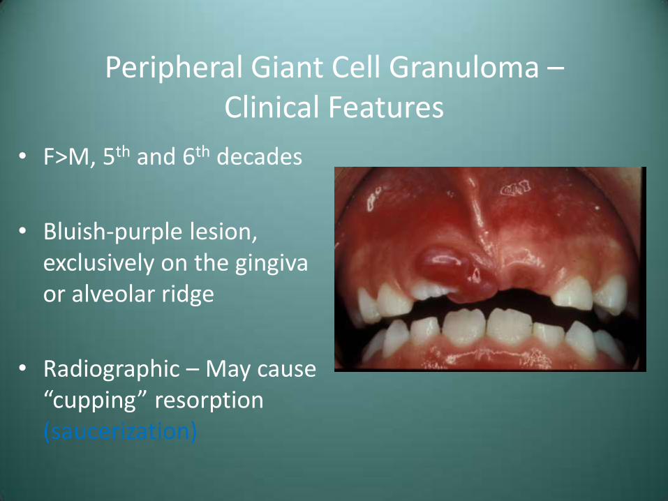
• F>M, 5th and 6th decades
• Bluish-purple lesion, exclusively on the gingiva or alveolar ridge
• Radiographic – May cause “cupping” resorption (saucerization)
Peripheral Giant Cell Granuloma – Clinical Features
• F>M, 5th and 6th decades
• Bluish-purple lesion, exclusively on the gingiva or alveolar ridge
• Radiographic – May cause “cupping” resorption (saucerization)

Peripheral Giant Cell Granuloma – Clinical Features
• F>M, 5th and 6th decades
• Bluish-purple lesion, exclusively on the gingiva or alveolar ridge
• Radiographic – May cause “cupping” resorption (saucerization)

Peripheral Giant Cell Granuloma – Clinical Features
• F>M, 5th and 6th decades
• Bluish-purple lesion, exclusively on the gingiva or alveolar ridge
• Radiographic – May cause “cupping” resorption (saucerization)
Peripheral Giant Cell Granuloma – Treatment and Prognosis
• Local excision down to underlying bone
• Removal of local factors
• Approximately 10% recurrence rate

Additional Considerations
Fibroma (Irritation Fibroma, Traumatic Fibroma)
• The most common tumor of the oral cavity
• Probably not a true neoplasm
• Reactive lesion, secondary to trauma or chronic irritation
Fibroma – Clinical Features
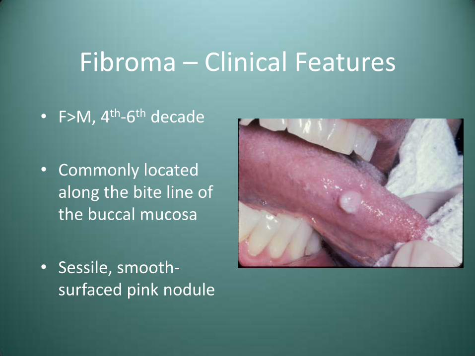
• F>M, 4th-6th decade
• Commonly located along the bite line of the buccal mucosa
• Sessile, smooth-surfaced pink nodule

Fibroma – Clinical Features
• F>M, 4th-6th decade
• Commonly located along the bite line of the buccal mucosa
• Sessile, smooth-surfaced pink nodule

Fibroma – Clinical Features
• F>M, 4th-6th decade
• Commonly located along the bite line of the buccal mucosa
• Sessile, smooth-surfaced pink nodule

Fibroma – Clinical Features
• F>M, 4th-6th decade
• Commonly located along the bite line of the buccal mucosa
• Sessile, smooth-surfaced pink nodule
Fibroma – Clinical Features
• F>M, 4th-6th decade
• Commonly located along the bite line of the buccal mucosa
• Sessile, smooth-surfaced pink nodule

Fibroma – Treatment
• Conservative surgical excision
• Prognosis – Recurrence is rare

Differential Diagnosis
• Pyogenic Granuloma
• Peripheral Ossifying Fibroma
• Peripheral Giant Cell Granuloma

Diagnosis Case #1 – Pyogenic Granuloma

Diagnosis Case #2 – Peripheral Ossifying Fibroma
Other Soft Tissue Considerations
Lipoma
• Benign tumor of fat
• Although rare in the oral/maxillofacial area, the lipoma is the most common mesenchymal neoplasm
• Unrelated to metabolism/body fat
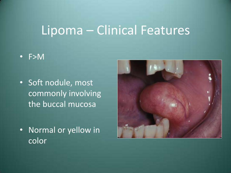
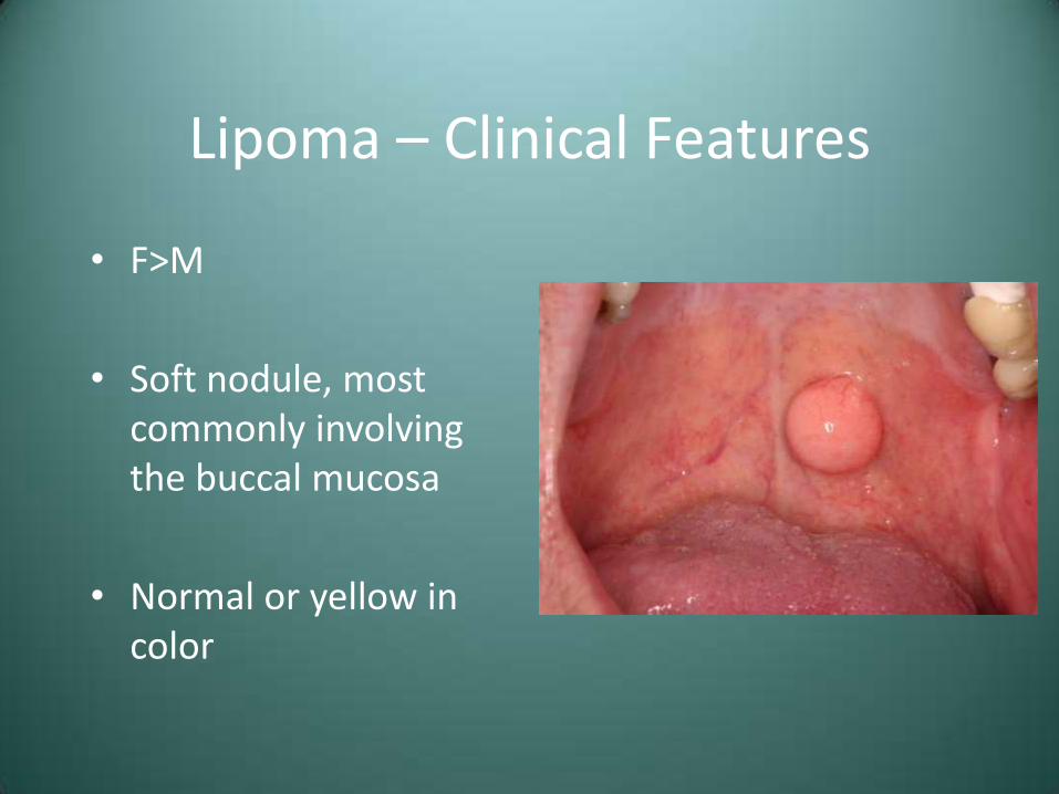
Lipoma – Clinical Features
• F>M
• Soft nodule, most commonly involving the buccal mucosa
• Normal or yellow in color
Lipoma – Clinical Features
• F>M
• Soft nodule, most commonly involving the buccal mucosa
• Normal or yellow in color

Lipoma – Clinical Features
• F>M
• Soft nodule, most commonly involving the buccal mucosa
• Normal or yellow in color
Lipoma – Clinical Features
• F>M
• Soft nodule, most commonly involving the buccal mucosa
• Normal or yellow in color

Lipoma – Clinical Features
• F>M
• Soft nodule, most commonly involving the buccal mucosa
• Normal or yellow in color

Lipoma – Treatment and Prognosis
• Conservative surgical excision
• Recurrence is rare
Granular Cell Tumor
• Uncommon tumor that appears to be of Schwann cell origin
• Significant predilection for the oral cavity
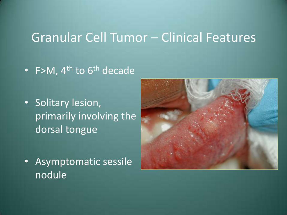
Granular Cell Tumor – Clinical Features
• F>M, 4th to 6th decade
• Solitary lesion, primarily involving the dorsal tongue
• Asymptomatic sessile nodule

Granular Cell Tumor – Clinical Features
• F>M, 4th to 6th decade
• Solitary lesion, primarily involving the dorsal tongue
• Asymptomatic sessile nodule
Granular Cell Tumor – Clinical Features
• F>M, 4th to 6th decade
• Solitary lesion, primarily involving the dorsal tongue
• Asymptomatic sessile nodule
Granular Cell Tumor – Treatment and Prognosis
• Conservative surgical excision
• Recurrence is rare, even with incomplete removal

Traumatic Neuroma
• Reactive proliferation of neural tissue
• Not necessarily a true neoplasm
• Secondary to disruption of Schwann cell tube

Traumatic Neuroma – Clinical Features
• F>M, middle-aged adults
• Smooth surfaced, submucosal nodule
• Commonly involve the mental foramen area
• May be symptomatic

Traumatic Neuroma – Clinical Features
• F>M, middle-aged adults
• Smooth surfaced, submucosal nodule
• Commonly involve the mental foramen area
• May be symptomatic

Traumatic Neuroma – Clinical Features
• F>M, middle-aged adults
• Smooth surfaced, submucosal nodule
• Commonly involve the mental foramen area
• May be symptomatic

Traumatic Neuroma – Clinical Features
• F>M, middle-aged adults
• Smooth surfaced, submucosal nodule
• Commonly involve the mental foramen area
• May be symptomatic

Traumatic Neuroma – Treatment and Prognosis
• Surgical excision, including a portion of the involved nerve bundle
• Recurrence is not expected

Schwannoma (Neurilomoma)
• Benign neural tumor of Schwann cell origin
• Uncommon, but often involve the head and neck

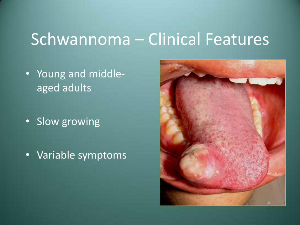
Schwannoma – Clinical Features
• Young and middle-aged adults
• Slow growing
• Variable symptoms
Schwannoma – Clinical Features
• Young and middle-aged adults
• Slow growing
• Variable symptoms

Schwannoma – Clinical Features
• Young and middle-aged adults
• Slow growing
• Variable symptoms
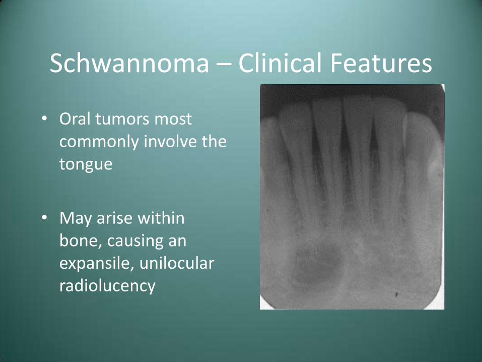
Schwannoma – Clinical Features
• Oral tumors most commonly involve the tongue
• May arise within bone, causing an expansile, unilocular radiolucency

Schwannoma – Clinical Features
Schwannoma – Treatment and Prognosis
• Surgical excision
• Recurrence is not expected
• Malignant transformation is rare
– Malignant peripheral nerve sheath tumor, malignant schwannoma, neurofibrosarcoma

Neurofibroma
• The most common peripheral nerve neoplasm
• Tumor cells are a mixture of Schwann cells and fibroblasts
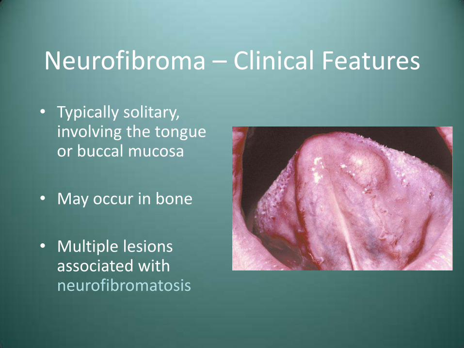
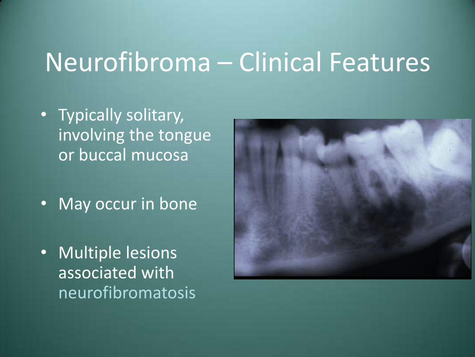
Neurofibroma – Clinical Features
• Typically solitary, involving the tongue or buccal mucosa
• May occur in bone
• Multiple lesions associated with neurofibromatosis
Neurofibroma – Clinical Features
• Typically solitary, involving the tongue or buccal mucosa
• May occur in bone
• Multiple lesions associated with neurofibromatosis

Neurofibroma – Clinical Features
• Typically solitary, involving the tongue or buccal mucosa
• May occur in bone
• Multiple lesions associated with neurofibromatosis

Neurofibroma – Clinical Features
• Typically solitary, involving the tongue or buccal mucosa
• May occur in bone
• Multiple lesions associated with neurofibromatosis

Neurofibroma – Clinical Features
• Typically solitary, involving the tongue or buccal mucosa
• May occur in bone
• Multiple lesions associated with neurofibromatosis
Neurofibroma – Clinical Features
• Typically solitary, involving the tongue or buccal mucosa
• May occur in bone
• Multiple lesions associated with neurofibromatosis

Neurofibroma – Treatment and Prognosis
• Solitary lesions – Surgical excision
• Multiple (neurofibromatosis) – Removal of symptomatic lesions
• Malignant transformation is possible, much more so
in patients with neurofibromatosis
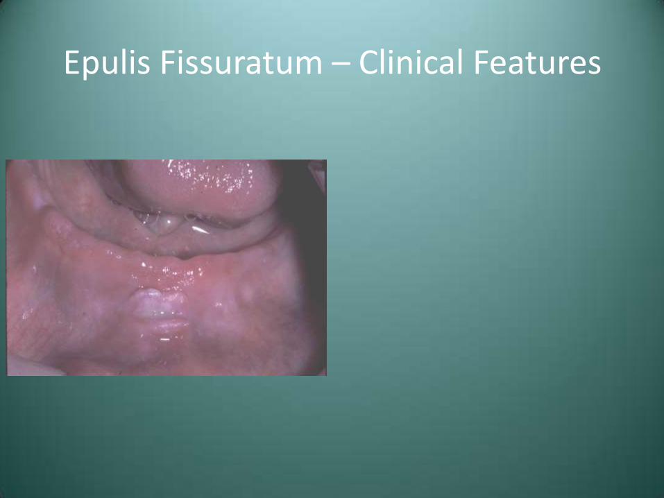
Epulis Fissuratum (Inflammatory Fibrous Hyperplasia, “Denture Epulis”)
• Reactive lesion that occurs secondary to irritation from an ill-fitting denture
• Epulis – Any tumor of the gingiva or alveolar mucosa
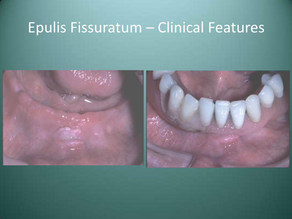
Epulis Fissuratum – Clinical Features
• F>M, middle aged and older
• Single or multiple folds of firm, fibrous tissue located in the alveolar vestibule (usually anterior)

Epulis Fissuratum – Clinical Features
• F>M, middle aged and older
• Single or multiple folds of firm, fibrous tissue located in the alveolar vestibule (usually anterior)
Epulis Fissuratum – Clinical Features
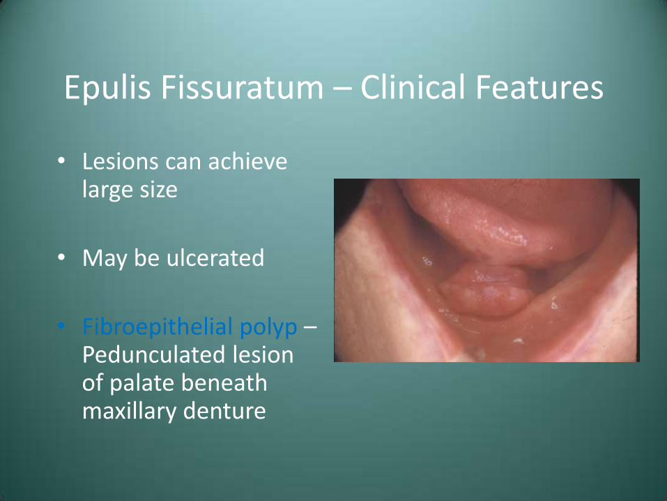
• Lesions can achieve large size
• May be ulcerated
• Fibroepithelial polyp – Pedunculated lesion of palate beneath maxillary denture
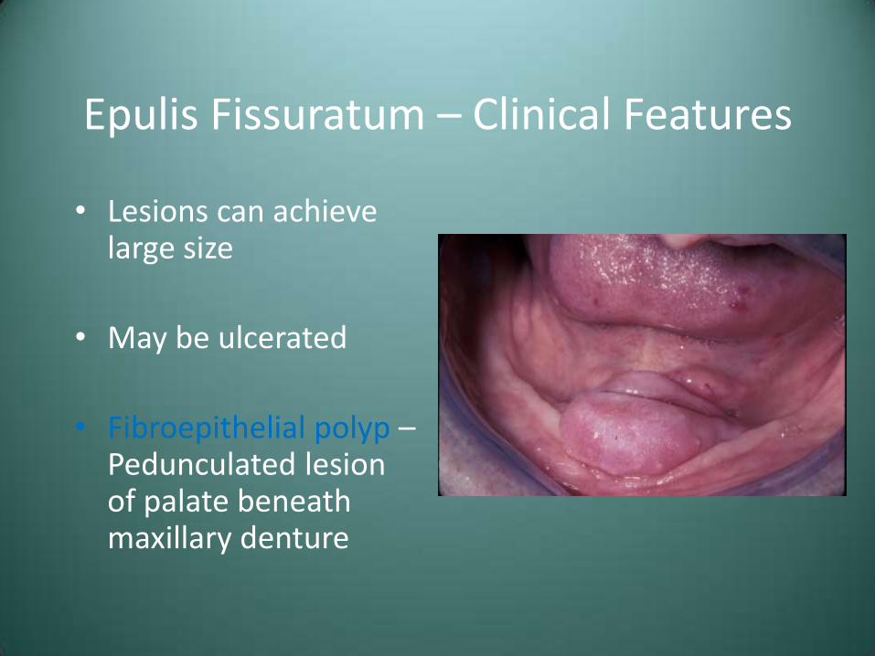
Epulis Fissuratum – Clinical Features
• Lesions can achieve large size
• May be ulcerated
• Fibroepithelial polyp – Pedunculated lesion of palate beneath maxillary denture

Epulis Fissuratum – Clinical Features
• Lesions can achieve large size
• May be ulcerated
• Fibroepithelial polyp – Pedunculated lesion of palate beneath maxillary denture
Epulis Fissuratum – Clinical Features
Epulis Fissuratum – Clinical Features
Epulis Fissuratum – Treatment and Prognosis
• Surgical removal
• Refabrication of the associated denture or relign
Erythematous Candidiasis - Denture Stomatitis
• Often referred to as “chronic atrophic candidiasis”
• Denture is often contaminated with candidal organisms, but no invasion of mucosa is seen
• Erythema of palatal denture-bearing area-typically asymptomatic
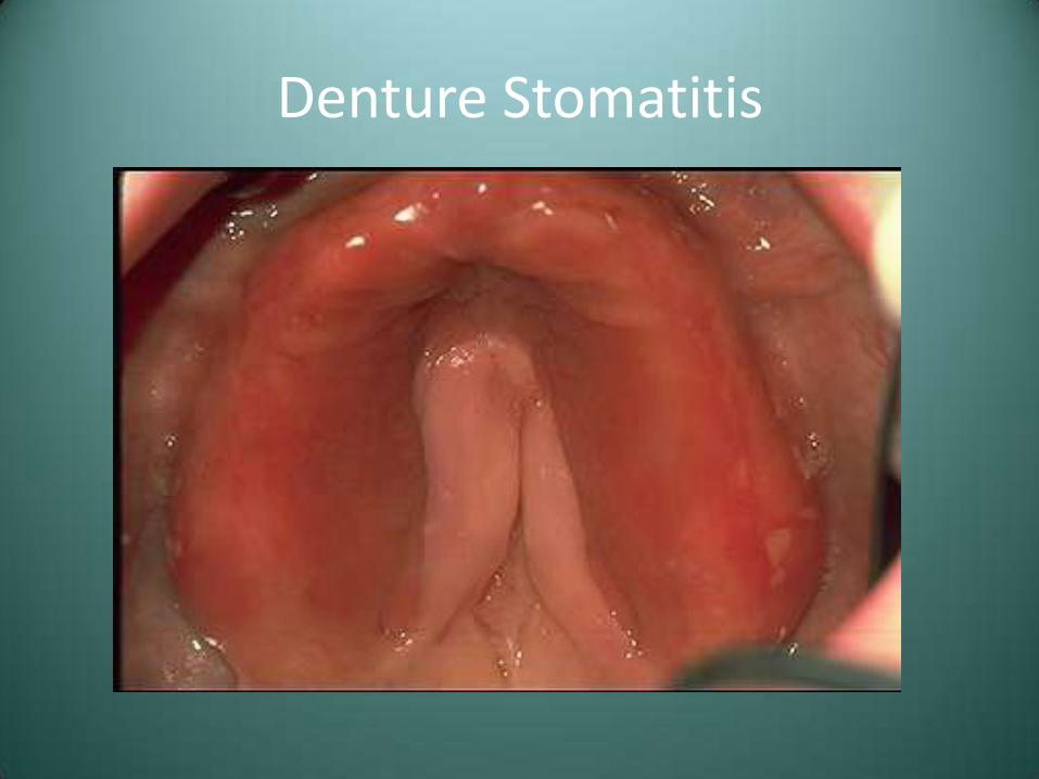
Denture Stomatitis
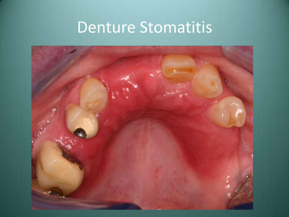
Denture Stomatitis

Inflammatory Papillary Hyperplasia
• Reactive process of the palate underneath a maxillary denture
• Variable involvement of the hard palate
• Asymptomatic, erythematous lesion with a pebbly surface
• Has been seen on edentulous mandibular ridge or on epulis
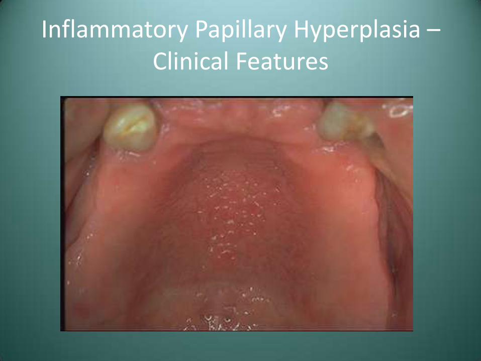
Inflammatory Papillary Hyperplasia – Clinical Features
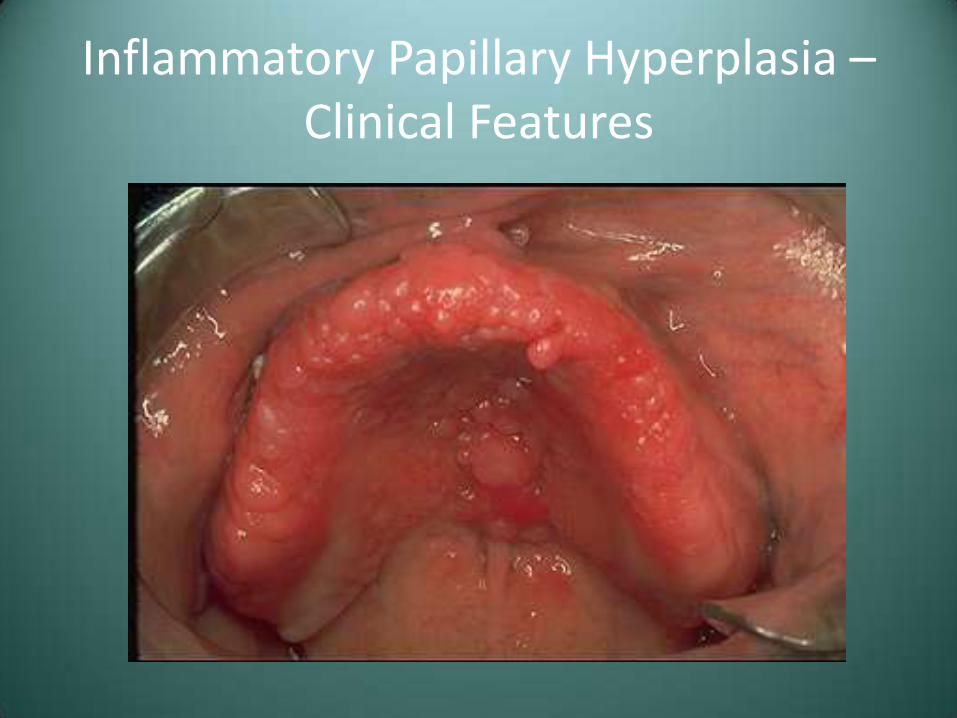
Inflammatory Papillary Hyperplasia – Clinical Features
Oral Squamous Papilloma
• Probably caused by human papillomavirus (HPV)
– Over 100 HPV types identified
– Types 6 and 11 are most commonly associated with oral papillomas
Squamous Papilloma – Clinical Features
• Any site, with the tongue and soft palate most frequently involved
• Typically solitary
• Usually pedunculated
• Variable color

Squamous Papilloma – Clinical Features
• Any site, with the tongue and soft palate most frequently involved
• Typically solitary
• Usually pedunculated
• Variable color

Squamous Papilloma – Clinical Features
• Any site, with the tongue and soft palate most frequently involved
• Typically solitary
• Usually pedunculated
• Variable color

Squamous Papilloma – Clinical Features
• Any site, with the tongue and soft palate most frequently involved
• Typically solitary
• Usually pedunculated
• Variable color

Squamous Papilloma – Clinical Features
• Any site, with the tongue and soft palate most frequently involved
• Typically solitary
• Usually pedunculated
• Variable color

Squamous Papilloma – Clinical Features
• Any site, with the tongue and soft palate most frequently involved
• Typically solitary
• Usually pedunculated
• Variable color

Squamous Papilloma - Treatment
• Surgical excision
• Recurrence is not expected, although lesions of the larynx may behave differently
– Laryngeal papillomatosis

Verruca Vulgaris (Common Wart)
• Typically a benign skin lesion induced by HPV types 2,4, 6, and 40
• Relatively contagious, with potential for autoinoculation
Verruca Vulgaris – Clinical Features
• Most commonly in children
• Skin of hands
• More commonly sessile
• Variable color

Verruca Vulgaris – Clinical Features
• Most commonly in children
• Skin of hands
• More commonly sessile
• Variable color
Verruca Vulgaris – Clinical Features
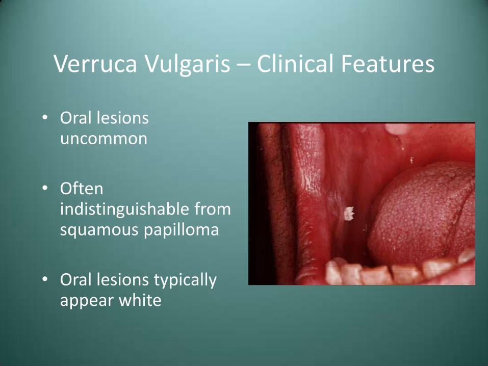
• Oral lesions uncommon
• Often indistinguishable from squamous papilloma
• Oral lesions typically appear white

Verruca Vulgaris - Treatment
• Surgical excision or curettage
• Liquid nitrogen, cryotherapy, or keratinolytic agents
• May spontaneously resolve
• Small rate of recurrence
Condyloma Acuminatum
• Also known as “venereal warts”
• Caused by several strains of HPV, including types 2, 6,11,16,18

Condyloma Acuminatum – Clinical Features
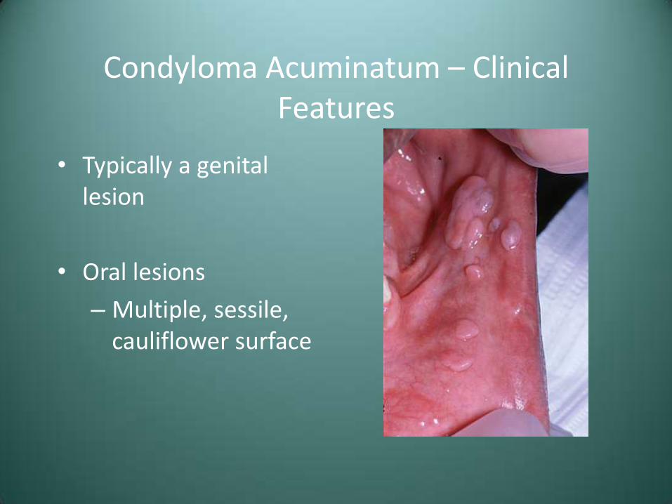
• Typically a genital lesion
• Oral lesions
– Multiple, sessile, cauliflower surface
Condyloma Acuminatum – Clinical Features
• Typically a genital lesion
• Oral lesions
– Multiple, sessile, cauliflower surface

Condyloma Acuminatum – Clinical Features
• Typically a genital lesion
• Oral lesions
– Multiple, sessile, cauliflower surface

Condyloma Acuminatum – Clinical Features
• Typically a genital lesion
• Oral lesions
– Multiple, sessile, cauliflower surface
Condyloma Acuminatum
• Excision, cryotherapy, laser excision
• Recurrence is common-30% of patients have recurrent lesions after each treatment episode
• Associated with squamous cell carcinoma of the uterine cervix

Ulcerative Conditions of the Oral Regions

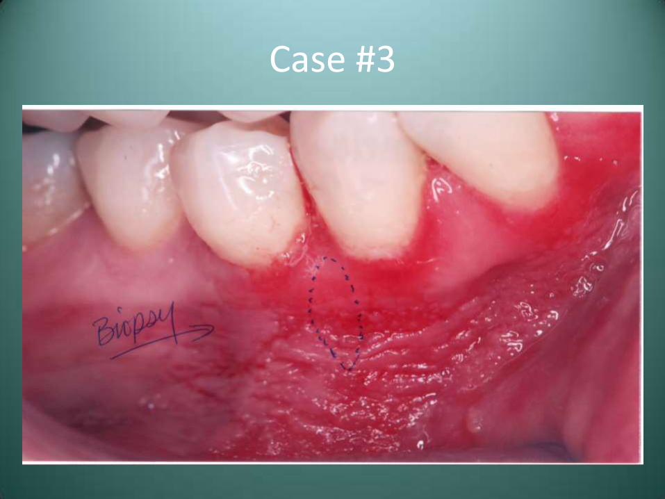
Case #3
• A 47 year old female presented with a history of these painful lesions

Case #3
Case #3

Case #3
• Clinical Diagnosis – “Desquamative Gingivitis”
• Differential Diagnosis
– Lichen Planus
– Cicatricial Pemphigoid
– Pemphigus Vulgaris
Lichen Planus
• Common chronic mucocutaneous disease
• Probably immune-mediated
• May have only skin, only oral, or both
Lichen Planus – Clinical Features
• F>M, Adults
• Skin lesions-purple, polygonal, pruritic papules

Lichen Planus – Clinical Features
• F>M, Adults
• Skin lesions-purple, polygonal, pruritic papules

Lichen Planus – Clinical Features
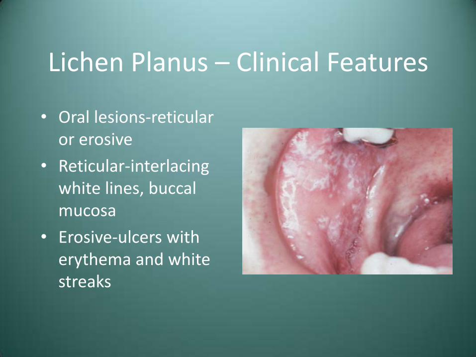
• Oral lesions-reticular or erosive
• Reticular-interlacing white lines, buccal mucosa
• Erosive-ulcers with erythema and white streaks
Lichen Planus – Clinical Features
• Oral lesions-reticular or erosive
• Reticular-interlacing white lines, buccal mucosa
• Erosive-ulcers with erythema and white streaks

Lichen Planus – Clinical Features
• Oral lesions-reticular or erosive
• Reticular-interlacing white lines, buccal mucosa
• Erosive-ulcers with erythema and white streaks

Lichen Planus – Clinical Features
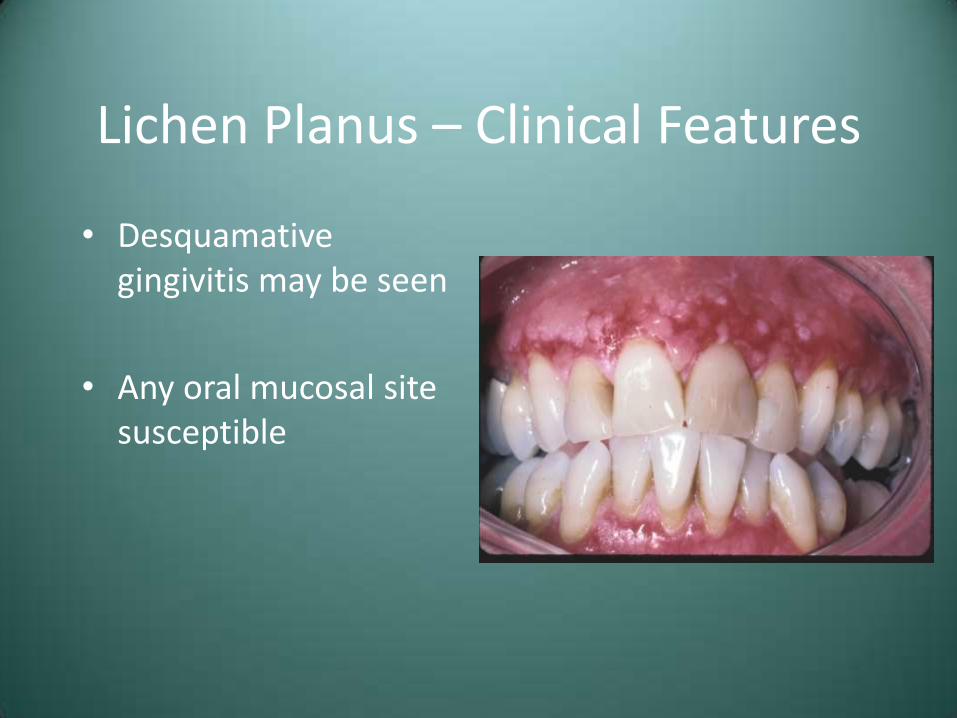
• Desquamative gingivitis may be seen
• Any oral mucosal site susceptible
Lichen Planus – Clinical Features
• Desquamative gingivitis may be seen
• Any oral mucosal site susceptible
Lichen Planus -Treatment
• 25% have superimposed candidiasis, so anti-fungal Tx may be necessary
• No treatment for reticular
• Topical corticosteroids for erosive
– Betemethasone Gel or Temovate (clobetasol) Gel
Lichen Planus -Prognosis
• Skin lesions may resolve spontaneously
• Oral lesions persist
• Malignant potential is controversial
• If premalignant, risk of transformation is probably small
Cicatricial Pemphigoid (Mucous Membrane Pemphigoid)
• Group of autoimmune disease characterized by antibodies directed against one or more components of the basement membrane
• Clinically resembles pemphigus due to blister formation
• About 2x more common than pemphigus
Cicatricial Pemphigoid – Clinical Features
• F>M, Avg. age 60
• Desquamative gingivitis
• May see intact blisters intraorally

Cicatricial Pemphigoid – Clinical Features
• F>M, Avg. age 60
• Desquamative gingivitis
• May see intact blisters intraorally

Cicatricial Pemphigoid – Clinical Features
• F>M, Avg. age 60
• Desquamative gingivitis
• May see intact blisters intraorally

Cicatricial Pemphigoid – Clinical Features
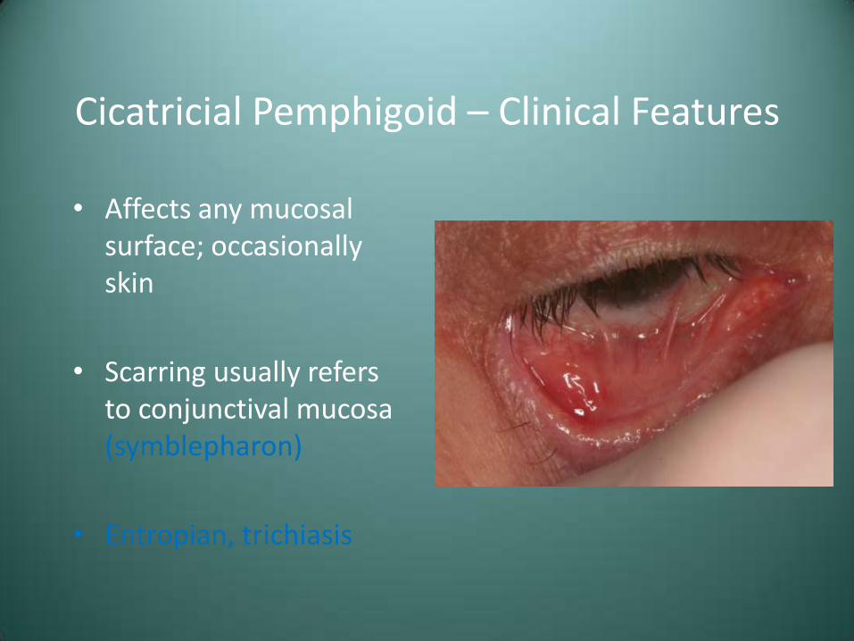
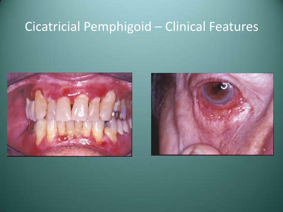
• Affects any mucosal surface; occasionally skin
• Scarring usually refers to conjunctival mucosa (symblepharon)
• Entropian, trichiasis
Cicatricial Pemphigoid – Clinical Features
• Affects any mucosal surface; occasionally skin
• Scarring usually refers to conjunctival mucosa (symblepharon)
• Entropian, trichiasis

Cicatricial Pemphigoid – Clinical Features
Cicatricial Pemphigoid – Clinical Features
Pemphigoid-Treatment
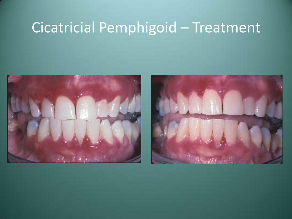
• Depends on extent of involvement
• Oral only-topical corticosteroids or dapsone
• Ocular lesions require systemic immunosuppressive therapy or human immunoglobulin therapy

Cicatricial Pemphigoid – Treatment
Cicatricial Pemphigoid – Treatment

Pemphigoid-Prognosis
• Rarely fatal
• Blindness results with untreated ocular disease
• Condition can usually be controlled
• Rarely undergoes spontaneous resolution

Pemphigus (Pemphigus Vulgaris)
• Autoimmune disorder characterized by antibodies directed against components of the epithelial desmosome complex
• Oral signs are often the first manifestations of the disease and the most difficult to resolve

Pemphigus-Clinical Features
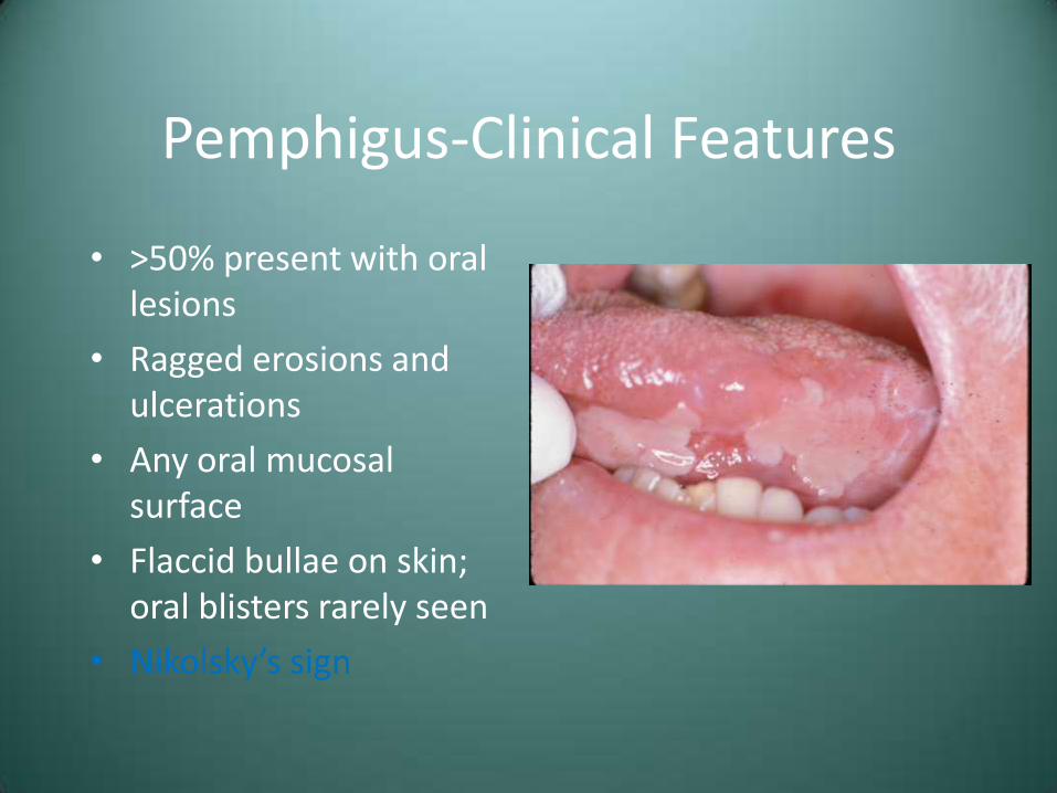
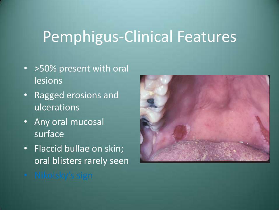
• >50% present with oral lesions
• Ragged erosions and ulcerations
• Any oral mucosal surface
• Flaccid bullae on skin; oral blisters rarely seen
• Nikolsky’s sign

Pemphigus-Clinical Features
• >50% present with oral lesions
• Ragged erosions and ulcerations
• Any oral mucosal surface
• Flaccid bullae on skin; oral blisters rarely seen
• Nikolsky’s sign
Pemphigus-Clinical Features
• >50% present with oral lesions
• Ragged erosions and ulcerations
• Any oral mucosal surface
• Flaccid bullae on skin; oral blisters rarely seen
• Nikolsky’s sign
Pemphigus-Clinical Features
• >50% present with oral lesions
• Ragged erosions and ulcerations
• Any oral mucosal surface
• Flaccid bullae on skin; oral blisters rarely seen
• Nikolsky’s sign

Pemphigus - Treatment and Prognosis
• Systemic corticosteroids, often with azathioprine
• Prior to corticosteroid therapy, 60-80% mortality
• Today, 5-10% mortality

Case #3
• Clinical Diagnosis – “Desquamative Gingivitis”
• Differential Diagnosis
– Lichen Planus
– Cicatricial Pemphigoid
– Pemphigus Vulgaris

Diagnosis Case #3 – Pemphigus Vulgaris
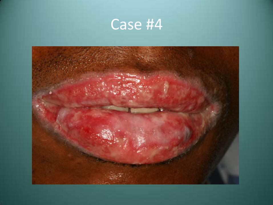
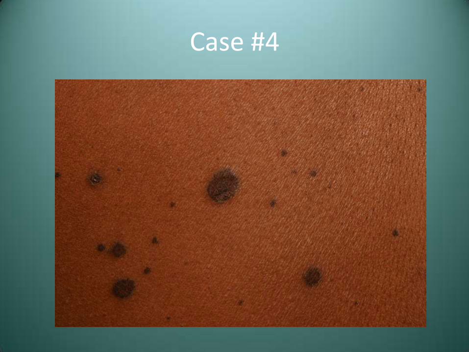
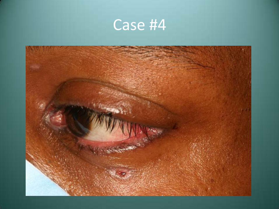
Case #4
• A 42 year old male presented with the lesions seen here as well as genital lesions
Case #4

Case #4
Case #4
Case #4

Case #4 – Differential Diagnosis
• Erythema Multiforme
• Paraneoplastic Pemphigus
Erythema Multiforme (EM)
• Acute, self-limiting ulcerative disorder
• Probably immune-mediated
• 50%-unknown; 25%-drugs (particularly antibiotics or analgesics); 25%-infection (herpes/Mycoplasma)

EM - Spectrum of Clinical Disease
• Erythema multiforme minor - skin and/or mucosa only
• Erythema multiforme major (Stevens-Johnson syndrome)
– At least two mucosal sites plus skin involvement
• Toxic epidermal necrolysis (Lyell’s disease)
EM-Clinical Features
• M>F
• Young adults
• May experience prodrome

EM-Clinical Features
• M>F
• Young adults
• May experience prodrome
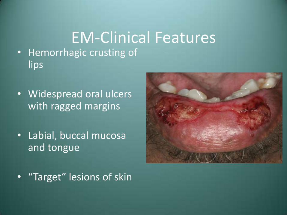
EM-Clinical Features • Hemorrhagic crusting of
lips
• Widespread oral ulcers with ragged margins
• Labial, buccal mucosa and tongue
• “Target” lesions of skin

EM-Clinical Features
• Outbreak typically clears in 2-6 weeks
• Often recurs in spring and fall
EM-Treatment
Supportive or topical corticosteroids for mild cases
Systemic corticosteroids for EM major
TEN managed in burn unit, possibly with pooled immunoglobulin

EM Prognosis
• Good for mild to moderate cases
• EM major-2-10% mortality
• TEN-34% mortality
Paraneoplastic Pemphigus • Serious vesiculobullous disorder affecting patients
with neoplastic disease, typically a lymphoreticular malignancy (CLL and lymphoma)
• Antibodies in response to the tumor probably cross react with components of the epithelial layer
• Cytotoxic T lymphocytes may also play a role in cutaneous and mucosal damage

Paraneoplastic Pemphigus – Clinical Features
• Clinically resembles a number of conditions
– Erythema multiforme
– Pemphigus
– Lichen planus
– Pemphigoid

Paraneoplastic Pemphigus – Clinical Features
• Clinically resembles a number of conditions
– Erythema multiforme
– Pemphigus
– Lichen planus
– Pemphigoid

Paraneoplastic Pemphigus – Clinical Features
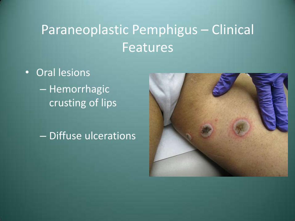
• Oral lesions
– Hemorrhagic crusting of lips
– Diffuse ulcerations

Paraneoplastic Pemphigus – Clinical Features
• Oral lesions
– Hemorrhagic crusting of lips
– Diffuse ulcerations

Paraneoplastic Pemphigus – Clinical Features
• Oral lesions
– Hemorrhagic crusting of lips
– Diffuse ulcerations
Paraneoplastic Pemphigus – Clinical Features
• Oral lesions
– Hemorrhagic crusting of lips
– Diffuse ulcerations

Paraneoplastic Pemphigus – Treatment and Prognosis
• Systemic corticosteroids plus azathioprine
• Topical corticosteroids
• Generally poor prognosis, high mortality due to sepsis or malignant progression

Case #4 – Differential Diagnosis
• Erythema Multiforme
• Paraneoplastic Pemphigus
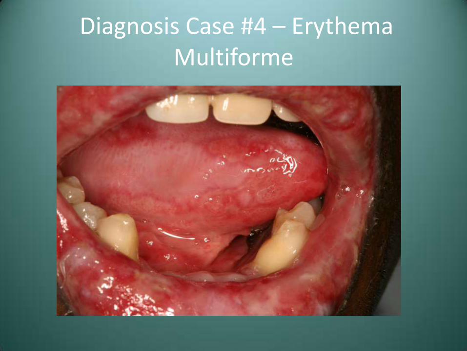
Diagnosis Case #4 – Erythema Multiforme

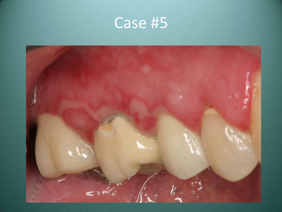
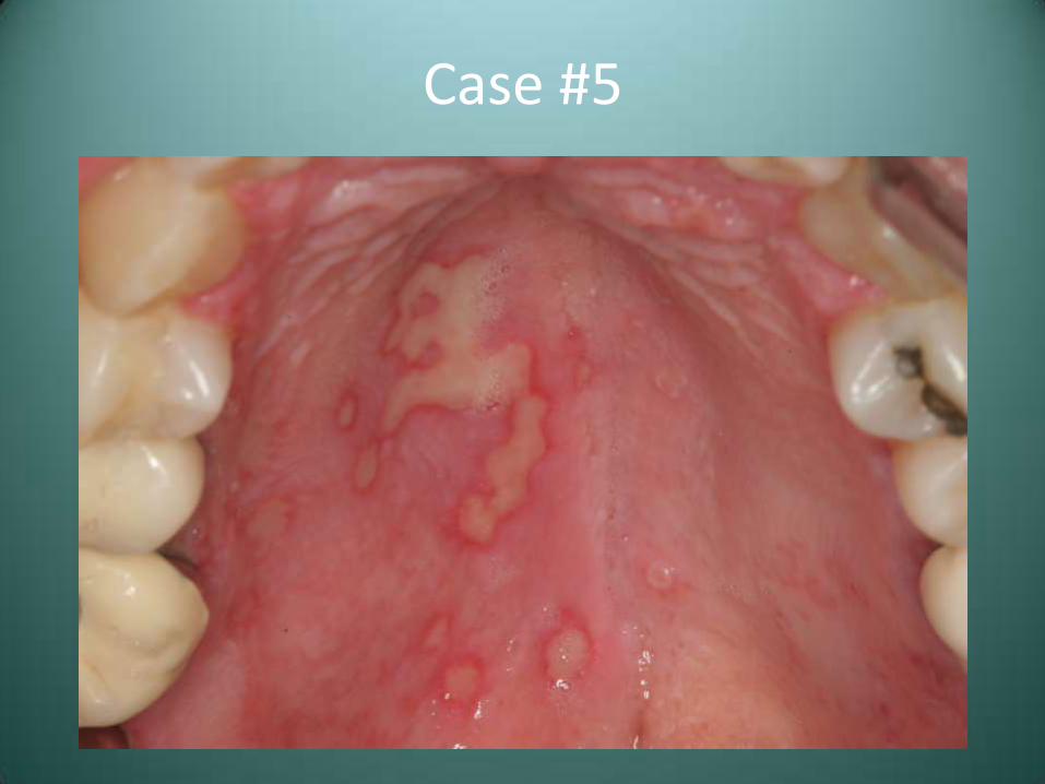
Case #5
• An adult male presents with ulcerations distributed as seen
Case #5

Case #5
Case #5
Case #7 – Differential Diagnosis
• Herpes Simplex Type 1
• Recurrent Aphthous Stomatitis
• Erythema multiforme

Herpes Simplex Virus (HSV)
• DNA virus in the herpesvirus family
– HHV-1 – oral herpes
– HHV-2 – genital herpes
– HHV-3 – chicken pox and shingles (Varicella-Zoster virus)
– HHV-4 – mononucleosis (Epstein-Barr virus)
– HHV-5 – cytomegalovirus (CMV)
– HHV-8 – Kaposi’s sarcoma-associated
Herpes Simplex Virus
• Two clinical patterns
– Primary herpetic infection
– Secondary or recurrent HSV

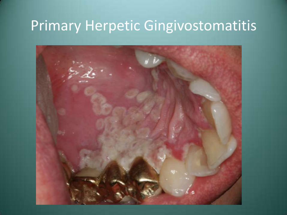
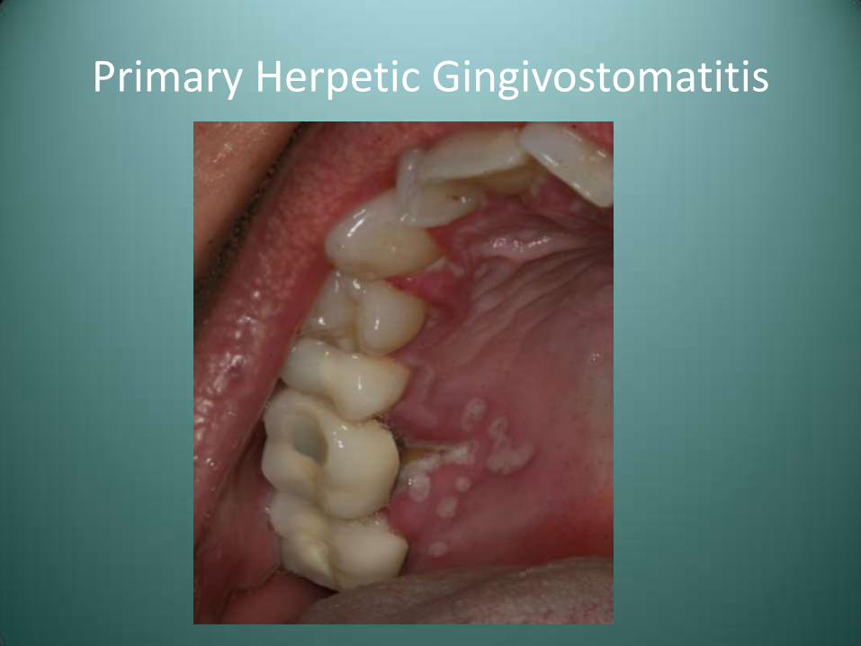
Primary Herpetic Gingivostomatitis – Clinical Features
• Children, sometimes adults
• Diffuse painful shallow ulcers
• Fever, malaise
• Lymphadenopathy
• One episode-10 to 14 days
• Virus remains dormant in sensory or autonomic ganglia

Primary Herpetic Gingivostomatitis

Primary Herpetic Gingivostomatitis
Primary Herpetic Gingivostomatitis
Primary Herpetic Gingivostomatitis

Primary Herpetic Gingivostomatitis

Primary Herpetic Gingivostomatitis
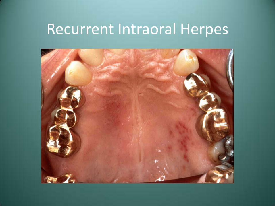
Recurrent Intraoral Herpes
• Relatively uncommon
• Usually few symptoms
• Cluster of shallow ulcers - intact vesicles rare
• Mucosa bound to periosteum
– Hard palate and attached gingiva
• Heal within one week

Recurrent Intraoral Herpes
Recurrent Intraoral Herpes
Primary Herpes-Treatment
• Restrict contact with lesions
• Topical anesthetics
– Dyclonine HCL or viscous lidocaine
• Ibuprofen or other NSAID’s
• Soft diet with fluids
• Antiviral medications of recognized early (1st 72 hours)

Recurrent Aphthous Stomatitis
• Very common condition of unknown etiology and pathogenesis
• Likely an immunologically mediated condition
• Numerous potential contributing factors
– HLA types
– Trauma
– Foods
– Stress
– HIV
Recurrent Aphthous Stomatitis – Clinical Features
• Three major forms
– Minor
– Major
– Herpetiform

Recurrent Aphthous Stomatitis – Clinical Features
• Minor aphthae
– 3-mm ulcer with yellow-white membrane and erythematous halo
– Unattached mucosa

Recurrent Aphthous Stomatitis – Clinical Features
• Minor aphthae
– 3-mm ulcer with yellow-white membrane and erythematous halo
– Unattached mucosa

Recurrent Aphthous Stomatitis – Clinical Features
• Minor aphthae
– 3-mm ulcer with yellow-white membrane and erythematous halo
– Unattached mucosa

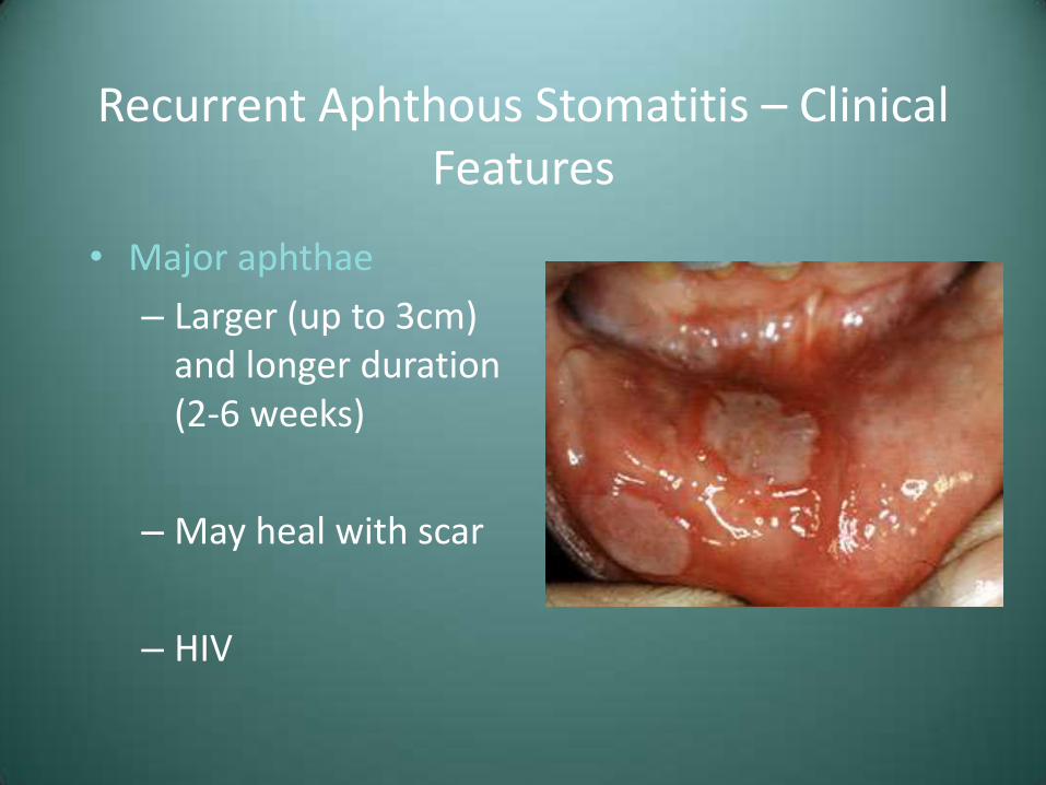
Recurrent Aphthous Stomatitis – Clinical Features
• Major aphthae
– Larger (up to 3cm) and longer duration (2-6 weeks)
– May heal with scar
– HIV
Recurrent Aphthous Stomatitis – Clinical Features
• Major aphthae
– Larger (up to 3cm) and longer duration (2-6 weeks)
– May heal with scar
– HIV

Recurrent Aphthous Stomatitis – Clinical Features
• Major aphthae
– Larger (up to 3cm) and longer duration (2-6 weeks)
– May heal with scar
– HIV

Recurrent Aphthous Stomatitis – Clinical Features
• Major aphthae
– Larger (up to 3cm) and longer duration (2-6 weeks)
– May heal with scar
– HIV

Recurrent Aphthous Stomatitis - Treatment
• Topical corticosteroids
– Betamethasone 0.05%
– Clobetasol propionate 0.05% (Temovate gel)
• Elixirs or syrup preparations for numerous and/or ulcerations in inaccessible areas
• If unresponsive, investigate possible underlying cause

Case #7 – Differential Diagnosis
• Herpes Simplex Type 1
• Recurrent Aphthous Stomatitis
• Erythema multiforme
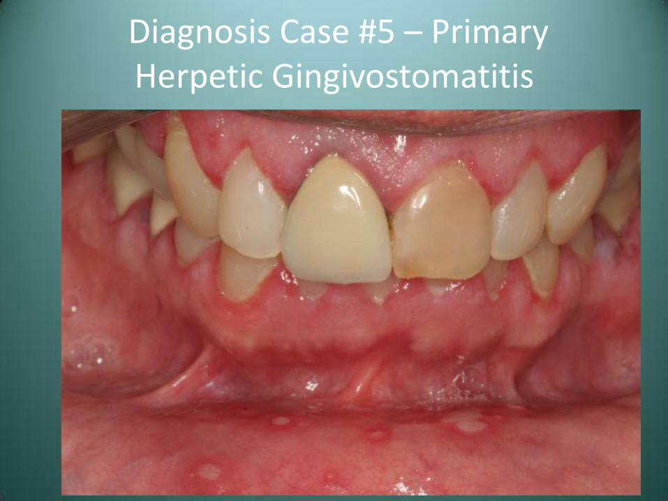
Diagnosis Case #5 – Primary Herpetic Gingivostomatitis

White, Red and Malignant Lesions
Smokeless Tobacco Use/Tobacco Pouch Keratosis
• Mucosal lesion secondary to the presence of chronic irritation from smokeless tobacco
• These products are currently used by approximately 4.5% of US males
• Also associated with gingival/periodontal destruction and tooth decay

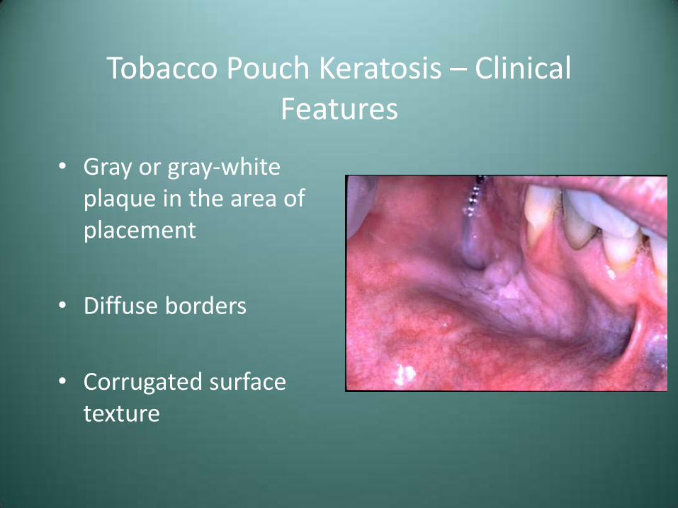
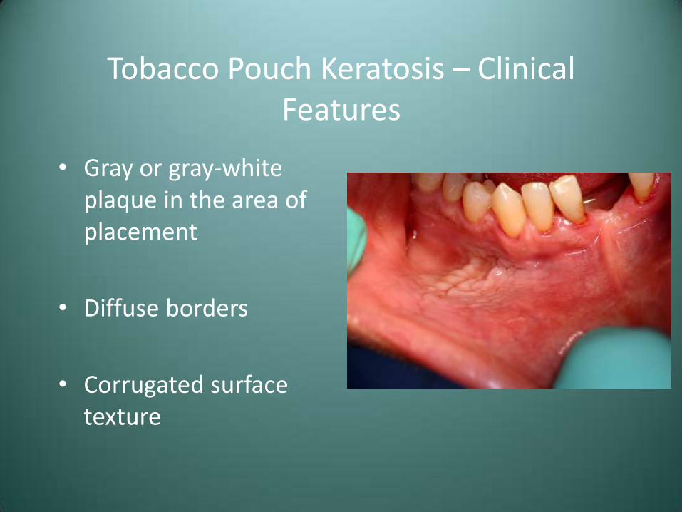
Tobacco Pouch Keratosis – Clinical Features
• Gray or gray-white plaque in the area of placement
• Diffuse borders
• Corrugated surface texture

Tobacco Pouch Keratosis – Clinical Features
• Gray or gray-white plaque in the area of placement
• Diffuse borders
• Corrugated surface texture
Tobacco Pouch Keratosis – Clinical Features
• Gray or gray-white plaque in the area of placement
• Diffuse borders
• Corrugated surface texture
Tobacco Pouch Keratosis – Clinical Features
• Gray or gray-white plaque in the area of placement
• Diffuse borders
• Corrugated surface texture

Tobacco Pouch Keratosis – Treatment and Prognosis
• Have patient stop or move the tobacco to another location to observe for resolution (2-4 weeks)
• If the lesion persists (after 6 weeks), biopsy for histologic diagnosis
• Controversy over true carcinogenicity of smokeless tobacco

Nicotine Stomatitis
• Benign hyperkeratotic change to the palatal mucosa secondary to tobacco smoking
• Most common in pipe and cigar smokers
• Similar changes may be induced by drinking hot beverages

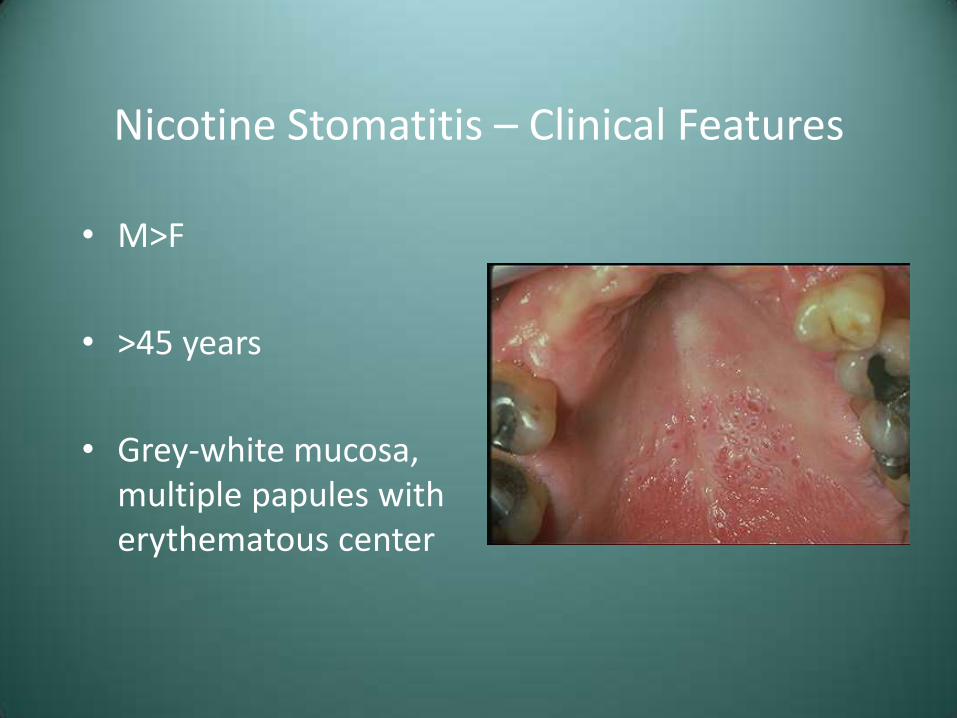
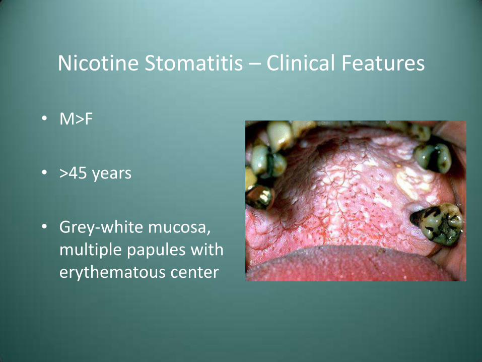
Nicotine Stomatitis – Clinical Features
• M>F,
• >45 years
• Grey-white mucosa, multiple papules with erythematous center
Nicotine Stomatitis – Clinical Features
• M>F
• >45 years
• Grey-white mucosa, multiple papules with erythematous center

Nicotine Stomatitis – Clinical Features
• M>F
• >45 years
• Grey-white mucosa, multiple papules with erythematous center

Nicotine Stomatitis – Clinical Features
• M>F
• >45 years
• Grey-white mucosa, multiple papules with erythematous center
Nicotine Stomatitis – Clinical Features
• M>F
• >45 years
• Grey-white mucosa, multiple papules with erythematous center

Nicotine Stomatitis – Treatment
• None
• If patient quits, changes will normally resolve within 1-2 weeks
• Persistent changes should be biopsied
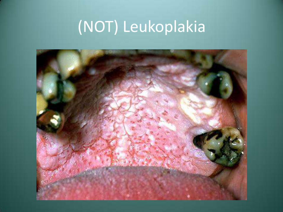
Leukoplakia
• Definition (WHO)-A white patch or plaque which cannot be characterized clinically or pathologically as any other disease
• Considered premalignant
– Most common precancerous oral lesion

Leukoplakia
• Etiology-Technically unknown
– Tobacco smoking
– Alcohol is not necessarily associated with leukoplakia
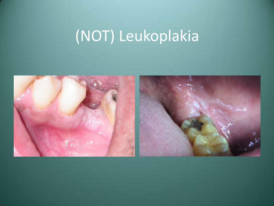
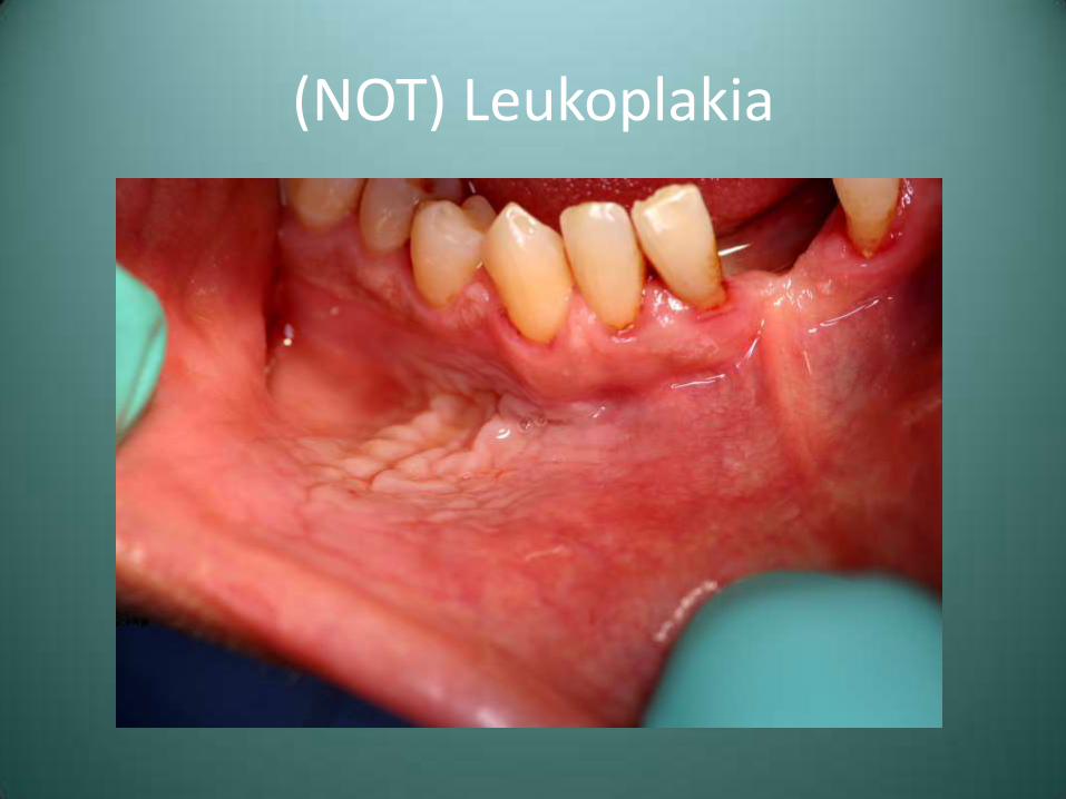
• Lesions that are not leukoplakia
– Nicotine stomatitis
– Frictional keratosis
– Lichen planus
– Amalgam reactions
(NOT) Leukoplakia
(NOT) Leukoplakia

(NOT) Leukoplakia
(NOT) Leukoplakia
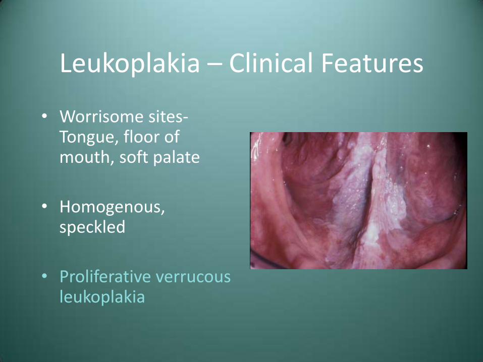
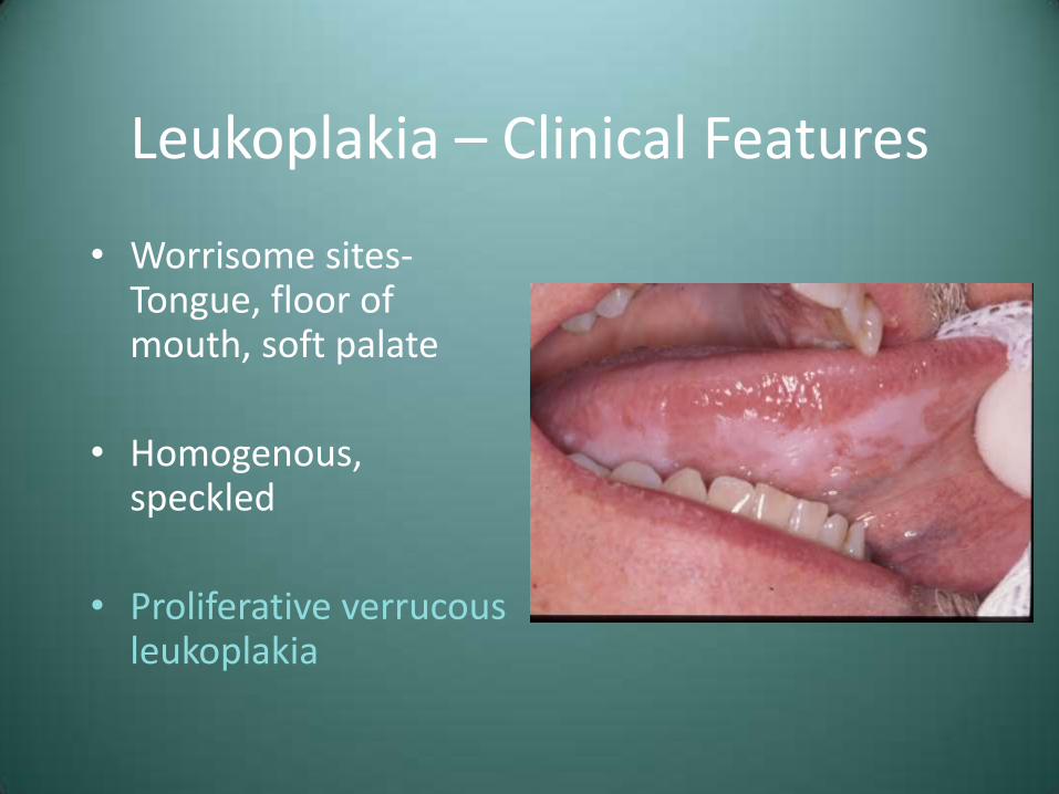
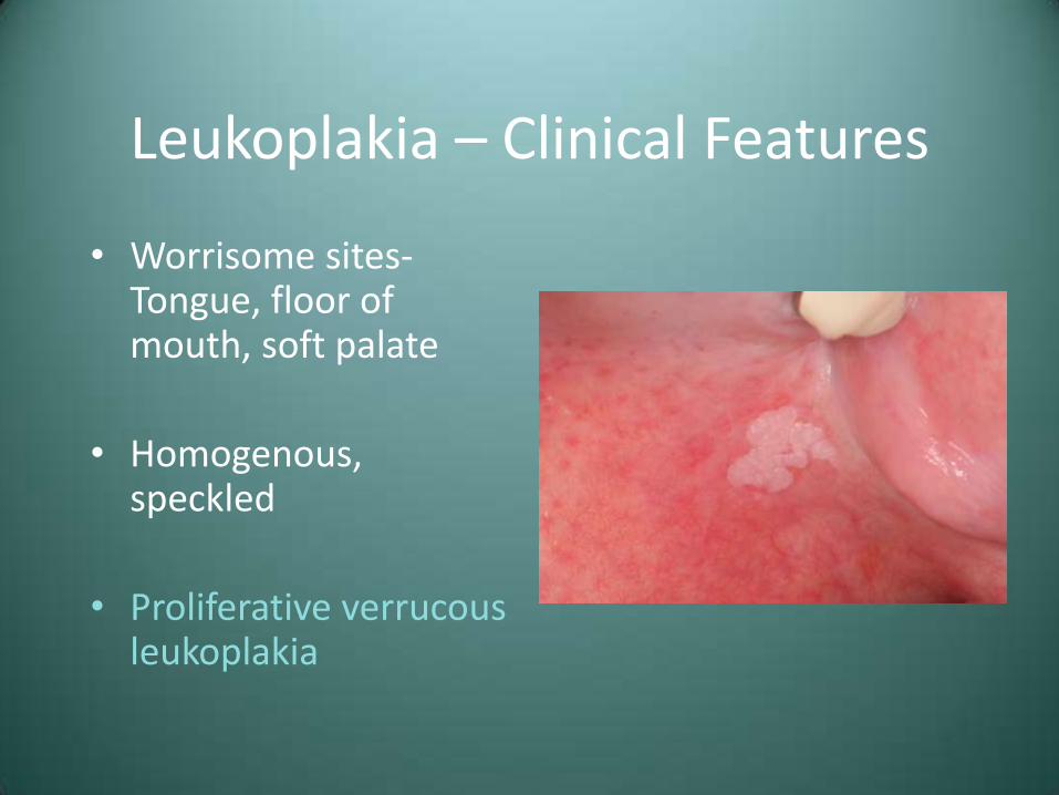
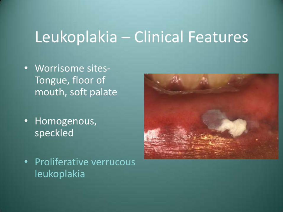
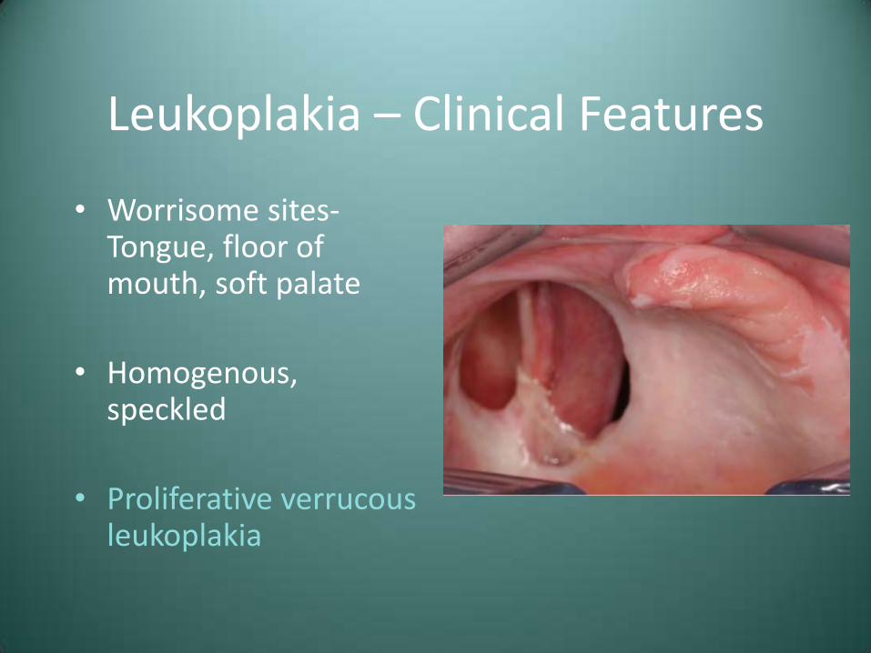
Leukoplakia – Clinical Features
• Worrisome sites-Tongue, floor of mouth, soft palate
• Homogenous, speckled
• Proliferative verrucous leukoplakia
Leukoplakia – Clinical Features
• Worrisome sites-Tongue, floor of mouth, soft palate
• Homogenous, speckled
• Proliferative verrucous leukoplakia

Leukoplakia – Clinical Features
• Worrisome sites-Tongue, floor of mouth, soft palate
• Homogenous, speckled
• Proliferative verrucous leukoplakia

Leukoplakia – Clinical Features
• Worrisome sites-Tongue, floor of mouth, soft palate
• Homogenous, speckled
• Proliferative verrucous leukoplakia
Leukoplakia – Clinical Features
• Worrisome sites-Tongue, floor of mouth, soft palate
• Homogenous, speckled
• Proliferative verrucous leukoplakia

Leukoplakia – Clinical Features
• Worrisome sites-Tongue, floor of mouth, soft palate
• Homogenous, speckled
• Proliferative verrucous leukoplakia
Leukoplakia – Clinical Features
• Worrisome sites-Tongue, floor of mouth, soft palate
• Homogenous, speckled
• Proliferative verrucous leukoplakia
Leukoplakia – Clinical Features
• Worrisome sites-Tongue, floor of mouth, soft palate
• Homogenous, speckled
• Proliferative verrucous leukoplakia

Leukoplakia – Clinical Features
• Worrisome sites-Tongue, floor of mouth, soft palate
• Homogenous, speckled
• Proliferative verrucous leukoplakia

Leukoplakia – Clinical Features
• Worrisome sites-Tongue, floor of mouth, soft palate
• Homogenous, speckled
• Proliferative verrucous leukoplakia

Leukoplakia – Clinical Features
• Worrisome sites-Tongue, floor of mouth, soft palate
• Homogenous, speckled
• Proliferative verrucous leukoplakia

Leukoplakia – Clinical Features
• Worrisome sites-Tongue, floor of mouth, soft palate
• Homogenous, speckled
• Proliferative verrucous leukoplakia

Leukoplakia – Clinical Features
• Worrisome sites-Tongue, floor of mouth, soft palate
• Homogenous, speckled
• Proliferative verrucous leukoplakia
Leukoplakia – Clinical Features
• Worrisome sites-Tongue, floor of mouth, soft palate
• Homogenous, speckled
• Proliferative verrucous leukoplakia

Leukoplakia – Treatment and Prognosis
• Biopsy is mandatory
• Treatment will then depend upon the histologic findings
• 4% risk of transformation to SCC
• With or without removal, follow-up is essential
• Recurrences are common (about 1/3)

Erythroplakia
• Red patch that cannot be clinically or pathologically diagnosed as any other condition
• Greater presence of dysplasia than leukoplakia
• Same etiology as SCC (tobacco, alcohol)

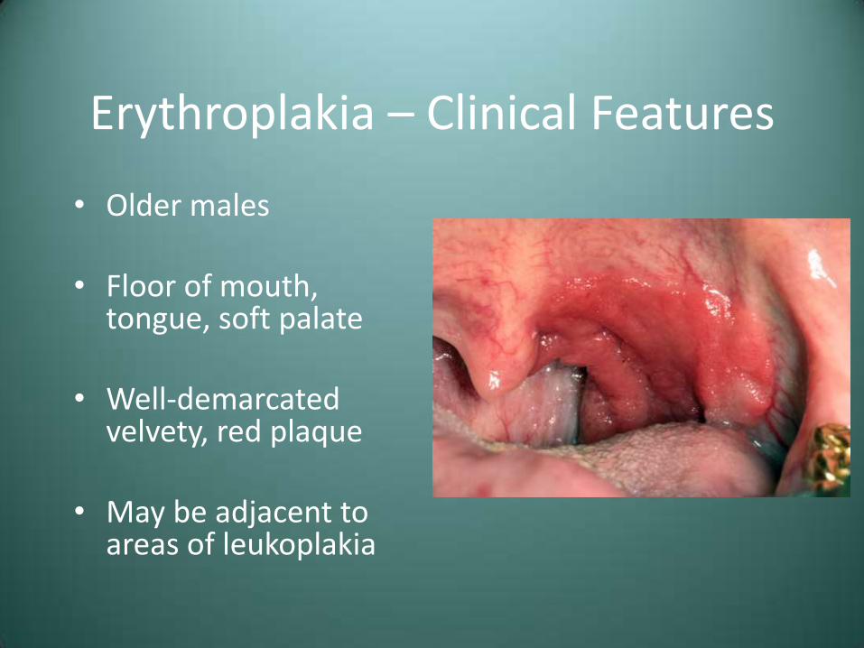
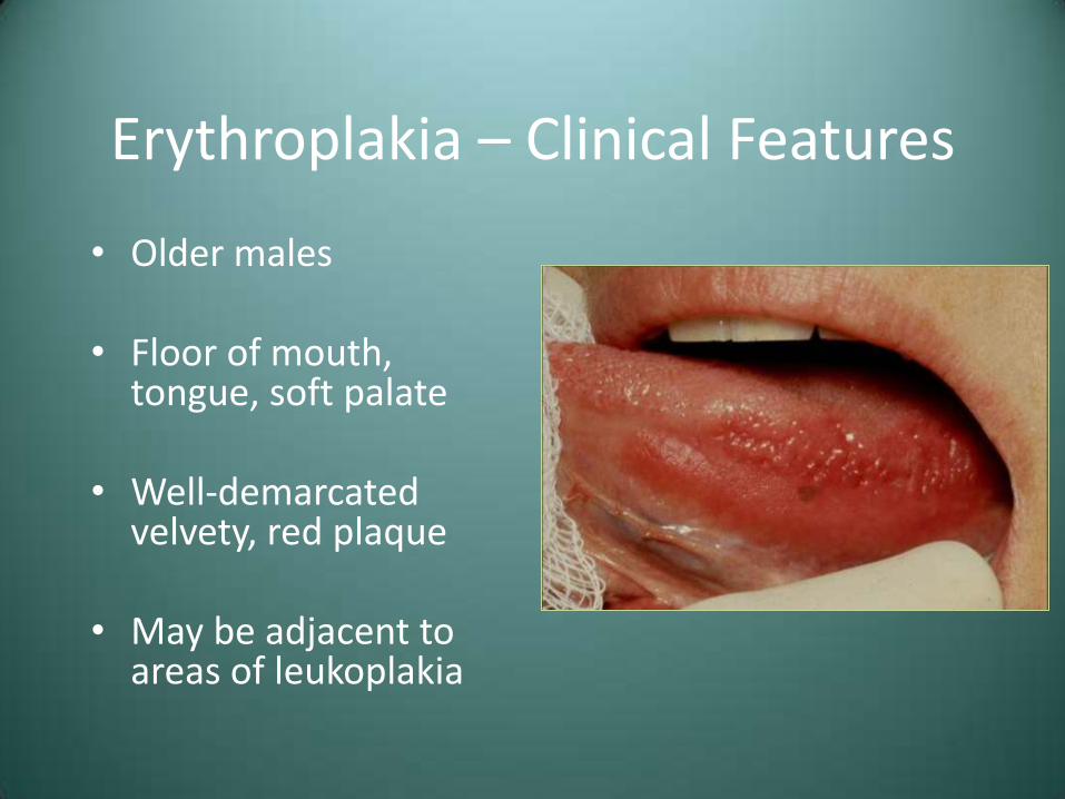
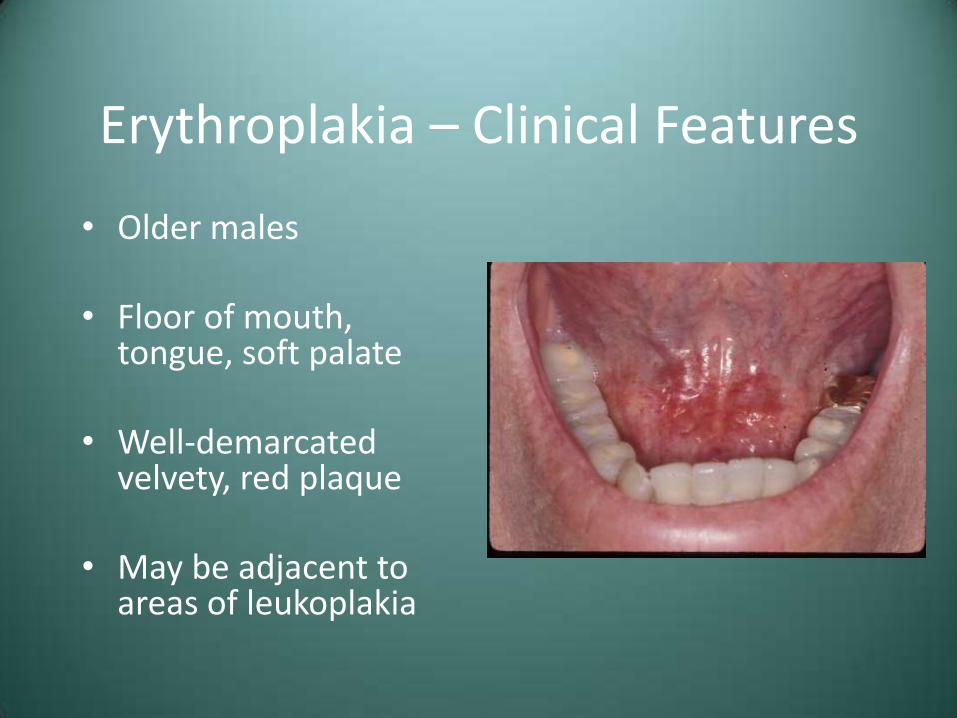
Erythroplakia – Clinical Features
• Older males
• Floor of mouth, tongue, soft palate
• Well-demarcated velvety, red plaque
• May be adjacent to areas of leukoplakia

Erythroplakia – Clinical Features
• Older males
• Floor of mouth, tongue, soft palate
• Well-demarcated velvety, red plaque
• May be adjacent to areas of leukoplakia
Erythroplakia – Clinical Features
• Older males
• Floor of mouth, tongue, soft palate
• Well-demarcated velvety, red plaque
• May be adjacent to areas of leukoplakia
Erythroplakia – Clinical Features
• Older males
• Floor of mouth, tongue, soft palate
• Well-demarcated velvety, red plaque
• May be adjacent to areas of leukoplakia
Erythroplakia – Clinical Features
• Older males
• Floor of mouth, tongue, soft palate
• Well-demarcated velvety, red plaque
• May be adjacent to areas of leukoplakia

Erythroplakia – Clinical Features
• Older males
• Floor of mouth, tongue, soft palate
• Well-demarcated velvety, red plaque
• May be adjacent to areas of leukoplakia
Erythroplakia - Histology
• 90% will show severe dysplasia or CIS
• Epithelial atrophy with lack of keratin production
• Chronic inflammation

Erythroplakia – Treatment and Prognosis
• Biopsy is mandatory, with treatment dependant upon the degree of dysplasia
• Close follow-up is necessary, since recurrence and the development of separate lesions are common
Oral Squamous Cell Carcinoma
• 22,000 cases per year, with about 1 in four dying of the disease
• Males-8th most common cancer (Females-15th)
• M>F
• Blacks>Whites
• Carcinoma of the lip should be considered in a different context
Oral Squamous Cell Carcinoma - Etiology
• Tobacco (especially combustible)
• Alcohol (works synergistically with tobacco)
• Radiation
• Plummer-Vinson syndrome (iron deficiency anemia, glossitis, dysphagia)
• Viruses (HPV)
• Immunosuppression

Oral Squamous Cell Carcinoma – Clinical Features
• Varied
– Exophytic
– Endophytic
– Ulcerated
– Erythroplakic
– Leukoplakic

Oral Squamous Cell Carcinoma – Clinical Features
• Varied
– Exophytic
– Endophytic
– Ulcerated
– Erythroplakic
– Leukoplakic
Oral Squamous Cell Carcinoma – Clinical Features
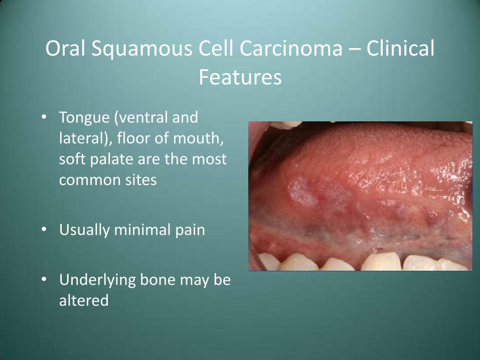
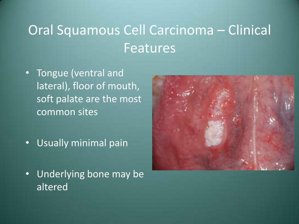
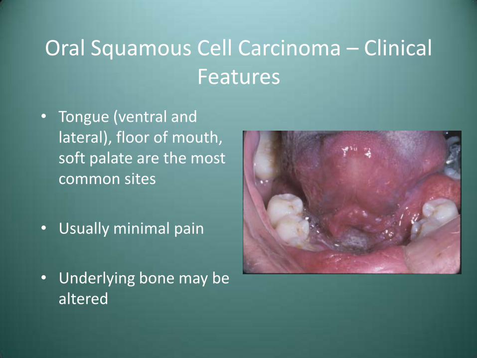
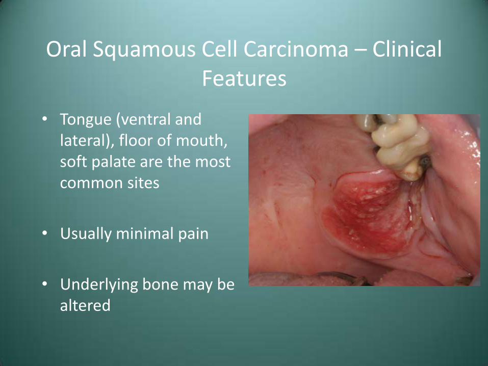
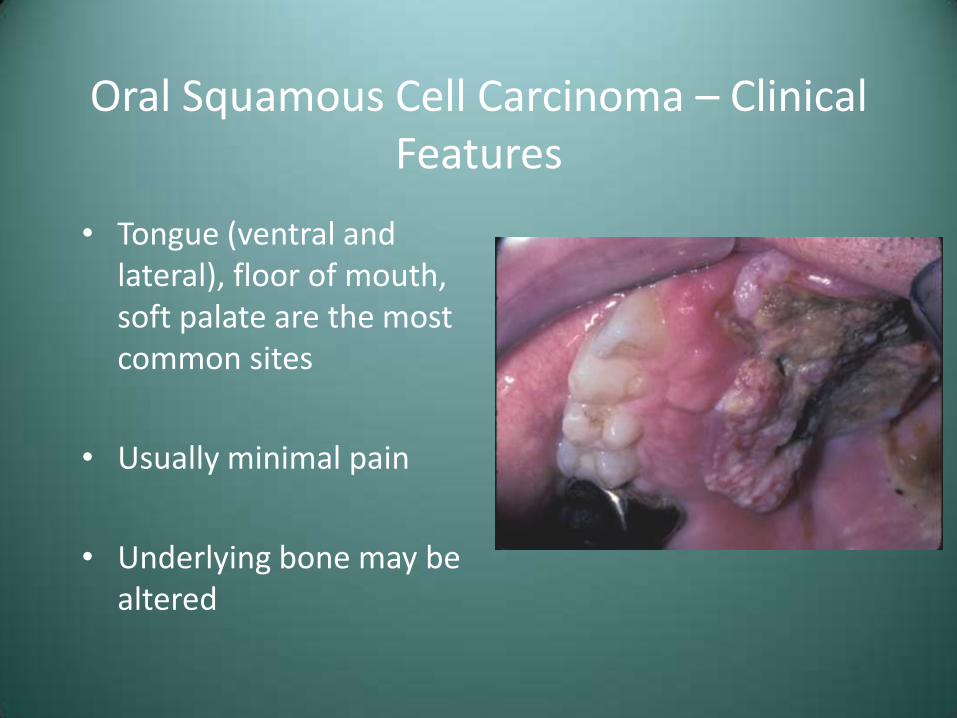
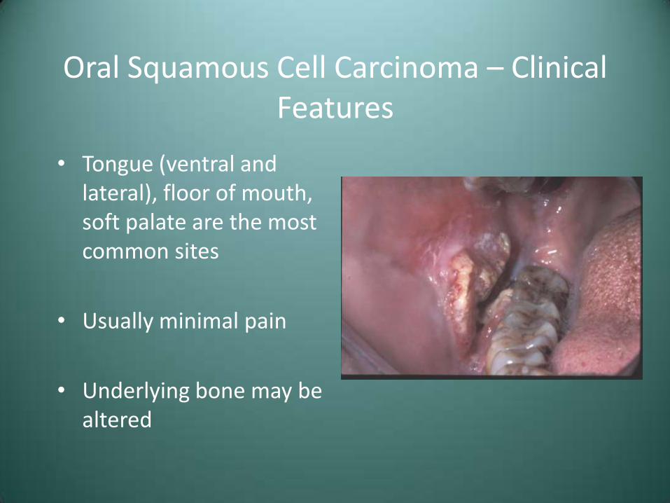
• Tongue (ventral and lateral), floor of mouth, soft palate are the most common sites
• Usually minimal pain
• Underlying bone may be altered
Oral Squamous Cell Carcinoma – Clinical Features
• Tongue (ventral and lateral), floor of mouth, soft palate are the most common sites
• Usually minimal pain
• Underlying bone may be altered

Oral Squamous Cell Carcinoma – Clinical Features
• Tongue (ventral and lateral), floor of mouth, soft palate are the most common sites
• Usually minimal pain
• Underlying bone may be altered

Oral Squamous Cell Carcinoma – Clinical Features
• Tongue (ventral and lateral), floor of mouth, soft palate are the most common sites
• Usually minimal pain
• Underlying bone may be altered
Oral Squamous Cell Carcinoma – Clinical Features
• Tongue (ventral and lateral), floor of mouth, soft palate are the most common sites
• Usually minimal pain
• Underlying bone may be altered

Oral Squamous Cell Carcinoma – Clinical Features
• Tongue (ventral and lateral), floor of mouth, soft palate are the most common sites
• Usually minimal pain
• Underlying bone may be altered
Oral Squamous Cell Carcinoma – Clinical Features
• Tongue (ventral and lateral), floor of mouth, soft palate are the most common sites
• Usually minimal pain
• Underlying bone may be altered

Oral Squamous Cell Carcinoma – Clinical Features
• Tongue (ventral and lateral), floor of mouth, soft palate are the most common sites
• Usually minimal pain
• Underlying bone may be altered

Oral Squamous Cell Carcinoma – Clinical Features
• Tongue (ventral and lateral), floor of mouth, soft palate are the most common sites
• Usually minimal pain
• Underlying bone may be altered
Oral Squamous Cell Carcinoma – Clinical Features
• Tongue (ventral and lateral), floor of mouth, soft palate are the most common sites
• Usually minimal pain
• Underlying bone may be altered

Oral Squamous Cell Carcinoma – Clinical Features
• Tongue (ventral and lateral), floor of mouth, soft palate are the most common sites
• Usually minimal pain
• Underlying bone may be altered
Oral Squamous Cell Carcinoma – Clinical Features
• Tongue (ventral and lateral), floor of mouth, soft palate are the most common sites
• Usually minimal pain
• Underlying bone may be altered

Oral Squamous Cell Carcinoma – Clinical Features
• Tongue (ventral and lateral), floor of mouth, soft palate are the most common sites
• Usually minimal pain
• Underlying bone may be altered
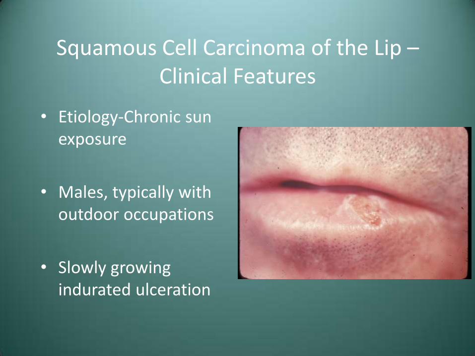
Squamous Cell Carcinoma of the Lip – Clinical Features
• Etiology-Chronic sun exposure
• Males, typically with outdoor occupations
• Slowly growing indurated ulceration
Squamous Cell Carcinoma - Metastasis
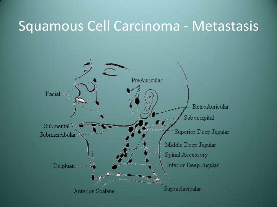
• Spread through lymphatics
• Firm nodes
• Movable or fixed
• Distant spread to lungs, liver, bones
• TNM staging
– Stage at diagnosis is the most important prognostic indicator

Squamous Cell Carcinoma - Metastasis
• TNM staging system
– T-Tumor size
– N-Local node involvement
– M-Distant metastasis
Squamous Cell Carcinoma - Metastasis

Squamous Cell Carcinoma – Treatment and Prognosis
• Surgical excision/resection
• Radiation
• Chemotherapy – Squamous cell carcinoma rarely responds well
• Stage I – 85% 5 year survival
• Stage II – 66%
• Stage III – 41%
• Stage IV – 9%

Squamous Cell Carcinoma – Treatment and Prognosis
• National Comprehensive Cancer Network
• http://www.nccn.org/professionals/physician_gls/f_guidelines.asp
Squamous Cell Carcinoma – Treatment and Prognosis
• Carcinoma of the lip carries a much better prognosis
• Prognosis is better for Whites than Blacks
• “Field cancerization” – Persons with one carcinoma are at increased risk of developing a second mucosal tumor

Odds and Ends

Case #6
• This patient presented with recent onset of the pigmentation seen here

Case #6
Case #5 – Differential Diagnosis
• Normal Physiologic Pigmentation
• Smoker’s Melanosis
• Medication-Associated
• Addison’s Disease

Smoker’s Melanosis
• Rather common melanocytic response found in heavy smokers
• Probably a protective response to the harmful aspects (polycyclic aromatic hydrocarbons) of tobacco smoke
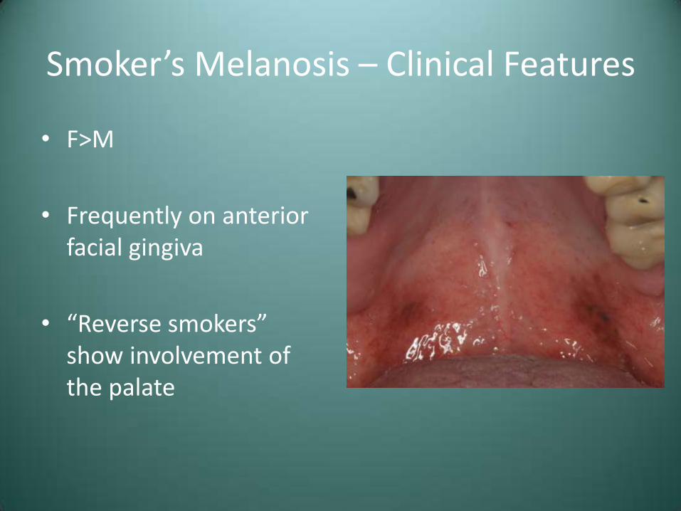
Smoker’s Melanosis – Clinical Features
• F>M
• Frequently on anterior facial gingiva
• “Reverse smokers” show involvement of the palate
Smoker’s Melanosis – Clinical Features
• F>M
• Frequently on anterior facial gingiva
• “Reverse smokers” show involvement of the palate

Smoker’s Melanosis – Clinical Features
• F>M
• Frequently on anterior facial gingiva
• “Reverse smokers” show involvement of the palate

Smoker’s Melanosis – Diagnosis and Treatment
• Clinical, tobacco, and medical history
• May need to rule out systemic cause
• Cessation of smoking will result in gradual resolution

Drug-Related Discolorations of the Oral Mucosa
• Discoloration secondary to melanocytic stimulation or direct deposition into tissue
• Antimalarial meds, minocycline, estrogen, chemotherapeutic agents, AIDS medications

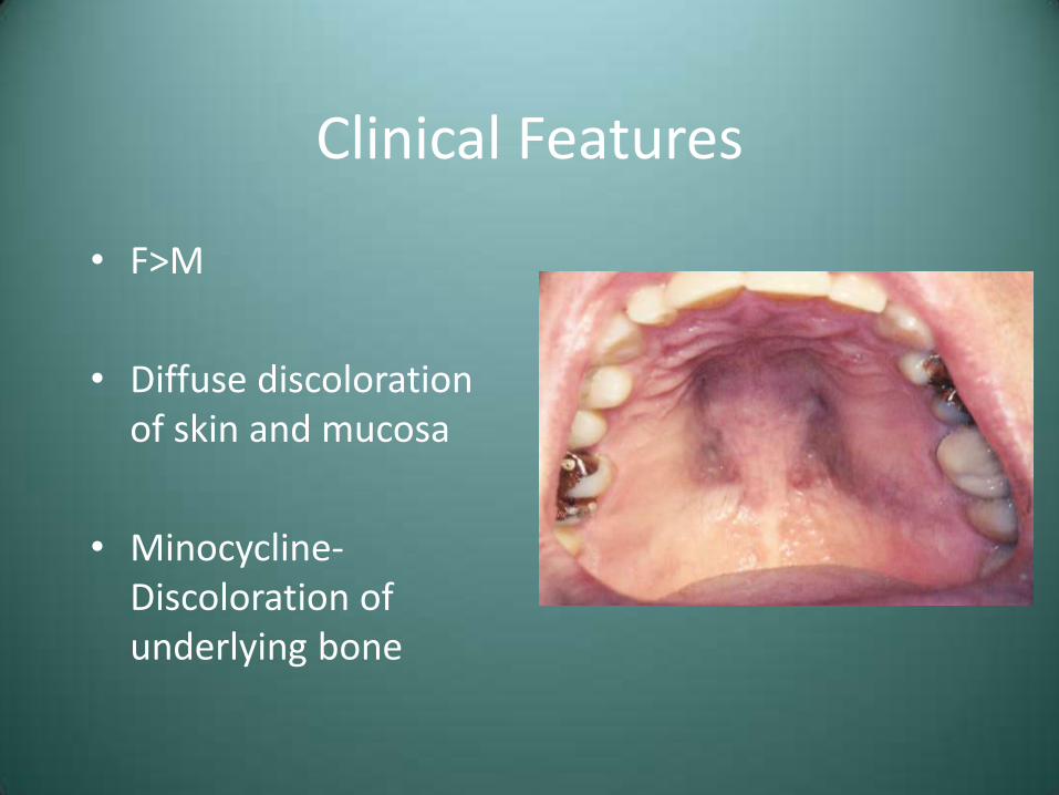
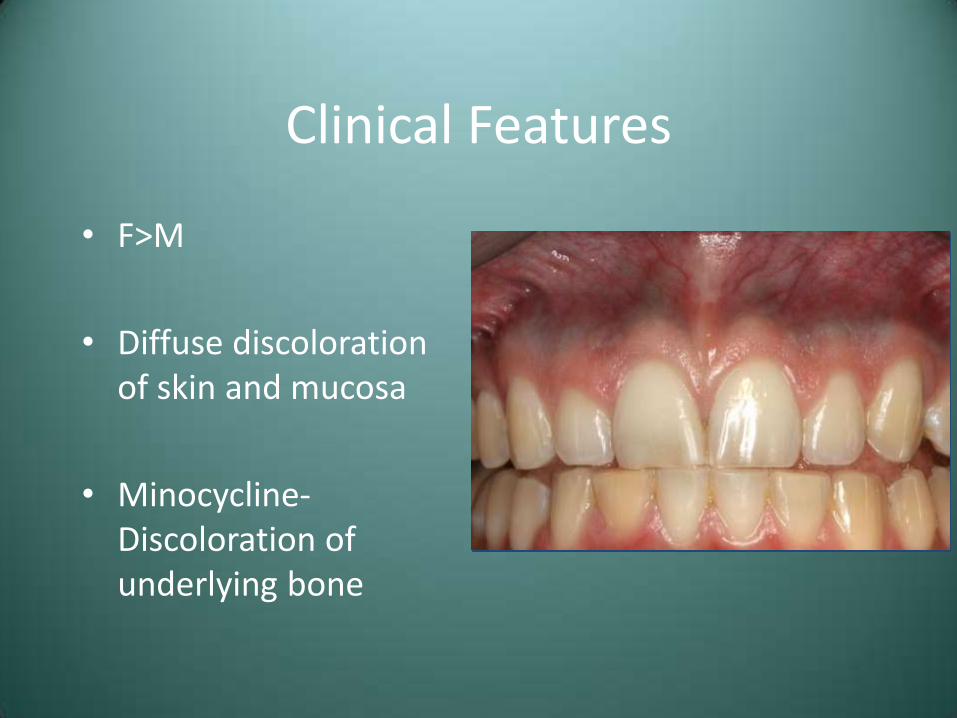
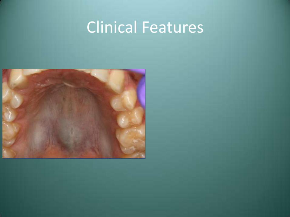
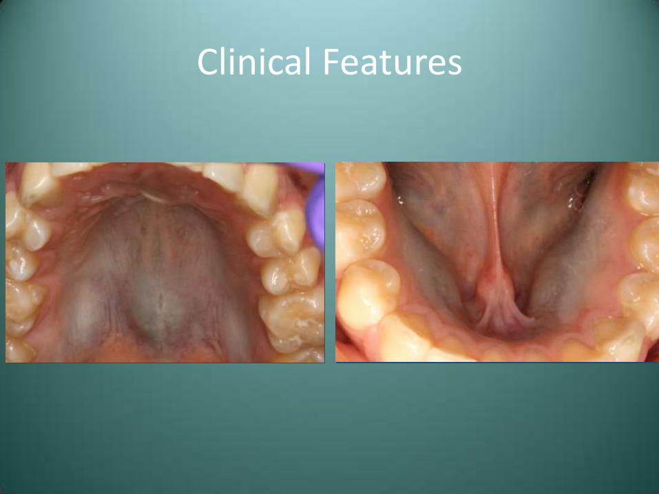
Clinical Features
• F>M
• Diffuse discoloration of skin and mucosa
• Minocycline-Discoloration of underlying bone

Clinical Features
• F>M
• Diffuse discoloration of skin and mucosa
• Minocycline-Discoloration of underlying bone
Clinical Features
• F>M
• Diffuse discoloration of skin and mucosa
• Minocycline-Discoloration of underlying bone
Clinical Features
• F>M
• Diffuse discoloration of skin and mucosa
• Minocycline-Discoloration of underlying bone
Clinical Features
Clinical Features

Treatment
• Gradual resolution upon discontinuation of medication
• Strictly and esthetic issue
• No long term complications

Addison’s Disease (Hypoadrenocorticism)
• Insufficient production of adrenal corticosteroid hormones
• Primary – Secondary to adrenal destruction
• Secondary – Due to malfunctioning pituitary gland

Addison’s Disease – Clinical Features
• Fatigue, irritability, depression, weakness, and hypotension
• Hyperpigmentation (may be seen intraorally)
• GI symptoms, salt-craving
Addison’s Disease – Lab Findings
• Primary – High plasma ACTH
• Secondary – Low plasma ACTH

Addison’s Disease - Treatment
• Corticosteroid replacement therapy
• Preplan dental and oral surgical procedures
• Good prognosis, with patients typically living a normal life span
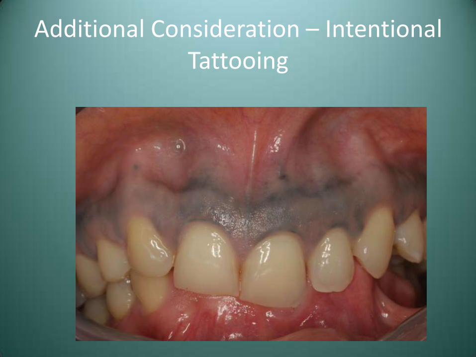
Additional Consideration – Intentional Tattooing

Case #6 – Differential Diagnosis
• Normal Physiologic Pigmentation
• Smoker’s Melanosis
• Medication-Associated
• Addison’s Disease

Diagnosis Case #6 – Addison’s Disease
Diagnosis Case #6 – Addison’s Disease
• Further questioning revealed a one month history of nausea, vomiting and intermittent weakness

Case #7
• This patient presents with the abnormality seen

Case #7

Case #7
Case #7 – Differential Diagnosis
• Angioedema
• Cheilitis Granulomatosis (Orofacial Granulomatosis)
Angioedema (Quincke’s Disease)
• Diffuse, often intermittent swelling of the soft tissue
• Three primary mechanisms-
– Hypersensitivity reaction due to IgE mediated mast cell degranulation
– Associated with ACE inhibitor antihypertensives, secondary to increased bradykinin levels
– Lack of or inactive C1 esterase inhibitor (inherited or acquired)

Angioedema – Clinical Features
• Enlargement of relatively rapid onset
• Pruritis, erythema
• Respiratory involvement may be life threatening
Angioedema – Diagnosis
• Allergic - Clinical presentation in association with suspected antigen
• Inciting cause often not determined
• Evaluate functional C1-INH

Angioedema - Treatment
• Antihistamines for allergic form
• IM epinephrine
• ACE inhibitor-related and C1-INH deficient do not respond to antihistamines
– C1-INH concentrate administration or esterase inhibiting drugs
Cheilitis Granulomatosis (Orofacial Granulomatosis)
• Granulomatous inflammation of unknown etiology or the orofacial presentation of Crohn’s, sarcoidosis, TB, or any other granulomatous process
Orofacial Granulomatosis – Clinical Features
• Highly variable presentation
• Involvement of lips-cheilitis granulomatosa

Orofacial Granulomatosis – Clinical Features
• Highly variable presentation
• Involvement of lips-cheilitis granulomatosa

Orofacial Granulomatosis – Clinical Features
• Highly variable presentation
• Involvement of lips-cheilitis granulomatosa
Treatment and Prognosis
• Intralesional corticosteroids
• Multiple treatments
• Good prognosis; requires thorough work-up
• Primarily a cosmetic problem

Case #7 – Differential Diagnosis
• Angioedema
• Cheilitis Granulomatosis (Orofacial Granulomatosis)
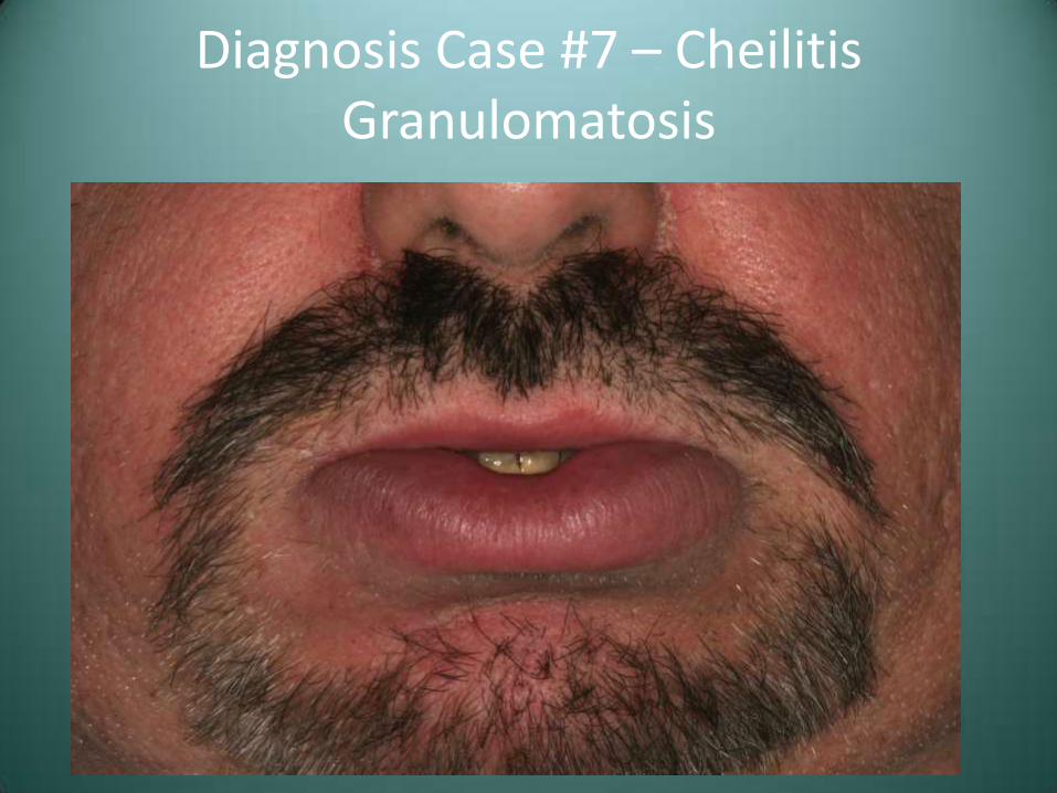
Diagnosis Case #7 – Cheilitis Granulomatosis