Embed Size (px)
Citation preview
1
Doctoral Thesis for the degree of Doctor of Philosophy, Sahlgren´s academy
Consequences of Arterial Switch Operation in
Children Born with Transposition of the Great
Arteries
- A clinical and experimental study of the autonomous
nervous system in the heart
Cecilia Falkenberg
Department of Paediatrics
Institute of Clinical Sciences
Sahlgrenska Academy at University of Gothenburg
Gothenburg 2013
2
Cover illustration: “Heart of my life” by Evelina Falkenberg
Figures 1, 2, 4, 5, 6 and 7 by Evelina Falkenberg
Consequences of Arterial Switch Operation in Children Born with Transposition of the Great Arteries
– A clinical and experimental study of the autonomous nervous system in the heart
©Cecilia Falkenberg 2013
ISBN 978-91-628-8542-7
Printed in Gothenburg, Sweden 2013
Kompendiet, Aidla Trading AB
3
To my ”Winter star”, my ”Autumn leaf” and my ”Summer flower”
Julia, Evelina and Martha
With you everything makes sense
“It´s hard to make a “come-back” if you haven´t been anywhere!!”
-the Road House “The Centre of the Universe”, Western Australia
“The thing about growing up with Fred and George, ´said Ginny thoughtfully´, is
that you sort of start thinking that anything´s possible if you´ve got enough
nerve.”
-Ginny Weasley in “Harry Potter and the Order of the Phoenix”,
by J.K. Rowling (2003)

4

5
Abstract Background: The introduction of the arterial switch operation (ASO) made it the procedure of choice for surgical correction of transposition of the great arteries. A majority of the sympathetic nerves innervate the heart alongside the great vessels; these are therefore likely to be damaged during the surgical procedure; imposing new challenges and questions that need to be addressed. The main aim for this thesis was to assess the long-term cardiac consequences on the autonomic nervous system after surgery (paper I and II) and to create an animal model allowing for cardiac physiological studies (paper III and IV). Methods: Long-term follow-up in adolescents who had undergone ASO as neonates (n=17, 1 female, mean fractional shorting 32±5%) was performed. This included sympathetic nervous system function assessed through infusion of tritiated Norepinephrine ([3H]NE) during heart catheterisation (n=8)(controls n=15) and blood samples analysed with high performance liquid chromatography. Samples were obtained both before and after adenosine stimulation as a response to sympathetic excitation. 24-hour heart rate variability (HRV)(n=15 in both groups) was measured both during the day and night using different algorithms. Baroreflex sensitivity and QT variability index (QTVI) (n=17 in both groups) were measured in awake patients. An animal model was developed using complex open heart surgery during cardiopulmonary bypass to mimick the arterial switch operation in piglets 8 weeks of age. The piglets surviving at least 5 to 6 weeks post-operation had follow-up of physiological response to catecholamines and were studied in vivo and in vitro using the Langendorff perfusion system. Results: In both groups the specific activity of [3H]NE decreased from the artery to the coronary sinus, but to a lesser extent in the ASO group. The extraction fraction in the ASO group was 56±10% compared to 82±9% in the healthy subjects (p<0.001). The arterial to coronary sinus plasma concentration of [3H] dihydroxyphenylglycol (DHPG) was significantly increased in the healthy group (70%, p=<0.0001) but was not so in the ASO group (8%, p=0.5). The difference of endogenous DHPG increase from the arterial to the coronary sinus was significantly smaller in the ASO group (p=0.008). After adenosine infusion, the total body NE spillover increased in the ASO group (p=0.002), reflecting major sympathetic activation. [3H]DHPG step-up from the artery to the coronary sinus increased 4-fold following adenosine. HRV frequency-domain at night-time, when cardio-parasympathetic drive is likely to be most pronounced, showed a significant decrease of normalized high frequency in the ASO group (52±20) compared to healthy subjects (68±15)(p=0.018). Time-domain showed no statistical difference between the two groups, neither during day-time nor night-time. Baroreflex sensitivity and QTVI did not show significant differences between groups. The animal model resulted in 14 out of 19 piglets surviving the mimicked ASO. Piglets operated with mimicked ASO had a significantly higher basal heart rate both in vivo (p=0.042) and in vitro (p=0.0056). Conclusion: A disturbed but functioning sympathetic cardiac innervation was found in the ASO patients at long-term follow-up. The vagal tone seemed normal in terms of BRS, however, frequency-domain analysis showed a decreased parasympathetic tone at night time in the ASO group. The surgical challenges due to translocation of the coronary arteries and the consequences of an injured autonomic nervous system impose risks of decreased myocardial perfusion and arrhythmias. Thus, the present data suggest that these patients ought to have follow-up that includes autonomic nervous system assessment. Key Words: Transposition of the great arteries, arterial switch operation, autonomous nerves system, norepinephrine, heart rate variability, cardiopulmonary bypass, piglets ISBN 978-91-628-8542-7

6
List of Papers
This thesis is based on the following papers, which will be referred to in the
text by their Roman numerals:
I Falkenberg C, Östman-Smith I, Gilljam T, Lambert G, Friberg P. Cardiac autonomic function in adolescents operated by arterial switch
surgery. Accepted for publication in International Journal of Cardiology 25-Dec- 2012. In press. DOI:10.1016/j.ijcard.2012.12.063
II Falkenberg C, Ekman M, Gilljam T, Friberg P. Heart rate variability in
adolescents who as neonates underwent neonatal arterial switch
operation. (Manuscript)
III Falkenberg C, Hallhagen S, Nilsson K, Östman-Smith I. Anaesthetic,
surgical and bypass techniques allowing long term survival after
complex cardiac surgery in piglets. (Manuscript)
IV Falkenberg C, Hallhagen S, Nilsson K, Nilsson B, Östman-Smith I. A study
of the physiological consequences of sympathetic denervation of the
heart caused by the arterial switch procedure. Cardiology in the Young
(2010), 20, 150–158
7
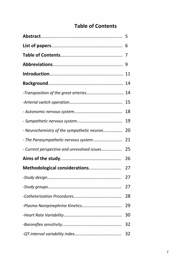
Table of Contents
Abstract............................................................... 5
List of papers....................................................... 6
Table of Contents................................................ 7
Abbreviations...................................................... 9
Introduction......................................................... 11
Background.......................................................... 14
-Transposition of the great arteries............................. 14
-Arterial switch operation......................................... 15
- Autonomic nervous system..................................... 18
- Sympathetic nervous system................................... 19
- Neurochemistry of the sympathetic neuron............... 20
- The Parasympathetic nervous system....................... 21
- Current perspective and unresolved issues................ 25
Aims of the study............................................... 26
Methodological considerations......................... 27
-Study design......................................................... 27
-Study groups......................................................... 27
-Catheterization Procedures..................................... 28
-Plasma Norepinephrine Kinetics............................... 29
-Heart Rate Variability............................................. 30
-Baroreflex sensitivity.............................................. 32
-QT-interval variability index.................................... 32

8
-Complex cardiac surgery during cardiopulmonary
bypass in piglets...................................................... 33
-Animal study group................................................. 33
-In Vivo study........................................................... 35
-In Vitro study………………………………………………………. 36
-Statistical analysis………………………………………………. 37
Results and Discussions....................................... 38
-Sympathetic neurochemistry in humans..................... 39
-Heart rate, baroreceptor sensitivity and
QT interval variability index....................................... 43
-Physiological consequences of sympathetic
denervation in piglets.............................................. 47
-Coronary perfusion.................................................. 52
General Conclusions ............................................ 53
Clinical Implications.............................................. 54
Acknowledgements.............................................. 56
References............................................................ 59

9
Abbrivations
[3H]DHPG Tritiated dihydroxyphenylglycol
[3H]NE Tritiated norepinephrine
ANS Autonomic nervous system
ASD Atrial septum defect
ASO Arterial switch operation
AV-node Atrio-ventricular node
BRS Baroreceptor sensitivity
CHD Congenital heart disease
CPB Cardiopulmonary bypass
DHPG Dihydroxyphenylglycol
ECG Electrocardiogram
EPI Epinephrine
EXcardiac Cardiac extraction fraction
HF High frequency
HRV Heart rate variability
Hz Hertz
LF Low frequency
LV Left ventricle
MAO Monoamine oxidase
NE Norephinephrine
NEA Arterial norepinephrine concentration
NEV Coronary sinus norepinephrine concentration
n.u Normalized
PDA Patent ductus arteriousus
PNMT Phenyethanolamine-N-metyltransferas
QTVI QT variability index

10
RMSSD The square root of the mean of the sum of the squares of
differences between adjacent NN intervals
SA Specific activity
SBP Systolic blood pressure
SDNN Standard deviation of all RR intervals
SNS Sympathetic nervous system
SVR Systemic vascular resistance
TB Total body
TGA Transposition of the great arteries
U1 Uptake-1
U2 Uptake-2
VLF Very low frequency
VSD Ventricular septum defect
11
Introduction
Transposition of the great arteries (TGA)is a congenital heart defect that is
found in approximately 5% of all newborns with congenital heart disease1. In
this defect, the pulmonary artery arises from the left ventricle and the aorta
from the right ventricle, a parallel instead of a sequential circulation exists. As a
consequence, oxygenated blood from the lungs is prevented from reaching the
systemic circulation unless there is mixing of venous and arterial circulation.
Without points of mixing, such as an atrial septum defect (ASD) or patent
ductus arteriousis (PDA), post-natal survival is not possible 2.
During the 20th century the quest for a feasible treatment for TGA was ongoing
despite the many challenges the surgeons encountered. In the 50s, methods
were developed for palliation procedures without extra-corporal circuits in
addition to methods for corrections of heart defects in older children such as
ventricular septum defect (VSD). Early pioneering attempts at correcting TGA
by retransposing the arteries were quickly abandoned due to technical
difficulties and the realisation that such surgery had to be performed in the
neonatal period which at the time was not feasible3. The creation of an ASD in
these patients proved to be a possible palliation as early as 1948. The idea of
redirecting blood at the atrial level can be seen as a further development of
that technique. Atrial redirection procedures were thus developed in children
who either had a congenital ASD or a surgically created ASD in TGA patients. In
1958, Senning, in Sweden, published the first series of survivals3 after the atrial
redirection procedure. His method proved to be too surgical challenging and
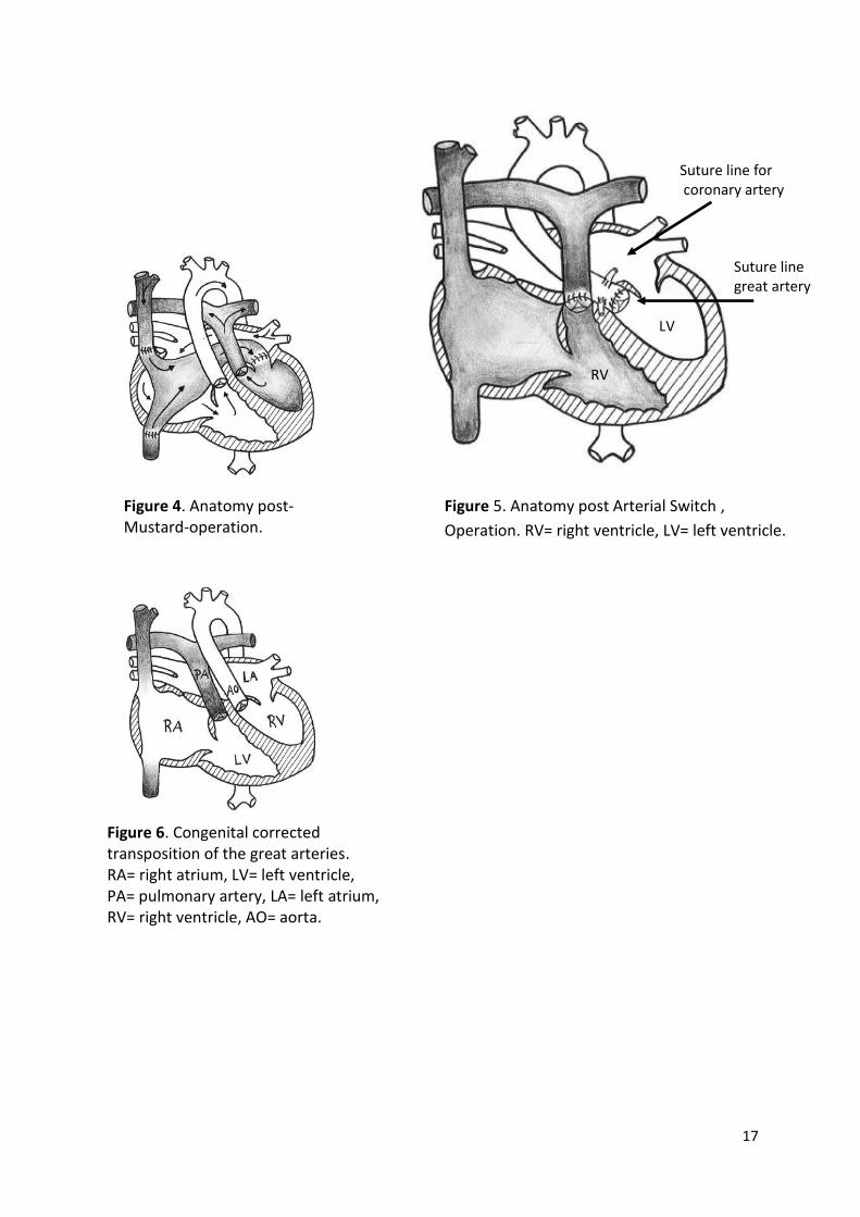
therefore Mustard designed a somewhat simpler technique in 19644 which
became the preferred method throughout the “atrial redirection” era (figure
4).

12
Simultaneously, neonatal survival was improved by percutaneous balloon
arterial septostomy5 and by prostaglandin infusion for patency of the arterial
duct in the late 1970s6.
Nevertheless long-term complications after the Mustard procedure proved
considerable. The technical difficulties with systemic and pulmonary venous
obstruction could be overcome, whereas the post operative arrhythmias and
systemic ventricular dysfunction proved fatal with a high degree of sudden
cardiac death as well as terminal heart failure. Concurrently, infant cardiac
surgery was developed in the early 1970s. The attention was re-focused on
correction of TGA by re-transposing the great arteries, i.e. the arterial switch
operation (ASO). Initial success was reported by Jatene, who operated in
infants past the neonatal period7; a procedure which was further developed by
Castaneda in neonates in 19828.
The early high mortality could be improved and ASO demonstrated fewer
sequelae than previous methods9 10. Hence, new questions arose that needed
to be addressed.
During an ASO, the arterial trunks are transected above the valvar complex,
and two of the three sinuses are incised to accommodate the coronary arterial
buttons. Transferral of the coronaries without obstructing flow is crucial when
performing ASO.
The transection and translocation of the great arteries as well as translocation
of the coronary arteries involve, as a consequence, transection of the cardiac
sympathetic nerves. Jane et al (1986)11 showed that a vast majority of the
cardiac sympathetic nerves enter the heart alongside the great arteries and are
therefore likely to be injured during the ASO. This may then cause denervation
hypersensitivity of norepinephrine (NE) receptors and a disruption of the NE
reuptake into the sympathetic nerve terminals (uptake-1)12. With time,
13
sympathetic nerves will to various degrees re-innervate the heart13, although
the sympathetic nerves re-innervating the coronary vascular bed will be
imposed with the challange of passing not only the suture line crossing the wall
of the great arteries, but also an additional suture line crossing the insertion of
the coronary arteries at their new location in the root of the neo-aorta. The
implication of an initial transection and a reinnervation of sympathetic nerves
may impede the future function and possibly lead to deterioration of the heart
after an ASO.
The autonomic nervous system (ANS) is a main regulator of the heart14. It has
been shown that autonomic dysfunction, of either the sympathetic and
parasympathetic division, is associated with clinical disorders such as ischemic
heart disease and heart failure, and that it also predicts mortality15-17. Humans
have low sympathetic activity (tone) and a dominant parasympathetic drive to
the heart during resting conditions. Cardiac response to a parasympathetic
burst allows for a quick dynamic vagal modulation of the cardiac rhythm. An
impaired parasympathetic tone has been demonstrated to contribute to
increased cardiovascular mortality and morbidity18. The anatomic innervation
of postganglionic parasympathetic nerves after ASO is assumed to not be
substantially injured, although considering the impact of the ANS in the
regulation of heart rate, there might be a possibility also for disturbed cardiac
vagal function.
The increased systemic vascular resistance (SVR) observed in patients with a
Fontan circulation has been incriminated as a responsible factor for late failure
of Fontan circulation. The mechanisms behind the increased SVR are, however,
uncertain. Lambert et al. (2012)19 investigated the muscle sympathetic nerve
activity in Fontan patients and demonstrated that the increased sympathetic
activity found was similar to that observed in patients with heart failure20.
14
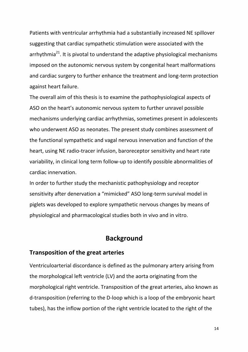
Patients with ventricular arrhythmia had a substantially increased NE spillover
suggesting that cardiac sympathetic stimulation were associated with the
arrhythmia21. It is pivotal to understand the adaptive physiological mechanisms
imposed on the autonomic nervous system by congenital heart malformations
and cardiac surgery to further enhance the treatment and long-term protection
against heart failure.
The overall aim of this thesis is to examine the pathophysiological aspects of
ASO on the heart’s autonomic nervous system to further unravel possible
mechanisms underlying cardiac arrhythmias, sometimes present in adolescents
who underwent ASO as neonates. The present study combines assessment of
the functional sympathetic and vagal nervous innervation and function of the
heart, using NE radio-tracer infusion, baroreceptor sensitivity and heart rate
variability, in clinical long term follow-up to identify possible abnormalities of
cardiac innervation.
In order to further study the mechanistic pathophysiology and receptor
sensitivity after denervation a “mimicked” ASO long-term survival model in
piglets was developed to explore sympathetic nervous changes by means of
physiological and pharmacological studies both in vivo and in vitro.
Background
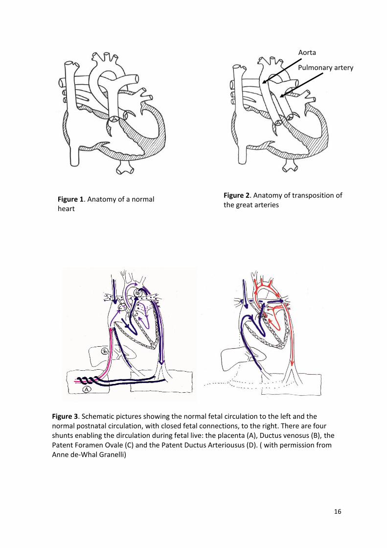
Transposition of the great arteries
Ventriculoarterial discordance is defined as the pulmonary artery arising from
the morphological left ventricle (LV) and the aorta originating from the
morphological right ventricle. Transposition of the great arteries, also known as
d-transposition (referring to the D-loop which is a loop of the embryonic heart
tubes), has the inflow portion of the right ventricle located to the right of the

15
morphological left ventricle. The great arteries become parallel rather than
crossing as in a normal heart, as the aorta tends to be on the right and anterior
(figure 2)22.
In a d-transposition, the systemic and pulmonary circulations run in parallel,
preventing oxygenation of systemic blood through the pulmonary circuit. It is
not compatible with extra-uterine life unless there is a mixing between the
pulmonary artery and the systemic circulation (figure 3). An ASD is generally
required since a patent duct alone has been proven to be insufficient to
alleviate cyanosis. Consequently, in cases without an ASD a Rashkind balloon
arterial septostomy is required if surgery is not performed within the first days
of life5.
TGA is not to be confused with congenital corrected transposition of the great
arteries, where there is atrial-ventricular discordance and ventricular-arterial
discordance, which, unless there are additional defects, is a non-cyanotic
condition, (figure 6).
Arterial Switch Operation
The ASO starts by transecting the pulmonary arteries and aorta above the
sinuses, followed by detaching the coronary arteries with a “button” from the
aortic wall. The great arteries are switched and sewn into their new positions
and the coronaries are sutured into the “neo-aorta”7, (figure 5). The “French
manoeuvre”, in which the pulmonary artery bifurcation is transferred in front
of the distal ascending aorta23, improved the technique and therefore also the
outcome.
16
Figure 2. Anatomy of transposition of the great arteries
Figure 1. Anatomy of a normal heart
Figure 3. Schematic pictures showing the normal fetal circulation to the left and the normal postnatal circulation, with closed fetal connections, to the right. There are four shunts enabling the dirculation during fetal live: the placenta (A), Ductus venosus (B), the Patent Foramen Ovale (C) and the Patent Ductus Arteriousus (D). ( with permission from Anne de-Whal Granelli)
Aorta
Pulmonary artery
17
Figure 4. Anatomy post- Mustard-operation.
Figure 5. Anatomy post Arterial Switch ,
Operation. RV= right ventricle, LV= left ventricle.
Figure 6. Congenital corrected transposition of the great arteries. RA= right atrium, LV= left ventricle, PA= pulmonary artery, LA= left atrium, RV= right ventricle, AO= aorta.
Suture line for coronary artery
Suture line great artery
RV
LV

18
A potential complication is kinking, distortion compression and complicated
coronary artery anatomy such as intramural coronaries which all might cause
obstruction of the coronary flow causing ischemia. It has been shown that
many ASO patients with obstructions in the coronary arteries and ischemia are
asymtomatic or have diffuse symptoms therefore routinely investigations
needs to be performed to detect potential ischemia even if there are no
traditionally symptoms24, 25
Autonomic nervous system
The autonomic nervous system, the internal regulatory nervous system,
regulates a large part of our organs and their functions. It includes the insular
cortex, amygdala, hypothalamus, periaqueductal gray matter, parabrachial
complex, nucleus of the tractus solitarius, and ventrolateral medulla26,
(figure 7). The ANS consists of three entities: the sympathetic nervous system
(SNS), the parasympathetic nervous system and the enteric nervous system
with plexus myentericus and plexus submucosus27. The latter system will not be
discussed in this thesis.
The aim of the ANS and its regulation of cardiovascular functions is to make
appropriate adjustments for the diverse physiological demands of the body and
maintain homeostasis. The moment-to-moment regulation is achieved by
integration of the sympathetic and parasympathetic components of the ANS.
Main neurotransmitters in the ANS are NE and epinephrine (EPI) and
acetylcholine. Dopamine (also an endogenous catecholamine) and other
transmitters (for example neuro peptide Y) and their roles will be discussed
briefly in this context

19
Sympathetic nervous system
According to Goldstein (1990, 1995) the regional innervation in the heart is
heterogenic and it is especially dense in the atria and the base of the
ventricles28-30
Fibres from the SNS also innervate the adrenal medulla and trigger release of
EPI and, to a substantially lesser extent, release of NE.
Figure 7. A schematic picture of the sympathetic and parasympathetic
innervation of the heart.

20
Neurochemistry of the sympathetic neuron
U1
U2
NE
NE
DHPG
MAO
DOPA
NE NE
Cardiac myocyte
DHPG
Tyrosine
NE
Venous side Arterial side
Sympatheticneve ending
The precursor to NE is dopamine31. The enzyme β-hydoxylase hydroxylates
dopamine into NE. The final stage of synthesis is in the synaptic vesicles. An
action potential triggers the nerve-terminal to release NE into the synaptic cleft
after which NE binds to NE-receptors postsynaptically, (figure 8). The vast
majority of released NE will be taken up into the sympathetic nerve-terminals
and its vesicles -- the so called neuronal re-uptake-1. A leakage from the
vesicles of NE into the terminals will expose NE to monoamine oxidase (MAO)
that degrades NE to dihydroxyphenylglycol (DHPG) -- the predominant intra-
neuronal metabolite. This metabolite reflects the turnover of NE and will be
released into the plasma and can, as well as NE, be measured in the blood32-34.
The majority of NE entering the sympathetic axoplasm by reuptake or by
leakage from vesicles will be transferred back into storage vesicles. A small
amount will escape and be exposed to the above described metabolism35.
Figure 8. A schematic picture of the sympathetic nerve terminal and
the release of norepinephrine (NE). DHPG=dihydroxyphenylglycol,
U1=re-uptake-1, U2=re-uptake-2, MAO=monoamine oxidase.

21
The re-uptake of NE into the nerve terminal can either be as uptake-1 (which in
the heart is almost 92 % of released NE)36, 37 or as uptake-2 (inactivated by the
post-synaptic cell), and a few % spills over into the blood stream.
Sympathetic preganglionic nerves release acetylcholine in the adrenal medulla,
which will trigger the phenyethanolamine-N-methyltransferas (PNMT) to
catalyse the transformation of NE into EPI in the adrenomedullary cells; EPI is
then released directly into the blood and is an agonist for both α-and β-
receptors38
NE and EPI have various actions at the different receptors; in general α-
receptors promote vasoconstriction while β-receptors affect the inotropic and
chronotropic capacity of the myocardium and produce vascular relaxation38.
Denervation owing to transection of sympathetic nerves by ASO is apt to cause
a disruption of the NE neuronal turnover, which may lead to a denervation
hypersensitivity of the NE- receptors12. To explore the consequences of nerve
denervation, an animal model was developed in the present study. The
advantages of such a model are that the SNS can be studied through
experimental approaches including dose-response patterns to catecholamine in
vivo. In an isolated heart preparation, in vitro, function and sensitivity of
sympathetic nerves and receptors can be studied, excluding any other
regulatory and counter-regulatory mechanisms such as vagal or humoral
responses.
The Parasympathetic nervous system
The parasympathetic ganglia are usually located close to or inside the
innervated organ, for that reason, vagal innervation consists of preganglionic
fibres; it innervates the heart and lowers the heart rate. The right vagal nerve
for the most part innervates the sinus node, whereas the left vagal nerve

22
mainly innervates the AV-node. The vagal nerve also innervates the atrial
muscle39. The vagal nervous system could be described as “rest and digest”.
Otto Loewin won (together with Sir Henry Dale) the Nobel Prize in physiology
or medicine in 1936 for the identification of a endogenous neurotransmitter,
acetylcholine40. The receptors that mediate the effects of acetylcholine are
nicotinic and muscarinic receptors. Muscarinic agonists inhibit NE release from
sympathetic nerve terminals and are selectively blocked by atropine.
Cholinergic transmission in the parasympathetic ganglia and cholinergic
stimulation of the EPI secretion from the adreno-medullary cells are mediated
by nicotinic receptors38.
Most of the inactivation of acetylcholine occurs extracellularly through
acetylcholinesterase, immediately after the release from the nerve endings.
This instantaneous inactivation makes it almost impossible to measure
acetylcholine in blood. Other methods have been developed to determine
parasympathetic nervous system function.
The pressure-sensitive nerve endings in the walls of the atria of the heart, the
carotid sinuses and the aortic arch, are called baroreceptors. They are
stimulated by central reflex mechanisms allowing physiological adjustment
through close interaction between the sympathetic and parasympathetic
nervous systems, with the aim of regulating changes in blood pressure via
altering heart rate, vasoconstriction and vasodilatation. Baroreceptors are
crucial for maintaining homeostasis41. The adjustment to alterations in blood
pressure causes beat-to-beat oscillations in RR-intervals. The oscillations can be
measured as heart rate variability42 (HRV) (figure 9).

23
Heart rate variability can be assessed by various means. Time domain analysis
measures the changes in heart rate over time or the intervals between
successive normal cardiac cycles. Frequency domain (power spectral density)
analysis describes the periodic oscillations of the heart rate signal as different
frequencies and amplitudes; and provides information regarding the amount of
their relative intensity (termed variance or power) in the heart's sinus rhythm.
Different frequencies have been identified to represent different parts of the
autonomous nervous system. High frequency (HF) (range 0.15-0.4Hz) is
considered to be driven by respiration and originates mainly from the
parasympathetic nervous system. Parasympathetic blockage, by for example
atropine, will reduce HF but it also reduces a significant part of the low
frequency (LF, range 0.04-0.15 Hz). Ganglionic blockade eliminates the residual
LF fluctuation, which implies that both sympathetic and parasympathetic
modulations contribute to LF43, 44 (figure 10). There is also very low frequency
(VLF), considered to reflect thermoregulatory cycles45 or plasma rennin
activity46.
Figure 9. Heart rate variability.

24
By measuring baroreceptor sensitivity (BRS), the entire baroreflex loop, from
spontaneous blood pressure oscillation to heart rate adjustment, is evaluated
(figure 11). BRS is a non-invasive method to assess the tone of the vagal
nervous system. A normal modulation of cardiac parasympathetic nervous
activity is considered to protect against ventricular arrhythmia47.
.
The electrocardiogram (ECG) can also be used to assess the QT variability index
(QTVI) that evaluates the repolarization of the heart and also considers also the
RR-interval48, 49. A high value of QTVI indicates a repolarization disturbance that
is a factor contributing to arrhythmogenesis.
Figure 11. Showing how systolic blood pressure (SBP) modulates
the pulse (RR) interval.
Figure 10. A schematic picture of heart rate variability with RR-interval in ms per
beats and a diagram showing the peaks of low frequency (LF) and high frequency
(HF) variability.
25
Current perspectives and unresolved issues
For patients who underwent ASO as correction for TGA in their neonatal lives,
usually within the first 2 weeks of life, cardiac problems such as arrhythmias,
heart failure and ischemic events have been reported. This may be related to
disturbances of the autonomic nervous system.
How can the ANS function in ASO patients be assessed in a feasible
clinical context, and what do the results predict in regard to future
potential heart failure?
To what extent can potential denervation hypersensitivity promote
arrhythmia in the recently ASO-operated neonate?
Is re-innervation of the heart heterogeneous and potentially pro-
arrhythmic?
Is an animal model creating the same transection of nerves that
innervates the heart possible in order to gain more knowledge of ANS
post-ASO.

26
Hypothesis
Arterial switch operation performed in neonates impairs cardiac autonomic
nervous function.
Aims of the study
To determine cardiac sympathetic neuronal function, cardiac
baroreceptor sensitivity and QT variability index in long-term follow-up
after arterial switch operation in adolescents.
To assess heart rate variability during night and day, respectively,
expressed both in time and frequency domains in long-term follow-up
after arterial switch surgery in adolescents.
Create a long-term survival piglet model, mirroring the ASO operation in
neonatal humans, to enable physiological and pharmacological studies of
sympathetic function.
To explore sympathetic responsiveness, determined as heart rate, to
catecholamines after “mimicked ASO” in piglets in vivo and ad modum
Langendorff perfusion.

27
Methodological Considerations
Study design
This thesis is based on two projects. The first one is a clinical long-term follow-
up of teenagers born with d-TGA. The second project is to develop a new
experimental piglet model, mimicking arterial switch surgery in humans to
enable us to do physiological and pharmacological studies.
Study groups
Teenagers:
Twenty-eight children born between May 1983 and October 1991 who
underwent surgery in the neonatal period using the ASO at the Sahlgrenska
University Hospital were in consecutive order of birth offered to participate in a
clinical long-term follow-up study. Seventeen of these participated in the
follow-up. Two additional children, born 1994 and 1995 were added upon
request from their paediatricians.
Healthy subjects:
Controls for cardiac neurochemical studies with tritiated NE were obtained
from a database at the Baker IDI Heart & Diabetes Institute, Melbourne,
Australia. BRS and QTVI were assessed in controls derived from a database at
the Department of Clinical Physiology at the Sahlgrenska University Hospital.
HRV controls were recruited from a database of healthy adolescents at the
Department of Clinical Physiology at the Sahlgrenska University Hospital/Östra
Sjukhuset.
Ethics: The protocols were approved by the ethics board of the University of
Gothenburg (no Ö379-02, and at the Alfred Hospital, Melbourne, Australia) and
conducted in accordance with the declaration of Helsinki (initial statement and
amendments found on the homepage of the World Medical Association,
28
www.wma.net). All subjects, and if below 18 years, also their parents, gave
informed consent.
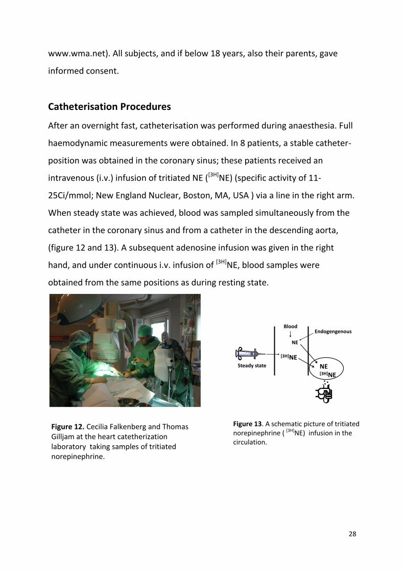
Catheterisation Procedures
After an overnight fast, catheterisation was performed during anaesthesia. Full
haemodynamic measurements were obtained. In 8 patients, a stable catheter-
position was obtained in the coronary sinus; these patients received an
intravenous (i.v.) infusion of tritiated NE ([3H]NE) (specific activity of 11-
25Ci/mmol; New England Nuclear, Boston, MA, USA ) via a line in the right arm.
When steady state was achieved, blood was sampled simultaneously from the
catheter in the coronary sinus and from a catheter in the descending aorta,
(figure 12 and 13). A subsequent adenosine infusion was given in the right
hand, and under continuous i.v. infusion of [3H]NE, blood samples were
obtained from the same positions as during resting state.
[3H]NE
Blood
NE
Endogengenous
Steady state NE[3H]NE
Figure 12. Cecilia Falkenberg and Thomas Gilljam at the heart catetherization laboratory taking samples of tritiated norepinephrine.
Figure 13. A schematic picture of tritiated norepinephrine ( [3H]NE) infusion in the circulation.

29
Plasma Norepinephrine Kinetics
Depolarisation of the nerve membrane releases NE from the sympathetic nerve
terminals. When released into the plasma NE is efficiently removed by
neuronal and extra-neuronal uptake36, 37. Only a small amount will spill over
into the circulation(figure 8)37. When estimating the release of NE from an
organ into plasma, the NE extraction has to be considered. Esler et al. (1979,
1984)32, 50 showed that the concentration of arterial NE is determined by the
ratio of the fraction of endogenously released NE that appears in plasma (the
spill over rate) to the removal of NE from plasma per unit time (clearance) from
the arterial plasma.
U1NE
NE
DHPG
MAO
DOPA
NE
DHPG
Tyrosine
NE
Venous side Arterial side
Sympatheticneve ending
3HNE
U1
3HDHPG
3HDHPG
Figure 14. Schematic picture of sympathetic nerve ending with norepinephrine turn over, including uptake of tritiated norepinephrine ([3H]NE) and its metabolising by monoamine oxidase (MAO) to tritiated dihydroxyphenylglycol ([3H]DHPG). U1= re-uptake-1, U2= re-uptake-2, DHPG= dihydroxyphenylglycol.

30
Plasma clearance can be calculated as:
NE clearance= [3H]NE infusion rate/arterial plasma [3H]NE
concentration
Whole body NE spillover can be calculated as:
Whole-body NE spillover= arterial plasma NE concentration x NA
plasma clearance
Cardiac fractional extraction of NE (EXcardiac) is calculated as:
EXcardiac = ([3H]NEA - [3H]NEV) / [3H]NEA
NEV is coronary sinus NE concentration (pmol/mL) and [3H]NEA is arterial
concentration of [3H]NE (dpm/mL).
Specific activity (SA)(isotope dilution) across an organ can be used to estimate
the endogenous release of NE37.
SANE= [3H]NEAV/NEAV
[3H]NEAV and NEAV are the respective arterial-coronary venous difference in
plasma concentration of [3H]NE (dmp/mL) and endogenous NE (pml/mL).
The gradient of [3H]DHPG, [3H]NE and DOPA between the descending aorta and
coronary sinus was also calculated (paper I).
Heart Rate Variability
Heart rate variability and blood pressure variability are believed to be the result
of a dynamic interplay of uncountable variations of the cardiovascular situation
and the response of the cardiovascular regulatory systems to these variations41.
HRV is usually evaluated from the RR-interval changes on beat-to-beat basis (a
Tachogram) and may be expressed in time-domain or frequency-domain units.
The latter requires transformation. Time-domain description is based on RR

31
changes over a period of time or number of beats. The frequency-domain
description is based on the strength (amplitude) of a set of harmonics, i.e. sine-
wave like oscillations, reflecting corresponding variability of the tachogram.
Different algorithms are used to translate heart rate and blood pressure
variability into a range of frequencies with different amplitues46, 51. In humans,
recorded frequencies usually range between 0 to 0.4 Hz and are divided into
high frequency (HF)( range 0.15-0.4Hz) and low frequency (LF)( range 0.04 to
0.15), while very low frequency (VLF)( range ≤0.04Hz) has also been
identified52. Heart rate variability evaluates the degree of autonomic
modulations rather than the level of autonomic tone53.
All 24-hour ECG analyses were performed using the ASPECT Holter System
3.80/3.81 (Danica Biomedical AB, Borlänge, Sweden). An automatic algorithm
detecting the QRS-complex was applied to the data-set54, (paper II). ECG data
were recorded using 100Hz sampling frequency. An automatic algorithm
detecting the QRS-complex onset was applied to the data set54. Prior to data
analysis, the data-set was corrected automatically for missing or ectopic beats.
Periods of more than 4% corrected beats were rejected from further analysis.
On a beat-to-beat basis, the QRS onset was defined to a nominal resolution of 1
ms using an envelope interpolation delineation technique described by Nygård
et al (1983)54.
The above procedure was carried out for the entire recording, resulting in
consecutive QRS onset intervals with 1 ms nominal resolution. From the QRS-
complex positions, an RR-tachogram was generated with a 5 Hz resolution from
which the frequency analysis was performed.
Each individual continued with his/her everyday life with no restrictions during
the recording. We aimed at analysing periods when cardiac vagal drive was
assumed to be maximal, i.e. during the night-time. Hence, HRV results from

32
such recordings would reveal the maximal cardiac vagal drive that can be
obtained at resting conditions. Day-time was also obtained for comparison. The
highest (day-time) and the lowest (night-time) heart rates, during 5 min
episodes, were identified for further analysis.
Cardiac baroreflex sensitivity
Increases or decreases in arterial blood pressure generate a response by
mutual changes in the sympathetic and parasympathetic activity30. The
baroreflex control of heart rate evaluates the sensitivity of the response in the
entire baroreflex loop to spontaneous blood pressure oscillations55, 56, and is a
sensitive method to detect cardiac vagal dysfunction that convey risk of
cardiovascular morbidity and mortality18.
ECG (RR-intervals) and beat-to-beat systolic blood pressure (SBP) recordings
were registered during 20 min using Portapres equipment in awake patients
and healthy subjects57-59, (paper I). The time series of SBP and RR-intervals were
scanned by the computer to identify baroreflex sequences, which are defined
as three or more consecutive beats in which successive SBP and RR-intervals
concordantly increase or decrease by the classical criteria suggested by
Bertinieri et al56. A change in SBP of 1mmHg and in RR-interval of 5 msec is
recorded.
QT-interval variability index
A period of 5 min with less than 5% atrial/ventricular ectopic beats was chosen
for the temporal QT interval variability analysis using a computer algorithm48, 49.
A template QT interval was defined, for one beat which was used for finding
the QT intervals of all other beats. The QT variability is measured and
33
calculated as the QT variability index adjusting to the RR-interval and
logarithmic transformation60, (paper I).
Complex cardiac surgery during cardiopulmonary bypass in piglets
Animal study groups:
Piglets of the Swedish Landrace -Yorkshire x Hampshire breed from a
commercial breeder were used. They were purchased in groups of four
littermates. Male piglets were abandoned early in the series because they had
to be castrated, in order to be kept for long term survival. Unless castrated,
they tend to become too aggressive to be kept at the research facility. In
accordance with Swedish Animal Welfare guidelines the piglets were 8.5 weeks
of age at the time of surgery.
Ethics: The study protocol was approved by the ethics committee for animal
studies at Gothenburg University (no 150/2002 and 150/2005). It conformed to
the principles of the American Physiological Society.
At the start of this study there was, to the best of our knowledge, limited
information regarding complex heart surgery during cardiopulmonary bypass
(CPB) in piglets of only 8 weeks of age61, 62.
The experimental protocol combined the paediatric cardiac surgery procedures
at the Queen Silvia Children’s Hospital with available information from a
literature search on this topic and veterinary experience at from Experimental
BioMedicine at Gothenburg University. If, for a particular drug, no veterinary
use or pharmaceutical dosing for pigs was available in the literature, paediatric
doses in mg/kg were used (picture 15-17). As this study progressed, the
equipment used for CPB changed to improve the procedure (figure 18)(paper
III).

34
Figure 15. From the left, Stefan Hallhagen,
Krister Nilsson, Nadia Karlsson, Cecilia
Falkenberg, and Ingegerd Östman-Smith.
Figure 16. The cardiopulmonary bypass set-
up.
Figure 17. Piglets post-arterial switch
surgery, picture taken the same day as the
surgery was performed.

35
In Vivo Study
A dose-response study was performed at an average age of 6 weeks post-
surgery12, 13 in the anaesthetised animal (figure 19) there was a 60 to 90 min
recovery period to prevent possible confounding factors by residual NE
response when starting the second dose-response curve with EPI between the
dose-response studies of NE and EPI. Doses of NE and EPI corresponding to
0.01, 0.02, 0.05, 0.1, 0.2, 0.4, 0.8, and 1.6 µg/kg were given. In order to be able
to measure the response of every dose in relation to basal heart rate and blood
pressure, heart rate and blood pressure responses were recorded until baseline
levels were regained. Subsequent dosing schedules were given once more in
the same fashion. Heart rate and blood pressure were monitored continuously
Arterial line
Venous line
Blood reservoir
Oxygenator
O2
Suction
Roller pump
Modified Ultra Filtration
Heater/ Cooler
oC
Substance removed
Cardioplegia 4 oC
Cardioplegia
Figure 18. Schematic set-up of the cardiopulmonary bypass.

36
using a Datex-Ohmeda S/5 (Datex-Ohmeda Division Instrumentarium Corp.,
Madison, Wisconsin, USA (paper IV).
In Vitro study
In the Intact body both nerves and humoral influences naturally affect
regulation of heart rate. By extracting the heart and performing a perfusion
with Krebs solution63 ad modum Langendorff64, 65 on the beating isolated heart,
(figure 20), a dose-response study of NE allowed assessment of the sensitivity
of alpha and beta receptors, as well as coronary flow without interaction by
nerves and humoral effects (paper IV).
Figure 19. The set-up for in vivo physiological study

37
Statistical analysis
Statistical analysis was performed on commercial software
“Statgraphics Plus v5.2 and Graph Pad Prism 4). Numerical
distributions are presented by their mean±SD. Mann-Whitney U-test
was used for inter-group comparisons, and Wilcoxon rank sum test
was used for paired comparisons given that the distribution of the
data from switch-patients were not normally distributed. The dose-
response curve from the Langendorff perfusions was assessed by
Graph Pad Prism comparing four-criteria analysis (bottom, top, Hill
slope, effective concentration for 50 percent of maximal response).
Statistical significance was defined as p<0.05.
Figure 20. Langendorff perfusion. To the left the polygrass, in the middle the Langendorff perfusion device, and to the right the heat exchanger from a cardiopulmonary bypass machine.
38
Results and Discussion
Congenital heart diseases (CHD) occur in approximately 0.8% of live births.
They are a heterogeneous group of patients who have due to surgical and
medical advances experienced incremental increases in survival beyond
childhood66. Estimates based on the current surgical mortality rate predict that
almost 1 in 150 young adults will have some form of CHD during the next
decade67, 68. Despite excellent surgical results long-term survival may lead to
patophysiological challenges and hence morbidity and mortality66.
Studies convey results of good early outcomes for TGA patients69. However, it is
important to study long-term consequences regarding plausible impairment of
both the sympathetic and parasympathetic division of the ANS66, development
of the coronary arteries70 and also myocardial ischaemia69. These can all
potentially be detrimental to the health of the TGA patient.
Long-term follow-up protocols with feasible methods for investigating and
assessing the post-operative situation and prognosis are clearly of great value.
Non-invasive methods are preferred due to the risk imposed with invasive
methods; however, they do not always have the desired sensitivity and
specificity and therefore need to be complemented with clinical invasive
methods, such as heart catheterization. In spite of all the scientific and
methodological developments it is not possible to study all potential
consequences of ASO, and the detrimental effects of imbalances in the ANS on
morbidity and mortality in humans18. The need for an animal model to enable
post-surgical investigations of injury and repair of the ANS to unravel some of
the research questions is imperative.

39
Sympathetic neurochemistry in humans
Sympathetic nervous system activation has been associated with the
development and complications of cardiovascular diseases19, 71, 72.
In heart failure, increased sympathetic activity is seen as a compensatory
mechanism73. With the use of “state of the art NE kinetics technique”50 32 used
in the present study, the ASO group demonstrated clear-cut evidence of
functioning cardiac sympathetic nerve terminals releasing NE at resting
conditions.
The substantial fall in specific activity across the heart supports the existence of
functioning cardiac sympathetic nerve terminals; however a trend was noted
towards a smaller fall in the ASO group, (figure 21). The greater the dilution of
endogenous NE across the coronary vascular bed, the lower the specific
activity. This indicates specifically endogenous NE release from cardiac
sympathetic neurons.
Figure 21. A statistically significant decrease of specific activity for tritiated norepinephrine (NE) from the arterial to the coronary sinus in healthy subjects (left panel) and switch operated patients (right panel).
40
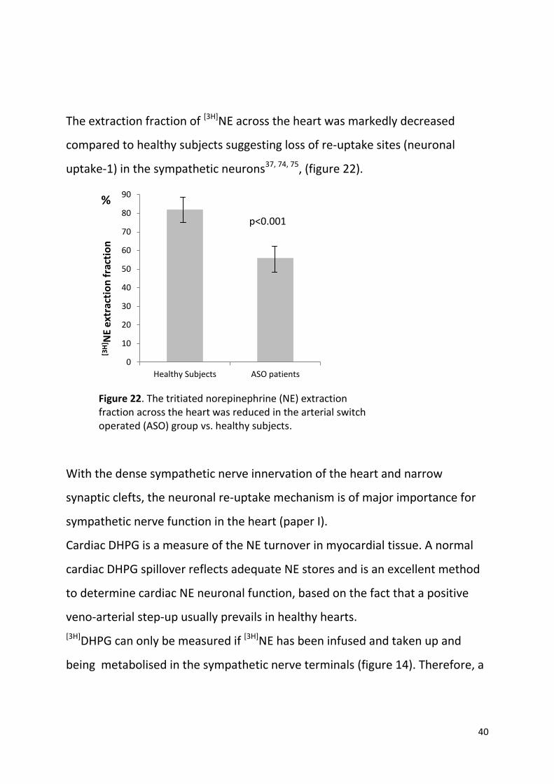
The extraction fraction of [3H]NE across the heart was markedly decreased
compared to healthy subjects suggesting loss of re-uptake sites (neuronal
uptake-1) in the sympathetic neurons37, 74, 75, (figure 22).
With the dense sympathetic nerve innervation of the heart and narrow
synaptic clefts, the neuronal re-uptake mechanism is of major importance for
sympathetic nerve function in the heart (paper I).
Cardiac DHPG is a measure of the NE turnover in myocardial tissue. A normal
cardiac DHPG spillover reflects adequate NE stores and is an excellent method
to determine cardiac NE neuronal function, based on the fact that a positive
veno-arterial step-up usually prevails in healthy hearts.
[3H]DHPG can only be measured if [3H]NE has been infused and taken up and
being metabolised in the sympathetic nerve terminals (figure 14). Therefore, a
0
10
20
30
40
50
60
70
80
90
Healthy Subjects ASO patients
[3H
] NE
extr
acti
on
fra
ctio
n
Figure 22. The tritiated norepinephrine (NE) extraction fraction across the heart was reduced in the arterial switch operated (ASO) group vs. healthy subjects.
%
p<0.001

41
measurable concentration of [3H]DHPG reflects functioning sympathetic nerve
terminals.
Both the endogenous DHPG and the [3H]DHPG had a lower gradient between
the arterial and coronary sinus in the ASO group (figure 23). These findings are
consistent with a lower NE extraction fraction, suggesting that cardiac
sympathetic neuronal function is disturbed in ASO patients at least to a limited
degree, given also the clear-cut fall in NE specific activity across the heart.
To further activate the sympathetic nerve system in general an adenosine
infusion was given to these ASO patients, which resulted in massive increase of
total body NE spillover supporting the contention of a substantial generalised
sympathetic activation, (figure 24).
Figure 23. The healthy subjects demonstrated a positive step-up between the arterial and coronary sinus for tritiated DHPG (left panel), a finding which could not be demonstrated in the arterial switch operated group (right panel).

42
After the adenosine infusion, the [3H]DHPG step up from the arterial to the
coronary sinus was increased 4-fold in the ASO group, pre-adenosin [3H]DHPG,
3.86±16.9 dpm/mL vs. post-adenosin 16.1±10 dpm/mL.
We speculate that the loss of cardiac sympathetic neurons, due to a
heterogenous innervation, is compensated for by increased NE release per
neuron of the remaining cardiac neurons.
Patients with heart failure demonstrate increased cardiac release of NE and a
decreased uptake-1. They also have a decreased leakage of NE from the
vesicles to the axoplasma of the sympathetic nerve terminals, aiding in
preventing depletion of NE in the vesicles. The increased cardiac release of NE
in such patients leads to an amplified stimulation of NE in the adrengenic post-
synaptic receptor resulting in a down regulation of these receptors35.
Consistent with such reasoning, the impairment of the sympathetic nerve
terminals caused by the ASO in the present study could impose an augmented
risk for these patients to subsequently develop heart failure and or arrythmia.
0
200
400
600
800
1000
1200
1400
1600
Pre-Adenosine Post-Adenosine
TB
NE
sp
illo
ver
pm
ol/
min
Figure 24. Total body (TB) Norepinephrine (NE) spillover increased in the arterial switch operated group after sympathetic stimulation following adenosine-infusion.
p=0.002

43
Heart rate variability, baroreceptor sensitivity and the QT interval
variability index
The regulation of changes in sequential beat-to-beat intervals reflects the
interaction between the parasympathetic and sympathetic nervous system; i.e.
the physiological regulation of the heart76 77. A high level of variability is
considered to reflect a healthy, responsive system able to regulate
physiological processes; i.e. the ability to respond to internal or external
stressors by diminishing parasympathetic activity and allowing sympathetic
activity to dominate followed by a recommencement of parasympathetic
dominance after the stress is relieved78, 79 On the other hand, a low level of
variation reflects an ANS that is less responsive to changing conditions80 A way
to evaluate LF and HF is the ratio between them which is considered a marker
of change in the relative balance between sympathetic and parasympathetic
activity81, 82.
Heart rate variability is easily assessed in a patient using ECGs and computer
algorithms. However, interpretation of the HRV variables and the implications
of the results are intriguing. A survey of the topic resulted in a so called “Task
Force” which had to develop an appropriate standard and also of give an
exposé of both the usefulness of HRV and addressing the difficulties in
interpretation52. HRV can be influenced by several factors other than
innervation and function of the ANS of the heart, such as the release of
adrenomedullary catecholamines and variations in the renin-angiotensin
system46, 83. Patients who have received heart transplants and therefore have
had a complete denervation of the heart show variation of HRV attributed to
humoral effects and to respiration84, clearly showing that analysis of HRV has
potential pit-falls.

44
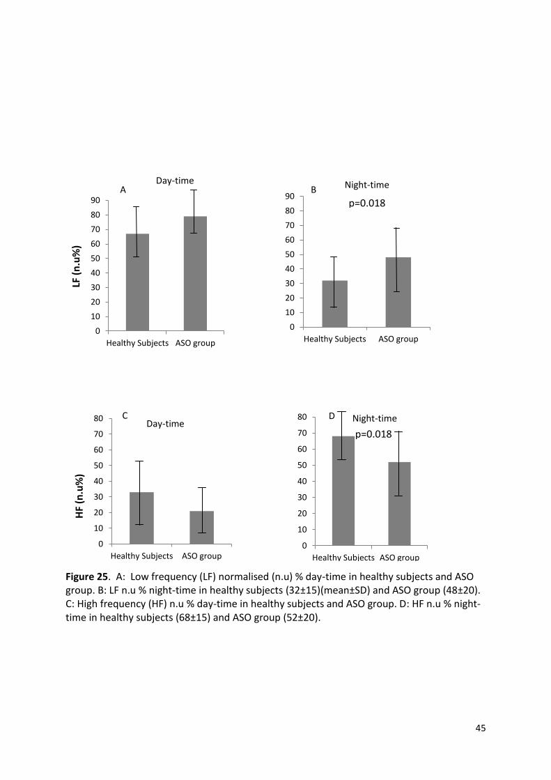
In the present study, the ASO group showed a statistically significant difference
for normalized (n.u) LF and HF, but not for LF/HF ratio, in the 24-hour
frequency analysis. In the 5 min recording during night-time, which
represented the lowest recorded values of mean heart rate for 5 min (healthy
subjects heart rate 56±8 and ASO group 52±10) there was a statistically
significant difference between the healthy group and the ASO group LF n.u, HF
n.u and LFn.u/HFn.u ration (table 2, paper II). The incremental increase of the
HF in the night-time compared to daytime for both groups and a corresponding
decrease in LF for both groups, (figure 25) suggests that ASO patients in long-
term follow-up may have a moderately reduced cardiac vagal influence that is
only detectable during sleep when the cardiac parasympathetic drive is
considered higher than during day-time.
In adults with acquired cardiac disease, sympathetic activation and
parasympathetic withdrawal have been studied85. Adults undergoing coronary
bypass surgery have reduced HF power, reflecting a reduced ability to regulate
cardiac ANS physiological function. The HF power subsequently returns to pre-
surgical levels within 30-60 days86.
Feeding during infancy is a challenge to homeostasis and a reduction in
parasympathetic activity (measured by HF power) during feeding in healthy
infants has been studied87. Adaptive reduction in HF power during feeding was
reduced in infants early after surgical correction for TGA compared to matched
healthy infants88. This study may suggest that vagal tone is affected by surgery
which would be consistent with findings in our study demonstrating a lower
45
Figure 25. A: Low frequency (LF) normalised (n.u) % day-time in healthy subjects and ASO group. B: LF n.u % night-time in healthy subjects (32±15)(mean±SD) and ASO group (48±20). C: High frequency (HF) n.u % day-time in healthy subjects and ASO group. D: HF n.u % night-time in healthy subjects (68±15) and ASO group (52±20).
0
10
20
30
40
50
60
70
80
90
Healthy Subjects ASO group
LF (
n.u
%)
Day-time
0
10
20
30
40
50
60
70
80
90
Healthy Subjects ASO group
Night-time
0
10
20
30
40
50
60
70
80
Healthy Subjects ASO group
HF
(n.u
%)
0
10
20
30
40
50
60
70
80
Healthy Subjects ASO group
Day-time Night-time C
A B
D
p=0.018
p=0.018
46
parasympathetic tone during night-time in the ASO group compared to healthy
subjects. In neonatal studies, however, one must consider that results may be
affected by examination in the hospital setting (such as the TGA infants prior to
surgery) versus the home environment.
In alignment with Kaltman et al (2006)88, our study could be interpreted as
showing that the passage of time after surgery improves the parasympathetic
function of cardiac rhythm regulation. Therefore detecting any impairment is
challenging and is mainly shown during circumstances when the
parasympathetic system tone is more influential than the sympathetic, i.e.
during night-time and sleep, (paper II).
HRV can has also been measured and analysed in time-domain. SDNN is
considered to respond to total power of the frequency domain and RMSSD to
HF. However, the present study showed no difference in regard to time-domain
between the two groups.
Baroreceptor reflexes impose a strong inhibitory effect on sympathetic outflow
and stimulate the vagal neurons. It is relevant to study these reflexes because
they operate within very short time frames and also with high frequencies of
heart rate. Baroreceptor afferents can transmit blood pressure fluctuations
accurately within high ranges of HRV. Cardiac vagal function, estimated by BRS,
revealed only a trend toward a reduction in the ASO group vs. controls. The
anatomical differences in innervation by the vagal nerve vs. the sympathetic
nerves of the heart impose less risk for vagal innervation to be damaged by the
surgical technique. This could be an explanation for preserved cardiac vagal
function, (paper I). The BRS was recorded at rest and analysed during day-time
with a more prominent sympathetic and less vagal tone prevailing, while
parasympathetic efferent activity is known to be the most influential for
governing heart rate during sleep. Since significant differences existed between
47
the groups at night-time in regard to high frequency HRV, the BRS results might
have been different had the examination been performed at night.
The QT interval variability index represents myocardial repolarisation and is
changed in patients with heart failure89. In our study, there was no statistical
difference between the two groups, although ultrasound examination revealed
signs of heart failure based on measurements of fractional shorting of the LV in
some of the patients, (see supplement, paper I). Regardless of the cause of
heart failure, it is of the outmost importance for long-term survival to detect
and treat it. The QT variability index could therefore be useful in the clinical
long-term follow-up programme.
Physiological consequences of sympathetic denervation in piglets
To our knowledge, a mimicked arterial switch operation had not been
performed in 8 week old piglets before (paper III). The logistic challenges were
many. It was essential to have highly experienced paediatric cardiac surgeons,
paediatric anaesthetists, perfusionists and the experience of animal models by
the staff at the research centre present. With, to our knowledge, limited
available information about this type of cardiac surgery in piglets we had to
resolve several problems. Piglets’ hearts are very sensitive and cardiac surgery
can easily cause arrhythmias. Hence, every detail from pre-operative
medications to post-operative care had to be scrutinised and reflected upon in
order to succeed. We usually performed surgery two days in a row and I stayed
at the research centre to observe and care for the animals, including
administration of drugs such as antibiotics and morphine, for at least the first
24 hour postoperatively. If their status then was satisfactory the staff at the
research centre would continue with the observation at that point. The lack of
a post-operative intensive care unit was solved in such a manner that the piglet

48
was extubated on the table and was observed and cared for until midnight on
the day of operation, and thereafter, at least every two hours or more if
needed. There was a protocol for post-operative pain relief and, if needed,
other medical treatments. After the introduction of blood transfusion of the
“ASO” piglets at the time of surgery the general condition of the piglets
improved after surgery. They were able to be mobilised and I could feed them
within the first few hours after they had been extubated and moved to the
post-operative pre-heated cubicles used for observation. All piglets had pleural
drains; this had to be removed at the first sign of the piglet wanting to move.
Initially, we also performed transection of either the pulmonary or the aortic
artery. The aim was to study the consequences of denervation and re-
innervation if only one of the great arteries had been transected; resulting in a
lesser degree of injury to the autonomic nervous system of the heart than
through an ASO. This had to be abandoned due to the vast amount of
resources needed; therefore we prioritized the mimicked ASO. As we
developed the ASO model in piglets we aimed to accomplish the same
denervation that the children experience after an ASO (paper IV). It became
obvious that, if successful, the model as such could be used not only for
mimicking ASO, but for a range of complex cardiac surgery procedures
unravelling potential deficiencies.
We performed open heart surgery during CPB on 23 piglets, out of which 2
underwent pulmonary transection and 2 underwent aortic resection. Of these
4, all survived. The ASO imposed a critical challenge compared to these 4
previously mentioned piglets due to translocation of the coronary arteries.
With every additional task, the risk for complications and decreased survival is
enhanced.
49
Combining the paediatric protocol used at the Queen Silvia Children’s with the
experience of the veterinarians and what we could obtain from the literature
proved successful with survival of 14 out of 19 mimicked ASO in piglets (paper
III).
Physiological studies of heart rate and blood pressure during a dose-response
curve of NE and EPI were performed in all surviving piglets, including all those
with a sham operation.
To execute the physiological study in piglets, we commenced with a pilot of 6
pigs to gain experience with the device built for the Langendorff perfusion. The
great amount of Krebs’ solution needed, 50 L, was warmed to 37 degrees °C
through a CPB heat exchanger connected to the device and was oxygenated.
The Krebs’ solution was pumped up to a container 60 cm above the heat
chamber containing the beating isolated heart. Hence, the perfusion pressure
was 60 cmH2O. This perfusion pressure was chosen because we wanted a good
perfusion at the same time as the tissues would be preserved for perfusion
fixation with formaldehyde, and not damaged, for future histology. Between
the container and the heat chamber, turbulence was created of the flowing
Krebs’ solution to ensure that the infused drug was properly mixed in the
Krebs’ solution before it reached the heart in order to be evenly distributed
into the coronary arteries. The polygrass was connected to the pipe between
the container and heat chamber and every heart beat created a pulsatile wave
in the pipe that could be recorded by the polygrass. In this manner, we could
measure the effect on heart rate by NE infused into the isolated heart.
Both the in vivo and in vitro experiments were performed in pigs up to 6
months of age. However, the size of the heart at that age made the challenges
of a successful Langendorff perfusion not feasible despite the very large device
built by us.
50
A total of 56 pigs were involved in the study of which 5 of the ASO operated
piglets succumbed; hence, the in vivo and in vitro studies were performed in 51
animals.
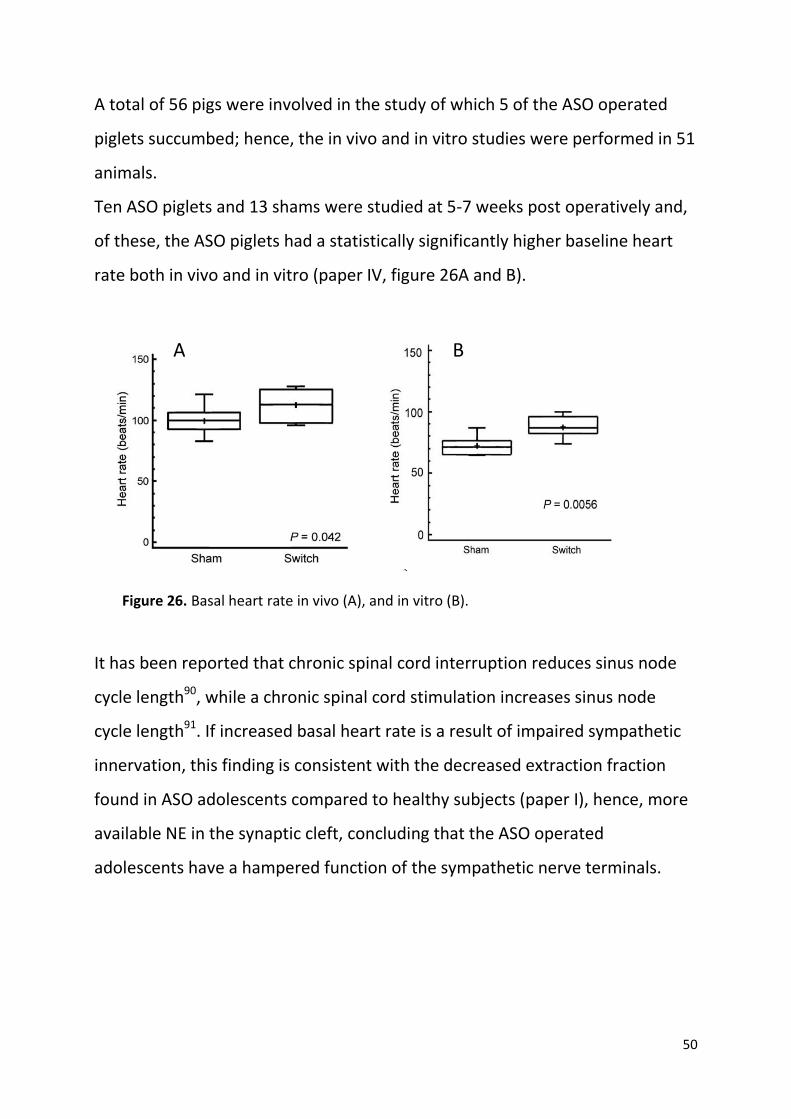
Ten ASO piglets and 13 shams were studied at 5-7 weeks post operatively and,
of these, the ASO piglets had a statistically significantly higher baseline heart
rate both in vivo and in vitro (paper IV, figure 26A and B).
It has been reported that chronic spinal cord interruption reduces sinus node
cycle length90, while a chronic spinal cord stimulation increases sinus node
cycle length91. If increased basal heart rate is a result of impaired sympathetic
innervation, this finding is consistent with the decreased extraction fraction
found in ASO adolescents compared to healthy subjects (paper I), hence, more
available NE in the synaptic cleft, concluding that the ASO operated
adolescents have a hampered function of the sympathetic nerve terminals.
Figure 26. Basal heart rate in vivo (A), and in vitro (B).
A B

51
The dose-response curve with NE in the Langendorff perfusion would have
been possible to optimize if more physiological doses could have been infused,
especially in the lower dose ranges. However, the challenges imposed by a
Langendorff perfusion are many, as mentioned above. If the osmotic pressure
of the solution cannot be maintained within physiological values, the heart will
quickly develop oedema, which further increases the risk of failure of the
experiment. Hence, we had a very limited timeframe of approximately 20 to 30
minutes in which to perform the experiments.
The results of the NE dose-response pattern in the isolated heart, using a four-
criteria statistical analysis (bottom, top, Hill slope and 50 percent of maximal
response) showed an increased sensitivity to catecholamine stimulation that is
consistent with other studies following denervation of sympathetic nerves12.
The amount of NE needed to increase the heart rate by 80% from baseline was
statistically significantly less in the “ASO” hearts compared to the controls. It
may be assumed that this was due to denervation hypersensitivity post ASO
operation. However, if adjusting baseline heart rate (given that the basal heart
rate was higher in the operated group) before every given dose of NE with the
Figure 27. Dose-response curve to norepinephrine (NE) in vitro. NE dose range used corresponded to 0.047-0.38 µmoles per litre perfusate. Statistical method used is four-criteria analysis (bottom, top, Hill slope and 50 % of maximal response).

52
relative increase of heart rate in percent after every dose, no statistical
difference between the two groups could be found. Limitations with an
experiment such as this, with the difficulties imposed by the procedure as well
as giving proper dosing, could affect the results in a manner not foreseen by us.
Possible denervation hypersensitivity is challenging from a clinical perspective;
increased sensitivity for catecholamines, which are used to increase inotropic
and chronotropic response in the heart, could be a potential hazard.
Coronary perfusion
The coronary flow at baseline, in vitro, was not different between the two
groups (Fig 6a, paper IV), although the increase in coronary flow per gram heart
weight after maximal NE stimulation was lower in stimulated ASO hearts.
Reports regarding myocardial perfusion have been controversial with
contradictory results regarding perfusion defects and coronary flow reserve92,
93. In addition, the methods to assess myocardial perfusion including control
groups have been debated94-96. Study of cardiac NE function, (paper I),
concluded that the ASO patients had functioning but likely loss of NE nerve
terminals. As angiogenesis can be prevented by chemical sympathectomy97 and
is stimulated by for example neuropeptide Y, a nerve growth factor and
transmitter released from sympathetic nerve terminals98, 99, it is essential to
have functioning sympathetic nerves for normal tissue growth. Reports of silent
ischemia in some ASO patients stress the importance of a thorough follow-up
of the coronary status in these patients.

53
General Conclusions
Survival and long-term good health in a child born with heart malformation
consist of a long sequence of challenges starting with the diagnosis and
continuing through treatments such as surgical and/or medical treatment and
follow-ups all through life.
The sympathetic nervous innervation of the heart, not only showed as
endogenous release of NE, but also showed as a functioning system while
exposed to sympathetic stimulation with adenosine in the ASO group. In
addition, the decreased extraction fraction could possibly result in a net
increase of NE concentration in the synaptic cleft leading to increased
stimulation of the post-synaptic receptors. This could potentially create the
same situation as for adults with heart failure where there is an increased
sympathetic stimulation to the heart. Tritiated DHPG can only be found if
tritiated NE has been extracted and metabolised in the sympathetic nerve
terminals. The endogenous release of NE and the decrease of tritiated DHPG in
the ASO group support the conclusion that the sympathetic nervous system in
the heart of the ASO patients is functioning, although moderately impaired.
The cardiac parasympathetic division of ANS, the effects of which are known to
be most pronounced during night-time, was found to functioning as well as in
the controls. However, when analysed using normalized HF at the lowest
measured heart rate at night, there was a difference between the two groups.
An imbalance of the parasympathetic nervous system is known to have
potential detrimental effect on cardiovascular prognosis. The impairment
found in the sympathetic system combined with a potentially decreased tone in
the parasympathetic system, makes the long-term prognosis challenging to
evaluate in order to avoid possible cardiac arrhythmias.

54
Animal studies are essential in exploring potential hazards of complex heart
surgery, which cannot be performed in humans. The increased basal heart rate,
the potentially increased sensitivity in the isolated heart to NE, and the
difference in coronary perfusion after adrenergic stimulation, support the
clinical findings of an altered heart innervation after ASO and moves the
frontier of knowledge forward.
Increased sympathetic activity in the heart is a compensatory mechanism in the
failing heart. The ASO patients’ prognosis is closely linked as to whether or not
they develop heart failure. If the children post-operatively have an increased
sensitivity to catecholamines, as shown in the animal model, there might be a
risk for arrhythmias. The neurochemical analysis reporting a functioning, but
impaired sympathetic nervous innervation and a potentially impaired cardiac
parasympathetic tone, also impose a future risk for arrhythmias and heart
failure.
Clinical Implications
Bonnet et al. concluded that a delay of post-birth diagnosis in TGA children
resulted in an increased incidence of metabolic acidosis and multi-organ
failure100. The introduction of pulse oximetry screening before discharge
improved total detection rate of duct dependent circulation to 92% 101 . This
approach could be an important tool for early detection of TGA.
The clinical challenges in long-term survival for these patients are many.
Sustained coronary perfusion is an important issue and silent ischemia makes it
important to follow myocardial perfusion continuously in these patients,
whether or not they are presented with ischemic symptoms. The debate, over
which methods are the best or the most feasible when it comes to autonomic
assessment, must also be taken into account the risk posed by invasive

55
methods. It is important to assess the ASN in ASO patients who, if they have an
imbalance of the ASN system, have a potential danger of developing
arrhythmias and heart failure. Early detection will enable them to receive
treatment to protect their hearts, and treat potential negative consequences
post-ASO during their lifetimes.

56
Acknowledgements
Peter Friberg: Thank you for your never ending support and for tutoring me
through the learning “roller-coaster”of writing a thesis with great knowledge
and a lot of laughter.
Ingegerd Östman-Smith: Thank you for creating the vision of this project and
for having the courage to believe that it was possible.
Krister Nilsson: Thank you - a true aesthetic; in anaesthesia, building the
“pluming” for heat-exchanger and “journalese” writing.
Stefan Hallhagen: Thank you for participating, sharing and performing your
surgical skills - in a slightly different setting than you normally operate in.
Christer Ericson: You “drive” the cardiopulmonary bypass machine with the
same artistic skills as you play your music. Wonderful!
Gavin Lambert: Thank you for generously allowing me to learn about
laboratory analysis in Melbourne and all the support.
Thomas Gilljam: Thank you for supporting me and sharing your knowledge
about transposition of the great arteries.
Mikael Ekberg: Thank you for revealing the mystery of computers to me and
responding to all my questions.
Nadia Karlsson: (at Experimental BioMedicine) Thank you for working all those
exceptional and extremely long hours to make sure this project would be
successful.
Gun Bodehed-Berg: Thank you for helping me and making sure that our
“samples” were well taken care of.
Elna Zetterberg: Thank you for helping me “having time” at the cath-lab, and
for encouraging me.
Boris Nilsson, for assisting with your surgical skills. The late Eva Oscarsson
whose passion to help PhD-students was always present.

57
The staff at Experimental BioMedicin for making sure that our “patients” were
excellently taken care of. Mats-Erik Nygård for helping me to up-grade the
heart rate analysis.
To my research co-worker and “sisters-in-arms” Ewa-Lena Bratt and Anne de-
Wahl Granelli for always encouraging me, all your jokes that made me laugh,
as well as assisting in this project.
All my collegues and friends at the Queen Silvia Children hospital.
All my friends around the globe - I want to thank all of you! Although, I do not
mention each of you for fear of mistakenly leaving someone out, I have to
especially thank Bodil Lindqvist who through a “massive” amount of years
through the social networks (northen to southern Sweden) relentlessly
supported me.
Thank you Khan Acadamy, Pub Med, Google and all the social networks for
helping me to find the answers I needed and to be able to keep in contact with
all my encouraging friends throughout this time.
To Nils and Armi Byström for all support through the years.
To my parents, my late father Torkel Falkenberg and my mother Karin
Falkenberg and my siblings Inger, Mårten and Bengt for all encouragement.
Maximilian, my grandson, who used his nervous system to learn how to walk
while I was using mine to write this thesis.
Björn Byström for teaching me how to skydive and how to take on the rest of
lives “rocken-roll” in the same manner, and for never hesitating to “tag along”
on the adventures that I have thrown your way.
My Winter Star, my Autumn Leaf and my Summer Flower, Julia, Evelina and
Martha, my wonderful daughters, whenever you enter the room I lack of
words.

58
This study was supported by LUA-ALF grants from Sahlgrenska University
Hospital and the Swedish Heart and Lung foundation.
59
References
1. Hoffman JI. Incidence of congenital heart disease: I. Postnatal incidence. Pediatric cardiology. 1995;16(3):103-113.
2. Paul M. Transposition of the great arteries. In: GC Emmanouilides TR, HA Allen , HP Gutgesell ed. Heart Disease in infants, Children, and Adolescents. Baltimore: Williams&Wilkins; 1995:1154-1224.
3. Senning Å. Transposition av aorta och arteria pulmonalis. Synpunkter på kirurgisk korrektion. Opuscula Medica. 1958;3(4):44-55.
4. Mustard WT. Successful Two-Stage Correction of Transposition of the Great Vessels. Surgery. 1964;55:469-472.
5. Rashkind WJ, Miller WW. Creation of an atrial septal defect without thoracotomy. A palliative approach to complete transposition of the great arteries. Jama. 1966;196(11):991-992.
6. Kachaner J. Role of prostaglandin infusion prior to surgery in complete transposition of the great arteries. In: M Vogel KB, ed. Transposition of the great arteries 25 years after Rashkind Balloon septostomy. New York: Springer; 1992:73-78.
7. Jatene AD, Fontes VF, Paulista PP, Souza LC, Neger F, Galantier M, Sousa JE. Anatomic correction of transposition of the great vessels. The Journal of thoracic and cardiovascular surgery. 1976;72(3):364-370.
8. Castaneda AR, Norwood WI, Jonas RA, Colon SD, Sanders SP, Lang P. Transposition of the great arteries and intact ventricular septum: anatomical repair in the neonate. The Annals of thoracic surgery. 1984;38(5):438-443.
9. JW Kirklin BB-B. Complete transposition of the great arteries. Cardiac surgery. 2nd ed. New York: Livingston Churchill; 1992:1383-1467.
10. Rhodes LA, Wernovsky G, Keane JF, Mayer JE, Jr., Shuren A, Dindy C, Colan SD, Walsh EP. Arrhythmias and intracardiac conduction after the arterial switch operation. The Journal of thoracic and cardiovascular surgery. 1995;109(2):303-310.
11. Janes RD, Brandys JC, Hopkins DA, Johnstone DE, Murphy DA, Armour JA. Anatomy of human extrinsic cardiac nerves and ganglia. The American journal of cardiology. 1986;57(4):299-309.
12. Trendelburg U, . Factors influencing the concentration of cathecholamines at the receptors. Handbook of Experimental Physiology 33Eds H. Blaschko and E. Muscholl. Springer Verlag, Berlin, heidelberg, New York 1972,726-761.
60
13. Kondo C, Nakazawa M, Momma K, Kusakabe K. Sympathetic denervation and reinnervation after arterial switch operation for complete transposition. Circulation. 1998;97(24):2414-2419.
14. Guyton A HJ. Nervous regulation of the circulation, and rapid control of arterial pressure. Textbook of Medical Physiology. Vol 2006. 10th ed. Philadelphia: Saunders; 2006:184-194.
15. Hasking GJ, Esler MD, Jennings GL, Burton D, Johns JA, Korner PI. Norepinephrine spillover to plasma in patients with congestive heart failure: evidence of increased overall and cardiorenal sympathetic nervous activity. Circulation. 1986;73(4):615-621.
16. Thomas JA, Marks BH. Plasma norepinephrine in congestive heart failure. The American journal of cardiology. 1978;41(2):233-243.
17. Schwartz PJ, Zaza A, Pala M, Locati E, Beria G, Zanchetti A. Baroreflex sensitivity and its evolution during the first year after myocardial infarction. Journal of the American College of Cardiology. 1988;12(3):629-636.
18. La Rover MT, Bigger JT, Jr., Marcus FI, Mortara A, Schwartz PJ. Baroreflex sensitivity and heart-rate variability in prediction of total cardiac mortality after myocardial infarction. ATRAMI (Autonomic Tone and Reflexes After Myocardial Infarction) Investigators. Lancet. 1998;351(9101):478-484.
19. Lambert E, d'Udekem Y, Cheung M, Sari CI, Inman J, Ahimastos A, Eikelis N, Pathak A, King I, Grigg L, Schlaich M, Lambert G. Sympathetic and vascular dysfunction in adult patients with Fontan circulation. International journal of cardiology. 2012, Apr 21. [Epub ahead of print]
20. Grassi G, Seravalle G, Cattaneo BM, Lanfranchi A, Vailati S, Giannattasio C, Del Bo A, Sala C, Bolla GB, Pozzi M. Sympathetic activation and loss of reflex sympathetic control in mild congestive heart failure. Circulation. 1995;92(11):3206-3211.
21. Meredith IT, Broughton A, Jennings GL, Esler MD. Evidence of a selective increase in cardiac sympathetic activity in patients with sustained ventricular arrhythmias. The New England journal of medicine. 1991;325(9):618-624.
22. Warnes CA. Transposition of the great arteries. Circulation. 2006;114(24):2699-2709.
23. Lecompte Y, Zannini L, Hazan E, Jarreau MM, Bex JP, Tu TV, Neveux JY. Anatomic correction of transposition of the great arteries. The Journal of thoracic and cardiovascular surgery. 1981;82(4):629-631.
24. Kampmann C, Kuroczynski W, Trubel H, Knuf M, Schneider M, Heinemann MK. Late results after PTCA for coronary stenosis after the

61
arterial switch procedure for transposition of the great arteries. The Annals of thoracic surgery. 2005;80(5):1641-1646.
25. Raisky O, Bergoend E, Agnoletti G, Ou P, Bonnet D, Sidi D, Vouhe PR. Late coronary artery lesions after neonatal arterial switch operation: results of surgical coronary revascularization. Eur J Cardiothorac Surg. 2007;31(5):894-898.
26. Benarroch EE. The central autonomic network: functional organization, dysfunction, and perspective. Mayo Clinic proceedings. 1993;68(10):988-1001.
27. Costa M BS, Henning GW. Anatomy and physiology of the enteric nervous system. Gut. 2000;SupplIV(47):iv15-iv19.
28. Goldstein DS, Chang PC, Eisenhofer G, Miletich R, Finn R, Bacher J, Kirk KL, Bacharach S, Kopin IJ. Positron emission tomographic imaging of cardiac sympathetic innervation and function. Circulation. 1990;81(5):1606-1621.
29. Goldstein DS, Eisenhofer G, Dunn BB, Armando I, Lenders J, Grossman E, Holmes C, Kirk KL, Bacharach S, Adams R, et al. Positron emission tomographic imaging of cardiac sympathetic innervation using 6-[18F]fluorodopamine: initial findings in humans. Journal of the American College of Cardiology. 1993;22(7):1961-1971.
30. Goldstein DS. Stress, catecholamines and cardiovascular disease. 1st ed. New York: Oxford University Press Inc; 1995.
31. Nagatsu T, Levitt M, Udenfriend S. Tyrosine Hydroxylase. the Initial Step in Norepinephrine Biosynthesis. The Journal of biological chemistry. 1964;239:2910-2917.
32. Esler M, Jennings G, Korner P, Blombery P, Sacharias N, Leonard P. Measurement of total and organ-specific norepinephrine kinetics in humans. The American journal of physiology. 1984;247(1 Pt 1):E21-28.
33. Esler M, Jennings G, Korner P, Willett I, Dudley F, Hasking G, Anderson W, Lambert G. Assessment of human sympathetic nervous system activity from measurements of norepinephrine turnover. Hypertension. 1988;11(1):3-20.
34. Goldstein DS. Historical perspective: Concepts about the autonomic nervous system and their originators. In: Goldstein DS, ed. The autonomic Nervous System in Health and Disease. New York, Basel: Marcel Edder, Inc; 2001:12
35. Eisenhofer G. Sympathetic nerve function--assessment by radioisotope dilution analysis. Clin Auton Res. 2005;15(4):264-283.
62
36. Eisenhofer G, Esler MD, Meredith IT, Dart A, Cannon RO, 3rd, Quyyumi AA, Lambert G, Chin J, Jennings GL, Goldstein DS. Sympathetic nervous function in human heart as assessed by cardiac spillovers of dihydroxyphenylglycol and norepinephrine. Circulation. 1992;85(5):1775-1785.
37. Eisenhofer G, Friberg P, Rundqvist B, Quyyumi AA, Lambert G, Kaye DM, Kopin IJ, Goldstein DS, Esler MD. Cardiac sympathetic nerve function in congestive heart failure. Circulation. 1996;93(9):1667-1676.
38. Goldstein DS. Overview of the Autonomic Naervous system. In: Goldstein DS, ed. The autonomic Nervous System in Health and Disease. New York, Basel: Marcel Dekker Inc; 2001:23-109.
39. Standish A, Enquist LW, Schwaber JS. Innervation of the heart and its central medullary origin defined by viral tracing. Science (New York, N.Y. 1994;263(5144):232-234.
40. Loewi O NE. Uber humorale Ubertragbarkeit der Herzenvenwirkung. X. Mitteilung. Uber das Schicksal des Bagusstoffs. Pflugers Arch. 1926;214:678-688.
41. Parati G, Saul JP, Di Rienzo M, Mancia G. Spectral analysis of blood pressure and heart rate variability in evaluating cardiovascular regulation. A critical appraisal. Hypertension. 1995;25(6):1276-1286.
42. Randall DC, Brown DR, Raisch RM, Yingling JD, Randall WC. SA nodal parasympathectomy delineates autonomic control of heart rate power spectrum. The American journal of physiology. 1991;260(3 Pt 2):H985-988.
43. Pomeranz B, Macaulay RJ, Caudill MA, Kutz I, Adam D, Gordon D, Kilborn KM, Barger AC, Shannon DC, Cohen RJ, et al. Assessment of autonomic function in humans by heart rate spectral analysis. The American journal of physiology. 1985;248(1 Pt 2):H151-153.
44. Rimoldi O, Pierini S, Ferrari A, Cerutti S, Pagani M, Malliani A. Analysis of short-term oscillations of R-R and arterial pressure in conscious dogs. The American journal of physiology. 1990;258(4 Pt 2):H967-976.
45. Kitney R. An analysis of the thermoregulatory influences of heart-rate variability. In: RI Kitney OR, ed. The study of heart-rate variability. Oxford: Clarendon Press.; 1980:81-106.
46. Akselrod S, Gordon D, Ubel FA, Shannon DC, Berger AC, Cohen RJ. Power spectrum analysis of heart rate fluctuation: a quantitative probe of beat-to-beat cardiovascular control. Science (New York, N.Y. 1981;213(4504):220-222.

63
47. Vanoli E, De Ferrari GM, Stramba-Badiale M, Hull SS, Jr., Foreman RD, Schwartz PJ. Vagal stimulation and prevention of sudden death in conscious dogs with a healed myocardial infarction. Circulation research. 1991;68(5):1471-1481.
48. Birkett C, Kienzle M, Myers G. Interpolation over ectopic beats increases low frequency power in heart rate variability spectra. IEEE Compter Society. Comput Cardiol. 1991:257-259.
49. Gao SA, Johansson M, Hammaren A, Nordberg M, Friberg P. Reproducibility of methods for assessing baroreflex sensitivity and temporal QT variability in end-stage renal disease and healthy subjects. Clin Auton Res. 2005;15(1):21-28.
50. Esler M JG, Bobik A, Kelleher D, Jennings G, Leonard P, Skews H, Korner P. Determination of norepinephrine apparent release rate and clearance in humans. Life Sci. 1979;25(17):1461-1470.
51. Appel ML, Berger RD, Saul JP, Smith JM, Cohen RJ. Beat to beat variability in cardiovascular variables: noise or music? Journal of the American College of Cardiology. 1989;14(5):1139-1148.
52. Malik M. Heart rate variability: standards of measurement, physiological interpretation and clinical use. Task Force of the European Society of Cardiology and the North American Society of Pacing and Electrophysiology. Circulation. 1996;93(5):1043-1065.
53. Malik M, Camm AJ. Components of heart rate variability--what they really mean and what we really measure. The American journal of cardiology. 1993;72(11):821-822.
54. Nygards ME, Sornmo L. Delineation of the QRS complex using the envelope of the e.c.g. Medical & biological engineering & computing. 1983;21(5):538-547.
55. Parlow J, Viale JP, Annat G, Hughson R, Quintin L. Spontaneous cardiac baroreflex in humans. Comparison with drug-induced responses. Hypertension. 1995;25(5):1058-1068.
56. Bertinieri G, Di Rienzo M, Cavallazzi A, Ferrari AU, Pedotti A, Mancia G. Evaluation of baroreceptor reflex by blood pressure monitoring in unanesthetized cats. The American journal of physiology. 1988;254 (2 Pt 2):H377-383.
57. Cloarec-Blanchard L. Heart rate and blood pressure variability in cardiac diseases: pharmacological implications. Fundamental & clinical pharmacology. 1997;11(1):19-28.
58. Eckberg DL. Sympathovagal balance: a critical appraisal. Circulation. 1997;96(9):3224-3232.

64
59. Imholz BP, Langewouters GJ, van Montfrans GA, Parati G, van Goudoever J, Wesseling KH, Wieling W, Mancia G. Feasibility of ambulatory, continuous 24-hour finger arterial pressure recording. Hypertension. 1993;21(1):65-73.
60. Berger RD, Kasper EK, Baughman KL, Marban E, Calkins H, Tomaselli GF. Beat-to-beat QT interval variability: novel evidence for repolarization lability in ischemic and nonischemic dilated cardiomyopathy. Circulation. 1997;96(5):1557-1565.
61. Swindle MM. Surgery, Anesthesia and Experimental Techniques in Swine: Iowa State University Press; 1998.
62. Glawischnig VE, Schlerka, G., Schuller, W., and Baumgartner, W. Arbeitswerte in der laboratoriumdiagnostik beim schwein,1977.
63. Krebs HA HK. Untersuchungen uber die Harnstoffbildung im Tierkörper. Hoppe-Seyler´s Zeitschrift fur Phsiol. Chemie. 1932;210:33-66.
64. Langendorff O. Untersuchungen am uberlebenden Säugenthiernherzen. Pflugers Arch. 1895;61:291-332.
65. Langendorff O. Geschichtliche Betrachtungen zur Methode des uberlebenden Warmbluterherzens. Munchener medizinische Wochenschrift. 1903;50:508-509.
66. Tanel RE. Preventing sudden death in the adult with congenital heart disease. Current cardiology reports. 2011;13(4):327-335.
67. Marelli AJ, Mackie AS, Ionescu-Ittu R, Rahme E, Pilote L. Congenital heart disease in the general population: changing prevalence and age distribution. Circulation. 2007;115(2):163-172.
68. Warnes CA, Liberthson R, Danielson GK, Dore A, Harris L, Hoffman JI, Somerville J, Williams RG, Webb GD. Task force 1: the changing profile of congenital heart disease in adult life. Journal of the American College of Cardiology. 2001;37(5):1170-1175.
69. Fricke TA, d'Udekem Y, Richardson M, Thuys C, Dronavalli M, Ramsay JM, Wheaton G, Grigg LE, Brizard CP, Konstantinov IE. Outcomes of the arterial switch operation for transposition of the great arteries: 25 years of experience. The Annals of thoracic surgery. 2012;94(1):139-145.
70. Pedra SR, Pedra CA, Abizaid AA, Braga SL, Staico R, Arrieta R, Costa JR, Jr., Vaz VD, Fontes VF, Sousa JE. Intracoronary ultrasound assessment late after the arterial switch operation for transposition of the great arteries. Journal of the American College of Cardiology. 2005;45(12):2061-2068.
71. Grassi G. Sympathetic neural activity in hypertension and related diseases. American journal of hypertension. 2010;23(10):1052-1060.
65
72. Lambert E, Sari CI, Dawood T, Nguyen J, McGrane M, Eikelis N, Chopra R, Wong C, Chatzivlastou K, Head G, Straznicky N, Esler M, Schlaich M, Lambert G. Sympathetic nervous system activity is associated with obesity-induced subclinical organ damage in young adults. Hypertension. 2010;56(3):351-358.
73. Gaffney TE, Braunwald E. Importance of the adrenergic nervous system in the support of circulatory function in patients with congestive heart failure. The American journal of medicine. 1963;34:320-324.
74. Kaye DM, Esler M, Kingwell B, McPherson G, Esmore D, Jennings G. Functional and neurochemical evidence for partial cardiac sympathetic reinnervation after cardiac transplantation in humans. Circulation. 1993;88(3):1110-1118.
75. Rundquist B, Eisenhofer G, Dakak NA, Elam M, Waagstein F, Friberg P. Cardiac noradrenergic function one year following cardiac transplantation. Blood pressure. 1993;2(4):252-261.
76. De Jong MJ, Randall DC. Heart rate variability analysis in the assessment of autonomic function in heart failure. The Journal of cardiovascular nursing. 2005;20(3):186-195; quiz 196-187.
77. Ohuchi H, Takasugi H, Ohashi H, Okada Y, Yamada O, Ono Y, Yagihara T, Echigo S. Stratification of pediatric heart failure on the basis of neurohormonal and cardiac autonomic nervous activities in patients with congenital heart disease. Circulation. 2003;108(19):2368-2376.
78. Porges SW, Byrne EA. Research methods for measurement of heart rate and respiration. Biological psychology. 1992;34(2-3):93-130.
79. Porges SW. Love: an emergent property of the mammalian autonomic nervous system. Psychoneuroendocrinology. 1998;23(8):837-861.
80. Pumprla J, Howorka K, Groves D, Chester M, Nolan J. Functional assessment of heart rate variability: physiological basis and practical applications. International journal of cardiology. 2002;84(1):1-14.
81. Malliani A, Pagani M, Lombardi F. Physiology and clinical implications of variability of cardiovascular parameters with focus on heart rate and blood pressure. The American journal of cardiology. 1994;73(10):3C-9C.
82. Montano N, Ruscone TG, Porta A, Lombardi F, Pagani M, Malliani A. Power spectrum analysis of heart rate variability to assess the changes in sympathovagal balance during graded orthostatic tilt. Circulation. 1994;90(4):1826-1831.
83. Bonaduce D, Marciano F, Petretta M, Migaux ML, Morgano G, Bianchi V, Salemme L, Valva G, Condorelli M. Effects of converting enzyme inhibition on heart period variability in patients with acute myocardial infarction. Circulation. 1994;90(1):108-113.

66
84. Berntson GG, Bigger JT, Jr., Eckberg DL, Grossman P, Kaufmann PG, Malik M, Nagaraja HN, Porges SW, Saul JP, Stone PH, van der Molen MW. Heart rate variability: origins, methods, and interpretive caveats. Psychophysiology. 1997;34(6):623-648.
85. Stein PK, Domitrovich PP, Huikuri HV, Kleiger RE. Traditional and nonlinear heart rate variability are each independently associated with mortality after myocardial infarction. Journal of cardiovascular electrophysiology. 2005;16(1):13-20.
86. Soares PP, Moreno AM, Cravo SL, Nobrega AC. Coronary artery bypass surgery and longitudinal evaluation of the autonomic cardiovascular function. Critical care (London, England). 2005;9(2):R124-131.
87. Lappi H, Valkonen-Korhonen M, Georgiadis S, Tarvainen MP, Tarkka IM, Karjalainen PA, Lehtonen J. Effects of nutritive and non-nutritive sucking on infant heart rate variability during the first 6 months of life. Infant behavior & development. 2007;30(4):546-556.
88. Kaltman JR, Hanna BD, Gallagher PR, Gaynor JW, Godinez RI, Tanel RE, Shah MJ, Vetter VL, Rhodes LA. Heart rate variability following neonatal heart surgery for complex congenital heart disease. Pacing Clin Electrophysiol. 2006;29(5):471-478.
89. Tomaselli GF, Beuckelmann DJ, Calkins HG, Berger RD, Kessler PD, Lawrence JH, Kass D, Feldman AM, Marban E. Sudden cardiac death in heart failure. The role of abnormal repolarization. Circulation. 1994;90(5):2534-2539.
90. Rodenbaugh DW, Collins HL, DiCarlo SE. Increased susceptibility to ventricular arrhythmias in hypertensive paraplegic rats. Clin Exp Hypertens. 2003;25(6):349-358.
91. Olgin JE, Takahashi T, Wilson E, Vereckei A, Steinberg H, Zipes DP. Effects of thoracic spinal cord stimulation on cardiac autonomic regulation of the sinus and atrioventricular nodes. Journal of cardiovascular electrophysiology. 2002;13(5):475-481.
92. Vogel M, Smallhorn JF, Gilday D, Benson LN, Ash J, Williams WG, Freedom RM. Assessment of myocardial perfusion in patients after the arterial switch operation. J Nucl Med. 1991;32(2):237-241.
93. Weindling SN, Wernovsky G, Colan SD, Parker JA, Boutin C, Mone SM, Costello J, Castaneda AR, Treves ST. Myocardial perfusion, function and exercise tolerance after the arterial switch operation. Journal of the American College of Cardiology. 1994;23(2):424-433.

67
94. Acar P, Maunoury C, Bonnet D, Sebahoun S, Bonhoeffer P, Saliba Z, Sidi D, Kachaner J. Comparison of myocardial perfusion single-photon emission computed tomography with coronary artery angiography after arterial switch operation. The American journal of cardiology. 2001;87(12):1425-1427.
95. Schwitter J, Nanz D, Kneifel S, Bertschinger K, Buchi M, Knusel PR, Marincek B, Luscher TF, von Schulthess GK. Assessment of myocardial perfusion in coronary artery disease by magnetic resonance: a comparison with positron emission tomography and coronary angiography. Circulation. 2001;103(18):2230-2235.
96. Manso B, Castellote A, Dos L, Casaldaliga J. Myocardial perfusion magnetic resonance imaging for detecting coronary function anomalies in asymptomatic paediatric patients with a previous arterial switch operation for the transposition of great arteries. Cardiology in the young.20(4):410-417.
97. Ostman-Smith I. Prevention of exercise-induced cardiac hypertrophy in rats by chemical sympathectomy (guanethidine treatment). Neuroscience. 1976;1(6):497-507.
98. Zukowska-Grojec Z, Karwatowska-Prokopczuk E, Fisher TA, Ji H. Mechanisms of vascular growth-promoting effects of neuropeptide Y: role of its inducible receptors. Regulatory peptides. 1998;75-76:231-238.
99. Lazarovici P, Marcinkiewicz C, Lelkes PI. Cross talk between the cardiovascular and nervous systems: neurotrophic effects of vascular endothelial growth factor (VEGF) and angiogenic effects of nerve growth factor (NGF)-implications in drug development. Current pharmaceutical design. 2006;12(21):2609-2622.
100. Bonnet D, Coltri A, Butera G, Fermont L, Le Bidois J, Kachaner J, Sidi D. Detection of transposition of the great arteries in fetuses reduces neonatal morbidity and mortality. Circulation. 1999;99(7):916-918.
101. de-Wahl Granelli A, Wennergren M, Sandberg K, Mellander M, Bejlum C, Inganas L, Eriksson M, Segerdahl N, Agren A, Ekman-Joelsson BM, Sunnegardh J, Verdicchio M, Ostman-Smith I. Impact of pulse oximetry screening on the detection of duct dependent congenital heart disease: a Swedish prospective screening study in 39,821 newborns. BMJ (Clinical research ed. 2009;338:a3037.