Embed Size (px)
Citation preview
Contralateral Effects and Binaural Interactionsin Dorsal Cochlear Nucleus
KEVIN A. DAVIS
Departments of Biomedical Engineering and Neurobiology and Anatomy and the Center for Navigation and CommunicationSciences, University of Rochester, Rochester, NY 14642, USA
Received: 26 January 2005; Accepted: 15 June 2005; Online publication: 2 August 2005
ABSTRACT
The dorsal cochlear nucleus (DCN) receives afferentinput from the auditory nerve and is thus usuallythought of as a monaural nucleus, but it also receivesinputs from the contralateral cochlear nucleus as wellas descending projections from binaural nuclei. Ev-idence suggests that some of these commissural andefferent projections are excitatory, whereas othersare inhibitory. The goals of this study were to invest-igate the nature and effects of these inputs in theDCN by measuring DCN principal cell (type IV unit)responses to a variety of contralateral monaural andbinaural stimuli. As expected, the results of contra-lateral stimulation demonstrate a mixture of excita-tory and inhibitory influences, although inhibitoryeffects predominate. Most type IV units are weakly, ifat all, inhibited by tones but are strongly inhibited bybroadband noise (BBN). The inhibition evoked byBBN is also low threshold and short latency. This in-hibition is abolished and excitation is revealed whenstrychnine, a glycine-receptor antagonist, is appliedto the DCN; application of bicuculline, a GABAA-receptor antagonist, has similar effects but does notblock the onset of inhibition. Manipulations ofdiscrete fiber bundles suggest that the inhibitory,but not excitatory, inputs to DCN principal cells en-ter the DCN via its output pathway, and that the shortlatency inhibition is carried by commissural axons.Consistent with their respective monaural effects, re-sponses to binaural tones as a function of interaural
level difference are essentially the same as responsesto ipsilateral tones, whereas binaural BBN responsesdecrease with increasing contralateral level. In com-parison to monaural responses, binaural responses tovirtual space stimuli show enhanced sensitivity to theelevation of a sound source in ipsilateral space butreduced sensitivity in contralateral space. These re-sults show that the contralateral inputs to the DCNare functionally relevant in natural listening condi-tions, and that one role of these inputs is to enhanceDCN processing of spectral sound localization cuesproduced by the pinna.
Keywords: dorsal cochlear nucleus, contralateralinputs, binaural interactions, sound localization
INTRODUCTION
The dorsal cochlear nucleus (DCN) receives afferentinput from the auditory nerve (Osen 1970; Ryugoand May 1993) and is thus usually thought of as amonaural nucleus. In response to ipsilateral stimula-tion, DCN principal cells in cat are well known toexhibit type IV unit response properties (Young andBrownell 1976; Young 1980). Type IV units areexcited by low-level best frequency (BF) tones andinhibited by high-level tones, but are excited bybroadband noise at all levels. Type IV units also re-spond with inhibition when a spectral notch is placedat their BF (Spirou and Young 1991). By virtue oftheir unusual sensitivity to narrowband peaks andnotches, such as those added to sounds by the di-rectionally dependent filtering properties of thepinna (Musicant et al. 1990; Rice et al. 1992), DCNprincipal cells are thought to serve a role in the
Correspondence to: Kevin A. Davis I Department of Neurobiology andAnatomy I University of Rochester I 601 Elmwood Ave., Box 603Rochester, NY 14642, USA. Telephone: (585) 273-4844; fax: (585)756-5334; email: [email protected]
JARO 6: 280–296 (2005)DOI: 10.1007/s10162-005-0008-5
280
JAROJournal of the Association for Research in Otolaryngology
detection of the spatial location of sound sources(Young et al. 1992; Imig et al. 2000). Consistent withthis interpretation, lesioning the output pathway ofthe DCN disrupts the sound orientation behaviors ofcats (Sutherland et al. 1998; May 2000).
Several lines of evidence, however, suggest thepotential for functionally significant binaural inter-actions at the level of the DCN. First, anatomicstudies have shown that the DCN receives inputsfrom the contralateral cochlear nucleus (Adams andWarr 1976; Cant and Gaston 1982; Shore et al. 1992;Schofield and Cant 1996; Alibardi 2000; Arnott et al.2004), as well as descending projections from binau-ral nuclei (superior olive: Brown et al. 1988; Ostapoffet al. 1997; inferior colliculus: Conlee and Kane 1982;Schofield 2001; auditory cortex: Weedman andRyugo 1996). Some of these projections are excitato-ry, whereas others are inhibitory. Moreover, theinhibitory inputs are both glycinergic and GABAergic(Wenthold 1987; Ostapoff et al. 1997; Alibardi 2000).Second, electrical stimulation of the contralateral au-ditory nerve in an in vitro whole brain preparationproduces inhibitory postsynaptic potentials in 80% ofDCN principal cells (Babalian et al. 1999). Thelatencies of these inhibitory responses are suggestiveof mono- and disynaptic connections from the con-tralateral cochlear nucleus. Finally, single-unit studieshave demonstrated that DCN neurons are sensitive toacoustic stimulation of the contralateral ear (Mast1970, 1973; Young and Brownell 1976; Evans andZhao 1993; Joris and Smith 1998; Imig et al. 2000).Some units are excited by monaural stimulation ofthe contralateral ear, but others are inhibited. Lim-ited data suggest that this inhibition is, at least inpart, glycinergic (i.e., it is blocked by the glycine an-tagonist strychnine; Evans and Zhao 1993). In somestudies, responses to binaural stimulation are mark-edly different from those to ipsilateral stimulation sug-gesting a functional relevance for these binauralinteractions (Mast 1970; Young and Brownell 1976),whereas in other studies, the effects are small (Jorisand Smith 1998; Imig et al. 2000).
The goals of this study were to gain further in-formation about the nature and effects of contrala-teral inputs to the DCN by sampling type IV unitresponses to a variety of contralateral and binauralstimuli. The results support prior observations thatmost, but not all, type IV units are inhibited by con-tralateral stimulation. This inhibition is stronger fornoise than for tones, and it is also lower in thresholdand shorter in latency. Application of either strych-nine or bicuculline (a GABAA antagonist) to therecording site blocks this inhibition and reveals un-derlying excitation; however, only strychnine blocksthe onset of inhibition. Manipulations of the acousticstriae suggest that the inhibitory, but not excitatory,
inputs to the DCN enter the DCN via its outputpathway, and that the short latency inhibition iscarried by axons from the contralateral cochlearnucleus. Consistent with the relative strengths ofmonaural effects, binaural inhibition is stronger fornoise than for tones. Compared to monauralresponses, binaural responses to virtual space stimulishow enhanced sensitivity to the location of a soundsource in ipsilateral space but reduced sensitivityin contralateral space. These results show that con-tralateral inputs to the DCN are functionally rele-vant in natural listening conditions, and that onerole of these inputs is to enhance DCN processingof spectral sound localization cues produced by thepinna.
METHODS
Experiments were performed on 11 adult cats (3–4 kg) with infection-free ears and clear tympanic mem-branes. The first nine of these experiments wereconducted at Johns Hopkins University (JHU); the lasttwo were conducted at the University of Rochester(UR). All of the following procedures were carried outusing similar protocols approved by the InstitutionalAnimal Care and Use Committee of JHU and theUniversity Committee on Animal Resources at theUR.
Surgical procedures
Cats were anesthetized with ketamine (40 mg/kg,im) and xylazine (0.5 mg/kg, im) and were givenatropine (0.05 mg/kg, im) to minimize respiratorysecretions and dexamethasone (2 mg/kg, im) to re-duce cerebral edema. Body temperature was main-tained at 39 T 0.5-C using a regulated heating blanket,and at the UR, breathing and heart rates were mon-itored. The cephalic vein was cannulated to allow in-travenous infusions of fluids, including supplementaldoses of ketamine (15 mg/kg) and xylazine (0.1 mg/kg) as needed (e.g., heart rate over 180 beats/min),and a tracheotomy was performed to facilitate quietbreathing.
A midline incision was made over the skull andthe temporalis muscles reflected to visualize the topof the skull and the ear canals. A craniotomy was per-formed over parietal cortex, and cats were made de-cerebrate by aspirating the brainstem between thesuperior colliculus and the thalamus. Anesthesiawas then discontinued. The ear canals were trans-ected near the tympanic membrane to accept hol-low ear bars for delivering closed-field acousticstimuli. The animal’s head was fixed in the record-ing position, 35- nose down with respect to stereo-
DAVIS: Contralateral Effects and Binaural Interactions in DCN 281
taxic horizontal coordinates, using a headpiece andtwo ear bars. The left DCN was visualized by removingthe skull about the nuchal ridge and aspirating theoverlying cerebellum. In some experiments, thecerebellum above the floor of the fourth ventricle wasalso aspirated to allow access to the acoustic striae. Atthe end of experiments, cats were euthanized with anoverdose of sodium pentobarbital (100 mg/kg, iv).Some cats were perfused to allow histological confir-mation of thecompleteness ofdecerebration andof theplacement of pharmacological electrodes.
Acoustic stimuli
Acoustic stimuli were delivered bilaterally via electro-static speakers that were coupled to hollow ear bars.At the start of each experiment, the frequency re-sponse of both systems was measured with a probetube microphone that was inserted into the ear barsnear the tympanic membrane. At JHU, the acousticcalibrations were relatively flat (õ100 dB SPL T 5 dB)across frequency from 40 Hz to 40 kHz and similar inboth ears (T2 dB). Therefore, applying equal atten-uation to binaural tones of the same frequency wasassumed to create a 0-dB interaural level difference(ILD). At the UR, the attenuations were adjustedseparately for the two ears to compensate for any dif-ferences in the calibration curves. At both institu-tions, interaural cross talk was at least 30 dB (andtypically 950 dB) down at all frequencies in the earopposite to the sound source. The physiologically ef-fective interaural cross talk was measured by means ofbinaural threshold differences to tones for single au-ditory nerve fibers and cochlear microphonics (JHUsystem: Gibson 1982) or for single DCN type IV units(UR system). In the latter case, acoustic crossoverwas easily detected when the response to high lev-el contralateral tones took the characteristic highlynonmonotonic form elicited by low-level ipsilateraltones.
All test stimuli, including tones, broadband noise,notch noise, and virtual space stimuli, were 200 ms induration, gated on and off with 10-ms rise/fall times,and presented once per second. The wideband stim-uli were synthesized on-line in the frequency domainand converted to time domain waveforms by takingthe inverse Fourier transform of the digitally creatednoise spectrum (Nelken and Young 1997). Generichead-related transfer functions (HRTFs; Rice et al.1992) were used to filter broadband noise spectra tosynthesize binaural virtual space (VS) stimuli at azi-muths ranging from _60 to +60- in 15- steps andelevations from _30 to +45- in 7.5- steps. At theUR, the magnitude spectra of all wideband stimuliwere corrected to compensate for nonflat calibrationcurves. Analog signals were created by playing the
waveforms through a 16-bit D/A converter at a (usual)sampling rate of 100 kHz.
Recording protocol
All experiments were carried out in a double-walledsound-attenuating booth. Single-unit activity was re-corded with platinum–iridium electrodes. At JHU,electrodes were advanced into the DCN using a hy-draulic microdrive. The signal from the electrode wasamplified (10,000–30,000�) and filtered from 0.3 to6 kHz. A variable-threshold Schmitt trigger was usedto discriminate action potentials from backgroundnoise. At the UR, an Alpha–Omega system was usedto advance the electrodes (via a motor-controlledmultielectrode positioning system or EPS), to condi-tion the signal (MCP-Plus), and to detect action po-tentials (template matching software, MSD). In allexperiments, spike times relative to stimulus onsetwere stored for off-line analysis. Sound-driven activitywas analyzed in terms of average discharge rates overthe final 150 ms of the stimulus-on interval to reflectsteady-state responses; spontaneous rates were com-puted over the last 400 ms of the stimulus-off intervalof each 1-s stimulation period.
Recording electrodes were advanced dorsoventral-ly through the DCN, whereas 50-ms search tones ornoise bursts were presented to the ipsilateral ear.When a single unit was isolated, its BF and thresholdwere determined using audiovisual feedback, and itsresponse type was determined from responses to 200-ms BF-tone and broadband noise bursts presentedacross a range of sound levels (100-dB range in 1-dBsteps). Units were classified as type IV, IV-T, III, II, orcomplex spiking using standard criteria (e.g., Shofnerand Young 1985; Zhang and Oertel 1993a; Maniset al. 1994). Results are primarily reported for type IVunits because DCN principal cells in cat usually ex-hibit these response properties (Young 1980). Once aunit was classified, rate-level functions were obtainedfor tones (at the ipsilateral BF) and broadband noisepresented to the contralateral ear. Frequency re-sponse maps for contralateral tones were created bysweeping the frequency of tone bursts over a three-octave range centered on the unit’s ipsilateral BF.These sweeps were presented at multiple sound lev-els, ranging from 0 to 60 dB above threshold. Eachfrequency–intensity combination was presented once.
The pharmacology of the contralateral inputs tothe DCN was investigated by infusing the recordingsite with either the glycine antagonist strychnine orthe GABAA antagonist bicuculline. Piggyback multi-barreled electrodes (after Havey and Caspary 1980)were used to deliver strychnine hydrochloride or bi-cuculline methiodide (each 10 mM, pH 3.5–4.0, Sig-ma) into the DCN (Davis and Young 2000). Electrode
282 DAVIS: Contralateral Effects and Binaural Interactions in DCN
negative retention currents of 20 nA and ejectioncurrents of 50 nA were produced with microionto-phoresis constant current generators. Rate-level func-tions for contralateral BF tones and broadband noisebursts were performed before and after pharmaco-logical manipulations to verify unit stability and toconfirm recovery from inhibitory blockade beforemoving on to the next unit.
The origin of contralateral effects in the DCN wasstudied using both pharmacological and surgical ma-nipulations of the dorsal output tracts (dorsal andintermediate acoustic striae). Hypodermic needles(30 gauge) were used to record from, and to deliversolutions of, lidocaine hydrochloride (2%; TechAmerica) into the contralateral DAS/IAS and DAS.Needles were coupled to a 1-ml syringe to allow reli-able injections of microliter amounts of lidocaine so-lution. The needles were either placed at the medialborder of the contralateral DCN, where the fibersof the DAS and IAS intermingle, or moved medial to-ward the midline of the floor of the fourth ventricle toisolate the DAS (Fernandez and Karapas 1967; Davis2002). Placement of the needle was guided by search-ing for noise-evoked background activity where theoutput tracts were expected to be found. The needlewas placed in a position judged to the center of thetract(s) (i.e., where there was the maximum back-ground activity), and then left there for the entire ex-periment. In some of these experiments, the medialborder of the ipsilateral DCN was aspirated to severfibers incoming through the ipsilateral DAS/IAS.
For binaural testing, two paradigms were used.First, the same pure tone or broadband noise stim-ulus was presented to both ears, but a 40-dB range ofILDs was created by varying the level of the contra-lateral stimulus relative to a fixed ipsilateral stimulus.The intensity of the ipsilateral stimulus was fixed at10-dB re threshold. Second, responses were obtainedto ipsilateral monaural and binaural VS stimulation.The sampling rate for these stimuli was set on a unit-by-unit basis to (BF/12) � 100 kHz. This stimulusmanipulation, which effectively shifted the BF of eachunit under study to 12 kHz, was performed because12 kHz is near the center of the frequency range (5–18 kHz) where cat HRTFs show directionally depen-dent spectral notches (Musicant et al. 1990; Riceet al. 1992). Furthermore, it increased the yield ofdata at one frequency and thereby allowed the datato be subjected to statistical analyses.
RESULTS
The effects of contralateral monaural stimulationwith pure tones and broadband noise (BBN) werestudied on 54 type IV units. To examine further the
nature and origin of the contralateral inputs to theDCN, different subsets of these units were tested un-der local blockade of inhibitory receptors (n = 8) andafter manipulations of the dorsal output tracts (n =18). Binaural interaction data were acquired on 20units.
Effects of contralateral monaural acousticstimulation on type IV units
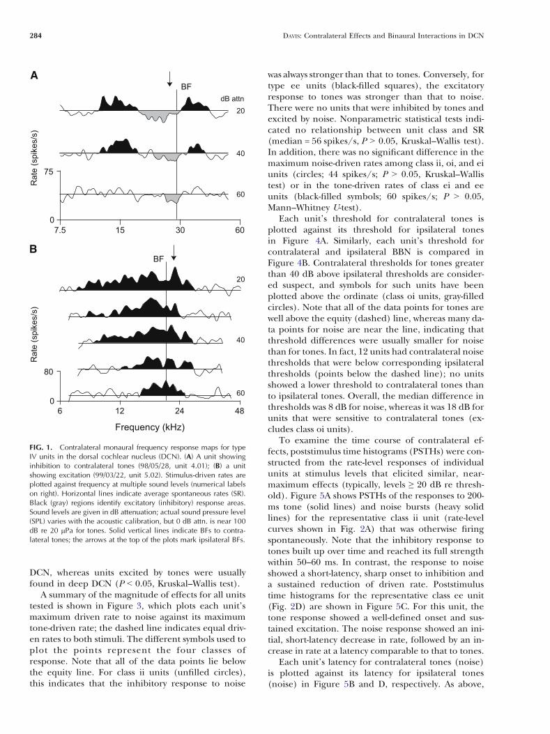
Frequency response maps for contralateral toneswere obtained for 10 type IV units. Five of theseunits showed evidence of inhibitory effects, threeshowed strictly excitatory effects, and two showed noeffects. Response maps for a unit inhibited bycontralateral tones and a unit excited by tones areshown in Figure 1A and B, respectively. In theseplots, inhibitory areas (gray fill) are defined as stim-ulus conditions that elicited responses Q1 standarddeviation (SD) below the average spontaneous rate(SR; horizontal lines); similarly, excitatory areas(black fill) indicate tone-driven rates Q1 SD abovethe SR. Inhibited units (Fig. 1A) have narrow V-shaped inhibitory areas that widen slightly about unitBF (vertical line) with increasing sound levels. Thisinhibition may, as shown here, or may not be flankedon both sides by excitation. Excited units (Fig. 1B)show broad V-shaped excitatory areas centered onBF. These units do not show inhibitory responses topure tones. The BFs for ipsilateral tones are indicatedby the arrows at the top of these plots. In all casesshowing a response, the BFs for contralateral andipsilateral tones differed by less than 0.2 octaves.
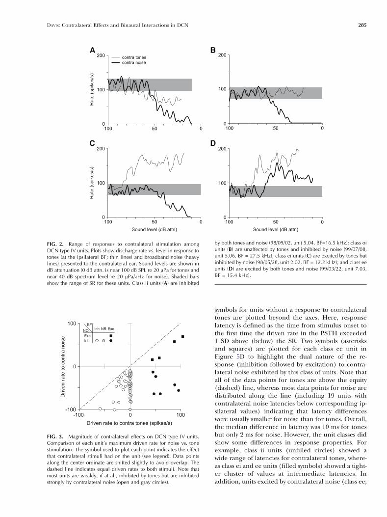
The responses of type IV units to stimulation ofthe contralateral ear can be divided into four groupsbased on the patterns of excitation and inhibitionrevealed in their rate versus level functions for puretones (at the ipsilateral BF) and BBN. Representativedata for each class are shown in Figure 2, whereresponses to tones are shown with thin solid lines andresponses to noise are shown with heavy solid lines.In these plots, a response was said to be excitatory(inhibitory) if the maximum driven rate was 1 SDabove (below) the SR. Responses with thresholdsgreater than 40 dB above ipsilateral thresholds to thesame stimulus were rejected because of the possibilityof cross talk. Most units were strongly inhibited byBBN (50/54 cases), and most of these were alsoweakly (16/50; class ii; Fig. 2A), if at all (28/50; classoi; Fig. 2B), inhibited by tones; the remainder wereexcited by tones (6/50; class ei; Fig. 2C). Only 4 of 54units were excited by noise, and all of these werealso excited by tones (class ee; Fig. 2D). No rela-tionship was found between unit class and BF (from 4to 40 kHz). However, units inhibited by tones weremore likely to be located closer to the surface of the
DAVIS: Contralateral Effects and Binaural Interactions in DCN 283
DCN, whereas units excited by tones were usuallyfound in deep DCN (P G 0.05, Kruskal–Wallis test).
A summary of the magnitude of effects for all unitstested is shown in Figure 3, which plots each unit’smaximum driven rate to noise against its maximumtone-driven rate; the dashed line indicates equal driv-en rates to both stimuli. The different symbols used toplot the points represent the four classes ofresponse. Note that all of the data points lie belowthe equity line. For class ii units (unfilled circles),this indicates that the inhibitory response to noise
was always stronger than that to tones. Conversely, fortype ee units (black-filled squares), the excitatoryresponse to tones was stronger than that to noise.There were no units that were inhibited by tones andexcited by noise. Nonparametric statistical tests indi-cated no relationship between unit class and SR(median = 56 spikes/s, P 9 0.05, Kruskal–Wallis test).In addition, there was no significant difference in themaximum noise-driven rates among class ii, oi, and eiunits (circles; 44 spikes/s; P 9 0.05, Kruskal–Wallistest) or in the tone-driven rates of class ei and eeunits (black-filled symbols; 60 spikes/s; P 9 0.05,Mann–Whitney U-test).
Each unit’s threshold for contralateral tones isplotted against its threshold for ipsilateral tonesin Figure 4A. Similarly, each unit’s threshold forcontralateral and ipsilateral BBN is compared inFigure 4B. Contralateral thresholds for tones greaterthan 40 dB above ipsilateral thresholds are consider-ed suspect, and symbols for such units have beenplotted above the ordinate (class oi units, gray-filledcircles). Note that all of the data points for tones arewell above the equity (dashed) line, whereas many da-ta points for noise are near the line, indicating thatthreshold differences were usually smaller for noisethan for tones. In fact, 12 units had contralateral noisethresholds that were below corresponding ipsilateralthresholds (points below the dashed line); no unitsshowed a lower threshold to contralateral tones thanto ipsilateral tones. Overall, the median difference inthresholds was 8 dB for noise, whereas it was 18 dB forunits that were sensitive to contralateral tones (ex-cludes class oi units).
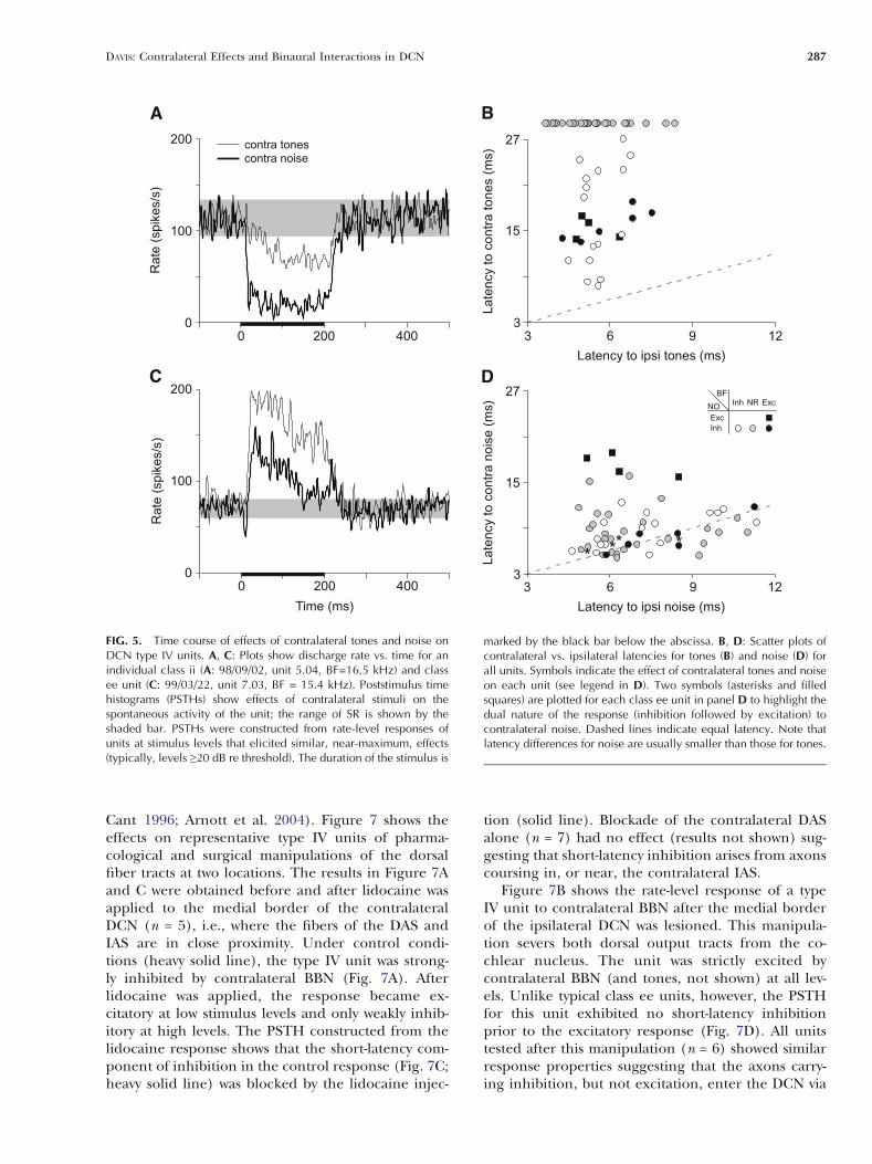
To examine the time course of contralateral ef-fects, poststimulus time histograms (PSTHs) were con-structed from the rate-level responses of individualunits at stimulus levels that elicited similar, near-maximum effects (typically, levels Q 20 dB re thresh-old). Figure 5A shows PSTHs of the responses to 200-ms tone (solid lines) and noise bursts (heavy solidlines) for the representative class ii unit (rate-levelcurves shown in Fig. 2A) that was otherwise firingspontaneously. Note that the inhibitory response totones built up over time and reached its full strengthwithin 50–60 ms. In contrast, the response to noiseshowed a short-latency, sharp onset to inhibition anda sustained reduction of driven rate. Poststimulustime histograms for the representative class ee unit(Fig. 2D) are shown in Figure 5C. For this unit, thetone response showed a well-defined onset and sus-tained excitation. The noise response showed an ini-tial, short-latency decrease in rate, followed by an in-crease in rate at a latency comparable to that to tones.
Each unit’s latency for contralateral tones (noise)is plotted against its latency for ipsilateral tones(noise) in Figure 5B and D, respectively. As above,
FIG. 1. Contralateral monaural frequency response maps for typeIV units in the dorsal cochlear nucleus (DCN). (A) A unit showinginhibition to contralateral tones (98/05/28, unit 4.01); (B) a unitshowing excitation (99/03/22, unit 5.02). Stimulus-driven rates areplotted against frequency at multiple sound levels (numerical labelson right). Horizontal lines indicate average spontaneous rates (SR).Black (gray) regions identify excitatory (inhibitory) response areas.Sound levels are given in dB attenuation; actual sound pressure level(SPL) varies with the acoustic calibration, but 0 dB attn. is near 100dB re 20 mPa for tones. Solid vertical lines indicate BFs to contra-lateral tones; the arrows at the top of the plots mark ipsilateral BFs.
284 DAVIS: Contralateral Effects and Binaural Interactions in DCN
symbols for units without a response to contralateraltones are plotted beyond the axes. Here, responselatency is defined as the time from stimulus onset tothe first time the driven rate in the PSTH exceeded1 SD above (below) the SR. Two symbols (asterisksand squares) are plotted for each class ee unit inFigure 5D to highlight the dual nature of the re-sponse (inhibition followed by excitation) to contra-lateral noise exhibited by this class of units. Note thatall of the data points for tones are above the equity(dashed) line, whereas most data points for noise aredistributed along the line (including 19 units withcontralateral noise latencies below corresponding ip-silateral values) indicating that latency differenceswere usually smaller for noise than for tones. Overall,the median difference in latency was 10 ms for tonesbut only 2 ms for noise. However, the unit classes didshow some differences in response properties. Forexample, class ii units (unfilled circles) showed awide range of latencies for contralateral tones, where-as class ei and ee units (filled symbols) showed a tight-er cluster of values at intermediate latencies. Inaddition, units excited by contralateral noise (class ee;
FIG. 2. Range of responses to contralateral stimulation amongDCN type IV units. Plots show discharge rate vs. level in response totones (at the ipsilateral BF; thin lines) and broadband noise (heavylines) presented to the contralateral ear. Sound levels are shown indB attenuation (0 dB attn. is near 100 dB SPL re 20 mPa for tones andnear 40 dB spectrum level re 20 mPa/¾Hz for noise). Shaded barsshow the range of SR for these units. Class ii units (A) are inhibited
by both tones and noise (98/09/02, unit 5.04, BF=16.5 kHz); class oiunits (B) are unaffected by tones and inhibited by noise (99/07/08,unit 5.06, BF = 27.5 kHz); class ei units (C) are excited by tones butinhibited by noise (98/05/28, unit 2.02, BF = 12.2 kHz); and class eeunits (D) are excited by both tones and noise (99/03/22, unit 7.03,BF = 15.4 kHz).
FIG. 3. Magnitude of contralateral effects on DCN type IV units.Comparison of each unit’s maximum driven rate for noise vs. tonestimulation. The symbol used to plot each point indicates the effectthat contralateral stimuli had on the unit (see legend). Data pointsalong the center ordinate are shifted slightly to avoid overlap. Thedashed line indicates equal driven rates to both stimuli. Note thatmost units are weakly, if at all, inhibited by tones but are inhibitedstrongly by contralateral noise (open and gray circles).
DAVIS: Contralateral Effects and Binaural Interactions in DCN 285
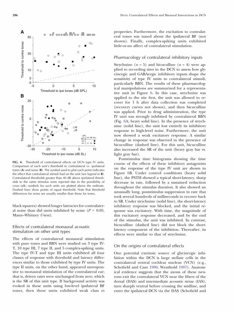
black squares) showed longer latencies for contralater-al noise than did units inhibited by noise (P 9 0.05,Mann–Whitney U-test).
Effects of contralateral monaural acousticstimulation on other unit types
The effects of contralateral monaural stimulationwith pure tones and BBN were studied on 3 type IV-T, 10 type III, 7 type II, and 5 complex-spiking units.The type IV-T and type III units exhibited all fourclasses of response with threshold and latency differ-ences similar to those exhibited by type IV units. Thetype II units, on the other hand, appeared unrespon-sive to monaural stimulation of the contralateral ear;that is, driven rates were unchanged from zero, whichis the SR of this unit type. If background activity wasevoked in these units using low-level ipsilateral BFtones, then these units exhibited weak class ei
properties. Furthermore, the excitation to contralat-eral tones was tuned about the ipsilateral BF (notshown). Finally, complex-spiking units exhibitedlittle-or-no affect of contralateral stimulation.
Pharmacology of contralateral inhibitory inputs
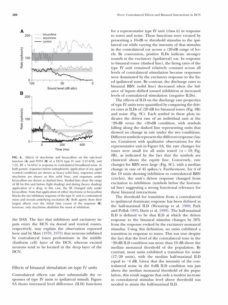
Strychnine (n = 5) and bicuculline (n = 4) were ap-plied to recording sites in the DCN to assess how gly-cinergic and GABAergic inhibitory inputs shape thesensitivity of type IV units to contralateral stimuli,particularly BBN. The results of these pharmacolog-ical manipulations are summarized for a representa-tive unit in Figure 6. In this case, strychnine wasapplied to the site first, the unit was allowed to re-cover for 1 h after data collection was completed(recovery curves not shown), and then bicucullinewas applied. Prior to drug administration, the typeIV unit was strongly inhibited by contralateral BBN(Fig. 6A; heavy solid line). In the presence of strych-nine (solid line), the unit lost entirely its inhibitoryresponse to high-level noise. Furthermore, the unitnow showed a weak excitatory response. A similarchange in response was observed in the presence ofbicuculline (dashed line). For this unit, bicucullinealso increased the SR of the unit (heavy gray bar vs.light gray bar).
Poststimulus time histograms showing the timecourse of the effects of these inhibitory antagonistson the response of the type IV unit are shown inFigure 6B. Under control conditions (heavy solidline), the PSTH showed a typical short-latency, sharpdecrease in rate, followed by a sustained reductionthroughout the stimulus duration. It also showed anunusually long, poststimulus suppression in rate thattook several hundreds of milliseconds to recover backto SR. Under strychnine (solid line), the short-latencyinhibitory response was blocked, and the initial re-sponse was excitatory. With time, the magnitude ofthis excitatory response decreased, and by the endof the stimulus, the unit was inhibited. In contrast,bicuculline (dashed line) did not block the short-latency component of the inhibition. Thereafter, itseffects were similar to that of strychnine.
On the origins of contralateral effects
One potential extrinsic source of glycinergic inhi-bition within the DCN is large stellate cells in thecontralateral ventral cochlear nucleus (VCN) (e.g.,Schofield and Cant 1996; Wenthold 1987). Anatom-ical evidence suggests that the axons of these neu-rons exit the contralateral VCN near the fibers of thedorsal (DAS) and intermediate acoustic striae (IAS),turn sharply ventral before crossing the midline, andenter the ipsilateral DCN via the DAS (Schofield and
FIG. 4. Threshold of contralateral effects on DCN type IV units.Comparison of each unit’s threshold to contralateral vs. ipsilateraltones (A) and noise (B). The symbol used to plot each point indicatesthe effect that contralateral stimuli had on the unit (see legend in B).Contralateral thresholds greater than 40 dB above ipsilateral thresh-olds to the same stimulus were rejected due to the possibility ofcross talk; symbols for such units are plotted above the ordinate.Dashed lines show points of equal threshold. Note that thresholddifferences for noise are usually smaller than those for tones.
286 DAVIS: Contralateral Effects and Binaural Interactions in DCN
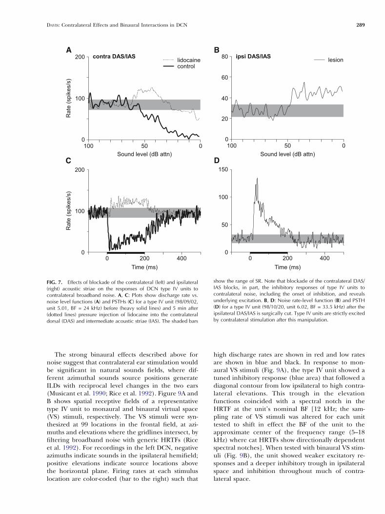
Cant 1996; Arnott et al. 2004). Figure 7 shows theeffects on representative type IV units of pharma-cological and surgical manipulations of the dorsalfiber tracts at two locations. The results in Figure 7Aand C were obtained before and after lidocaine wasapplied to the medial border of the contralateralDCN (n = 5), i.e., where the fibers of the DAS andIAS are in close proximity. Under control condi-tions (heavy solid line), the type IV unit was strong-ly inhibited by contralateral BBN (Fig. 7A). Afterlidocaine was applied, the response became ex-citatory at low stimulus levels and only weakly inhib-itory at high levels. The PSTH constructed from thelidocaine response shows that the short-latency com-ponent of inhibition in the control response (Fig. 7C;heavy solid line) was blocked by the lidocaine injec-
tion (solid line). Blockade of the contralateral DASalone (n = 7) had no effect (results not shown) sug-gesting that short-latency inhibition arises from axonscoursing in, or near, the contralateral IAS.
Figure 7B shows the rate-level response of a typeIV unit to contralateral BBN after the medial borderof the ipsilateral DCN was lesioned. This manipula-tion severs both dorsal output tracts from the co-chlear nucleus. The unit was strictly excited bycontralateral BBN (and tones, not shown) at all lev-els. Unlike typical class ee units, however, the PSTHfor this unit exhibited no short-latency inhibitionprior to the excitatory response (Fig. 7D). All unitstested after this manipulation (n = 6) showed similarresponse properties suggesting that the axons carry-ing inhibition, but not excitation, enter the DCN via
FIG. 5. Time course of effects of contralateral tones and noise onDCN type IV units. A, C: Plots show discharge rate vs. time for anindividual class ii (A: 98/09/02, unit 5.04, BF=16.5 kHz) and classee unit (C: 99/03/22, unit 7.03, BF = 15.4 kHz). Poststimulus timehistograms (PSTHs) show effects of contralateral stimuli on thespontaneous activity of the unit; the range of SR is shown by theshaded bar. PSTHs were constructed from rate-level responses ofunits at stimulus levels that elicited similar, near-maximum, effects(typically, levels Q20 dB re threshold). The duration of the stimulus is
marked by the black bar below the abscissa. B, D: Scatter plots ofcontralateral vs. ipsilateral latencies for tones (B) and noise (D) forall units. Symbols indicate the effect of contralateral tones and noiseon each unit (see legend in D). Two symbols (asterisks and filledsquares) are plotted for each class ee unit in panel D to highlight thedual nature of the response (inhibition followed by excitation) tocontralateral noise. Dashed lines indicate equal latency. Note thatlatency differences for noise are usually smaller than those for tones.
DAVIS: Contralateral Effects and Binaural Interactions in DCN 287
the DAS. The fact that inhibitory and excitatory in-puts enter the DCN via dorsal and ventral routes,respectively, may explain the observation reportedhere and by Mast (1970, 1973) that neurons inhibitedby contralateral tones predominate in the middle(fusiform cell) layer of the DCN, whereas excitedneurons tend to be located in the deep layer of theDCN.
Effects of binaural stimulation on type IV units
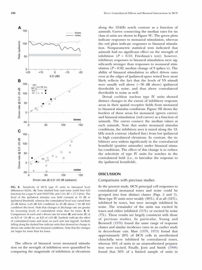
Contralateral effects can alter substantially the re-sponses of type IV units to ipsilateral stimuli. Figure8A shows interaural level difference (ILD) functions
for a representative type IV unit (class ii) in responseto tones and noise. These functions were created bypresenting a 10-dB re threshold stimulus to the ipsi-lateral ear while varying the intensity of that stimulusin the contralateral ear across a T20-dB range of lev-els. By convention, positive ILDs indicate strongersounds at the excitatory (ipsilateral) ear. In responseto binaural tones (dashed line), the firing rates of thetype IV unit remained relatively constant across alllevels of contralateral stimulation because responseswere dominated by the excitatory response to the fix-ed ipsilateral tone. By contrast, the discharge rates tobinaural BBN (solid line) decreased when the bal-ance of inputs shifted toward inhibition at increasedlevels of contralateral stimulation (negative ILDs).
The effects of ILD on the discharge rate propertiesof type IV units were quantified by comparing the driv-en rates at ILDs of T20 dB for binaural tones (Fig. 8B)and noise (Fig. 8C). Each symbol in these plots in-dicates the driven rate of an individual unit at the_20-dB versus the +20-dB condition, with symbolsfalling along the dashed line representing units thatshowed no change in rate under the two conditions.Different symbols represent the different response clas-ses. Consistent with qualitative observations for therepresentative unit in Figure 8A, the rate changes fortones were small for all units tested (n = 10; Fig.8B), as indicated by the fact that the symbols areclustered about the equity line. Conversely, ratechanges for BBN were large (Fig. 8C), with a medianchange in rate of 45 spikes/s. Furthermore, for 4 ofthe 19 units showing inhibition to contralateral BBN(circles), the unit’s driven response changed fromexcitation to inhibition (symbols below the horizon-tal line) suggesting a strong functional relevance forthese binaural interactions.
The threshold for transition from a contralateralto ipsilateral dominant response has been defined asthe half-maximal ILD (Wenstrup et al. 1988; Parkand Pollak 1993; Davis et al. 1999). The half-maximalILD is defined to be that ILD at which the drivenresponse to the binaural stimulus changes by 50%from the response evoked by the excitatory monauralstimulus. Using this definition, no units exhibited atransition in response to tones. This was true despitethe fact that the level of the contralateral tone in the_20-dB ILD condition was more than 10 dB above themedian monaural threshold of the population. Bycontrast, most units exhibited a transition for noise(17/20 units), with the median half-maximal ILDequal to _4 dB. Given that the intensity of the con-tralateral noise in the 0-dB ILD condition was justabove the median monaural threshold of the popu-lation, this result suggests that only a modest increasein contralateral stimulus level above threshold wasneeded to attain the half-maximal ILD.
FIG. 6. Effects of strychnine and bicuculline on the rate-levelfunction (A) and PSTH (B) of a DCN type IV unit (12/14/98, unit1.02, BF = 16 kHz) in response to contralateral broadband noise. Inboth panels, responses before iontophoretic application of any agent(control condition) are shown as heavy solid lines, responses understrychnine are shown as thin solid lines, and responses underbicuculline are shown as dashed lines. Shaded bars show the rangeof SR for this unit before (light shading) and during (heavy shading)application of a drug; in this case, the SR changed only underbicuculline. Note that application of either strychnine or bicucullineblocks the net inhibitory response of the type IV unit to contralateralnoise and reveals underlying excitation (A). Both agents show theirlargest affects over the initial time course of the response (B);however, only strychnine abolishes the onset of inhibition.
288 DAVIS: Contralateral Effects and Binaural Interactions in DCN
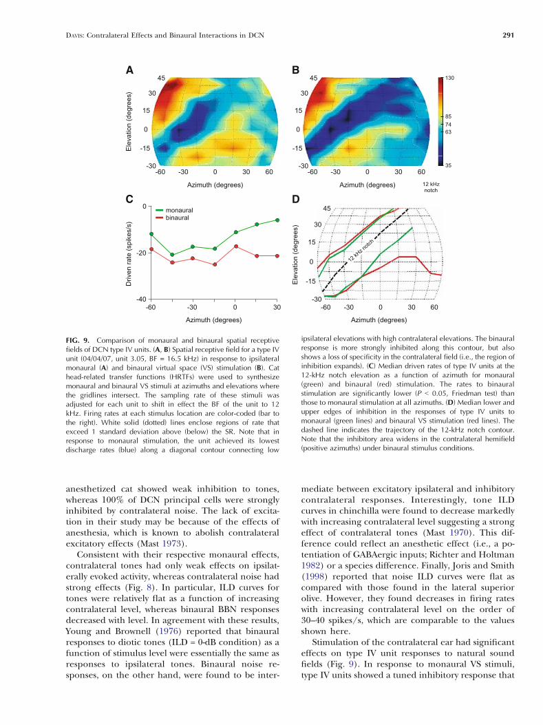
The strong binaural effects described above fornoise suggest that contralateral ear stimulation wouldbe significant in natural sounds fields, where dif-ferent azimuthal sounds source positions generateILDs with reciprocal level changes in the two ears(Musicant et al. 1990; Rice et al. 1992). Figure 9A andB shows spatial receptive fields of a representativetype IV unit to monaural and binaural virtual space(VS) stimuli, respectively. The VS stimuli were syn-thesized at 99 locations in the frontal field, at azi-muths and elevations where the gridlines intersect, byfiltering broadband noise with generic HRTFs (Riceet al. 1992). For recordings in the left DCN, negativeazimuths indicate sounds in the ipsilateral hemifield;positive elevations indicate source locations abovethe horizontal plane. Firing rates at each stimuluslocation are color-coded (bar to the right) such that
high discharge rates are shown in red and low ratesare shown in blue and black. In response to mon-aural VS stimuli (Fig. 9A), the type IV unit showed atuned inhibitory response (blue area) that followed adiagonal contour from low ipsilateral to high contra-lateral elevations. This trough in the elevationfunctions coincided with a spectral notch in theHRTF at the unit’s nominal BF [12 kHz; the sam-pling rate of VS stimuli was altered for each unittested to shift in effect the BF of the unit to theapproximate center of the frequency range (5–18kHz) where cat HRTFs show directionally dependentspectral notches]. When tested with binaural VS stim-uli (Fig. 9B), the unit showed weaker excitatory re-sponses and a deeper inhibitory trough in ipsilateralspace and inhibition throughout much of contra-lateral space.
FIG. 7. Effects of blockade of the contralateral (left) and ipsilateral(right) acoustic striae on the responses of DCN type IV units tocontralateral broadband noise. A, C: Plots show discharge rate vs.noise level functions (A) and PSTHs (C) for a type IV unit (98/09/02,unit 5.01, BF = 24 kHz) before (heavy solid lines) and 5 min after(dotted lines) pressure injection of lidocaine into the contralateraldorsal (DAS) and intermediate acoustic striae (IAS). The shaded bars
show the range of SR. Note that blockade of the contralateral DAS/IAS blocks, in part, the inhibitory responses of type IV units tocontralateral noise, including the onset of inhibition, and revealsunderlying excitation. B, D: Noise rate-level function (B) and PSTH(D) for a type IV unit (98/10/20, unit 6.02, BF = 33.5 kHz) after theipsilateral DAS/IAS is surgically cut. Type IV units are strictly excitedby contralateral stimulation after this manipulation.
DAVIS: Contralateral Effects and Binaural Interactions in DCN 289
The effects of binaural versus monaural stimula-tion on the strength of inhibition were quantified bycomparing the magnitude of inhibition at elevations
along the 12-kHz notch contour as a function ofazimuth. Curves connecting the median rates for sixclass oi units are shown in Figure 9C. The green plotsindicate responses to monaural stimulation, whereasthe red plots indicate responses to binaural stimula-tion. Nonparametric statistical tests indicated thatazimuth had no significant effect on the strength ofinhibition (P = 0.53; Friedman’s test); however,inhibitory responses to binaural stimulation were sig-nificantly stronger than responses to monaural stim-ulation (P = 0.02; median change of 9 spikes/s). Theability of binaural stimulation to affect driven rateseven at the edges of ipsilateral space tested here mostlikely reflects the fact that the levels of VS stimuliwere usually well above (õ30 dB above) ipsilateralthresholds to noise, and thus above contralateralthresholds to noise as well.
Dorsal cochlear nucleus type IV units showeddistinct changes in the extent of inhibitory responseareas in their spatial receptive fields from monauralto binaural stimulus conditions. Figure 9D shows theborders of these areas for monaural (green curves)and binaural stimulation (red curves) as a function ofazimuth. The curves connect the median values ateach azimuth. Note that under monaural stimulusconditions, the inhibitory area is tuned along the 12-kHz notch contour (dashed line) from low ipsilateralto high contralateral elevations. In contrast, the in-hibitory area widens significantly in the contralateralhemifield (positive azimuths) under binaural stimu-lus conditions. The effect of this change is to reducethe selectivity of type IV units for notches in thecontralateral field (i.e., to lateralize the response tothe ipsilateral hemifield).
DISCUSSION
Comparisons with previous studies
In the present study, DCN principal cell responses tocontralateral monaural tones and noise could begrouped into four distinct classes (Figs. 2 and 3).Most type IV units were weakly (30%), if at all (52%),inhibited by tones, but were strongly inhibited bynoise. The remainder of the units was excited bytones and either inhibited (11%) or excited by noise(7%). These results are largely consistent with thoseof previous studies. In particular, Young andBrownell (1976) found the same range of responseclasses and similar incidence rates in an earlier studyin decerebrate cats. Mast (1970, 1973) found thatapproximately 20% of DCN cells in anesthetizedchinchilla were inhibited by contralateral tones,whereas 10% of units in an unanesthetized prepara-tion were excited. Finally, Joris and Smith (1998)found that 50% of a limited sample of units in
FIG. 8. Sensitivity of DCN type IV units to interaural leveldifferences (ILDs). (A) Tone (dashed line) and noise (solid line) ILDfunctions for a type IV unit (99/07/08, unit 5.03, BF = 27.5 kHz). Thelevel of the ipsilateral stimulus was held constant at 10 dB reipsilateral threshold, whereas the contralateral level was varied from20 dB below (+20 dB ILD condition) to 20 dB above (
_20 dB ILD
condition) this level. Note that changes in discharge rate are greaterfor increasing levels of contralateral noise than for tones. B, C:Comparisons of each unit’s driven rate for tones (B) and noise (C) atan ILD of
_20 dB vs. an ILD of +20 dB. Symbols indicate the effect
of contralateral tones and noise on each unit (see legend). Symbolsfalling along the dashed line indicate units that showed no change indriven rate under the two binaural conditions. Note that the changesare larger for noise than for tones.
290 DAVIS: Contralateral Effects and Binaural Interactions in DCN
anesthetized cat showed weak inhibition to tones,whereas 100% of DCN principal cells were stronglyinhibited by contralateral noise. The lack of excita-tion in their study may be because of the effects ofanesthesia, which is known to abolish contralateralexcitatory effects (Mast 1973).
Consistent with their respective monaural effects,contralateral tones had only weak effects on ipsilat-erally evoked activity, whereas contralateral noise hadstrong effects (Fig. 8). In particular, ILD curves fortones were relatively flat as a function of increasingcontralateral level, whereas binaural BBN responsesdecreased with level. In agreement with these results,Young and Brownell (1976) reported that binauralresponses to diotic tones (ILD = 0-dB condition) as afunction of stimulus level were essentially the same asresponses to ipsilateral tones. Binaural noise re-sponses, on the other hand, were found to be inter-
mediate between excitatory ipsilateral and inhibitorycontralateral responses. Interestingly, tone ILDcurves in chinchilla were found to decrease markedlywith increasing contralateral level suggesting a strongeffect of contralateral tones (Mast 1970). This dif-ference could reflect an anesthetic effect (i.e., a po-tentiation of GABAergic inputs; Richter and Holtman1982) or a species difference. Finally, Joris and Smith(1998) reported that noise ILD curves were flat ascompared with those found in the lateral superiorolive. However, they found decreases in firing rateswith increasing contralateral level on the order of30–40 spikes/s, which are comparable to the valuesshown here.
Stimulation of the contralateral ear had significanteffects on type IV unit responses to natural soundfields (Fig. 9). In response to monaural VS stimuli,type IV units showed a tuned inhibitory response that
FIG. 9. Comparison of monaural and binaural spatial receptivefields of DCN type IV units. (A, B) Spatial receptive field for a type IVunit (04/04/07, unit 3.05, BF = 16.5 kHz) in response to ipsilateralmonaural (A) and binaural virtual space (VS) stimulation (B). Cathead-related transfer functions (HRTFs) were used to synthesizemonaural and binaural VS stimuli at azimuths and elevations wherethe gridlines intersect. The sampling rate of these stimuli wasadjusted for each unit to shift in effect the BF of the unit to 12kHz. Firing rates at each stimulus location are color-coded (bar tothe right). White solid (dotted) lines enclose regions of rate thatexceed 1 standard deviation above (below) the SR. Note that inresponse to monaural stimulation, the unit achieved its lowestdischarge rates (blue) along a diagonal contour connecting low
ipsilateral elevations with high contralateral elevations. The binauralresponse is more strongly inhibited along this contour, but alsoshows a loss of specificity in the contralateral field (i.e., the region ofinhibition expands). (C) Median driven rates of type IV units at the12-kHz notch elevation as a function of azimuth for monaural(green) and binaural (red) stimulation. The rates to binauralstimulation are significantly lower (P G 0.05, Friedman test) thanthose to monaural stimulation at all azimuths. (D) Median lower andupper edges of inhibition in the responses of type IV units tomonaural (green lines) and binaural VS stimulation (red lines). Thedashed line indicates the trajectory of the 12-kHz notch contour.Note that the inhibitory area widens in the contralateral hemifield(positive azimuths) under binaural stimulus conditions.
DAVIS: Contralateral Effects and Binaural Interactions in DCN 291
followed a diagonal contour from low ipsilateral tohigh contralateral elevations. This trough in the ele-vation functions coincided with a spectral notch inthe HRTF at the unit’s BF. When tested with binauralVS stimuli, type IV units showed a deeper inhibitorytrough in the ipsilateral hemifield and inhibitionthroughout much of contralateral space. Imig et al.(2000) also found that DCN principal cells inanesthetized cat showed response nulls in their azi-muth functions to free-field monaural and binauralnoise. Furthermore, spatial receptive fields showedthat these response nulls followed the expectedtrajectory of spectral notches. The depth of modula-tion was slightly greater for binaural than monauralnoise showing that contralateral inhibition had asmall but significant effect on unit directionality.
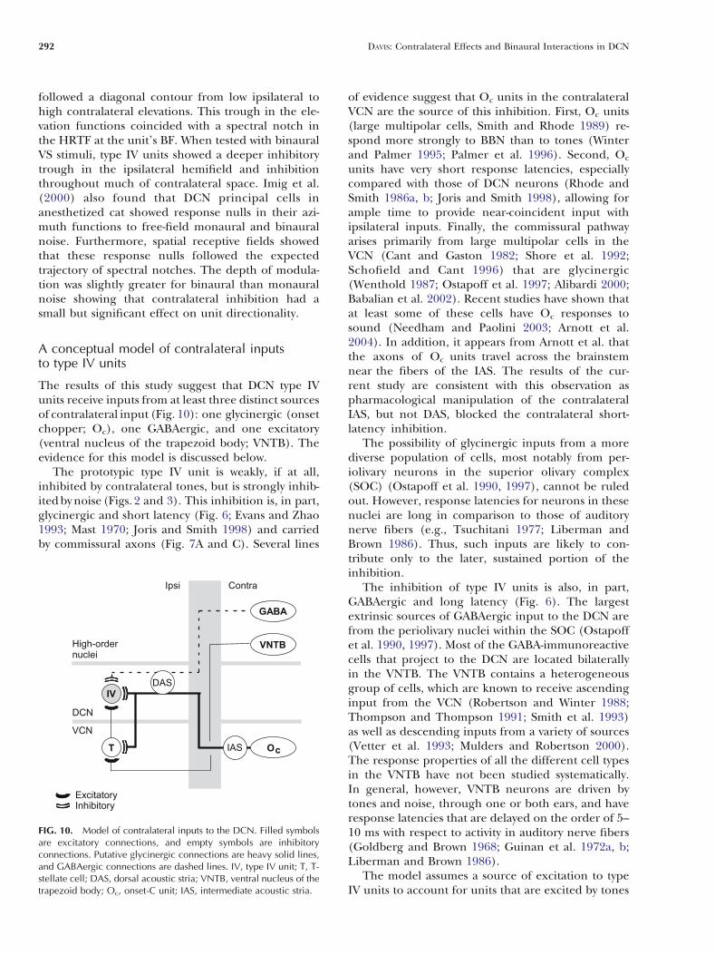
A conceptual model of contralateral inputsto type IV units
The results of this study suggest that DCN type IVunits receive inputs from at least three distinct sourcesof contralateral input (Fig. 10): one glycinergic (onsetchopper; Oc), one GABAergic, and one excitatory(ventral nucleus of the trapezoid body; VNTB). Theevidence for this model is discussed below.
The prototypic type IV unit is weakly, if at all,inhibited by contralateral tones, but is strongly inhib-ited by noise (Figs. 2 and 3). This inhibition is, in part,glycinergic and short latency (Fig. 6; Evans and Zhao1993; Mast 1970; Joris and Smith 1998) and carriedby commissural axons (Fig. 7A and C). Several lines
of evidence suggest that Oc units in the contralateralVCN are the source of this inhibition. First, Oc units(large multipolar cells, Smith and Rhode 1989) re-spond more strongly to BBN than to tones (Winterand Palmer 1995; Palmer et al. 1996). Second, Oc
units have very short response latencies, especiallycompared with those of DCN neurons (Rhode andSmith 1986a, b; Joris and Smith 1998), allowing forample time to provide near-coincident input withipsilateral inputs. Finally, the commissural pathwayarises primarily from large multipolar cells in theVCN (Cant and Gaston 1982; Shore et al. 1992;Schofield and Cant 1996) that are glycinergic(Wenthold 1987; Ostapoff et al. 1997; Alibardi 2000;Babalian et al. 2002). Recent studies have shown thatat least some of these cells have Oc responses tosound (Needham and Paolini 2003; Arnott et al.2004). In addition, it appears from Arnott et al. thatthe axons of Oc units travel across the brainstemnear the fibers of the IAS. The results of the cur-rent study are consistent with this observation aspharmacological manipulation of the contralateralIAS, but not DAS, blocked the contralateral short-latency inhibition.
The possibility of glycinergic inputs from a morediverse population of cells, most notably from per-iolivary neurons in the superior olivary complex(SOC) (Ostapoff et al. 1990, 1997), cannot be ruledout. However, response latencies for neurons in thesenuclei are long in comparison to those of auditorynerve fibers (e.g., Tsuchitani 1977; Liberman andBrown 1986). Thus, such inputs are likely to con-tribute only to the later, sustained portion of theinhibition.
The inhibition of type IV units is also, in part,GABAergic and long latency (Fig. 6). The largestextrinsic sources of GABAergic input to the DCN arefrom the periolivary nuclei within the SOC (Ostapoffet al. 1990, 1997). Most of the GABA-immunoreactivecells that project to the DCN are located bilaterallyin the VNTB. The VNTB contains a heterogeneousgroup of cells, which are known to receive ascendinginput from the VCN (Robertson and Winter 1988;Thompson and Thompson 1991; Smith et al. 1993)as well as descending inputs from a variety of sources(Vetter et al. 1993; Mulders and Robertson 2000).The response properties of all the different cell typesin the VNTB have not been studied systematically.In general, however, VNTB neurons are driven bytones and noise, through one or both ears, and haveresponse latencies that are delayed on the order of 5–10 ms with respect to activity in auditory nerve fibers(Goldberg and Brown 1968; Guinan et al. 1972a, b;Liberman and Brown 1986).
The model assumes a source of excitation to typeIV units to account for units that are excited by tones
FIG. 10. Model of contralateral inputs to the DCN. Filled symbolsare excitatory connections, and empty symbols are inhibitoryconnections. Putative glycinergic connections are heavy solid lines,and GABAergic connections are dashed lines. IV, type IV unit; T, T-stellate cell; DAS, dorsal acoustic stria; VNTB, ventral nucleus of thetrapezoid body; Oc, onset-C unit; IAS, intermediate acoustic stria.
292 DAVIS: Contralateral Effects and Binaural Interactions in DCN
and noise (Figs. 2 and 3). This excitatory source has along latency (Fig. 5B and D; Mast 1973), and itsaxons must enter the DCN via a ventral route (Fig. 7Band D). A strong candidate for this source is a circuitthat includes neurons in the VNTB and T stellatecells in the VCN. The VNTB contains two distinctgroups of cholinergic cells, medial olivocochlear(MOC) neurons and small cells, which provide allof the cholinergic input to the VCN (Sherriff andHenderson 1994). The efferent fibers of MOCneurons project to outer hair cells in the cochlea,but also give off collateral branches within the ves-tibular nerve root that contact the dendrites ofstellate cells in the VCN (Benson and Brown 1990;Brown and Benson 1992). The small cells project tothe magnocellular region of the VCN through thetrapezoid body (Sherriff and Henderson 1994). Inturn, in vitro studies suggest that T stellate cells exciteDCN principal cells (Oertel et al. 1990; Zhang andOertel 1993b, 1994).
Several lines of evidence suggest that contralateralacoustic stimulation could elicit long-latency excita-tion of T stellate cells (chopper units, Smith andRhode 1989). First, MOC neurons, and VNTBneurons in general, are driven by tones or noise,through one or both ears, and have long responselatencies (Goldberg and Brown 1968; Guinan et al.1972a, b; Liberman and Brown 1986; Liberman 1988;Brown et al. 1998). Second, T stellate cells are excitedby application of cholinergic agonists (Fujino andOertel 2001) suggesting that activity of either theMOC or small cells in the VNTB would likely ex-cite T stellate cells. Consistent with this hypothesis,Mulders et al. (2002) found that electrical stimula-tion of the MOC, after the olivocochlear effect waseliminated, elicited excitation in some VCN chopperunits. However, other chopper units were inhibitedsuggesting a dual action of MOC efferents. Third,Needham and Paolini (2003) found that contralater-al tones elicited sustained excitatory postsynapticpotentials in chopper units with a delay of 7–10 ms.Similarly, chopper units showed a short-latency mem-brane hyperpolarization after stimulation of the con-tralateral ear with noise that was soon exceeded byexcitatory inputs (latency of õ14 ms). Finally, Shoreet al. (2003) found that 30% of VCN units inanesthetized guinea pig were inhibited by contralat-eral stimulation, but that 4.5% of cells were excited.
The possibility of other sources of excitatory inputto DCN type IV units cannot be ruled out. For ex-ample, there is considerable heterogeneity in theneurons that contribute to the commissure (Shoreet al. 1992; Schofield and Cant 1996; Alibardi 2000).Some of these neurons are immunonegative for bothglycine and GABA (Alibardi 2000) and thus presum-ably excitatory. In addition, some projections from
the periolivary nuclei are also immunonegative forboth glycine and GABA (Ostapoff et al. 1997). Fi-nally, projections from the inferior colliculus may beglutamatergic and excitatory (Saint Marie 1996).
Functional implications
Behavioral studies have shown that cats are capableof localizing the source of broadband sounds in thefrontal field with considerable accuracy (May andHuang 1996). This accuracy is maintained when thebandwidth of stimuli is narrowed to contain energyfrom 5 to 18 kHz, but is lost when mid-frequency cuesare removed by high-pass filtering or narrowing thebandwidth of the stimulus to mid-frequency puretones (Huang and May 1996). Studies of the filterfunctions that describe the transformation of a free-field sound to the energy spectrum at the eardrumsuggest that a directionally dependent spectral notchat mid-frequencies may be the cue to localizingbroadband sounds (Musicant et al. 1990; Rice et al.1992). Interestingly, notch frequency does not re-present a single point in space, but rather a contourthat connects low ipsilateral to high contralateralelevations. The ambiguity along this line could beresolved by information from low and high frequen-cies, from binaural time and level disparity cues(Middlebrooks and Green 1991), or knowledge ofthe notch frequency in the contralateral ear (thebinaural first notch direction code or BiFiND code;Rice et al. 1992).
Electrophysiological studies in cats have shownthat the projection neurons of the DCN are especiallysensitive to spectral notches (Young et al. 1992; Imiget al. 2000; this study). Type IV units are inhibited bytones, but give excitatory responses to broadbandnoise (Young and Brownell 1976). When a spectralnotch is added to noise near BF, type IV units showan inhibitory response (Spirou and Young 1991).Thus, type IV units show a tuned inhibition for aspectral notch at their BF. Furthermore, the spatialreceptive fields of DCN principal cells show responsenulls that follow the expected diagonal trajectory ofspectral notches (Imig et al. 2000; this study). Theseobservations suggest that the DCN serves as an earlystage in the processing of HRTF-based sound local-ization cues. Consistent with this interpretation,lesioning the output pathway of the DCN disruptsthe sound localization performance of cats, particu-larly their orientation to sound source elevation(Sutherland et al. 1998; May 2000).
The results of present study show that DCNprincipal cells are generally inhibited by contralateralstimuli, particularly by broadband noise. The effectsof the contralateral inputs on the spatial receptivefields of type IV units are twofold: stronger inhibitory
DAVIS: Contralateral Effects and Binaural Interactions in DCN 293
responses to notches in ipsilateral space and primar-ily inhibitory responses in contralateral space. Theformer effect increases DCN sensitivity to spectralnotches and is thus likely to enhance upstream pro-cessing of spectral cues. For example, type O units inthe inferior colliculus are known to receive DCNinputs (Davis 2002) and to show tuned excitatory res-ponses for notches located just below BF (Davis et al.2003). The stronger inhibition exhibited by DCNunits for notches at BF under binaural stimulusconditions would likely prevent the upward spreadof excitation in type O units. The latter effect largelyconfines the sensitivity of DCN principal cells tosounds in ipsilateral space. This has clear implica-tions for the BiFiND hypothesis because it restrictstoward the midline the region of space where notchfrequency may be known from both ears. This effectcould also explain, in part, preliminary observationsthat type O units show spatial tuning for soundsprimarily in one hemifield (Davis et al. 2003). Morestudies are needed to determine the extent to whichbinaural interactions in DCN impact the processingof spectral-dependent information in higher-orderneurons.
ACKNOWLEDGMENTS
The author thanks Dr. Eric D. Young for support and en-couragement, Drs. Nell Cant and Brett Schofield for help-ful discussions, Oleg Lomakin for software developmentand for participating in some of the experiments, and Yue-Houng Hu and Oleg Lomakin for help in data analysis andfigure preparation. This work was supported by NationalInstitute of Deafness and Other Communication Disordersgrants DC00979 (to E.D.Y.) and DC05161 (to K.A.D.).
REFERENCES
ADAMS JC, WARR WB. Origins of axons in the cat’s acoustic striaedetermined by injection of horseradish peroxidase into severedtracts. J. Comp. Neurol. 170:107–121, 1976.
ALIBARDI L. Cytology, synaptology and immunocytochemistry ofcommissural neurons and their putative axonal terminals in thedorsal cochlear nucleus of the rat. Anat. Anz. 182:207–220,2000.
ARNOTT RH, WALLACE MN, SHACKLETON TM, PALMER AR. Onsetneurones in the anteroventral cochlear nucleus project to thedorsal cochlear nucleus. J. Assoc. Res. Otolaryngol. 5:153–170,2004.
BABALIAN AL, RYUGO DK, VISCHER MW, ROUILLER EM. Inhibitorysynaptic interactions between cochlear nuclei: evidence froman in vitro whole brain study. NeuroReport 10:1913–1917,1999.
BABALIAN AL, JACOMME AV, DOUCET JR, RYUGO DK, ROUILLER EM.Commissural glycinergic inhibition of bushy and stellate cells inthe anteroventral cochlear nucleus. NeuroReport 13:555–558,2002.
BENSON TE, BROWN MC. Synapses formed by olivocochlear axonbranches in the mouse cochlear nucleus. J. Comp. Neurol.295:52–70, 1990.
BROWN MC, BENSON TE. Transneuronal labeling of cochlearnucleus neurons by HRP-labeled auditory nerve fibers andolivocochlear branches in mice. J. Comp. Neurol. 321:645–665, 1992.
BROWN MC, LIBERMAN MC, BENSON TE, RYUGO DK. Brainstembranches from olivocochlear axons in cats and rodents. J.Comp. Neurol. 278:591–603, 1988.
BROWN MC, KUJAWA SG, DUCA ML. Single olivocochlear neurons inthe guinea pig. I. Binaural facilitation of responses to high-levelnoise. J. Neurophysiol. 79:3077–3087, 1998.
CANT NB, GASTON KC. Pathways connecting the right and leftcochlear nuclei. J. Comp. Neurol. 212:313–326, 1982.
CONLEE JW, KANE ES. Descending projections from the inferiorcolliculus to the dorsal cochlear nucleus in the cat: anautoradiographic study. Neuroscience 7:161–178, 1982.
DAVIS KA. Evidence of a functionally segregated pathway fromdorsal cochlear nucleus to inferior colliculus. J. Neurophysiol.87:1824–1835, 2002.
DAVIS KA, YOUNG ED. Pharmacological evidence of inhibitory anddisinhibitory neuronal circuits in dorsal cochlear nucleus. J.Neurophysiol. 83:926–940, 2000.
DAVIS KA, RAMACHANDRAN R, MAY BJ. Single-unit responses inthe inferior colliculus of decerebrate cats. II. Sensitivity tointeraural level differences. J. Neurophysiol. 82:164–175,1999.
DAVIS KA, RAMACHANDRAN R, MAY BJ. Auditory processing of spectralcues for sound localization in the inferior colliculus. J. Assoc.Res. Otolaryngol. 4:148–163, 2003.
EVANS EF, ZHAO W. Varieties of inhibition in the processing andcontrol of processing in the mammalian cochlear nucleus.Prog. Brain Res. 97:117–126, 1993.
FERNANDEZ C, KARAPAS F. The course and termination of the striae ofMonakow and Held in the cat. J. Comp. Neurol. 131:371–386,1967.
FUJINO K, OERTEL D. Cholinergic modulation of stellate cells in themammalian ventral cochlear nucleus. J. Neurosci. 21:7372–7383, 2001.
GIBSON DJ. Interaural crosstalk in the cat. Hear. Res. 7:325–333,1982.
GOLDBERG JM, BROWN PB. Functional organization of the dogsuperior olivary complex: an anatomical and electrophysiolog-ical study. J. Neurophys. 31:619–656, 1968.
GUINAN J J J, GUINAN SS, NORRIS BE. Single auditory units in thesuperior olivary complex. I. Responses to sounds and classifica-tion based on physiological properties. Int. J. Neurosci. 4:101–120, 1972a.
GUINAN J J J, GUINAN SS, NORRIS BE. Single auditory units in thesuperior olivary complex. II. Locations of unit categories andtonotopic organization. Int. J. Neurosci. 4:147–166, 1972b.
HAVEY DC, CASPARY DM. A simple technique for constructing Fpiggy-back_ multibarrel microelectrodes. Electroencephalogr. Clin.Neurophysiol. 48:249–251, 1980.
HUANG AY, MAY BJ. Sound orientation behavior in cats. II. Mid-frequency spectral cues for sound localization. J. Acoust. Soc.Am. 100:1070–1080, 1996.
IMIG TJ, BIBIKOV NG, POIRIER P, SAMSON FK. Directionality derivedfrom pinna-cue spectral notches in cat dorsal cochlear nucleus.J. Neurophysiol. 83:907–925, 2000.
JORIS PX, SMITH PH. Temporal and binaural properties in dorsalcochlear nucleus and its output tract. J. Neurosci. 18:10157–10170, 1998.
LIBERMAN MC. Response properties of cochlear efferent neurons:monaural vs. binaural stimulation and the effects of noise.J. Neurophysiol. 60:1779–1798, 1988.
294 DAVIS: Contralateral Effects and Binaural Interactions in DCN
LIBERMAN MC, BROWN MC. Physiology and anatomy of singleolivocochlear neurons in the cat. Hear. Res. 24:17–36, 1986.
MANIS PB, SPIROU GA, WRIGHT DD, PAYDAR S, RYUGO DK. Physiologyand morphology of complex spiking neurons in the guineapig dorsal cochlear nucleus. J. Comp. Neurol. 348:261–276,1994.
MAST TE. Binaural interaction and contralateral inhibition indorsal cochlear nucleus of the chinchilla. J. Neurophysiol.33:108–115, 1970.
MAST TE. Dorsal cochlear nucleus of the chinchilla : excitation bycontralateral sound. Brain Res. 62:61–70, 1973.
MAY BJ. Role of the dorsal cochlear nucleus in the soundlocalization behavior of cats. Hear. Res. 148:74–87, 2000.
MAY BJ, HUANG AY. Sound orientation behavior in cats. I.Localization of broadband noise. J. Acoust. Soc. Am.100:1059–1069, 1996.
MIDDLEBROOKS JC, GREEN DM. Sound localization by humanlisteners. Annu. Rev. Psychol. 42:135–159, 1991.
MULDERS WH, ROBERTSON D. Evidence for direct cortical innervationof medial olivocochlear neurones in rats. Hear. Res. 144:65–72, 2000.
MULDERS WH, WINTER IM, ROBERTSON D. Dual action of olivoco-chlear collaterals in the guinea pig cochlear nucleus. Hear. Res.174:264–280, 2002.
MUSICANT AD, CHAN JC, HIND JE. Direction-dependent spectralproperties of cat external ear: new data and cross-speciescomparisons. J. Acoust. Soc. Am. 87:757–781, 1990.
NEEDHAM K, PAOLINI AG. Fast inhibition underlies the transmissionof auditory information between cochlear nuclei. J. Neurosci.23:6357–6361, 2003.
NELKEN I, YOUNG ED. Linear and nonlinear spectral integration intype IV neurons of the dorsal cochlear nucleus. I. Regions oflinear interaction. J. Neurophysiol. 78:790–799, 1997.
OERTEL D, WU S, GARB MW, DIZAK C. Morphology and physiologyof cells in slice preparations of the posteroventral cochlearnucleus of mice. J. Comp. Neurol. 295:136–154, 1990.
OSEN KK. Course and termination of the primary afferents in thecochlear nuclei of the cat. Arch. Ital. Biol. 108:21–51, 1970.
OSTAPOFF EM, MOREST DK, POTASHNER SJ. Uptake and retrogradetransport of [3H]GABA from the cochlear nucleus to the super-ior olive in the guinea pig. J. Chem. Neuroanat. 3:285–289, 1990.
OSTAPOFF EM, BENSON CG, SAINT MARIE RL. GABA- and glycine-immunoreactive projections from the superior olivary complexto the cochlear nucleus in guinea pig. J. Comp. Neurol.381:500–511, 1997.
PALMER AR, JIANG D, MARSHALL DH. Responses of ventral cochlearnucleus onset and chopper units as a function of signalbandwidth. J. Neurophysiol. 75:780–794, 1996.
PARK TJ, POLLAK GD. GABA shapes sensitivity to interaural inten-sity disparities in the mustache bat’s inferior colliculus: im-plications for encoding sound location. J. Neurosci. 13:2050–2067, 1993.
RHODE WS, SMITH PH. Encoding timing and intensity in theventral cochlear nucleus of the cat. J. Neurophysiol. 56:261–286, 1986a.
RHODE WS, SMITH PH. Physiological studies on neurons in thedorsal cochlear nucleus of cat. J. Neurophysiol. 56:287–307,1986b.
RICE JJ, MAY BJ, SPIROU GA, YOUNG ED. Pinna-based spectral cues forsound localization in cat. Hear. Res. 58:132–152, 1992.
RICHTER JA, HOLTMAN JR. Barbiturates: their in vivo effects andpotential biochemical mechanisms. Neurobiology 18:275–319,1982.
ROBERTSON D, WINTER IM. Cochlear nucleus inputs to olivoco-chlear neurones revealed by combined anterograde and ret-
rograde labeling in the guinea pig. Brain Res. 462:47–55,1988.
RYUGO DK, MAY BJ. The projections of intracellularly labeledauditory nerve fibers to the dorsal cochlear nucleus of cat. J.Comp. Neurol. 329:20–35, 1993.
SAINT MARIE RL. Glutamatergic connections of the auditorymidbrain: selective uptake and axonal transport of D-[3H]as-partate. J. Comp. Neurol. 373:255–270, 1996.
SCHOFIELD BR. Origins of projections from the inferior colliculus tothe cochlear nucleus in guinea pigs. J. Comp. Neurol. 429:206–220, 2001.
SCHOFIELD BR, CANT NB. Origins and targets of commissuralconnections between the cochlear nuclei in guinea pigs. J.Comp. Neurol. 375:128–146, 1996.
SHERRIFF FE, HENDERSON Z. Cholinergic neurons in the ventraltrapezoid nucleus project to the cochlear nuclei in the rat.Neuroscience 58:627–633, 1994.
SHOFNER WP, YOUNG ED. Excitatory/inhibitory response types in thecochlear nucleus: relationships to discharge patterns andresponses to electrical stimulation of the auditory nerve. J.Neurophysiol. 54:917–939, 1985.
SHORE SE, GODFREY DA, HELFERT RH, ALTSCHULER RA, BLEDSOE SC JR.Connections between the cochlear nuclei in guinea pig. Hear.Res. 62:16–26, 1992.
SHORE SE, SUMNER CJ, BLEDSOE SC, LU J. Effects of contralateralsound stimulation on unit activity of ventral cochlear nucleusneurons. Exp. Brain Res. 153:427–435, 2003.
SMITH PH, RHODE WS. Structural and functional propertiesdistinguish two types of multipolar cells in the ventral cochlearnucleus. J. Comp. Neurol. 282:595–616, 1989.
SMITH PH, JORIS PX, YIN TC. Projections of physiologically charac-terized spherical bushy cell axons from the cochlear nucleus ofthe cat: evidence for delay lines to the medial superior olive. J.Comp. Neurol. 331:245–260, 1993.
SPIROU GA, YOUNG ED. Organization of dorsal cochlear nucleustype IV unit response maps and their relationship to activ-ation by bandlimited noise. J. Neurophysiol. 66:1750–1768,1991.
SUTHERLAND DP, MASTERTON RB, GLENDENNING KK. Role of acousticstriae in hearing: reflexive responses to elevated sound-sources.Behav. Brain Res. 97:1–12, 1998.
THOMPSON AM, THOMPSON GC. Projections from the posteroventralcochlear nucleus to the superior olivary complex in guinea pig:light and EM observations with the PHA-L method. J. Comp.Neurol. 311:495–508, 1991.
TSUCHITANI C. Functional organization of lateral cell groups ofcat superior olivary complex. J. Neurophysiol. 40:296–318,1977.
VETTER DE, SALDANA E, MUGNAINI E. Input from the inferiorcolliculus to medial olivocochlear neurons in the rat: a doublelabel study with PHA-L and cholera toxin. Hear. Res. 70:173–186, 1993.
WEEDMAN DL, RYUGO DK. Projections from auditory cortex to thecochlear nucleus in rats: synapses on granule cell dendrites. J.Comp. Neurol. 371:311–324, 1996.
WENSTRUP JJ, FUZESSERY ZM, POLLAK GD. Binaural neurons in themustache bat’s inferior colliculus. II. Determinants of spatialresponses among 60-kHz EI units. J. Neurophysiol. 60:1384–1404, 1988.
WENTHOLD RJ. Evidence for a glycinergic pathway connectingthe two cochlear nuclei: an immunocytochemical and retro-grade transport study. Brain Res. 415:183–187, 1987.
WINTER IM, PALMER AR. Level dependence of cochlear nucleusonset unit responses and facilitation by second tones orbroadband noise. J. Neurophysiol. 73:141–159, 1995.
DAVIS: Contralateral Effects and Binaural Interactions in DCN 295

YOUNG ED. Identification of response properties of ascendingaxons from dorsal cochlear nucleus. Brain Res. 200:23–38,1980.
YOUNG ED, BROWNELL WE. Responses to tone and noise of singlecells in dorsal cochlear nucleus of unanesthetized cats. J.Neurophysiol. 39:282–300, 1976.
YOUNG ED, SPIROU GA, RICE J J, VOIGT HF. Neural organization andresponses to complex stimuli in the dorsal cochlear nucleus.Philos. Trans. R. Soc. Lond., B 336:407–413, 1992.
ZHANG S, OERTEL D. Cartwheel and superficial stellate cells of thedorsal cochlear nucleus of mice: intracellular recordings inslices. J. Neurophysiol. 69:1384–1397, 1993a.
ZHANG S, OERTEL D. Giant cells of the dorsal cochlear nucleus ofmice: intracellular recordings in slices. J. Neurophysiol.69:1398–1408, 1993b.
ZHANG S, OERTEL D. Neuronal circuits associated with the output ofthe dorsal cochlear nucleus through fusiform cells. J. Neuro-physiol. 71:914–930, 1994.
296 DAVIS: Contralateral Effects and Binaural Interactions in DCN