Embed Size (px)
Citation preview
JOURNAL OF BACTERIOLOGY, Nov. 2011, p. 6020–6031 Vol. 193, No. 210021-9193/11/$12.00 doi:10.1128/JB.05436-11Copyright © 2011, American Society for Microbiology. All Rights Reserved.
Coordinated Regulation by AgrA, SarA, and SarR To Control agrExpression in Staphylococcus aureus�†
Dindo Reyes,1 Diego O. Andrey,2 Antoinette Monod,2 William L. Kelley,2Gongyi Zhang,3 and Ambrose L. Cheung1*
Department of Microbiology, Dartmouth Medical School, Hanover, New Hampshire 037551; Service of Infectious Diseases,University Hospital and Medical School of Geneva, 4 Rue Gabrielle-Perret-Gentil, CH-1211 Geneva 14,
Switzerland2; and Department of Immunology, National Jewish Hospital, Denver, Colorado3
Received 1 June 2011/Accepted 25 August 2011
The agr locus of Staphylococcus aureus is composed of two divergent transcripts (RNAII and RNAIII) drivenby the P2 and P3 promoters. The P2-P3 intergenic region comprises the SarA/SarR binding sites and the fourAgrA boxes to which AgrA binds. We reported here the role of AgrA, SarA, and SarR on agr P2 and P3transcription. Using real-time reverse transcription (RT)-PCR and promoter fusion studies with selectedsingle, double, triple, and complemented mutants, we showed that AgrA is indispensable to agr P2 and P3transcription, whereas SarA activates and SarR represses P2 transcription. In vitro runoff transcription assaysrevealed that AgrA alone promoted transcription from the agr P2 promoter, with SarA enhancing it and SarRinhibiting agr P2 transcription in the presence of AgrA or with SarA and AgrA. Electrophoretic mobility shiftassay (EMSA) analysis disclosed that SarR binds more avidly to the agr promoter than SarA and displacesSarA from the agr promoter. Additionally, SarA and AgrA bend the agr P2 promoter, whereas SarR does not.Collectively, these data indicated that AgrA activates agr P2 and P3 promoters while SarA activates the P2promoter, presumably via bending of promoter DNA to bring together AgrA dimers to facilitate engagement ofRNA polymerase (RNAP) to initiate transcription.
Staphylococcus aureus is an opportunistic pathogen thatcauses a broad range of human infections in both communityand hospital settings (17, 22). In most cases, infections begin asa localized lesion and then spread to the bloodstream. Oncethe infection is in the bloodstream, patients are at risk fordeveloping endocarditis and other metastatic complications(11).
The pathogenicity of S. aureus is a complex process thatinvolves the coordinated expression of many virulence factors,which can be divided into two main categories, based on theirfunctions either as surface protein adhesins or as secretedtoxins and enzymes. Typically, surface protein adhesins areproduced during the exponential phase of growth. The secondstage of infection, analogous to the post-exponential phase ofgrowth, is characterized by enhanced toxin and enzyme pro-duction, eventually leading to tissue destruction and bacterialspread (22). The regulatory events controlling the transitionfrom the exponential phase to the post-exponential phase ofgrowth are mediated in part by agr, which, upon activation,represses surface protein expression and promotes secretion ofextracellular toxins (25).
The agr locus is composed of two divergent transcripts calledRNAII and RNAIII. RNAII encodes four genes (agrDBCA),with AgrD encoding a 46-residue peptide which is processedinto a cyclic autoinducing peptide (AIP) and then exported byAgrB (25). Upon accumulation of AIP with increasing cell
density, it induces phosphorylation of AgrC, a membrane sen-sor within a two-component regulatory system, followed by asecond-step phosphorylation of the response regulator AgrA(25). AgrA can then bind to specific direct repeats in theintergenic region between the RNAII and RNAIII promotersto modulate agr transcription (18). Activation of RNAIII, theagr effector molecule, results in repression of many surface-associated proteins while promoting exoprotein gene transcrip-tion and, to a lesser extent, translation (13, 25).
Besides AgrA, a number of regulators controlling agr ex-pression have been described (5, 7). Among these is SarA, a14.7-kDa DNA binding protein that is a prototypic memberof a family called the SarA protein family (6, 7). Most, if notall, members of the SarA protein family are winged helixproteins that bind to target promoters to alter virulencegene expression (7, 21). Indeed, DNA binding studies re-vealed that SarA can bind to the agr P2 and P3 promoters (8,30). Transcription studies of S. aureus cells in vivo haveconsistently shown that SarA upregulates RNAII expression(8, 9); however, in vitro transcription analysis of the agr P2promoter in the presence of SarA alone revealed repressionrather than activation (3). This discrepancy between in vivoactivation and in vitro repression of the agr P2 promoter bySarA has not been explained until now.
SarR is a 13.6-kDa protein that is also a member of the SarAprotein family. In vitro, SarR can bind to the agr promoter toinfluence agr transcription in S. aureus strain RN6390 (23), astrain with a defective rsbU gene which is required for theoptimal expression of the stress-induced alternative sigma fac-tor called SigB (19, 28). Using real-time reverse transcription(RT)-PCR and transcriptional fusion analyses of a deletionmutant (without an antibiotic marker) in strain SH1000 (rsbU-
* Corresponding author. Mailing address: Vail 210, DartmouthMedical School, Hanover, NH 03755. Phone: (603) 650-1340. Fax:(603) 650-1362. E-mail: [email protected].
† Supplemental material for this article may be found at http://jb.asm.org/.
� Published ahead of print on 9 September 2011.
6020
on March 25, 2018 by guest
http://jb.asm.org/
Dow
nloaded from
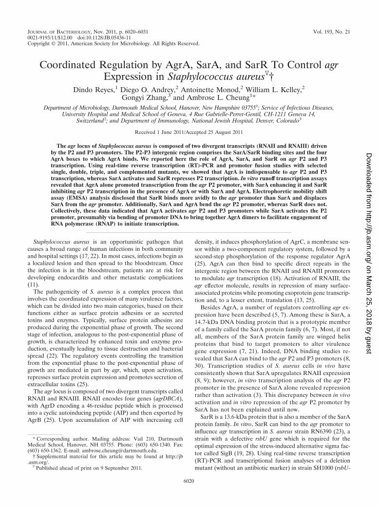
restored variant of RN6390), we have recently shown thatSarR can downregulate agr RNAII expression in strain SH1000(34), different from what we have discerned with the sarRmutant of strain RN6390 (rsbU mutant) in which RNAII tran-scription was slightly elevated compared with that of the par-ent. The binding site of SarR on the agr promoter, locatedbetween the P2 and P3 promoters, shares an overlap with thatof SarA (8, 24) (Fig. 1).
Given the temporal regulation of agr by SarR and SarA andthe effect of AgrA on its cognate promoter, we wanted toinvestigate the role of AgrA, SarA, and SarR on transcriptionfrom the agr P2 and P3 promoters, using deletion mutantsgenerated without any replacement antibiotic marker but con-taining intact promoter and transcription termination signals.Our data showed that AgrA is essential to transcriptions fromthe agr P2 and P3 promoters. More specifically, the AgrApromoter boxes and the overlapping SarA and SarR bindingsites between the P2 and P3 promoters (Fig. 1) are critical toagr P2 transcription but not P3 transcription. Using in vitrorunoff transcription assays, we found that SarA, in the presenceof AgrA, promotes transcription from the agr P2 promoter invitro, while SarR acts as a repressor. Electrophoretic mobilityshift assay (EMSA) and promoter DNA bending assays suggestthat SarA and AgrA bind and bend the agr P2 promoter,whereas SarR displaces SarA and binds but does not bend thepromoter, thus providing a mechanism by which SarA andSarR modulates the agr P2 promoter activity via DNA bendingin the presence of AgrA.
MATERIALS AND METHODS
Bacterial strains, plasmids, and growth conditions. S. aureus strains used inthis study, listed in Table 1, are derivatives of SH1000, an rsbU-restored strain ofthe 8325-4 strain (15). Growth of S. aureus was conducted in tryptic soy broth
(TSB; Difco) supplemented with erythromycin (3 or 10 �g/ml, depending on thecopy number of the erythromycin cassette) as needed. Gene deletions of strainSH1000 for sarA and sarR were carried out using the temperature-sensitivepMAD plasmid as described previously (1, 36). For the agrA mutant, we intro-duced translation stops (TAATGA) right next to the start codon to ensure thatAgrA was not translated without any interruption of RNAII transcription, usingpMAD. A similar technique was used to replace the deleted gene with a wild-type copy. Escherichia coli DH5� was the vehicle for routine DNA manipulation.Shuttle vector pALC1484, a derivative of pSK236 containing the gfpuvr reportergene (16), was used to construct promoter DNA-green fluorescent protein(GFP) reporter fusions. For growth in E. coli, Luria-Bertani (LB) broth wasroutinely used; ampicillin was added to a final concentration of 50 �g/ml asneeded. For growth in S. aureus, chloramphenicol at 10 �g/ml was used in TSB.Growth was monitored at 37°C at an optical density at 650 nm (OD650) with aSpectronic 20D� spectrophotometer (Spectronic Analytical Instruments, Gar-forth, England), using 18-mm borosilicate glass tubes.
Promoter DNA-GFP reporter assay. Promoter-GFP reporter fusions wereconstructed by inserting the agr promoter fragments into the EcoRI and XbaIsites of pALC1484 (16). Recombinant plasmids were then transformed first intoS. aureus strain RN4220 for proper methylation followed by introduction into thewild-type, mutant, and restored strains of SH1000 derivatives as described pre-viously (33). Overnight cultures were diluted 1:500 into 10 ml of TSB containingchloramphenicol (10 �g/ml) to yield an OD650 of �0.01 and grown at 37°C withconstant shaking at 250 rpm. Fluorescence (excitation/emission � 485/515 nm)was then monitored with an FL600 fluorescence spectrophotometer (Biotek,Winooski, VT) every 2 h and reported as fluorescence units/OD650 values.
RNAP purification. For purification of native RNA polymerase (RNAP) fromS. aureus, a DNA insert bearing the His10 tag at the C terminus of rpoC wascloned into the pMAD vector and introduced into the chromosome of a sarAagrA sarR triple mutant of SH1000. RNAP was then purified from 3 liters of cellsgrown in TSB to an OD600 of �1.0. Briefly, cells were harvested by centrifuga-tion, followed by freezing overnight at �80°C. Cells were then suspended in 30ml lysis buffer (50 mM Tris-HCl [pH 8.0], 100 mM NaCl, 5 mM MgCl2, 5 mM�-mercaptoethanol, 1 mM phenylmethylsulfonyl fluoride [PMSF], and 20% glyc-erol) and lysed with four passages through a French press followed by clarifica-tion of the lysate with centrifugation (14,000 rpm, 30 min). RNAP was thenpurified by passaging the clear lysate through 10 ml of Ni-nitrilotriacetic acid(Ni-NTA) in a column (Qiagen), washing the resin with 10� volume of lysisbuffer with 50 mM imidazole, followed by elution with 50 ml of elution buffer(similar to lysis buffer but with 250 mM NaCl) containing 500 mM imidazole. Theeluted RNAP was concentrated with Amicon Ultracel-10K (Millipore), dialyzedwith buffer containing 10 mM Tris-HCl (pH 8.0), 100 mM KCl, 5 mM MgCl2, and20% glycerol using a Slide-A-Lyzer dialysis cassette (Pierce), and stored at�20°C. The purities of various subunits within RNAP were confirmed by SDS-gel analysis.
Purification of SarA, SarR, and AgrA proteins. Both sarA and sarR codingregions are cloned into pET11b and pET14b, respectively, in E. coliBL21(DE3)pLysS. Protein expression and purification were conducted as de-scribed previously (8, 23). For AgrA protein purification, the agrA coding regionwas cloned into the NdeI and SmaI sites of pTYB2 (New England BioLabs),followed by transformation into BL21(DE3)pLysS cells. Cells grown in 1 liter ofLB and induced with 0.5 mM IPTG (isopropyl-�-D-thiogalactopyranoside) for5 h were harvested by centrifugation, passed through a French press thrice, andclarified by a second centrifugation step. The clarified lysate was loaded on achitin bead (NEB) column, and the AgrA protein was purified according to themanufacturer’s protocol. For additional purification, the fractions of interestwere applied to a Bio-Rad High Q column and eluted with an NaCl gradient of0.1 to 0.5 M. Fractions containing the AgrA were concentrated with AmiconUltracel-10K (Millipore) and dialyzed with 10 mM Tris-HCl (pH 8.0), 50 mMNaCl, and 5% glycerol using a Slide-A-Lyzer dialysis cassette (Pierce).
In vitro runoff transcription assay. Linear DNA templates of rpsD (controltemplate) and agr P2 and P3 promoters were generated by PCR. In vitro tran-scription experiments were carried out as follows: the promoter DNA template(20 nM) was incubated with 50 nM RNAP with or without AgrA, SarA, or SarR(as indicated) in buffer containing 10 mM Tris-HCl (pH 8.0), 50 mM NaCl, 5 mMMgCl2, and 50 �g/ml of bovine serum albumin (BSA) for 10 min at roomtemperature. To initiate the reaction, a nucleotide mixture containing 200 �M(each) ATP, GTP, and CTP, 10 �M UTP, and 10 �Ci [�-32P]UTP was added tothe reaction mix and incubated at 37°C, and then the reaction was terminatedafter 15 min with the addition of 10 �l of stop solution (1 M NH4CH3COO, 30mM EDTA, and 100 �g/ml yeast tRNA). The reaction mixture was ethanolprecipitated and resolved in an 8% urea-PAGE gel. Images were scanned using
FIG. 1. The intergenic sequence between the agr P2 and P3 pro-moters. There are 186 bp between the two transcription start sites(labeled with bent arrows). The �10 and �35 promoter boxes for P2and P3 promoters are depicted on top of the sequence. The AgrAtandem repeats are indicated by long bold arrows (the set below iscomplementary to the top set). The SarA and SarR binding sites,located between the two sets of tandem repeats, share a partial over-lap, with the SarA binding site (palindromic sequence) highlighted bydotted lines and the SarR binding site boxed. The �10 promoter boxfor the P3 promoter has an extended �10 sequence (TGT), which mayreduce the requirement for the canonical �35 promoter motif (14).The intergenic agr P2-P3 sequence here is conserved in 6 published S.aureus genomes. The difference between this sequence and divergentones in other S. aureus genomes resides primarily in one base change(marked with an asterisk) downstream of the �10 promoter box of the agrP2 promoter, with a C replaced by a T.
VOL. 193, 2011 AgrA, SarA, AND SarR ON THE agr PROMOTER 6021
on March 25, 2018 by guest
http://jb.asm.org/
Dow
nloaded from
Molecular Dynamics Typhoon 8600 (GE Healthcare) and processed using Im-ageQuant version 5.2.
RNA extraction and real-time RT-PCR. Cells of wild-type or mutant strainsgrown in TSB to mid-, late, and post-exponential phases were harvested at 4,000rpm for 10 min (4°C) and stored overnight at �80°C. Cells were then suspendedin 350 �l of TRIzol (Invitrogen), mixed with glass/zirconia beads, and lysed witha reciprocating shaker (BioSpec). Final steps of RNA extraction and purificationwere carried out using a RiboPure-bacteria RNA purification kit (Ambion/Applied Biosystems, Austin, TX) as described in the manufacturer’s insert. Toremove residual DNA, RNAs were treated with RNase-free DNase I, providedin the kit.
For real-time RT-PCR, 1 �g each of RNA was used to generate cDNA viareverse transcription using a Roche first-strand transcriptor kit (Roche, Mann-heim, Germany). Using a SYBR green I master kit (Roche, Mannheim, Ger-many), cDNAs were analyzed and quantified in a LightCycler 480 instrument(Roche) according to the manufacturer’s instructions, using gene-specific prim-ers. The list of primers is available from the authors upon request. Data werereported as agrB and RNAIII expression levels normalized to those of gyrB.
EMSA analysis. The agr promoter fragment (102 bp), generated by PCR, waslabeled with [-32P]ATP (Perkin Elmer) by end labeling one of the PCR primersusing T4 polynucleotide kinase (NEB) prior to PCR. The PCR product was thenresolved in a 6% nondenaturing gel and purified using an Elutip-d column(Schleicher and Schuell). Binding reaction was carried out by mixing the labeledprobe (�10,000 cpm) with increasing concentrations of the protein and incubat-ing for 20 min at room temperature. For displacement analysis, the labeled probewas allowed to bind initially either SarA or SarR prior to the addition of differentconcentrations of the competing protein and incubated for another 20 min.Complexes were resolved in a denaturing 6% urea-PAGE gel, and images werescanned using Molecular Dynamics Typhoon 8600 (GE Healthcare).
DNA bending assays. Hybridized oligonucleotides containing the SarA andSarR binding sites in the agr promoter or the AgrA P2 tandem site were clonedinto the SacI-BglII site of the bending vector pCY7 (31) to yield plasmids
pAM1163 and pAM1847, respectively. Plasmids were digested with EcoRI,HindIII, BstNI, EcoRV, NheI, and BamHI, and the fragments were purified andlabeled with [-32P]ATP using T4 polynucleotide kinase. Labeled fragments(�5,000 cpm) were then mixed with and without SarA (0.5 �mol), SarR (0.5�mol), or AgrA (25 nmol) in binding buffer containing 25 mM Tris (pH 7.5), 0.1mM EDTA, 75 mM NaCl, 10% glycerol, 1 mM dithiothreitol (DTT), and 0.5 �gof sonicated calf thymus at room temperature for 20 min. The reaction mixtureswere then resolved in a 5% acrylamide gel, dried, and autoradiographed.
RESULTS
RNAII transcription in agrA, sarA, and sarR mutant strains.Previous studies on transcriptional control of the agr locus in S.aureus revealed that AgrA, SarA, and SarR likely play impor-tant roles in regulating RNAII and RNAIII transcription.However, the detailed events on how these proteins exert con-trol on the agr P2 and P3 promoters (yielding RNAII andRNAIII, respectively) and subsequently on downstream effec-tor genes are not well understood. To address this question, weconstructed, using pMAD (1), single, double, and triple mu-tants of agrA, sarA, and sarR in strain SH1000, a derivative ofstrain 8325-4 with restored rsbU activity (15), by deleting therespective coding regions without any antibiotic marker andleaving the promoter and the transcription termination signalintact. As for the agrA mutant, we introduced nonsense muta-tions into Lys and Ile at positions 2 and 3 in the AgrA codingregion. We then analyzed by quantitative real-time RT-PCR,
TABLE 1. Strains and plasmids used in this study
Strain orplasmid Description Reference
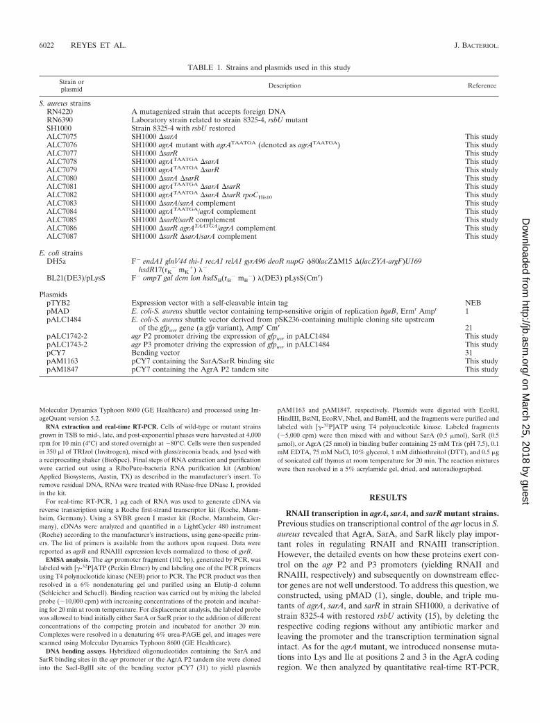
S. aureus strainsRN4220 A mutagenized strain that accepts foreign DNARN6390 Laboratory strain related to strain 8325-4, rsbU mutantSH1000 Strain 8325-4 with rsbU restoredALC7075 SH1000 sarA This studyALC7076 SH1000 agrA mutant with agrATAATGA (denoted as agrATAATGA) This studyALC7077 SH1000 sarR This studyALC7078 SH1000 agrATAATGA sarA This studyALC7079 SH1000 agrATAATGA sarR This studyALC7080 SH1000 sarA sarR This studyALC7081 SH1000 agrATAATGA sarA sarR This studyALC7082 SH1000 agrATAATGA sarA sarR rpoCHis10 This studyALC7083 SH1000 sarA/sarA complement This studyALC7084 SH1000 agrATAATGA/agrA complement This studyALC7085 SH1000 sarR/sarR complement This studyALC7086 SH1000 sarR agrATAATGA/agrA complement This studyALC7087 SH1000 sarR sarA/sarA complement This study
E. coli strainsDH5a F� endA1 glnV44 thi-1 recA1 relA1 gyrA96 deoR nupG �80lacZM15 (lacZYA-argF)U169
hsdR17(rK� mK
�) ��
BL21(DE3)/pLysS F� ompT gal dcm lon hsdSB(rB� mB
�) �(DE3) pLysS(Cmr)
PlasmidspTYB2 Expression vector with a self-cleavable intein tag NEBpMAD E. coli-S. aureus shuttle vector containing temp-sensitive origin of replication bgaB, Ermr Ampr 1pALC1484 E. coli-S. aureus shuttle vector derived from pSK236-containing multiple cloning site upstream
of the gfpuvr gene (a gfp variant), Ampr Cmr 21pALC1742-2 agr P2 promoter driving the expression of gfpuvr in pALC1484 This studypALC1743-2 agr P3 promoter driving the expression of gfpuvr in pALC1484 This studypCY7 Bending vector 31pAM1163 pCY7 containing the SarA/SarR binding site This studypAM1847 pCY7 containing the AgrA P2 tandem site This study
6022 REYES ET AL. J. BACTERIOL.
on March 25, 2018 by guest
http://jb.asm.org/
Dow
nloaded from
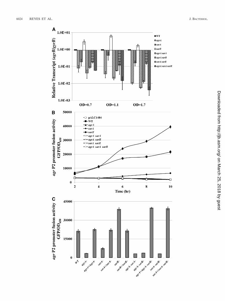
using agrB and the sequence coding RNAIII as probes, thetranscription of RNAII and RNAIII in TSB-grown cells duringexponential, late exponential, and stationary phases (OD650 of0.7, 1.1, and 1.7, respectively, as determined with 18-mm boro-silicate glass tubes in a Spectronic 20D� spectrophotometer[Garforth, England]). As illustrated in Fig. 2A, the deletion ofagrA or sarA alone resulted in a significant decrease in RNAIItranscription in all three growth phases compared with that ofthe parent. However, the effect was more dramatic with theagrA mutant than with the sarA mutant, especially during thelate exponential and post-exponential phases (OD650 of 1.1and 1.7). This finding is consistent with the notion that AgrA isan important factor in expression from the agr P2 promoter.The fact that deletion of sarA alone did not render the agr P2promoter completely inactive implies additional genetic re-quirement besides SarA for the optimal transcription from theP2 promoter (37). Notably, the downregulatory effect of thedouble sarA agrA mutation on RNAII transcription was similarto that of the single agrA mutant, suggesting that the majoreffect of sarA on RNAII expression is likely to be mediated viaAgrA. Interestingly, deletion of sarR triggered an upregulationin RNAII transcription, with the effect more pronounced dur-ing the late exponential phase (OD650 of 1.1), thus suggestingthat SarR acts as a repressor of the agr locus in the SH1000background. However, strains bearing the sarR deletion incombination with the agrA and/or sarA deletions did not ex-hibit the aforementioned derepression, especially during thelate exponential phase, in contrast to what has been found withthe single sarR mutant, thus implying that derepression ofRNAII in the sarR mutant strain is likely dependent on intactagrA and/or sarA loci.
We also confirmed these data by analyzing agr P2 promoterfusion with GFPuvr (16). For the promoter fusion construct, wehave cloned a 235-bp P2 promoter fragment encompassing thepreviously reported 4 putative AgrA boxes, the putative SarAand SarR binding sites, and the core promoter elements (Fig.1) upstream of gfpuvr in pALC1484. The recombinant plasmidwas introduced into various isogenic mutants of SH1000. Tominimize the variation in growth density, we normalized thesedata to fluorescence units/OD650 values. As shown in Fig. 2B,the activity of the agr P2 promoter was elevated in the sarRmutant but was reduced in the isogenic sarA mutant comparedwith that of the parental strain. In contrast, the agrA mutant,the double agrA sarA, agrA sarR, and sarA sarR mutants, as wellas the triple mutant, all exhibited low levels of agr P2 promoteractivity, similar to that of the empty vector control.
To verify these data, we complemented agrA, sarA, and sarRsingle mutants as well as the double agrA sarR and sarA sarRmutants by replacing the deleted gene with a native copy usingpMAD and analyzing the ensuing agr P2 promoter activity withGFP-mediated fluorescence. Our data showed that the pro-portionately reduced P2 promoter activity levels in agrA andsarA mutants were restored to wild-type levels upon comple-mentation, while that of the sarR mutant was reduced to theparental level (Fig. 2C). More importantly, both the agrA sarRand sarA sarR double mutants, upon respective complementa-tion with agrA and sarA, restored agr P2 promoter activity levelto that of the sarR mutant, at a level much higher than theSH1000 parental strain. Taken together, the above results con-cur with those of the quantitative RT-PCR data, strongly sug-
gesting that AgrA and, to a lesser extent, SarA activate agrRNAII transcription, whereas SarR acts as a repressor, exert-ing a greater impact on the later stage of growth. It should beclarified that our data here on RNAII expression with the sarRmutant of SH1000 (rsbU�) differed from that reported forRN6390 (rsbU mutant), where the sarR mutant exhibitedslightly lower RNAII expression than the isogenic parent (24).Indeed, a direct comparison of P2 promoter activities usingpromoter DNA-reporter fusions confirmed the discrepancy inRNAII expression between sarR mutants of RN6390 andSH1000 (data not shown). We speculate that reduced SigBactivity in strain RN6390 may conceivably impact RNAII ex-pression and hence becomes a contributing factor to slightlylower agr P2 activity in the sarR mutant in RN6390. In contrastto RN6390, we have repeatedly found that RNAII transcrip-tion in strain SH1000 was consistently elevated in the sarRmutant compared with that of the isogenic parent.
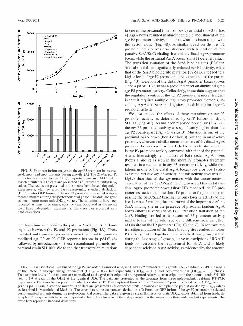
SarR-mediated repression impacts the agr P2 but not theagr P3 promoter. We also examined the effect of AgrA, SarA,and SarR on the agr P3 promoter driving transcription ofRNAIII, which is the agr effector molecule (27). As with the agrP2 promoter fusion constructs, the 219-bp agr P3 promoterfragment encompassing the AgrA, SarA, and SarR bindingsites was fused upstream of the GFPuvr reporter gene inpALC1484; the recombinant plasmid was then introduced intovarious mutant strains of SH1000. Surprisingly, a mutation insarR, unlike the effect on the agr P2 promoter, did not lead toany significant increase in agr P3 promoter activity comparedwith that of the parent (Fig. 3A). A deletion of sarA resulted ina notable decrease in agr P3 promoter activity in comparison tothe parent, whereas an agrA mutation rendered the same pro-moter completely inactive. Reconstitution of the mutated sarAor agrA gene with a wild-type copy in the chromosome restoredP3 promoter activity to the wild-type level, while complemen-tation of the sarR mutant at the chromosomal level had noeffect, as determined by GFP-mediated fluorescence (Fig. 3B).When agrA and sarA were restored in the respective agrA sarRand sarA sarR double mutants, the agr P3 promoter exhibitedwild-type promoter activity similar to that found in the sarRmutant, strongly implying that a deletion in sarR has no directrole in influencing RNAIII transcription. RT-PCR of RNAIIIexpression in these mutants and complemented mutants alsosupported results of the promotion fusion studies (data notshown). Collectively, these results clearly imply that AgrA pos-itively regulates both agr P2 and P3 promoters, whereas SarRimpacts on P2 but not P3 transcription. While SarA also up-regulates the P2 promoter, it is likely that the effect on the agrP3 promoter may be indirect (i.e., via upregulation of RNAIIand subsequently AgrA), because the double sarA agr mutant,similar to the single agr mutant, had little, if any, P3 promoteractivity.
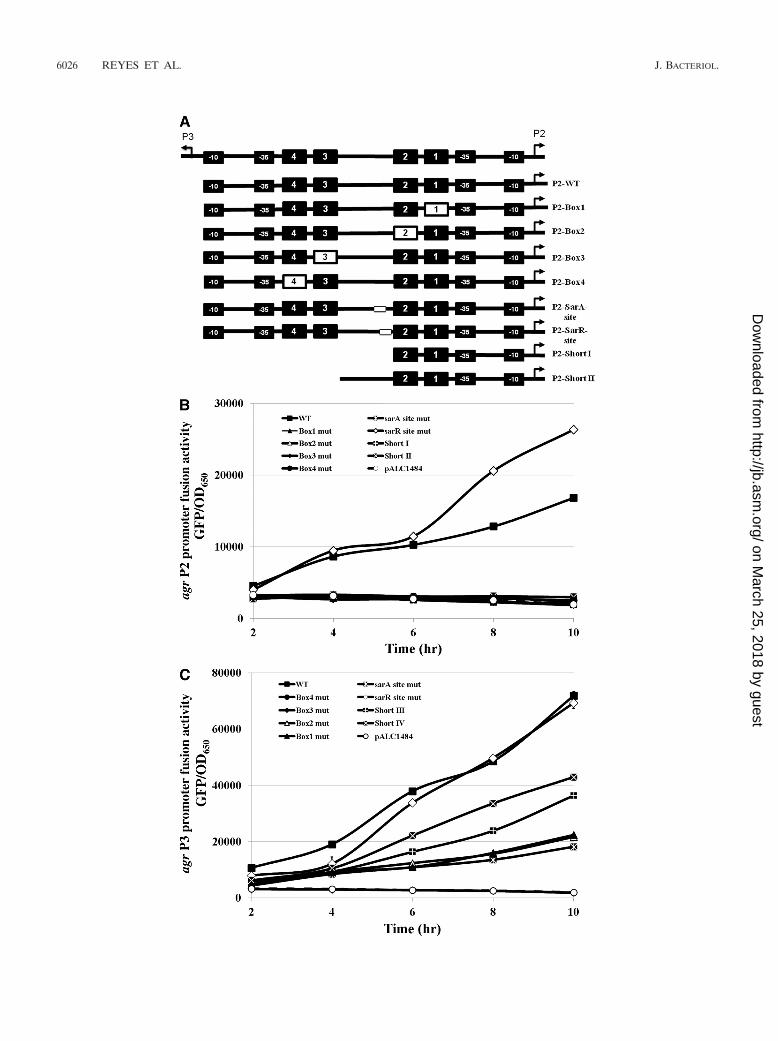
The effect of AgrA box and SarA/SarR binding sites on agrP2 and P3 transcription. To establish the crucial promoterelements relevant to agr transcription, we focused on fourAgrA promoter boxes to which AgrA binds (32) as well as thesarA and sarR binding sites shown to be important for agrtranscription (Fig. 1) (8, 24). As AgrA exists as a dimer (32)and hence binds to two tandem AgrA promoter boxes (18), weintroduced transversion mutations on individual AgrA boxeswithin each of the tandem AgrA boxes as well as truncation
VOL. 193, 2011 AgrA, SarA, AND SarR ON THE agr PROMOTER 6023
on March 25, 2018 by guest
http://jb.asm.org/
Dow
nloaded from
6024 REYES ET AL. J. BACTERIOL.
on March 25, 2018 by guest
http://jb.asm.org/
Dow
nloaded from
and transition mutations to the putative SarA and SarR bind-ing sites between the P2 and P3 promoters (Fig. 4A). Thesemutated and truncated promoters were then used to generatemodified agr P2 or P3 GFP reporter fusions in pALC1484followed by introduction of these recombinant plasmids intoparental strain SH1000. We found that transversion mutations
to one of the proximal (box 1 or box 2) or distal (box 3 or box4) AgrA boxes resulted in almost complete abolishment of theagr P2 promoter activity, similar to what has been found withthe vector alone (Fig. 4B). A similar trend on the agr P2promoter activity was also observed with truncation of theputative SarA/SarR binding sites and the distal AgrA promoterboxes, while the proximal AgrA boxes (short I) were left intact.The transition mutation of the SarA binding sites (P2-SarAsite) also exhibited significantly reduced agr P2 activity, whilethat of the SarR binding site mutation (P2-SarR site) led to ahigher level of agr P2 promoter activity than that of the parent(Fig. 4B). Deletion of the distal AgrA promoter boxes (boxes3 and 4 [short II]) also has a profound effect on diminishing theagr P2 promoter activity. Collectively, these data suggest thatthe regulatory control of the agr P2 promoter is more stringentin that it requires multiple regulatory promoter elements, in-cluding AgrA and SarA binding sites, to exhibit optimal agr P2promoter activity.
We also studied the effects of these mutations on agr P3promoter activity as determined by GFP fusions in strainSH1000 (Fig. 4C). As has been reported previously (2, 4, 26),the agr P3 promoter activity was significantly higher than theagr P2 counterpart (Fig. 4C versus B). Mutation in one of theproximal AgrA boxes (box 4 or box 3) resulted in an inactivepromoter, whereas a similar mutation in one of the distal AgrApromoter boxes (box 2 or box 1) led to a moderate reductionin agr P3 promoter activity compared with that of the parentalstrain. Interestingly, elimination of both distal AgrA boxes(boxes 1 and 2) as seen in the short IV promoter fragmentresulted in a reduction in agr P3 promoter activity, while mu-tations in one of the distal AgrA boxes (box 2 or box 1) alsoexhibited reduced agr P3 activity, but this activity level was stillhigher than that of the agr mutant with the vector control.Truncation of the SarA/SarR binding sites and the distal tan-dem AgrA promoter boxes (short III) rendered the P3 pro-moter less active than the short IV promoter fragment encom-passing the SarA/SarR binding site but closer in activity to thebox 1 or box 2 mutant, thus indicative of the importance of theSarA binding site in the presence of proximal tandem AgrAboxes (short III versus short IV). Transition mutation of theSarR binding site led to a pattern of P3 promoter activitysimilar to that of the wild type, quite different from the effectof this site on the P2 promoter (Fig. 4B and C). In contrast, thetransition mutation of the SarA binding site resulted in lowerP3 activity. Taken together, these results strongly suggest thatduring the late stage of growth, active transcription of RNAIIItends to overcome the requirement for SarA and is likelydependent solely on AgrA activity, as evidenced by the absence
FIG. 2. Transcriptional analysis of the agr P2 promoter in assorted agrA, sarA, and sarR mutants during growth. (A) Real-time RT-PCR analysisof the RNAII transcript during exponential (OD650 � 0.7), late exponential (OD650 � 1.1), and post-exponential (OD650 � 1.7) phases.Transcription levels of the mutants are normalized to the gyrB transcript and are reported relative to transcriptions in the parental strain SH1000(set to 1.0 at each of the ODs) at the identical ODs. The data are presented as the averages from three independent, real-time RT-PCRexperiments. The error bars represent standard deviations. (B) Transcriptional fusions of the 235-bp agr P2 promoter fused to the GFPuvr reportergene in pALC1484 in assorted mutants. The data are presented as fluorescence units (obtained at multiple time points) divided by OD650 valuesas described in Materials and Methods. The error bars represent standard deviations. (C) Promoter GFP fusion of the agr P2 promoter in selectedcomplemented mutants during the post-exponential phase. The data are given as mean fluorescence units/OD650 values obtained from triplicatesamples. The experiments have been repeated at least three times, with the data presented as the means from three independent experiments. Theerror bars represent standard deviations.
FIG. 3. Promoter fusion analysis of the agr P3 promoter in assortedagrA, sarA, and sarR mutants during growth. (A) The 219-bp agr P3promoter was fused to the GFPuvr reporter gene in pALC1484 inassorted mutants. The data are presented as fluorescence units/OD650values. The results are presented as the means from three independentexperiments, with the error bars representing standard deviations.(B) Promoter GFP fusion of the agr P3 promoter in selected comple-mented mutants during the postexponential phase. The data are givenas mean fluorescence units/OD650 values. The experiments have beenrepeated at least three times, with the data presented as the meansfrom three independent experiments. The error bars represent stan-dard deviations.
VOL. 193, 2011 AgrA, SarA, AND SarR ON THE agr PROMOTER 6025
on March 25, 2018 by guest
http://jb.asm.org/
Dow
nloaded from
6026 REYES ET AL. J. BACTERIOL.
on March 25, 2018 by guest
http://jb.asm.org/
Dow
nloaded from
of agr P3 promoter activity when the proximal AgrA box (box3 or box 4) was mutated, even with the SarA binding site intact.
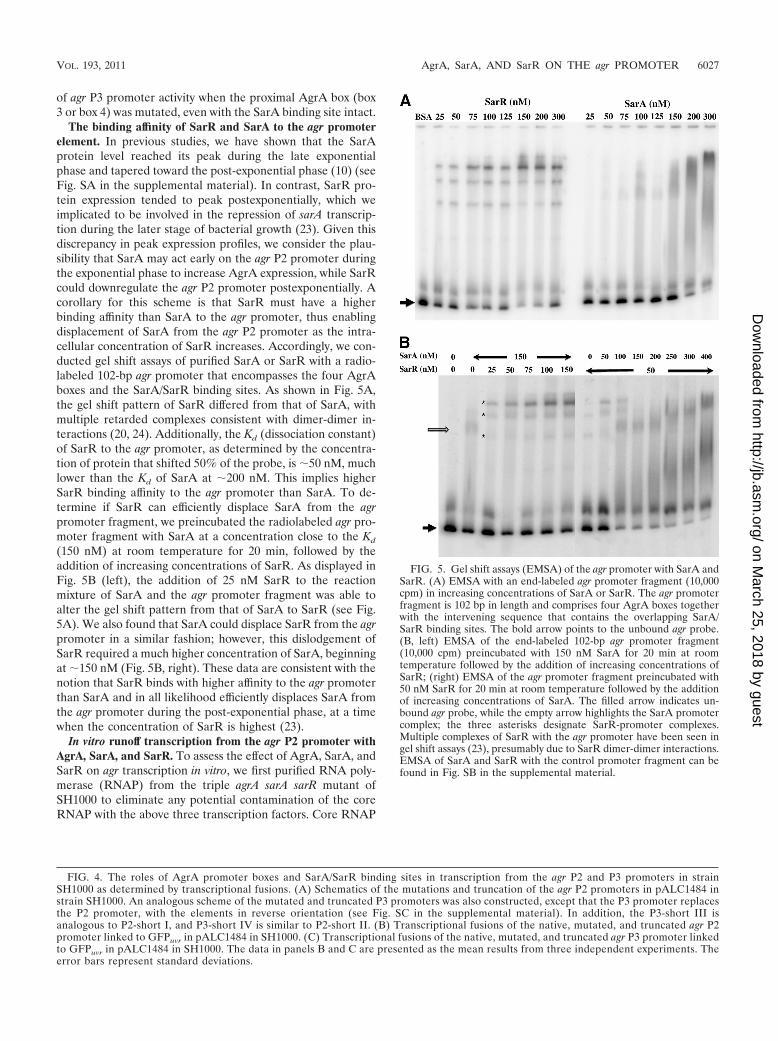
The binding affinity of SarR and SarA to the agr promoterelement. In previous studies, we have shown that the SarAprotein level reached its peak during the late exponentialphase and tapered toward the post-exponential phase (10) (seeFig. SA in the supplemental material). In contrast, SarR pro-tein expression tended to peak postexponentially, which weimplicated to be involved in the repression of sarA transcrip-tion during the later stage of bacterial growth (23). Given thisdiscrepancy in peak expression profiles, we consider the plau-sibility that SarA may act early on the agr P2 promoter duringthe exponential phase to increase AgrA expression, while SarRcould downregulate the agr P2 promoter postexponentially. Acorollary for this scheme is that SarR must have a higherbinding affinity than SarA to the agr promoter, thus enablingdisplacement of SarA from the agr P2 promoter as the intra-cellular concentration of SarR increases. Accordingly, we con-ducted gel shift assays of purified SarA or SarR with a radio-labeled 102-bp agr promoter that encompasses the four AgrAboxes and the SarA/SarR binding sites. As shown in Fig. 5A,the gel shift pattern of SarR differed from that of SarA, withmultiple retarded complexes consistent with dimer-dimer in-teractions (20, 24). Additionally, the Kd (dissociation constant)of SarR to the agr promoter, as determined by the concentra-tion of protein that shifted 50% of the probe, is �50 nM, muchlower than the Kd of SarA at �200 nM. This implies higherSarR binding affinity to the agr promoter than SarA. To de-termine if SarR can efficiently displace SarA from the agrpromoter fragment, we preincubated the radiolabeled agr pro-moter fragment with SarA at a concentration close to the Kd
(150 nM) at room temperature for 20 min, followed by theaddition of increasing concentrations of SarR. As displayed inFig. 5B (left), the addition of 25 nM SarR to the reactionmixture of SarA and the agr promoter fragment was able toalter the gel shift pattern from that of SarA to SarR (see Fig.5A). We also found that SarA could displace SarR from the agrpromoter in a similar fashion; however, this dislodgement ofSarR required a much higher concentration of SarA, beginningat �150 nM (Fig. 5B, right). These data are consistent with thenotion that SarR binds with higher affinity to the agr promoterthan SarA and in all likelihood efficiently displaces SarA fromthe agr promoter during the post-exponential phase, at a timewhen the concentration of SarR is highest (23).
In vitro runoff transcription from the agr P2 promoter withAgrA, SarA, and SarR. To assess the effect of AgrA, SarA, andSarR on agr transcription in vitro, we first purified RNA poly-merase (RNAP) from the triple agrA sarA sarR mutant ofSH1000 to eliminate any potential contamination of the coreRNAP with the above three transcription factors. Core RNAP
FIG. 4. The roles of AgrA promoter boxes and SarA/SarR binding sites in transcription from the agr P2 and P3 promoters in strainSH1000 as determined by transcriptional fusions. (A) Schematics of the mutations and truncation of the agr P2 promoters in pALC1484 instrain SH1000. An analogous scheme of the mutated and truncated P3 promoters was also constructed, except that the P3 promoter replacesthe P2 promoter, with the elements in reverse orientation (see Fig. SC in the supplemental material). In addition, the P3-short III isanalogous to P2-short I, and P3-short IV is similar to P2-short II. (B) Transcriptional fusions of the native, mutated, and truncated agr P2promoter linked to GFPuvr in pALC1484 in SH1000. (C) Transcriptional fusions of the native, mutated, and truncated agr P3 promoter linkedto GFPuvr in pALC1484 in SH1000. The data in panels B and C are presented as the mean results from three independent experiments. Theerror bars represent standard deviations.
FIG. 5. Gel shift assays (EMSA) of the agr promoter with SarA andSarR. (A) EMSA with an end-labeled agr promoter fragment (10,000cpm) in increasing concentrations of SarA or SarR. The agr promoterfragment is 102 bp in length and comprises four AgrA boxes togetherwith the intervening sequence that contains the overlapping SarA/SarR binding sites. The bold arrow points to the unbound agr probe.(B, left) EMSA of the end-labeled 102-bp agr promoter fragment(10,000 cpm) preincubated with 150 nM SarA for 20 min at roomtemperature followed by the addition of increasing concentrations ofSarR; (right) EMSA of the agr promoter fragment preincubated with50 nM SarR for 20 min at room temperature followed by the additionof increasing concentrations of SarA. The filled arrow indicates un-bound agr probe, while the empty arrow highlights the SarA promotercomplex; the three asterisks designate SarR-promoter complexes.Multiple complexes of SarR with the agr promoter have been seen ingel shift assays (23), presumably due to SarR dimer-dimer interactions.EMSA of SarA and SarR with the control promoter fragment can befound in Fig. SB in the supplemental material.
VOL. 193, 2011 AgrA, SarA, AND SarR ON THE agr PROMOTER 6027
on March 25, 2018 by guest
http://jb.asm.org/
Dow
nloaded from
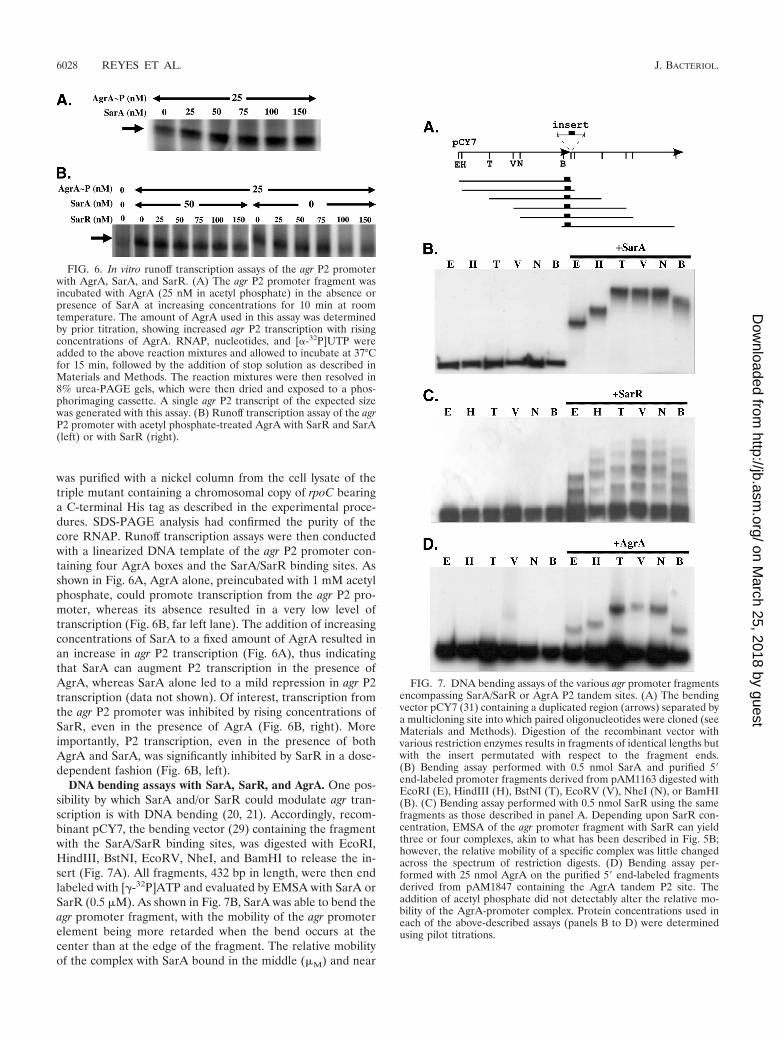
was purified with a nickel column from the cell lysate of thetriple mutant containing a chromosomal copy of rpoC bearinga C-terminal His tag as described in the experimental proce-dures. SDS-PAGE analysis had confirmed the purity of thecore RNAP. Runoff transcription assays were then conductedwith a linearized DNA template of the agr P2 promoter con-taining four AgrA boxes and the SarA/SarR binding sites. Asshown in Fig. 6A, AgrA alone, preincubated with 1 mM acetylphosphate, could promote transcription from the agr P2 pro-moter, whereas its absence resulted in a very low level oftranscription (Fig. 6B, far left lane). The addition of increasingconcentrations of SarA to a fixed amount of AgrA resulted inan increase in agr P2 transcription (Fig. 6A), thus indicatingthat SarA can augment P2 transcription in the presence ofAgrA, whereas SarA alone led to a mild repression in agr P2transcription (data not shown). Of interest, transcription fromthe agr P2 promoter was inhibited by rising concentrations ofSarR, even in the presence of AgrA (Fig. 6B, right). Moreimportantly, P2 transcription, even in the presence of bothAgrA and SarA, was significantly inhibited by SarR in a dose-dependent fashion (Fig. 6B, left).
DNA bending assays with SarA, SarR, and AgrA. One pos-sibility by which SarA and/or SarR could modulate agr tran-scription is with DNA bending (20, 21). Accordingly, recom-binant pCY7, the bending vector (29) containing the fragmentwith the SarA/SarR binding sites, was digested with EcoRI,HindIII, BstNI, EcoRV, NheI, and BamHI to release the in-sert (Fig. 7A). All fragments, 432 bp in length, were then endlabeled with [-32P]ATP and evaluated by EMSA with SarA orSarR (0.5 �M). As shown in Fig. 7B, SarA was able to bend theagr promoter fragment, with the mobility of the agr promoterelement being more retarded when the bend occurs at thecenter than at the edge of the fragment. The relative mobilityof the complex with SarA bound in the middle (�M) and near
FIG. 6. In vitro runoff transcription assays of the agr P2 promoterwith AgrA, SarA, and SarR. (A) The agr P2 promoter fragment wasincubated with AgrA (25 nM in acetyl phosphate) in the absence orpresence of SarA at increasing concentrations for 10 min at roomtemperature. The amount of AgrA used in this assay was determinedby prior titration, showing increased agr P2 transcription with risingconcentrations of AgrA. RNAP, nucleotides, and [�-32P]UTP wereadded to the above reaction mixtures and allowed to incubate at 37°Cfor 15 min, followed by the addition of stop solution as described inMaterials and Methods. The reaction mixtures were then resolved in8% urea-PAGE gels, which were then dried and exposed to a phos-phorimaging cassette. A single agr P2 transcript of the expected sizewas generated with this assay. (B) Runoff transcription assay of the agrP2 promoter with acetyl phosphate-treated AgrA with SarR and SarA(left) or with SarR (right).
FIG. 7. DNA bending assays of the various agr promoter fragmentsencompassing SarA/SarR or AgrA P2 tandem sites. (A) The bendingvector pCY7 (31) containing a duplicated region (arrows) separated bya multicloning site into which paired oligonucleotides were cloned (seeMaterials and Methods). Digestion of the recombinant vector withvarious restriction enzymes results in fragments of identical lengths butwith the insert permutated with respect to the fragment ends.(B) Bending assay performed with 0.5 nmol SarA and purified 5 end-labeled promoter fragments derived from pAM1163 digested withEcoRI (E), HindIII (H), BstNI (T), EcoRV (V), NheI (N), or BamHI(B). (C) Bending assay performed with 0.5 nmol SarR using the samefragments as those described in panel A. Depending upon SarR con-centration, EMSA of the agr promoter fragment with SarR can yieldthree or four complexes, akin to what has been described in Fig. 5B;however, the relative mobility of a specific complex was little changedacross the spectrum of restriction digests. (D) Bending assay per-formed with 25 nmol AgrA on the purified 5 end-labeled fragmentsderived from pAM1847 containing the AgrA tandem P2 site. Theaddition of acetyl phosphate did not detectably alter the relative mo-bility of the AgrA-promoter complex. Protein concentrations used ineach of the above-described assays (panels B to D) were determinedusing pilot titrations.
6028 REYES ET AL. J. BACTERIOL.
on March 25, 2018 by guest
http://jb.asm.org/
Dow
nloaded from
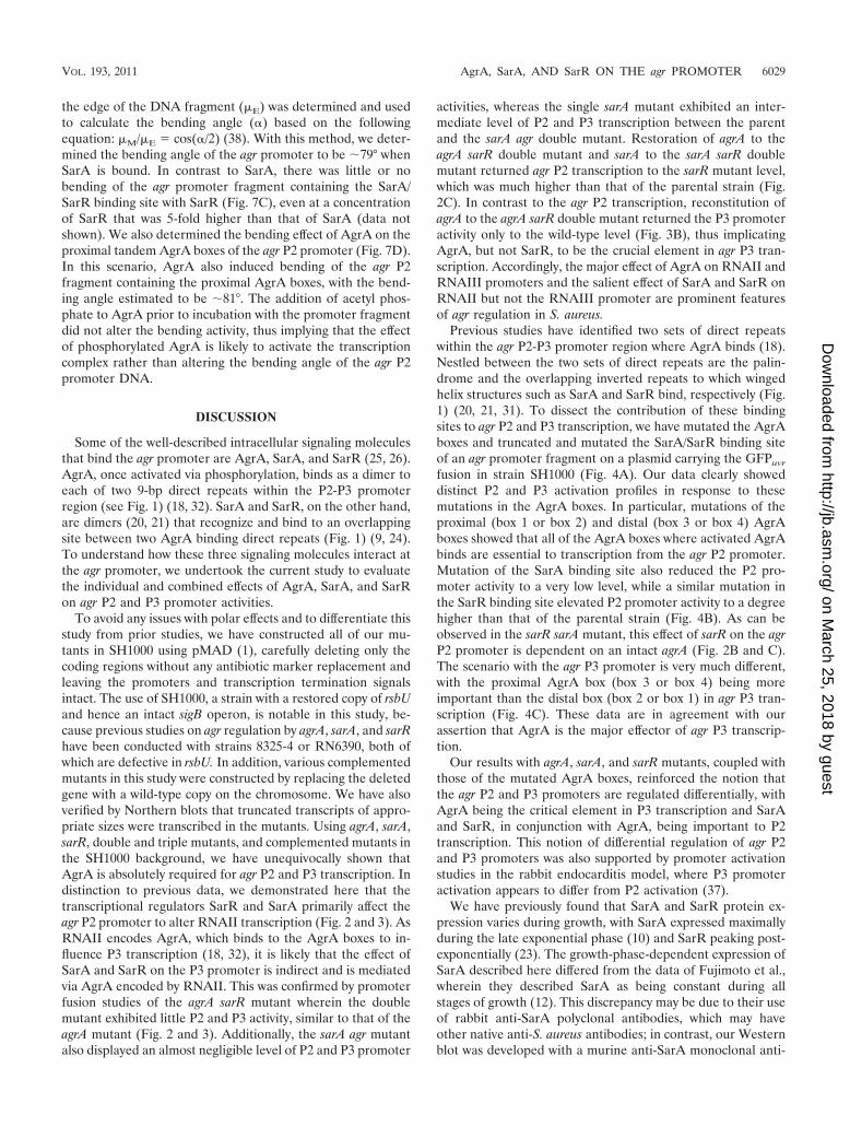
the edge of the DNA fragment (�E) was determined and usedto calculate the bending angle (�) based on the followingequation: �M/�E � cos(�/2) (38). With this method, we deter-mined the bending angle of the agr promoter to be �79° whenSarA is bound. In contrast to SarA, there was little or nobending of the agr promoter fragment containing the SarA/SarR binding site with SarR (Fig. 7C), even at a concentrationof SarR that was 5-fold higher than that of SarA (data notshown). We also determined the bending effect of AgrA on theproximal tandem AgrA boxes of the agr P2 promoter (Fig. 7D).In this scenario, AgrA also induced bending of the agr P2fragment containing the proximal AgrA boxes, with the bend-ing angle estimated to be �81°. The addition of acetyl phos-phate to AgrA prior to incubation with the promoter fragmentdid not alter the bending activity, thus implying that the effectof phosphorylated AgrA is likely to activate the transcriptioncomplex rather than altering the bending angle of the agr P2promoter DNA.
DISCUSSION
Some of the well-described intracellular signaling moleculesthat bind the agr promoter are AgrA, SarA, and SarR (25, 26).AgrA, once activated via phosphorylation, binds as a dimer toeach of two 9-bp direct repeats within the P2-P3 promoterregion (see Fig. 1) (18, 32). SarA and SarR, on the other hand,are dimers (20, 21) that recognize and bind to an overlappingsite between two AgrA binding direct repeats (Fig. 1) (9, 24).To understand how these three signaling molecules interact atthe agr promoter, we undertook the current study to evaluatethe individual and combined effects of AgrA, SarA, and SarRon agr P2 and P3 promoter activities.
To avoid any issues with polar effects and to differentiate thisstudy from prior studies, we have constructed all of our mu-tants in SH1000 using pMAD (1), carefully deleting only thecoding regions without any antibiotic marker replacement andleaving the promoters and transcription termination signalsintact. The use of SH1000, a strain with a restored copy of rsbUand hence an intact sigB operon, is notable in this study, be-cause previous studies on agr regulation by agrA, sarA, and sarRhave been conducted with strains 8325-4 or RN6390, both ofwhich are defective in rsbU. In addition, various complementedmutants in this study were constructed by replacing the deletedgene with a wild-type copy on the chromosome. We have alsoverified by Northern blots that truncated transcripts of appro-priate sizes were transcribed in the mutants. Using agrA, sarA,sarR, double and triple mutants, and complemented mutants inthe SH1000 background, we have unequivocally shown thatAgrA is absolutely required for agr P2 and P3 transcription. Indistinction to previous data, we demonstrated here that thetranscriptional regulators SarR and SarA primarily affect theagr P2 promoter to alter RNAII transcription (Fig. 2 and 3). AsRNAII encodes AgrA, which binds to the AgrA boxes to in-fluence P3 transcription (18, 32), it is likely that the effect ofSarA and SarR on the P3 promoter is indirect and is mediatedvia AgrA encoded by RNAII. This was confirmed by promoterfusion studies of the agrA sarR mutant wherein the doublemutant exhibited little P2 and P3 activity, similar to that of theagrA mutant (Fig. 2 and 3). Additionally, the sarA agr mutantalso displayed an almost negligible level of P2 and P3 promoter
activities, whereas the single sarA mutant exhibited an inter-mediate level of P2 and P3 transcription between the parentand the sarA agr double mutant. Restoration of agrA to theagrA sarR double mutant and sarA to the sarA sarR doublemutant returned agr P2 transcription to the sarR mutant level,which was much higher than that of the parental strain (Fig.2C). In contrast to the agr P2 transcription, reconstitution ofagrA to the agrA sarR double mutant returned the P3 promoteractivity only to the wild-type level (Fig. 3B), thus implicatingAgrA, but not SarR, to be the crucial element in agr P3 tran-scription. Accordingly, the major effect of AgrA on RNAII andRNAIII promoters and the salient effect of SarA and SarR onRNAII but not the RNAIII promoter are prominent featuresof agr regulation in S. aureus.
Previous studies have identified two sets of direct repeatswithin the agr P2-P3 promoter region where AgrA binds (18).Nestled between the two sets of direct repeats are the palin-drome and the overlapping inverted repeats to which wingedhelix structures such as SarA and SarR bind, respectively (Fig.1) (20, 21, 31). To dissect the contribution of these bindingsites to agr P2 and P3 transcription, we have mutated the AgrAboxes and truncated and mutated the SarA/SarR binding siteof an agr promoter fragment on a plasmid carrying the GFPuvr
fusion in strain SH1000 (Fig. 4A). Our data clearly showeddistinct P2 and P3 activation profiles in response to thesemutations in the AgrA boxes. In particular, mutations of theproximal (box 1 or box 2) and distal (box 3 or box 4) AgrAboxes showed that all of the AgrA boxes where activated AgrAbinds are essential to transcription from the agr P2 promoter.Mutation of the SarA binding site also reduced the P2 pro-moter activity to a very low level, while a similar mutation inthe SarR binding site elevated P2 promoter activity to a degreehigher than that of the parental strain (Fig. 4B). As can beobserved in the sarR sarA mutant, this effect of sarR on the agrP2 promoter is dependent on an intact agrA (Fig. 2B and C).The scenario with the agr P3 promoter is very much different,with the proximal AgrA box (box 3 or box 4) being moreimportant than the distal box (box 2 or box 1) in agr P3 tran-scription (Fig. 4C). These data are in agreement with ourassertion that AgrA is the major effector of agr P3 transcrip-tion.
Our results with agrA, sarA, and sarR mutants, coupled withthose of the mutated AgrA boxes, reinforced the notion thatthe agr P2 and P3 promoters are regulated differentially, withAgrA being the critical element in P3 transcription and SarAand SarR, in conjunction with AgrA, being important to P2transcription. This notion of differential regulation of agr P2and P3 promoters was also supported by promoter activationstudies in the rabbit endocarditis model, where P3 promoteractivation appears to differ from P2 activation (37).
We have previously found that SarA and SarR protein ex-pression varies during growth, with SarA expressed maximallyduring the late exponential phase (10) and SarR peaking post-exponentially (23). The growth-phase-dependent expression ofSarA described here differed from the data of Fujimoto et al.,wherein they described SarA as being constant during allstages of growth (12). This discrepancy may be due to their useof rabbit anti-SarA polyclonal antibodies, which may haveother native anti-S. aureus antibodies; in contrast, our Westernblot was developed with a murine anti-SarA monoclonal anti-
VOL. 193, 2011 AgrA, SarA, AND SarR ON THE agr PROMOTER 6029
on March 25, 2018 by guest
http://jb.asm.org/
Dow
nloaded from
body (see the supplemental data). As SarR and SarA bind tooverlapping sequences on the agr promoter, we conceptualizedthat SarR may have a higher binding affinity to the agr pro-moter than SarA, thus allowing displacement of SarA from theagr promoter in transition from the late exponential phase tothe post-exponential phase. For EMSA analysis with the agr P2promoter, we have elected to use the unphosphorylated formof SarA, because we have found that a pknB mutant of SH1000exhibited a higher level of RNAII expression than the parent(35), thus implying that the unphosphorylated form of SarAmay be more active on the agr P2 promoter than the phos-phorylated counterpart. Using these proteins, we have foundthat SarR indeed has a lower Kd for the agr P2 promoter thanSarA, thus implying higher binding affinity of SarR to the agrpromoter than that of SarA. Competition assays with SarRadded to a preincubated mixture of SarA with an agr promoterfragment also validated successful displacement of SarA bySarR at �50 nM, while the corresponding assay with SarAdisplacing SarR necessitated a much higher concentration of�150 nM (Fig. 5B).
In contrast to agr P3 transcription, we have shown that bothSarA and SarR required AgrA to exert its activation and re-pression of the agr P2 promoter, respectively. To validate thisfinding, we purified core RNAP from a triple agrA sarA sarRmutant of SH1000 to steer clear of any prior contamination ofRNAP with these three transcription factors. Using thisRNAP, we found that acetyl phosphate-treated AgrA can ac-tivate transcription from the agr P2 promoter (Fig. 6). Thisactivation can be further enhanced by SarA in a dose-depen-dent fashion. Importantly, this finding contrasts with the datafrom Chakrabarti and Misra, who reported that SarA repressesagr P2 transcription (3). This discrepancy can be explained by
the fact that SarA likely activates agr P2 transcription only inthe presence of AgrA. In the absence of AgrA, we also foundthat SarA can repress agr P2 transcription in a dose-dependentfashion. Interestingly, SarR can repress agr P2 transcription inthe presence of AgrA alone or a combination of AgrA andSarA (Fig. 6B). These data, together with those from the sarRmutant, highlighted the repressive role of SarR in the presenceof AgrA and SarA under conditions that more likely reflect thescenario in vivo.
We have also conducted the agr promoter bending assaywith these proteins. Interestingly, SarA, but not SarR, was ableto induce bending of the agr promoter fragment comprising theSarA/SarR binding sites (Fig. 7), with the estimated bendingangle of the agr promoter fragment induced by SarA alone tobe �80°. We also observed that AgrA could induce �80°bending of the tandem AgrA boxes (box 1 and box 2) proximalto the agr P2 promoter. Notably, Sidote et al. observed incrystallization studies that the C-terminal DNA binding do-main of AgrA, as a monomer, bends a single 9-bp agrA pro-moter fragment �40° (32). Our data here suggest that AgrA,as a dimer, can provoke additional bending upon binding tothe tandem repeat of AgrA boxes. Conceivably, induced bend-ing of the agr P2 promoter comprising the tandem AgrA boxesand the SarA/SarR binding sites would be close to �160° orhigher, given the limits of approximation in these assays. Thesedata, together with those from EMSA and in vitro transcriptionassays, suggest a model whereby SarA and SarR can modulatetranscription from the agr P2 promoter via interaction withAgrA on the agr promoter (Fig. 8). AgrA dimer, by virtue of itsbinding to each of the tandem AgrA boxes to induce DNAbending (32), is the main driving force behind agr P2 and P3transcription. While AgrA is the requisite factor for agr P3
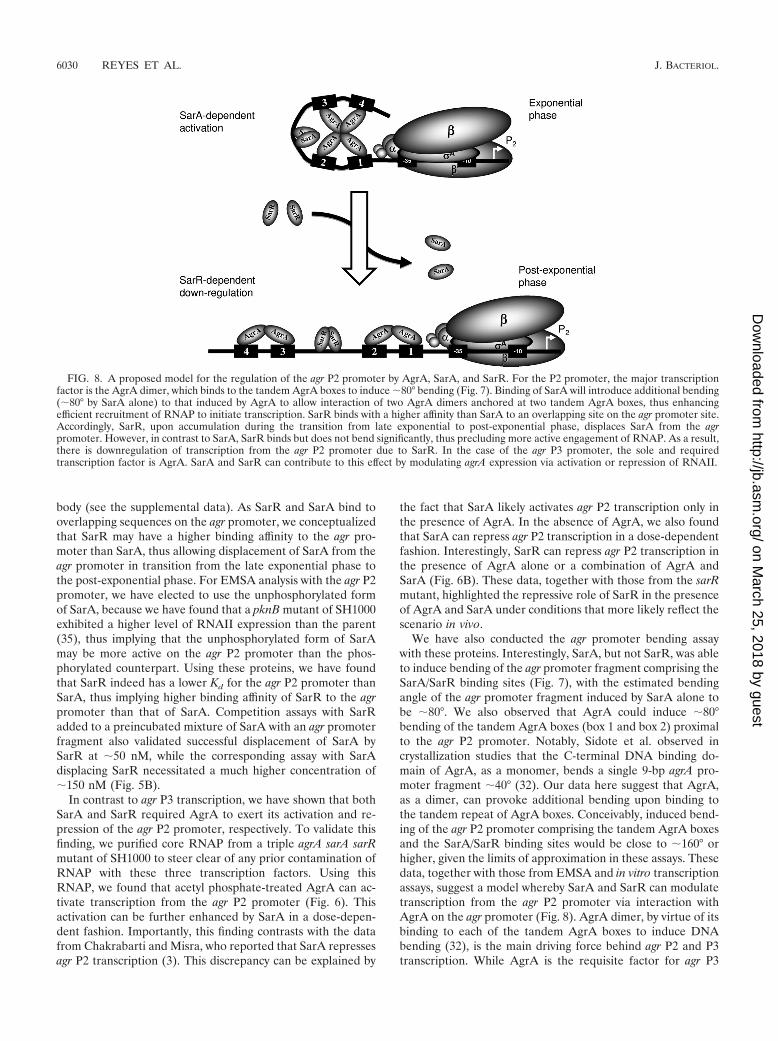
FIG. 8. A proposed model for the regulation of the agr P2 promoter by AgrA, SarA, and SarR. For the P2 promoter, the major transcriptionfactor is the AgrA dimer, which binds to the tandem AgrA boxes to induce �80° bending (Fig. 7). Binding of SarA will introduce additional bending(�80° by SarA alone) to that induced by AgrA to allow interaction of two AgrA dimers anchored at two tandem AgrA boxes, thus enhancingefficient recruitment of RNAP to initiate transcription. SarR binds with a higher affinity than SarA to an overlapping site on the agr promoter site.Accordingly, SarR, upon accumulation during the transition from late exponential to post-exponential phase, displaces SarA from the agrpromoter. However, in contrast to SarA, SarR binds but does not bend significantly, thus precluding more active engagement of RNAP. As a result,there is downregulation of transcription from the agr P2 promoter due to SarR. In the case of the agr P3 promoter, the sole and requiredtranscription factor is AgrA. SarA and SarR can contribute to this effect by modulating agrA expression via activation or repression of RNAII.
6030 REYES ET AL. J. BACTERIOL.
on March 25, 2018 by guest
http://jb.asm.org/
Dow
nloaded from
transcription, the scenario with agr P2 transcription is quitedifferent, with two AgrA dimers first binding to two sets oftandem AgrA boxes to induce bending of the agr promoter(Fig. 7) (18, 32) to facilitate P2 transcription. During the ex-ponential phase, the SarA protein level increases (10), leadingto binding of the SarA dimer to the SarA binding site situatedbetween the two tandem AgrA boxes (21). Binding of SarAinduces additional bending of the agr P2 promoter (Fig. 7 and8), presumably more than that of AgrA alone, thus furtherenhancing transcriptional activation of the agr P2 promoter byRNA polymerase (Fig. 6A). We surmise that this enhancementmay be due to additional interaction of the two AgrA dimersanchored on two sets of tandem AgrA boxes (Fig. 8), facili-tated by the �160° bending of the agr promoter by SarA andAgrA. In transition from the late exponential phase to thepost-exponential phase, the SarR protein accumulates (23). Asthe SarR protein binds to the agr promoter with higher affinitythan SarA, it would displace the SarA bound on the agr pro-moter (see Fig. 5B). In contrast to SarA, SarR binds but doesnot induce bending of the agr promoter, possibly unwrappingthe transcription competent complex, thus leading to down-regulation of transcription from the agr P2 promoter.
While our model does not support direct modulation of theagr P3 promoter by SarR and SarA, it does not preclude indi-rect effects whereby SarA and SarR would activate and repressthe agr P2 promoter, respectively, to modulate AgrA expres-sion, which in turn would dictate transcription from the P3promoter.
ACKNOWLEDGMENTS
This work was supported in part by NIH grant AI37142 to A.L.C.,Swiss National Science Foundation Grant 3100A0-120428 to W.L.K.,and an F. Hoffmann-La-Roche MD-Ph.D. training grant to D.O.A.
REFERENCES
1. Arnaud, M., A. Chastanet, and M. Debarbouille. 2004. New vector forefficient allelic replacement in naturally nontransformable, low-GC-content,Gram-positive bacteria. Appl. Environ. Microbiol. 70:6887–6891.
2. Arvidson, S., and K. Tegmark. 2001. Regulation of virulence determinants inStaphylococcus aureus. Int. J. Med. Microbiol. 291:159–170.
3. Chakrabarti, S. K., and T. K. Misra. 2000. SarA represses agr operonexpression in a purified in vitro Staphylococcus aureus transcription system.J. Bacteriol. 182:5893–5897.
4. Cheung, A. L. 2001. Global regulation of virulence determinants in Staphy-lococcus aureus, p. 295–322. In A. L. Honeyman, H. Friedman, and M.Bendinelli (ed.), Staphylococcus aureus infection and disease. Plenum Pub-lisher, New York, NY.
5. Cheung, A. L., A. S. Bayer, G. Zhang, H. Gresham, and Y.-Q. Xiong. 2004.Regulation of virulence determinants in vitro and in vivo in Staphylococcusaureus. FEMS Microbiol. Lett. 1649:1–9.
6. Cheung, A. L., M. G. Bayer, and J. H. Heinrichs. 1997. sar genetic determi-nants necessary for transcription of RNAII and RNAIII in the agr locus ofStaphylococcus aureus. J. Bacteriol. 179:3963–3971.
7. Cheung, A. L., K. A. Nishina, M. P. Trotonda, and S. Tamber. 2008. TheSarA protein family of Staphylococcus aureus. Int. J. Biochem. Cell Biol.40:355–361.
8. Chien, C.-T., A. C. Manna, S. J. Projan, and A. L. Cheung. 1999. SarA, aglobal regulator of virulence determinants in Staphylococcus aureus, binds toa conserved motif essential for sar dependent gene regulation. J. Biol. Chem.274:37169–37176.
9. Chien, Y., and A. L. Cheung. 1998. Molecular interactions between twoglobal regulators, sar and agr, in Staphylococcus aureus. J. Biol. Chem. 237:2645–2652.
10. Chien, Y. T., A. C. Manna, and A. L. Cheung. 1998. SarA level is a deter-minant of agr activation in Staphylococcus aureus. Mol. Microbiol. 31:991–1001.
11. Crossley, K. B., and G. L. Archer. 1997. The staphylococci in human disease.Churchill Livingstone, New York, NY.
12. Fujimoto, D. F., et al. 2009. Staphylococcus aureus SarA is a regulatoryprotein responsive to redox and pH that can support bacteriophage lambdaintegrase-mediated excision/recombination. Mol. Microbiol. 74:1445–1458.
13. Geisinger, E., R. P. Adhikari, R. Jin, H. F. Ross, and R. P. Novick. 2006.Inhibition of rot translation by RNAIII, a key feature of agr function. Mol.Microbiol. 61:1038–1048.
14. Helmann, J. D. 1995. Compilation and analysis of Bacillus subtilis sigmaA-dependent promoter sequences: evidence for extended contact betweenRNA polymerase and upstream promoter DNA. Nucleic Acids Res. 23:2351–2360.
15. Horsburgh, M. J., et al. 2002. sigmaB modulates virulence determinantexpression and stress resistance: characterization of a functional rsbU strainderived from Staphylococcus aureus 8325-4. J. Bacteriol. 184:5457–5467.
16. Kahl, B., et al. 2000. Staphylococcus aureus RN6390 replicates and inducesapoptosis in a pulmonary epithelial cell line derived from a cystic fibrosispatient. Infect. Immun. 68:5385–5392.
17. Klevens, R. M., et al. 2007. Invasive methicillin-resistant Staphylococcusaureus infections in the United States. J. Am. Med. Assoc. 298:1763–1771.
18. Koenig, R. L., J. L. Ray, S. J. Maleki, M. S. Smeltzer, and B. K. Hurlburt.2004. Staphylococcus aureus AgrA binding to the RNAIII-agr regulatoryregion. J. Bacteriol. 186:7549–7555.
19. Kullik, I., and P. Giachino. 1997. The alternative sigma factor sB in Staph-ylococcus aureus: regulation of the sigB operon in response to growth phaseand heat shock. Arch. Microbiol. 167:151–159.
20. Liu, Y., et al. 2001. Crystal structure of the SarR protein from Staphylococcusaureus. Proc. Natl. Acad. Sci. U. S. A. 98:6877–6882.
21. Liu, Y., et al. 2006. Structural and function analyses of the global regulatoryprotein SarA from Staphylococcus aureus. Proc. Natl. Acad. Sci. U. S. A.103:2392–2397.
22. Lowy, F. 1998. Staphylococcus aureus infections. N. Engl. J. Med. 339:520–532.
23. Manna, A. C., and A. L. Cheung. 2001. Characterization of sarR, a modulatorof sar expression in Staphylococcus aureus. Infect. Immun. 69:885–896.
24. Manna, A. C., and A. L. Cheung. 2006. Transcriptional regulation of the agrlocus and the identification of DNA binding residues of the global regulatoryprotein SarR in Staphylococcus aureus. Mol. Microbiol. 60:1289–1301.
25. Novick, R. P. 2003. Autoinduction and signal transduction in the regulationof staphylococcal virulence. Mol. Microbiol. 48:1429–1449.
26. Novick, R. P., and E. Geisinger. 2008. Quorum sensing in staphylococci.Annu. Rev. Genet. 42:541–564.
27. Novick, R. P., et al. 1993. Synthesis of staphylococcal virulence factors iscontrolled by a regulatory RNA molecule. EMBO J. 12:3967–3977.
28. Palma, M., and A. L. Cheung. 2001. SigB activity in Staphylococcus aureus iscontrolled by RsbU and additional factors during growth. Infect. Immun.69:7858–7865.
29. Prentki, P., M. H. Pham, and D. J. Galas. 1987. Plasmid permutation vectorsto monitor DNA bending. Nucleic Acids Res. 15:10060.
30. Rechtin, T. M., et al. 1999. Characterization of the SarA virulence generegulator of Staphylococcus aureus. Mol. Microbiol. 33:307–316.
31. Safo, M. K., et al. 2005. Crystal structures of the BlaI repressor from Staph-ylococcus aureus and its complex with DNA: insights into transcriptionalregulation of the bla and mec operons. J. Bacteriol. 187:1833–1844.
32. Sidote, D. J., C. M. Barbieri, T. Wu, and A. M. Stock. 2008. Structure of theStaphylococcus aureus AgrA LytTR domain bound to DNA reveals a betafold with an unusual mode of binding. Structure 16:727–735.
33. Tamber, S., and A. L. Cheung. 2009. SarZ promotes the expression ofvirulence factors and represses biofilm formation by modulating SarA andagr in Staphylococcus aureus. Infect. Immun. 77:419–428.
34. Tamber, S., et al. 2010. The staphylococcal specific gene, rsr, represses agrand virulence in Staphylococcus aureus. Infect. Immun. 78:4384–4391.
35. Tamber, S., J. Schwartzman, and A. L. Cheung. 2010. Role of PknB kinasein antibiotic resistance and virulence in community-acquired methicillin-resistant Staphylococcus aureus strain USA300. Infect. Immun. 78:3637–3646.
36. Trotonda, M. P., S. Tamber, G. Memmi, and A. L. Cheung. 2008. MgrArepresses biofilm formation in Staphylococcus aureus. Infect. Immun. 76:5645–5654.
37. Xiong, Y.-Q., et al. 2002. Activation and transcriptional interaction betweenagr RNAII and RNAIII in Staphylococcus aureus in vitro and in an experi-mental endocarditis model. J. Infect. Dis. 186:668–677.
38. Zwieb, C., and S. Adhya. 2009. Plasmid vectors for the analysis of protein-induced DNA bending, p. 547–562. In T. Moss and B. Lebanc (ed.), Methodsin molecular biology, DNA-protein interactions. Humana Press, NewYork, NY.
VOL. 193, 2011 AgrA, SarA, AND SarR ON THE agr PROMOTER 6031
on March 25, 2018 by guest
http://jb.asm.org/
Dow
nloaded from