Embed Size (px)
Citation preview
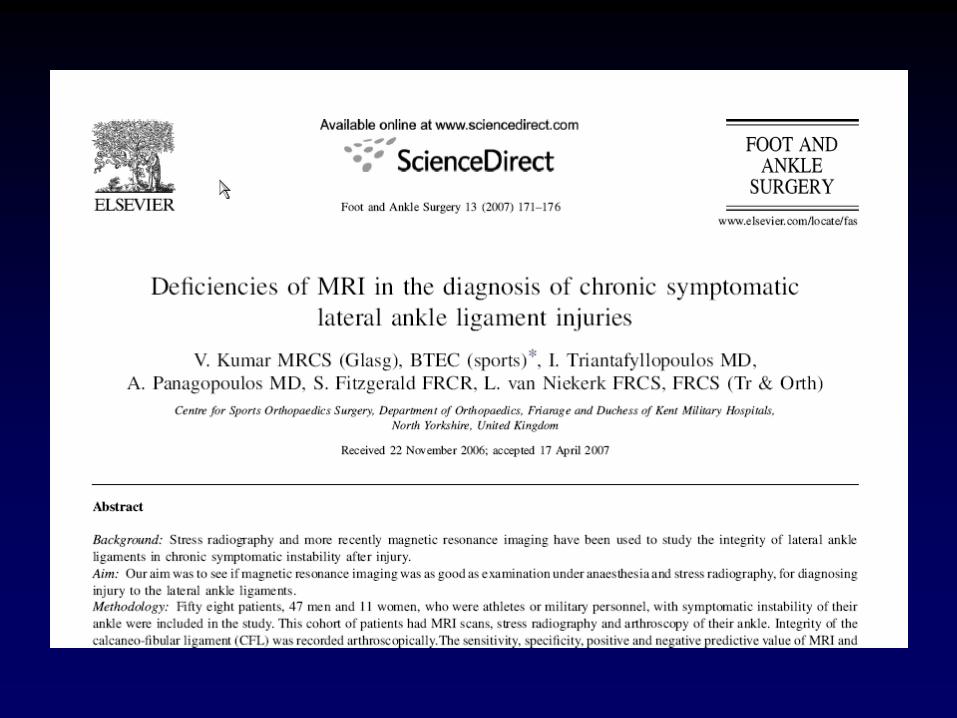
CORRELATION OF CLINICAL, RADIOLOGICAL AND ARTHROSCOPIC FINDINGS IN POST-TRAUMATIC INSTABILITY OF THE ANKLE JOINT:
THE CRITICAL ROLE OF THE CALCANEOFIBULAR LIGAMENT
A. Panagopoulos, MD, PhD Clinical Research Fellow V. Kumar, MRCS (Glas) SHO in Orthopaedics J. Triantafyllopoulos, MD, PhD Clinical Research Fellow S. Fitsgerald, FRCR, MRCP (Irl) Consultant Radiologist L. van Niekerk, MD, FRCS (Ed), FRCS (Orth) Consultant Orthopaedic Surgeon
Sports Orthopaedic Centre, Friarage Hospital & Duchess of Kent Military Hospital,
Northallerton, North Yorkshire, United Kingdom
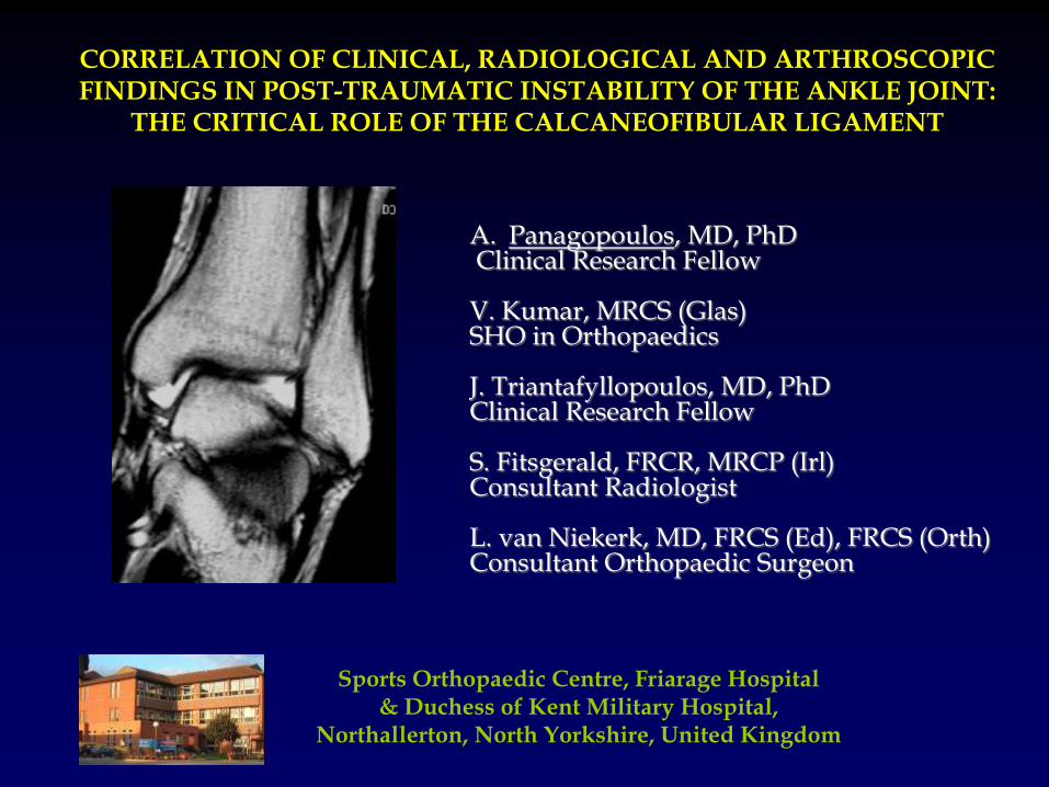
Natural history - prognosis
80% to 85% of acute ankle sprains are successfully treated with a functional ankle-rehabilitation program1
The remaining 15% to 20% have recurrent ankle instability and re-injury, necessitating surgical intervention2
25% to 95% incidence of chondral lesions3
(persistent pain after ligament reconstruction) degenerative changes after 10 years in patients with chronic ankle instability
1. Baumhauer J et al. Journal of Athletic Training, 2002 2. Colville MR, et al. Am J Sports Med, 1990 3. Hintermann B et al. Am J Sports Med, 2002 4. Harrington KD et al. JBJS Am, 1979
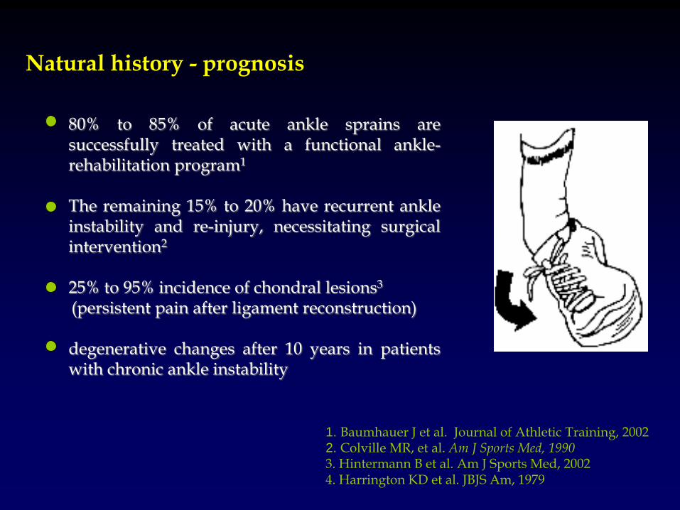
Anatomy
ATFL is the primary restraint to inversion and is usually torn in inversion, plantar flexion and internal rotation1 CFL stabilizes both the ankle & subtalar joint and tears primarily in inversion and ankle dorsiflexion1
In a cadaveric study ATFL was always torn when CFL was torn
1. Burks RT and Morgan J. Am J Sports Med, 1994 2. Heilman AE et al. Foot Ankle, 1990 3. Rasmussen O. Acta Orthop Scand Supl, 1985
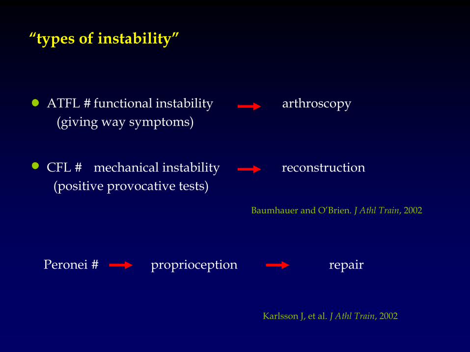
“types of instability”
Baumhauer and O’Brien. J Athl Train, 2002
ATFL # functional instability arthroscopy
(giving way symptoms)
CFL # mechanical instability reconstruction
(positive provocative tests)
Karlsson J, et al. J Athl Train, 2002
Peronei # proprioception repair
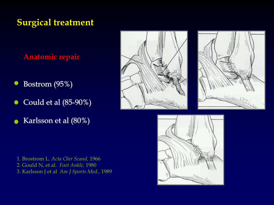
Anatomic repair Bostrom (95%) Could et al (85-90%) Karlsson et al (80%)
Surgical treatment
1. Brostrom L. Acta Chir Scand, 1966 2. Gould N, et al. Foot Ankle, 1980 3. Karlsson J et al Am J Sports Med., 1989
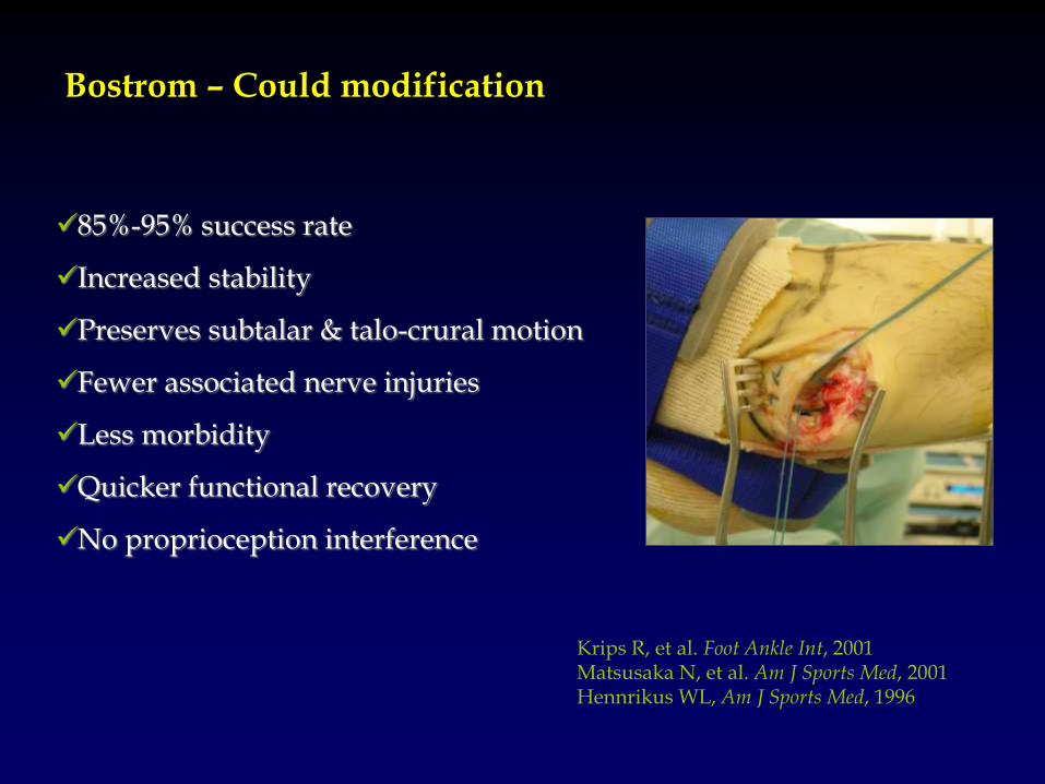
85%-95% success rate
Increased stability
Preserves subtalar & talo-crural motion
Fewer associated nerve injuries
Less morbidity
Quicker functional recovery
No proprioception interference
Krips R, et al. Foot Ankle Int, 2001 Matsusaka N, et al. Am J Sports Med, 2001 Hennrikus WL, Am J Sports Med, 1996
Bostrom – Could modification
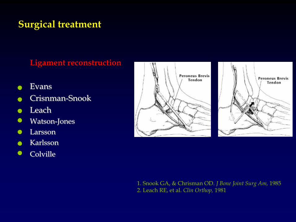
1. Snook GA, & Chrisman OD. J Bone Joint Surg Am, 1985 2. Leach RE, et al. Clin Orthop, 1981
Ligament reconstruction
Evans
Crisnman-Snook
Leach
Watson-Jones
Larsson
Karlsson
Colville
Surgical treatment
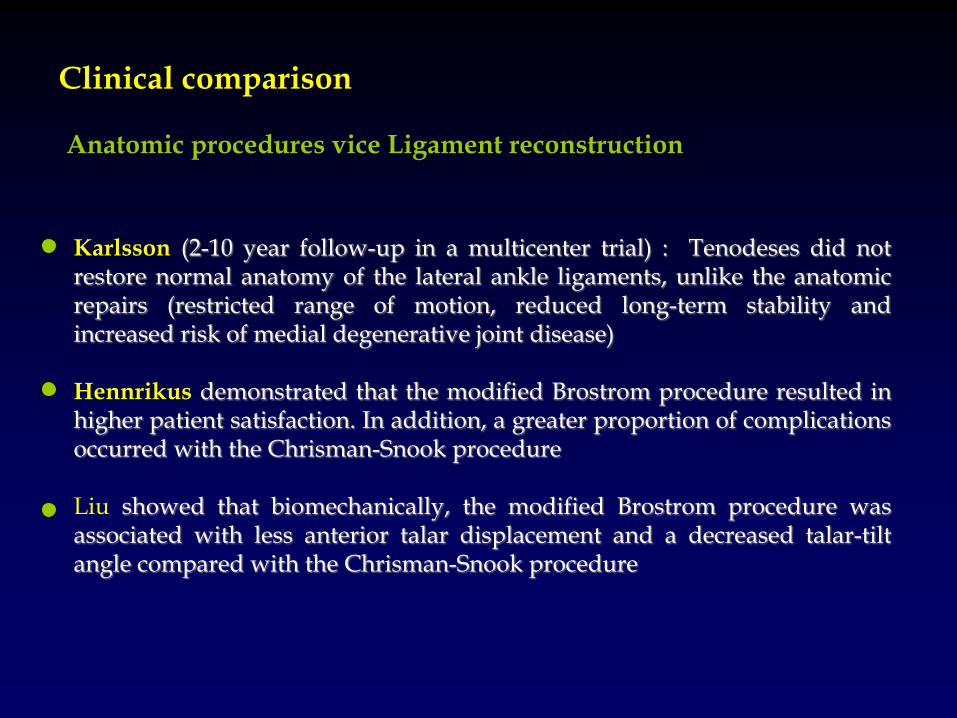
Karlsson (2-10 year follow-up in a multicenter trial) : Tenodeses did not restore normal anatomy of the lateral ankle ligaments, unlike the anatomic repairs (restricted range of motion, reduced long-term stability and increased risk of medial degenerative joint disease) Hennrikus demonstrated that the modified Brostrom procedure resulted in higher patient satisfaction. In addition, a greater proportion of complications occurred with the Chrisman-Snook procedure Liu showed that biomechanically, the modified Brostrom procedure was associated with less anterior talar displacement and a decreased talar-tilt angle compared with the Chrisman-Snook procedure
Clinical comparison
Anatomic procedures vice Ligament reconstruction
Ankle instability hypothesis
• Only unstable ankles with ligament rupture
(mechanical instability) require reconstruction
• Functional instability can be improved with arthroscopic treatment of the ankle
• Mechanical stability is not lost with isolated
ATFL rupture
1. To determine the sensitivity and specificity of MRI diagnosis of lateral ligamentous injuries as confirmed by EUA and arthroscopy findings
2. To define the prevalence of peronei tendons injury that coexist with lateral ligamentous injury
3. To compare the occurrence rates of other associated pathologies found on MRI to that of arthroscopy finding
Role of MRI?
History-symptoms Clinical evaluation X-rays and MRI scan Evaluation under anesthesia Stress views in comparison with the normal ankle Arthroscopic confirmation & treatment Bostrom-Could reconstruction (CFL rupture)
Ankle instability protocol (2004-)
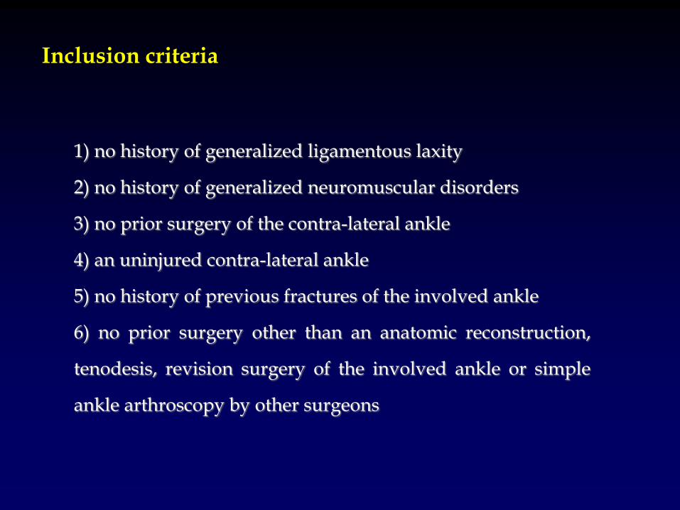
Inclusion criteria
1) no history of generalized ligamentous laxity
2) no history of generalized neuromuscular disorders
3) no prior surgery of the contra-lateral ankle
4) an uninjured contra-lateral ankle
5) no history of previous fractures of the involved ankle
6) no prior surgery other than an anatomic reconstruction,
tenodesis, revision surgery of the involved ankle or simple
ankle arthroscopy by other surgeons
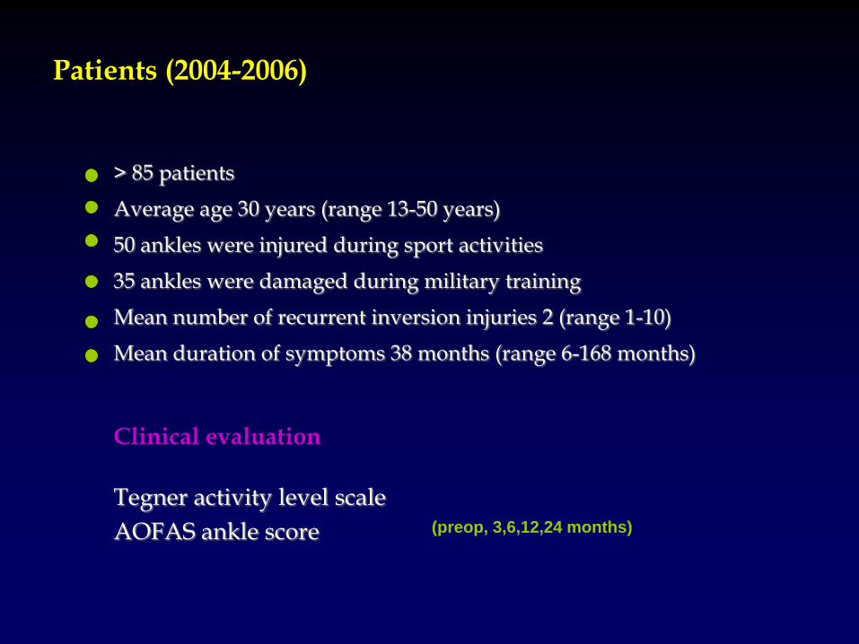
> 85 patients Average age 30 years (range 13-50 years) 50 ankles were injured during sport activities 35 ankles were damaged during military training Mean number of recurrent inversion injuries 2 (range 1-10) Mean duration of symptoms 38 months (range 6-168 months) Clinical evaluation
Tegner activity level scale
AOFAS ankle score
Patients (2004-2006)
(preop, 3,6,12,24 months)
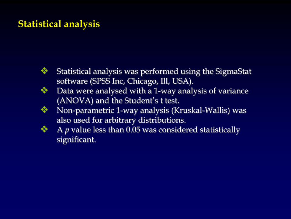
Statistical analysis was performed using the SigmaStat
software (SPSS Inc, Chicago, Ill, USA). Data were analysed with a 1-way analysis of variance
(ANOVA) and the Student’s t test. Non-parametric 1-way analysis (Kruskal-Wallis) was
also used for arbitrary distributions. A p value less than 0.05 was considered statistically
significant.
Statistical analysis
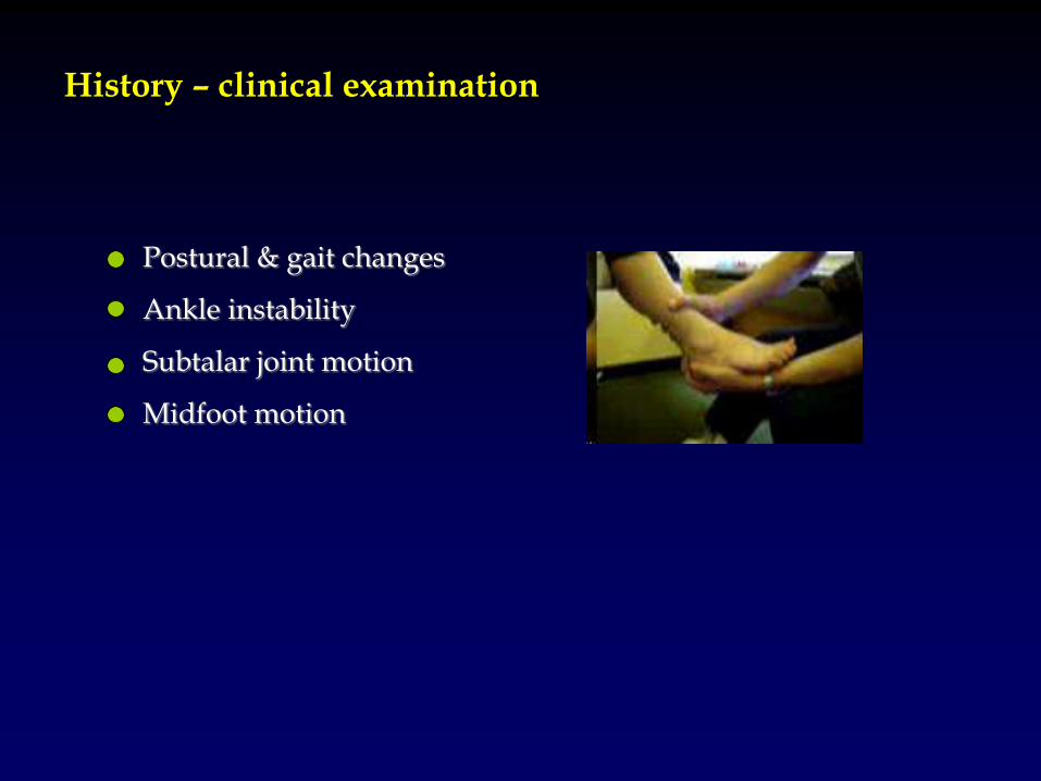
History – clinical examination
Postural & gait changes
Ankle instability
Subtalar joint motion
Midfoot motion
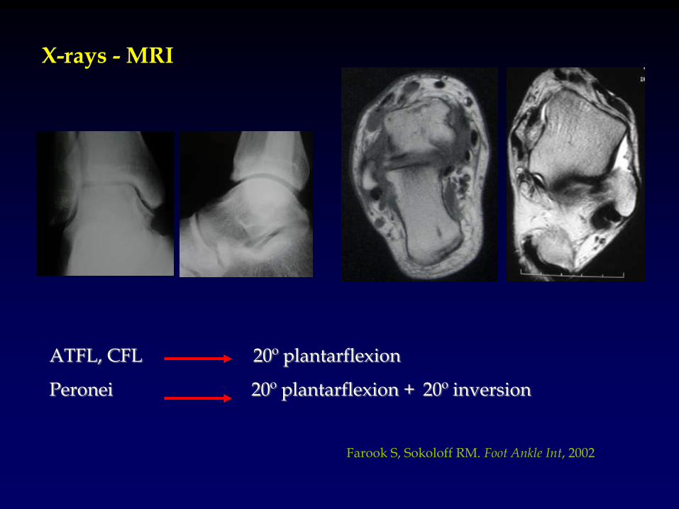
X-rays - MRI
Farook S, Sokoloff RM. Foot Ankle Int, 2002
ATFL, CFL 20º plantarflexion
Peronei 20º plantarflexion + 20º inversion
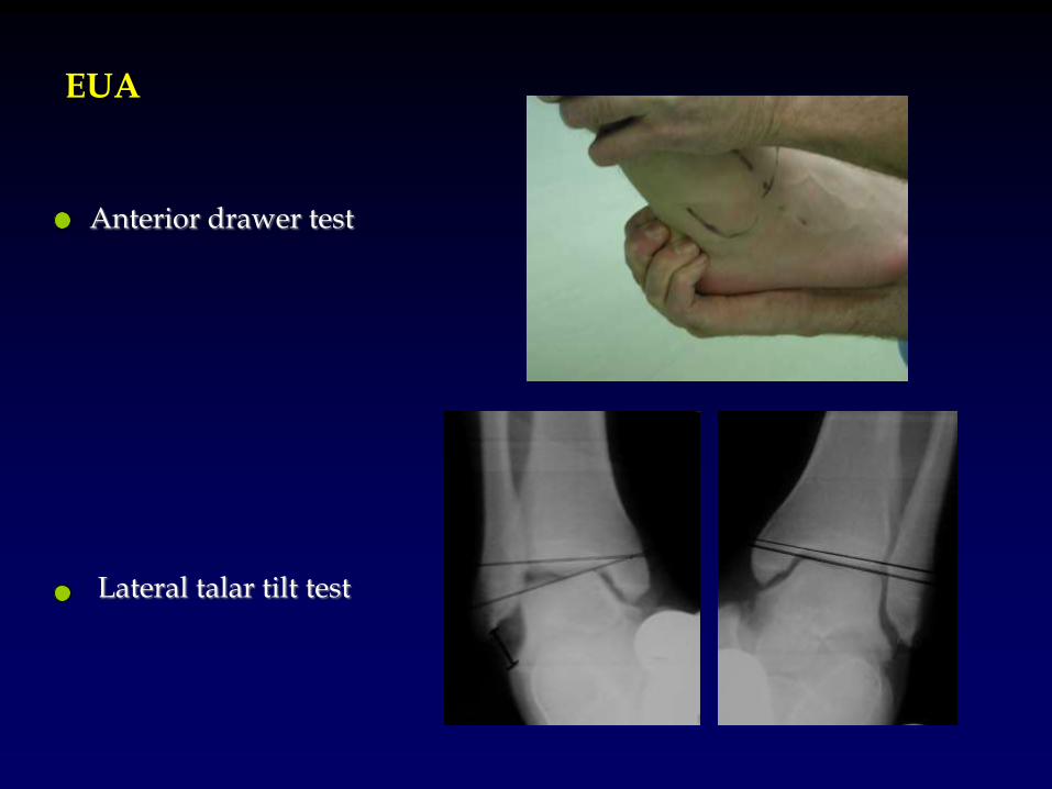
EUA
Anterior drawer test
Lateral talar tilt test
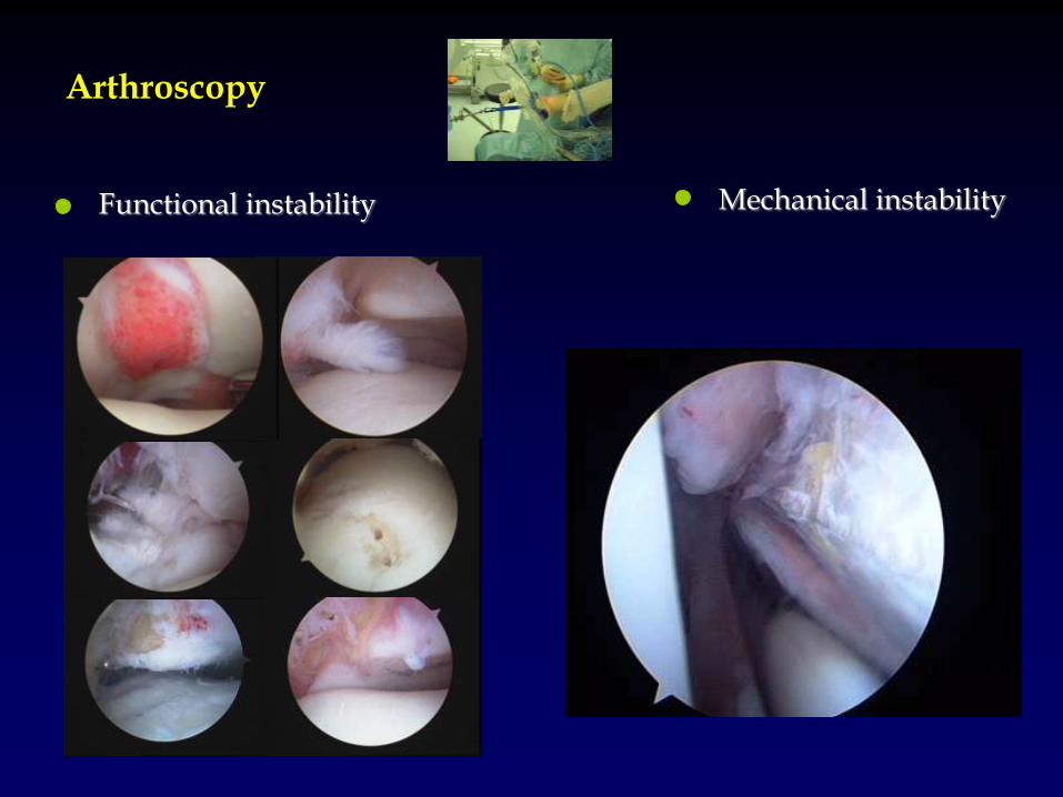
Arthroscopy
Mechanical instability Functional instability
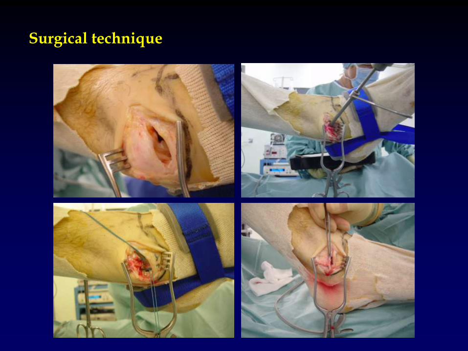
Surgical technique
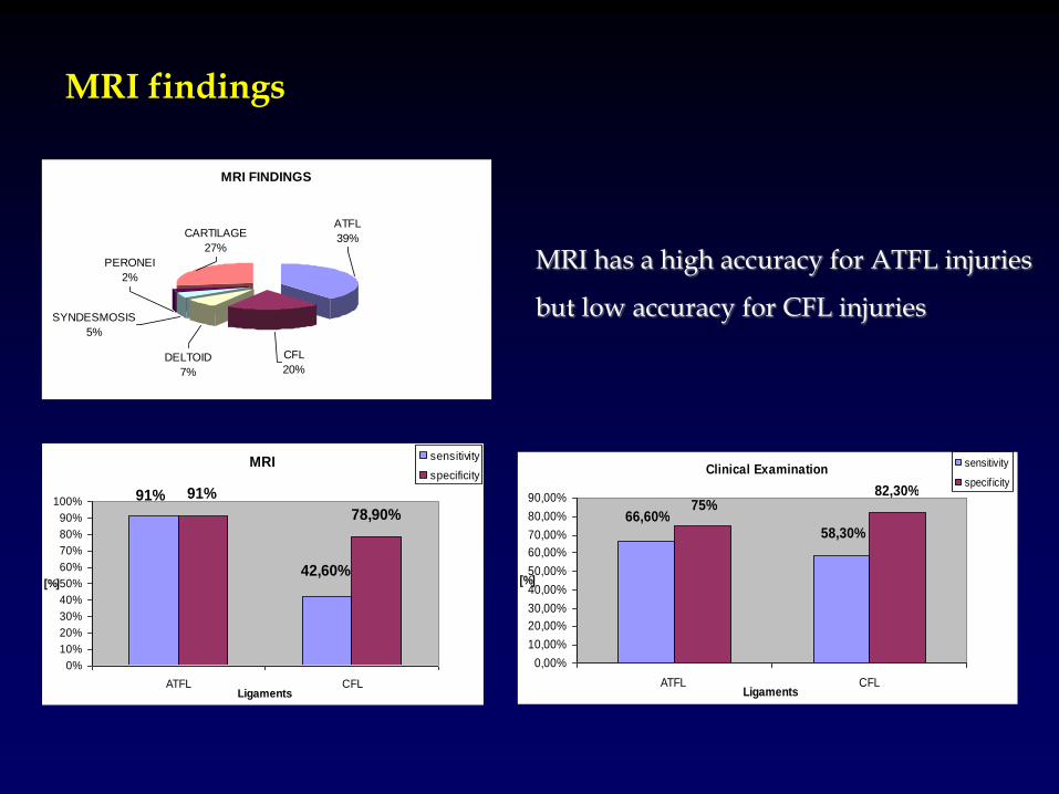
MRI
42,60%
91% 91%
78,90%
0%
10%
20%
30%
40%
50%
60%
70%
80%
90%
100%
ATFL CFLLigaments
[%]
sensitivity
specificity
MRI findings
Clinical Examination
58,30%66,60%
75%82,30%
0,00%
10,00%
20,00%
30,00%
40,00%
50,00%
60,00%
70,00%
80,00%
90,00%
ATFL CFLLigaments
[%]
sensitivity
specif icity
MRI FINDINGS
PERONEI
2%
CARTILAGE
27%
SYNDESMOSIS
5%
DELTOID
7%
CFL
20%
ATFL
39%
MRI has a high accuracy for ATFL injuries
but low accuracy for CFL injuries
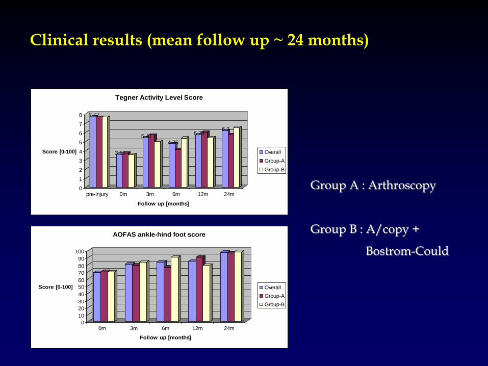
Clinical results (mean follow up ~ 24 months)
7.67
3.61
5.414.75
5.736.2
0
1
2
3
4
5
6
7
8
Score [0-100]
pre-injury 0m 3m 6m 12m 24m
Follow up [months]
Tegner Activity Level Score
Overall
Group-A
Group-B
0
10
20
30
40
50
60
70
80
90
100
Score [0-100]
0m 3m 6m 12m 24m
Follow up [months]
AOFAS ankle-hind foot score
Overall
Group-A
Group-B
Group A : Arthroscopy
Group B : A/copy +
Bostrom-Could
Conclusions
Our diagnostic and treatment algorithm for chronic ankle instability provides accurate evaluation of the deficient lateral ankle ligaments This study, demonstrated the excellent outcome of the anatomic lateral ankle ligaments repair in a high demand population The CFL ligament is the major restraint of lateral ankle instability and the pre-operative planning can be based on the integrity of this ligament. The associated intra-articular injuries may be a significant source of symptoms for this group of patients. Treatment of this associated pathology may provide excellent post-operative outcome.
Limitations
Could the final outcome be the same no matter the group of patients that treatment would be applied? Is arthroscopy only a sufficient treatment for both functional and mechanical ankle instability? A double blind study must be performed in which both treatment methods should be applied for both types of chronic ankle instability (ethical restrictions?)
Expectations (research)
The study will be completed (min. 3 years follow up) in 2008 Preliminary results have already been presented in Hellenic Orthopaedic Association Meeting (Athens 2004), ESSKA 2006 Congress (Austria 2006) and Effort 2007 (Florence) Three papers are planned for submission to AJSM (Overall clinical study), Knee Surgery Sports Traumatology and Arthroscopy (role of CFL) and the Foot and Ankle Surgery (role of MRI)
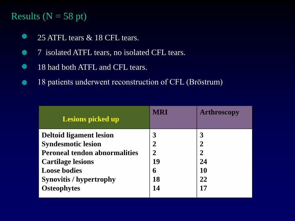
Results (N = 58 pt)
25 ATFL tears & 18 CFL tears.
7 isolated ATFL tears, no isolated CFL tears.
18 had both ATFL and CFL tears.
18 patients underwent reconstruction of CFL (Bröstrum)
MRI Arthroscopy
Deltoid ligament lesion
Syndesmotic lesion
Peroneal tendon abnormalities
Cartilage lesions
Loose bodies
Synovitis / hypertrophy
Osteophytes
3
2
2
19
6
18
14
3
2
2
24
10
22
17
Lesions picked up
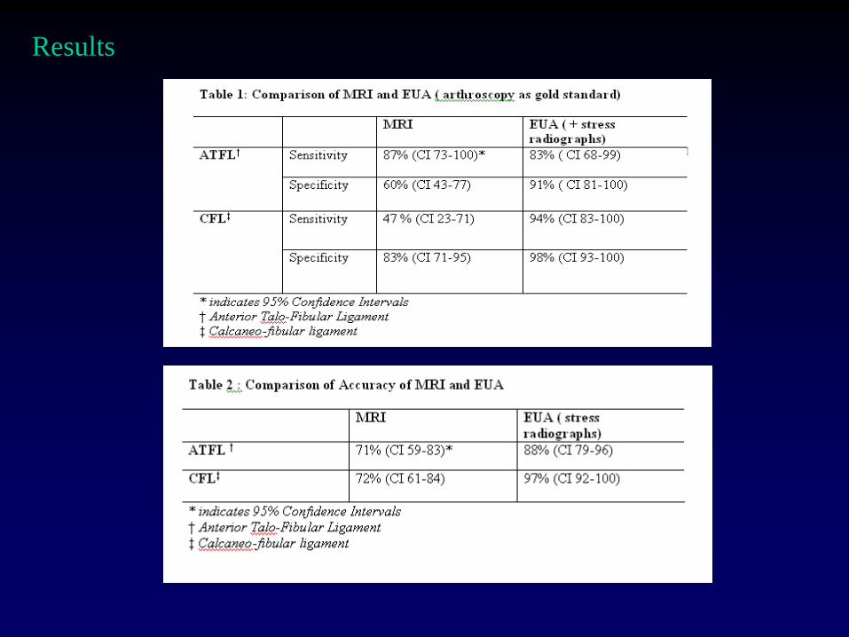
Results
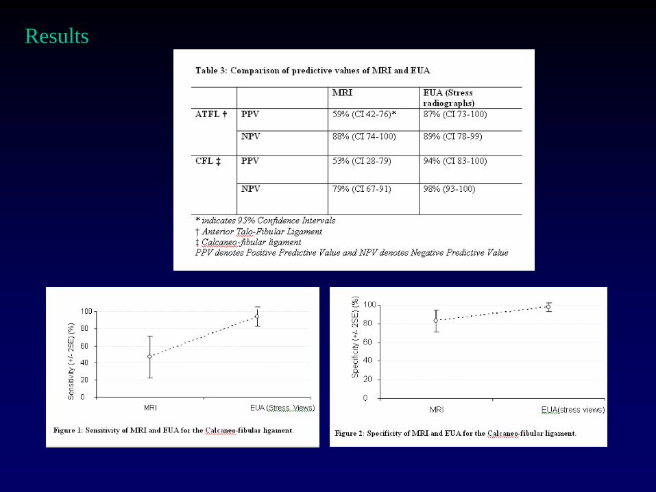
Results
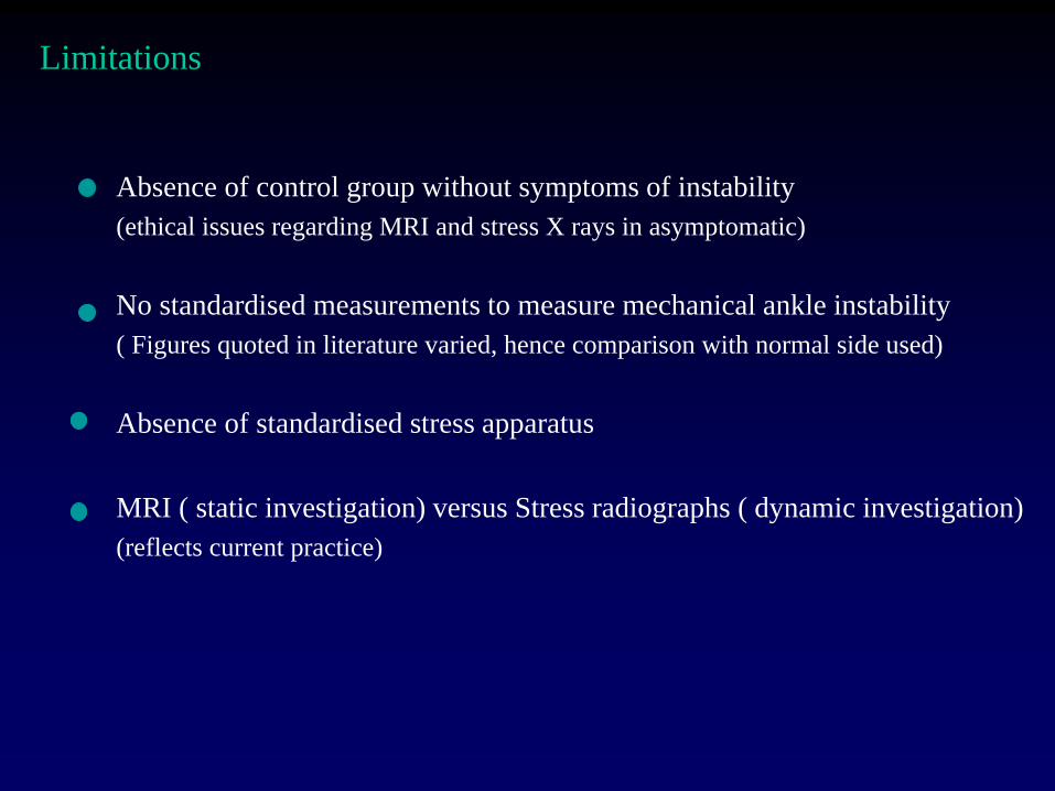
Absence of control group without symptoms of instability
(ethical issues regarding MRI and stress X rays in asymptomatic)
No standardised measurements to measure mechanical ankle instability
( Figures quoted in literature varied, hence comparison with normal side used)
Absence of standardised stress apparatus
MRI ( static investigation) versus Stress radiographs ( dynamic investigation)
(reflects current practice)
Limitations
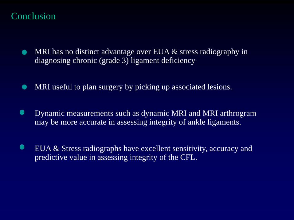
MRI has no distinct advantage over EUA & stress radiography in diagnosing chronic (grade 3) ligament deficiency MRI useful to plan surgery by picking up associated lesions. Dynamic measurements such as dynamic MRI and MRI arthrogram may be more accurate in assessing integrity of ankle ligaments. EUA & Stress radiographs have excellent sensitivity, accuracy and predictive value in assessing integrity of the CFL.
Conclusion
THANK YOU