Embed Size (px)
Citation preview
CASE REPORT Open Access
Crossed pulmonary arteries associated withsingle atrium in an adult: a case reportFengli Fu, Jiahu Yang, Jianjun Zhang and Yue Feng*
Abstract
Background: Crossed pulmonary arteries or single atrium is a rare form of cardiovascular anomaly. In previousstudies, the anomalies are detected in infant or early adolescence, and infrequently seen in adult population.
Case presentation: We presented a case of the coexistence of two congenital anomalies in a 44-year-old womanwho remained well tolerated and undiscovered until adulthood. Physical examination showed a grade III systolicmurmur at the cardiac apex, and a grade II/III systolic murmur at left 2–3 intercostal space. An echocardiographyrevealed absence of atrial septal tissue. Dual-source CT angiography was performed for further evaluation of thegreat vessel. Except an enlarged single atrium, the imaging showed that the origination of the left pulmonary arteryfrom the pulmonary trunk was superior to that of the right pulmonary artery. The branch pulmonary arteries thencrisscrossed as they coursed to their respective lungs. The findings were illustrated by the right heart catheterizationand then confirmed at surgery.
Conclusions: To our knowledge, this is the first case report of crossed pulmonary arteries with single atrium as theonly additional cardiac anomaly in an adult. Knowledge of this rare combination will be helpful in the differentialdiagnosis of congenital heart disease and assist the surgeon in treatment planning.
Keywords: Malposition of pulmonary arteries, Crossed pulmonary arteries, Single atrium, Computed tomography
Abbreviations: CTA, Computed tomography angiography; LPA, Left pulmonary artery; RPA, Right pulmonary artery
BackgroundCrossed pulmonary arteries are a rare form of pulmon-ary arterial malposition, characterized by an abnormalostium of the left pulmonary artery (LPA) that originatessuperior to the right pulmonary artery (RPA) and to itsright [1, 2]. Single atrium is regarded as the least com-mon variety among the malformations of the atrialseptum, and infrequently seen in adult [3]. In this anom-aly, atrial septum is complete absence, and often accom-panied by malformations of the atrioventricular valves,especially with a cleft in the mitral valve [4]. Crossedpulmonary arteries or single atrium is often associatedwith other congenital cardiac and extracardiac disease.Majority of patients with crossed pulmonary arteries orsingle atrium are symptomatic and poorly tolerated dur-ing infancy or early adolescence with dyspnea on effort,fatigue respiratory tract infection, cyanosis [1–4]. Very
few cases with crossed pulmonary arteries or singleatrium have been reported in adult population. In thefollowing case, we present the combination of two rarecongenital anomalies in an adult woman.
Case presentationA 44-year-old Chinese woman was referred to our hos-pital with heart murmur of 40 years duration. Shereported no cyanosis, chest congestion and anhelationafter activities. On auscultation, there were a grade 3systolic murmur on apex, and a grade 2/3 systolic mur-mur on left 2–3 intercostal space.Routine laboratory examinations were unremarkable.
Chest X-way revealed a mild cardiomegaly and promin-ent pulmonary trunk. On electrocardiogram, there wasnormal sinus rhythm, right and left ventricle hyper-trophy signs. Echocardiography revealed an absence ofatrial septal tissue (Fig. 1) and a small cleft in anteriormitral leaflet. Color doppler examination presented mod-erate mitral regurgitation and mild tricuspid regurgitation.
* Correspondence: [email protected] of Radiology, Zhejiang Hospital, No.12 Lingyin Rd, Hangzhou310013, China
© 2016 The Author(s). Open Access This article is distributed under the terms of the Creative Commons Attribution 4.0International License (http://creativecommons.org/licenses/by/4.0/), which permits unrestricted use, distribution, andreproduction in any medium, provided you give appropriate credit to the original author(s) and the source, provide a link tothe Creative Commons license, and indicate if changes were made. The Creative Commons Public Domain Dedication waiver(http://creativecommons.org/publicdomain/zero/1.0/) applies to the data made available in this article, unless otherwise stated.
Fu et al. BMC Cardiovascular Disorders (2016) 16:172 DOI 10.1186/s12872-016-0354-8
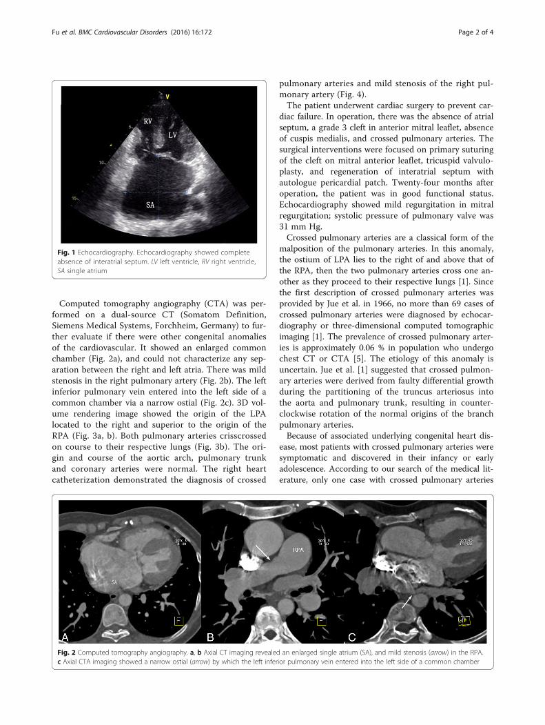
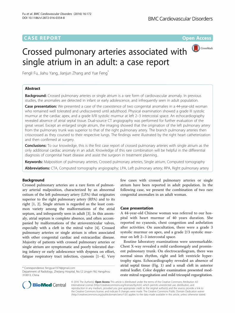
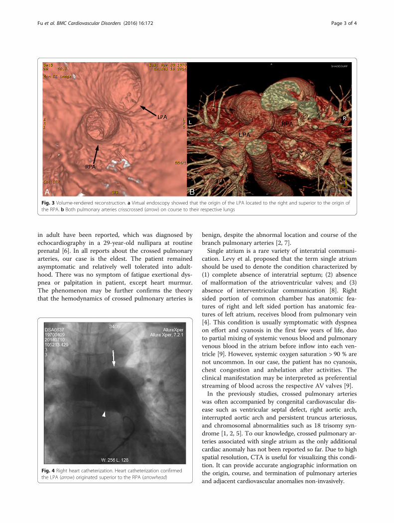
Computed tomography angiography (CTA) was per-formed on a dual-source CT (Somatom Definition,Siemens Medical Systems, Forchheim, Germany) to fur-ther evaluate if there were other congenital anomaliesof the cardiovascular. It showed an enlarged commonchamber (Fig. 2a), and could not characterize any sep-aration between the right and left atria. There was mildstenosis in the right pulmonary artery (Fig. 2b). The leftinferior pulmonary vein entered into the left side of acommon chamber via a narrow ostial (Fig. 2c). 3D vol-ume rendering image showed the origin of the LPAlocated to the right and superior to the origin of theRPA (Fig. 3a, b). Both pulmonary arteries crisscrossedon course to their respective lungs (Fig. 3b). The ori-gin and course of the aortic arch, pulmonary trunkand coronary arteries were normal. The right heartcatheterization demonstrated the diagnosis of crossed
pulmonary arteries and mild stenosis of the right pul-monary artery (Fig. 4).The patient underwent cardiac surgery to prevent car-
diac failure. In operation, there was the absence of atrialseptum, a grade 3 cleft in anterior mitral leaflet, absenceof cuspis medialis, and crossed pulmonary arteries. Thesurgical interventions were focused on primary suturingof the cleft on mitral anterior leaflet, tricuspid valvulo-plasty, and regeneration of interatrial septum withautologue pericardial patch. Twenty-four months afteroperation, the patient was in good functional status.Echocardiography showed mild regurgitation in mitralregurgitation; systolic pressure of pulmonary valve was31 mm Hg.Crossed pulmonary arteries are a classical form of the
malposition of the pulmonary arteries. In this anomaly,the ostium of LPA lies to the right of and above that ofthe RPA, then the two pulmonary arteries cross one an-other as they proceed to their respective lungs [1]. Sincethe first description of crossed pulmonary arteries wasprovided by Jue et al. in 1966, no more than 69 cases ofcrossed pulmonary arteries were diagnosed by echocar-diography or three-dimensional computed tomographicimaging [1]. The prevalence of crossed pulmonary arter-ies is approximately 0.06 % in population who undergochest CT or CTA [5]. The etiology of this anomaly isuncertain. Jue et al. [1] suggested that crossed pulmon-ary arteries were derived from faulty differential growthduring the partitioning of the truncus arteriosus intothe aorta and pulmonary trunk, resulting in counter-clockwise rotation of the normal origins of the branchpulmonary arteries.Because of associated underlying congenital heart dis-
ease, most patients with crossed pulmonary arteries weresymptomatic and discovered in their infancy or earlyadolescence. According to our search of the medical lit-erature, only one case with crossed pulmonary arteries
Fig. 1 Echocardiography. Echocardiography showed completeabsence of interatrial septum. LV left ventricle, RV right ventricle,SA single atrium
Fig. 2 Computed tomography angiography. a, b Axial CT imaging revealed an enlarged single atrium (SA), and mild stenosis (arrow) in the RPA.c Axial CTA imaging showed a narrow ostial (arrow) by which the left inferior pulmonary vein entered into the left side of a common chamber
Fu et al. BMC Cardiovascular Disorders (2016) 16:172 Page 2 of 4
in adult have been reported, which was diagnosed byechocardiography in a 29-year-old nullipara at routineprenatal [6]. In all reports about the crossed pulmonaryarteries, our case is the eldest. The patient remainedasymptomatic and relatively well tolerated into adult-hood. There was no symptom of fatigue exertional dys-pnea or palpitation in patient, except heart murmur.The phenomenon may be further confirms the theorythat the hemodynamics of crossed pulmonary arteries is
benign, despite the abnormal location and course of thebranch pulmonary arteries [2, 7].Single atrium is a rare variety of interatrial communi-
cation. Levy et al. proposed that the term single atriumshould be used to denote the condition characterized by(1) complete absence of interatrial septum; (2) absenceof malformation of the atrioventricular valves; and (3)absence of interventricular communication [8]. Rightsided portion of common chamber has anatomic fea-tures of right and left sided portion has anatomic fea-tures of left atrium, receives blood from pulmonary vein[4]. This condition is usually symptomatic with dyspneaon effort and cyanosis in the first few years of life, duoto partial mixing of systemic venous blood and pulmonaryvenous blood in the atrium before inflow into each ven-tricle [9]. However, systemic oxygen saturation > 90 % arenot uncommon. In our case, the patient has no cyanosis,chest congestion and anhelation after activities. Theclinical manifestation may be interpreted as preferentialstreaming of blood across the respective AV valves [9].In the previously studies, crossed pulmonary arteries
was often accompanied by congenital cardiovascular dis-ease such as ventricular septal defect, right aortic arch,interrupted aortic arch and persistent truncus arteriosus,and chromosomal abnormalities such as 18 trisomy syn-drome [1, 2, 5]. To our knowledge, crossed pulmonary ar-teries associated with single atrium as the only additionalcardiac anomaly has not been reported so far. Due to highspatial resolution, CTA is useful for visualizing this condi-tion. It can provide accurate angiographic information onthe origin, course, and termination of pulmonary arteriesand adjacent cardiovascular anomalies non-invasively.
Fig. 3 Volume-rendered reconstruction. a Virtual endoscopy showed that the origin of the LPA located to the right and superior to the origin ofthe RPA. b Both pulmonary arteries crisscrossed (arrow) on course to their respective lungs
Fig. 4 Right heart catheterization. Heart catheterization confirmedthe LPA (arrow) originated superior to the RPA (arrowhead)
Fu et al. BMC Cardiovascular Disorders (2016) 16:172 Page 3 of 4
ConclusionsIn summary, we present, to our knowledge, for the firsttime a case of coexistence of two rare congenital anom-alies in the form of crossed pulmonary arteries associ-ated with single atrium in an adult. Diagnostic key pointof the case is to reveal the true relationship between theLPA and RPA, and an absence of atrial septal tissue.Knowledge of this rare anomaly will help in the differen-tial diagnosis of congenital heart disease and assist thesurgeon in treatment planning.
AcknowledgementsThe authors appreciate all participants who were involved in the managementof the patient and the preparation of the manuscript.
FundingThis work was supported by Zhejiang Provincial administration of TraditionalChinese Medicine (2013ZA012), Zhejiang Provincial Health Department(2011KYB018) and Zhejiang Provincial Natural Science Foundation (LY15H180009).
Availability of data and materialsAll relevant data is contained within the manuscript.
Authors’ contributionsFLF analyzed and interpreted the data, drafted the manuscript and madecritical revision of the manuscript for important intellectual content. JHY andJJZ collected the patient data, carried out the post-procession after computedtomography angiography examination, and provided resources for literaturereview. YF provided cardiology consultation, participated in the design andcoordination of the manuscript, and conducted final edits of the manuscript.All authors read and approved the final manuscript.
Competing interestsThe authors declare that they have no competing interests.
Ethics approval and consent to participateNot applicable.
Consent for publicationWritten informed consent was obtained from the patient for publication ofthis case report and accompanying images.
Received: 4 April 2016 Accepted: 26 August 2016
References1. Jue KL, Lockman LA, Edwards JE. Anomalous origins of pulmonary arteries
from pulmonary trunk (“crossed pulmonary arteries”): observation in a casewith 18 trisomy syndrome. Am Heart J. 1966;71:807–12.
2. Becker AE, Becker MJ, Edwards JE. Malposition of pulmonary arteries (crossedpulmonary arteries) in persistent truncus arteriosus. Am J Roentgenol RadiumTher Nucl Med. 1970;110:509–14.
3. Avula S, Salazar M, Alturk N, Kukafka S, Ritter S, Grodman RS. Cor triatriatum withsingle atrium presenting in aduithhood. Echocardiography. 2005;22:839–43.
4. Munoz-Armas S, Gorin JRD, Anselmi OG, et al. Single Atrium. Am J Cardiol.1968;21:639.
5. Liu H, Juan YH, Wang Q, Xie Z, Chen J, Huang H, et al. Evaluation ofmalposition of the branch pulmonary arteries using cardiovascularcomputed tomography angiography. Eur Radiol. 2014;24:3300–7.
6. Xiong Y, Gan HJ, Liu T, Tao F, Wang HF, Wu Y. Prenatal diagnosis of crossedpulmonary arteries. Ultrasound Obstet Gynecol. 2010;36:776–7.
7. Chen J, Feng Y. A rare case of crossed pulmonary arteries in an infant-casereport. J Cardiothorac Surg. 2013;8:79.
8. Levy MJ, Salomon J, Vidne BA. Correction of single and common atrium,with reference to simplified terminology. Chest. 1974;66:444–6.
9. Hasanin AM, Kinsara AJ. Single atrium associated with persistent leftsuperior vena cava in asymptomatic adult: case report and review ofliterature. Congenit Heart Dis. 2008;3:368–71.
• We accept pre-submission inquiries
• Our selector tool helps you to find the most relevant journal
• We provide round the clock customer support
• Convenient online submission
• Thorough peer review
• Inclusion in PubMed and all major indexing services
• Maximum visibility for your research
Submit your manuscript atwww.biomedcentral.com/submit
Submit your next manuscript to BioMed Central and we will help you at every step:
Fu et al. BMC Cardiovascular Disorders (2016) 16:172 Page 4 of 4

![Developing Country of Pakistan Great Arteries in a ... · congenitally corrected transposition of the great arteries (CCTGA) [1]. CCTGA is a defect whereby the right atrium is connected](https://img.pdfslide.net/doc/110x75/5cace22d88c99376788cec5d/developing-country-of-pakistan-great-arteries-in-a-congenitally-corrected.jpg)


![Anatomie- [Compatibiliteitsmodus] · PDF fileAND GREAT AND MASSE. ASPECT Pulmonary Adery Coronaru arteru Left ventricle Right ventricle atrium Exterior structures of the heart Arteries](https://img.pdfslide.net/doc/110x75/5a8159477f8b9a0c748d293f/anatomie-compatibiliteitsmodus-great-and-masse-aspect-pulmonary-adery-coronaru.jpg)