Embed Size (px)
Citation preview
Cruciate Disease in Dogs
Improving your skills in diagnosis and treatment
Chris Preston FACVSc DACVS
Pet Emergency & Specialist Centre
1103 Dandenong Rd, Malvern East VIC 3145 Tel: (03) 9569 3677 Fax: (03) 9569 3688 E-mail: [email protected] Web: http://www.petemergency.com.au
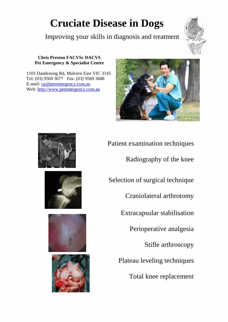
Patient examination techniques
Radiography of the knee
Selection of surgical technique
Craniolateral arthrotomy
Extracapsular stabilisation
Perioperative analgesia
Stifle arthroscopy
Plateau leveling techniques
Total knee replacement
Patient examination techniques Following a thorough general physical examination, assessment of the lame dog involves three sequential steps. The way a dog stands can be of great benefit in helping establish a short list of differential diagnoses. Assessment of standing conformation involves looking for weight shifts, evidence of muscle atrophy and looking at the standing angle of joints. Visual evaluation of gait can be challenging in companion animal practice as few owners are able to suitably control their pets. Typically the patient is walked then trotted outside and the trunk, head and limb movements observed. Finally a complete orthopaedic examination is performed with the patient adequately restrained.
Muscle atrophy Weight shift – ‘toeing off’ Medial periarticular fibrosis Incomplete stifle extension
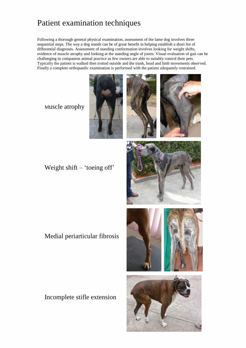
Hock hyperextension Tibial subluxation
Positive ‘sit test’ Limb malalignment (genu varus / tibial torsion) Tibial compression test
Palpation of the knee is used to achieve a diagnosis in most patients. You can assess pain, range of motion, crepitus (DJD), clicking (meniscal tear) and craniocaudal instability (drawer motion). It is wise to assess medial (and lateral) patellar movement as some dogs have both ACL disease and patellar instability. It is common to have a lame dog and no drawer motion – many of these dogs have a partial ACL tear. In such cases, forceful knee extension will elicit a pain response. Compare the patient’s response to this test on the ‘normal’ leg first to see what they do. Direct palpation of the medial and lateral joint line may elicit a focal region of pain if the patient has a meniscal tear.
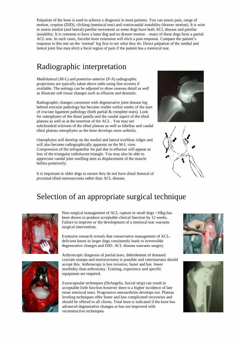
Radiographic interpretation Mediolateral (M-L) and posterior-anterior (P-A) radiographic projections are typically taken above table using fine-screens if available. The settings can be adjusted to show osseous detail as well as illustrate soft tissue changes such as effusion and desmitis. Radiographic changes consistent with degenerative joint disease leg behind articular pathology but become visible within weeks of the start of cruciate ligament pathology (both partial & complete tears). Look for osteophytes of the distal patella and the caudal aspect of the tibial plateau as well as at the insertion of the ACL . You may see subchondral sclerosis of the tibial plateau as well as fabellae and caudal tibial plateau osteophytes as the knee develops more arthritis. Osteophytes will develop on the medial and lateral trochlear ridges and will also become radiographically apparent on the M-L view. Compression of the infrapatellar fat pad due to effusion will appear as loss of the triangular radiolucent triangle. You may also be able to appreciate caudal joint swelling seen as displacement of the muscle bellies posteriorly. It is important in older dogs to ensure they do not have distal femoral of proximal tibial osteosarcoma rather than ACL disease.
Selection of an appropriate surgical technique
Non-surgical management of ACL rupture in small dogs <10kg has been shown to produce acceptable clinical function by 12 weeks. Failure to improve or the development of a meniscal tear warrants surgical intervention. Extensive research reveals that conservative management of ACL-deficient knees in larger dogs consistently leads to irreversible degenerative changes and DJD. ACL disease warrants surgery. Arthroscopic diagnosis of partial tears, debridement of diseased cruciate stumps and meniscectomy is possible and veterinarians should accept this. Arthroscopy is less invasive, faster and has lower morbidity than arthrotomy. Training, experience and specific equipment are required. Extracapsular techniques (DeAngelis, fascial strip) can result in acceptable limb function however there is a higher incidence of late onset meniscal tears. Progressive osteoarthritis develops too. Plateau leveling techniques offer faster and less complicated recoveries and should be offered to all clients. Total knee is indicated if the knee has advanced degenerative changes or has not improved with reconstructive techniques.
Craniolateral stifle arthrotomy
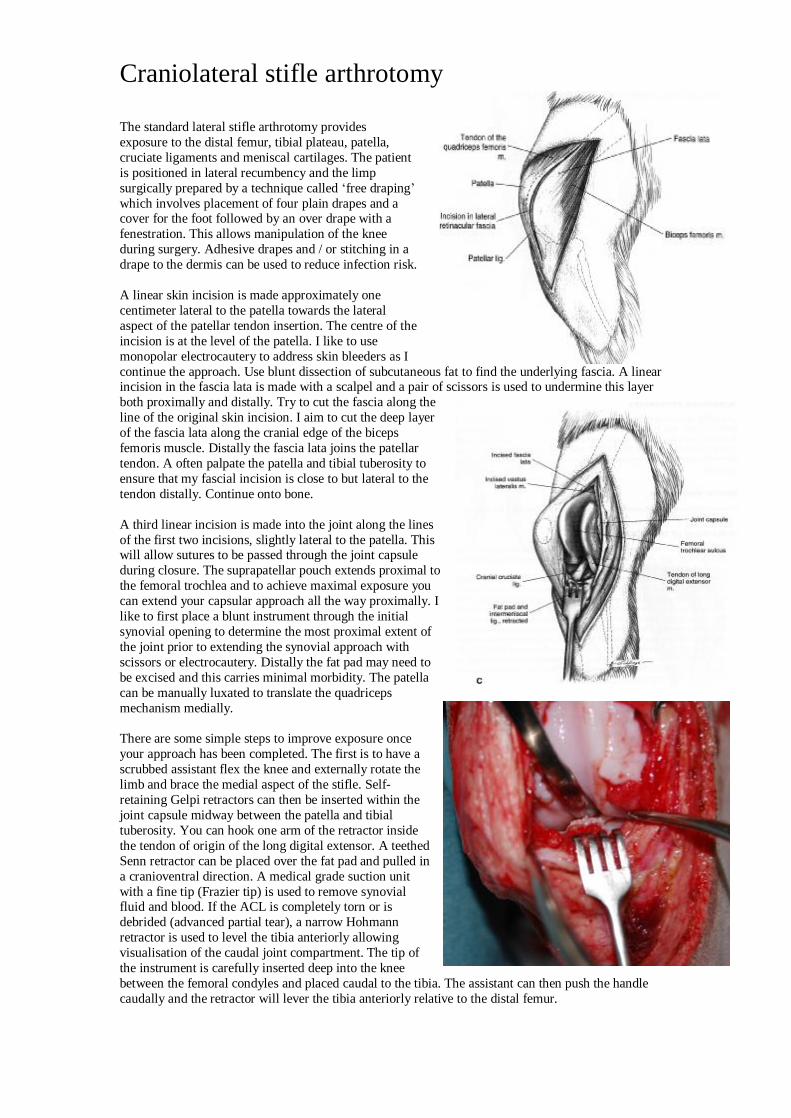
The standard lateral stifle arthrotomy provides exposure to the distal femur, tibial plateau, patella, cruciate ligaments and meniscal cartilages. The patient is positioned in lateral recumbency and the limp surgically prepared by a technique called ‘free draping’ which involves placement of four plain drapes and a cover for the foot followed by an over drape with a fenestration. This allows manipulation of the knee during surgery. Adhesive drapes and / or stitching in a drape to the dermis can be used to reduce infection risk. A linear skin incision is made approximately one centimeter lateral to the patella towards the lateral aspect of the patellar tendon insertion. The centre of the incision is at the level of the patella. I like to use monopolar electrocautery to address skin bleeders as I continue the approach. Use blunt dissection of subcutaneous fat to find the underlying fascia. A linear incision in the fascia lata is made with a scalpel and a pair of scissors is used to undermine this layer both proximally and distally. Try to cut the fascia along the line of the original skin incision. I aim to cut the deep layer of the fascia lata along the cranial edge of the biceps femoris muscle. Distally the fascia lata joins the patellar tendon. A often palpate the patella and tibial tuberosity to ensure that my fascial incision is close to but lateral to the tendon distally. Continue onto bone. A third linear incision is made into the joint along the lines of the first two incisions, slightly lateral to the patella. This will allow sutures to be passed through the joint capsule during closure. The suprapatellar pouch extends proximal to the femoral trochlea and to achieve maximal exposure you can extend your capsular approach all the way proximally. I like to first place a blunt instrument through the initial synovial opening to determine the most proximal extent of the joint prior to extending the synovial approach with scissors or electrocautery. Distally the fat pad may need to be excised and this carries minimal morbidity. The patella can be manually luxated to translate the quadriceps mechanism medially. There are some simple steps to improve exposure once your approach has been completed. The first is to have a scrubbed assistant flex the knee and externally rotate the limb and brace the medial aspect of the stifle. Self-retaining Gelpi retractors can then be inserted within the joint capsule midway between the patella and tibial tuberosity. You can hook one arm of the retractor inside the tendon of origin of the long digital extensor. A teethed Senn retractor can be placed over the fat pad and pulled in a cranioventral direction. A medical grade suction unit with a fine tip (Frazier tip) is used to remove synovial fluid and blood. If the ACL is completely torn or is debrided (advanced partial tear), a narrow Hohmann retractor is used to level the tibia anteriorly allowing visualisation of the caudal joint compartment. The tip of the instrument is carefully inserted deep into the knee between the femoral condyles and placed caudal to the tibia. The assistant can then push the handle caudally and the retractor will lever the tibia anteriorly relative to the distal femur.
Extracapsular techniques Numerous procedures have been reported to address stifle instability. These procedures can be broadly categorised as extracapsular or intracapsular techniques. With all of these techniques, recoveries are prolonged, joint range of motion is reduced long term, laxity returns in many cases and osteoarthritis is progressive. There appears to be little correlation between the development of recurrent laxity and limb function. Clinically, these techniques are worthwhile in most patients and can be performed by veterinarians who are familiar with stifle anatomy and who have basic surgical instrumentation.
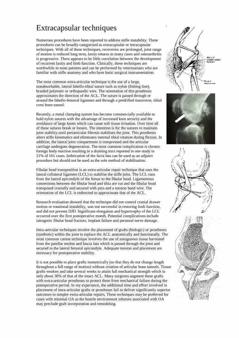
The most common extra-articular technique is the use of a large, nonabsorbable, lateral fabello-tibial suture such as nylon (fishing line), braided polyester or orthopaedic wire. The orientation of this prosthesis approximates the direction of the ACL. The suture is passed through or around the fabello-femoral ligament and through a predrilled transverse, tibial crest bone tunnel.
Recently, a metal clamping system has become commercially available to hold nylon sutures with the advantage of increased knot security and the avoidance of large knots which can cause soft tissue irritation. Over time all of these sutures break or loosen. The intention is for the sutures to maintain joint stability until periarticular fibrosis stabilises the joint. This prosthesis alters stifle kinematics and eliminates internal tibial rotation during flexion. In addition, the lateral joint compartment is compressed and the articular cartilage undergoes degeneration. The most common complication is chronic foreign body reaction resulting in a draining tract reported in one study in 21% of 161 cases. Imbrication of the facia lata can be used as an adjunct procedure but should not be used as the sole method of stabilisation.
Fibular head transposition is an extra-articular repair technique that uses the lateral collateral ligament (LCL) to stabilise the stifle joint. The LCL runs from the lateral epicondyle of the femur to the fibular head. Ligamentous connections between the fibular head and tibia are cut and the fibular head transposed cranially and secured with pins and a tension band wire. The orientation of the LCL is redirected to approximate that of the ACL.
Research evaluation showed that the technique did not control cranial drawer motion or rotational instability, was not successful in restoring limb function, and did not prevent DJD. Significant elongation and hypertrophy of the LCL occurred over the first postoperative month. Potential complications include iatrogenic fibular head fracture, implant failure and peroneal nerve damage.
Intra-articular techniques involve the placement of grafts (biologic) or prostheses (synthetic) within the joint to replace the ACL anatomically and functionally. The most common canine technique involves the use of autogenous tissue harvested from the patellar tendon and fascia lata which is passed through the joint and secured to the lateral femoral epicondyle. Adequate tension and placement are necessary for postoperative stability.
It is not possible to place grafts isometrically (so that they do not change length throughout a full range of motion) without creation of articular bone tunnels. Tissue grafts weaken and take several weeks to attain full mechanical strength which is only about 30% of that of the intact ACL. Many surgeons augment these grafts with extra-articular prostheses to protect them from mechanical failure during the postoperative period. In my experience, the additional time and effort involved in placement of intra-articular grafts or prostheses fail to deliver significantly superior outcomes to simpler extra-articular repairs. These techniques may be preferred for cases with minimal OA as the hostile environment inherent associated with OA may preclude graft incorporation and remodeling.
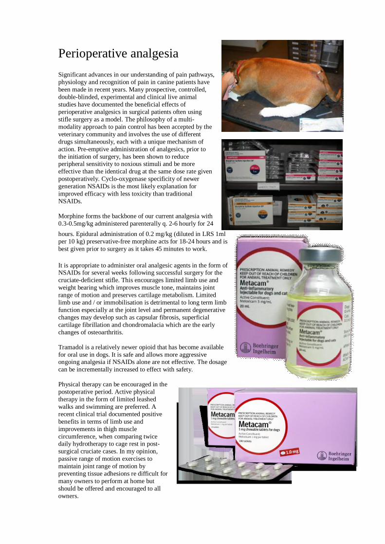
Perioperative analgesia Significant advances in our understanding of pain pathways, physiology and recognition of pain in canine patients have been made in recent years. Many prospective, controlled, double-blinded, experimental and clinical live animal studies have documented the beneficial effects of perioperative analgesics in surgical patients often using stifle surgery as a model. The philosophy of a multi-modality approach to pain control has been accepted by the veterinary community and involves the use of different drugs simultaneously, each with a unique mechanism of action. Pre-emptive administration of analgesics, prior to the initiation of surgery, has been shown to reduce peripheral sensitivity to noxious stimuli and be more effective than the identical drug at the same dose rate given postoperatively. Cyclo-oxygenase specificity of newer generation NSAIDs is the most likely explanation for improved efficacy with less toxicity than traditional NSAIDs. Morphine forms the backbone of our current analgesia with 0.3-0.5mg/kg administered parenterally q. 2-6 hourly for 24 hours. Epidural administration of 0.2 mg/kg (diluted in LRS 1ml per 10 kg) preservative-free morphine acts for 18-24 hours and is best given prior to surgery as it takes 45 minutes to work. It is appropriate to administer oral analgesic agents in the form of NSAIDs for several weeks following successful surgery for the cruciate-deficient stifle. This encourages limited limb use and weight bearing which improves muscle tone, maintains joint range of motion and preserves cartilage metabolism. Limited limb use and / or immobilisation is detrimental to long term limb function especially at the joint level and permanent degenerative changes may develop such as capsular fibrosis, superficial cartilage fibrillation and chondromalacia which are the early changes of osteoarthritis. Tramadol is a relatively newer opioid that has become available for oral use in dogs. It is safe and allows more aggressive ongoing analgesia if NSAIDs alone are not effective. The dosage can be incrementally increased to effect with safety. Physical therapy can be encouraged in the postoperative period. Active physical therapy in the form of limited leashed walks and swimming are preferred. A recent clinical trial documented positive benefits in terms of limb use and improvements in thigh muscle circumference, when comparing twice daily hydrotherapy to cage rest in post-surgical cruciate cases. In my opinion, passive range of motion exercises to maintain joint range of motion by preventing tissue adhesions re difficult for many owners to perform at home but should be offered and encouraged to all owners.
Knee arthroscopy
Arthroscopy offers improved visualisation, less pain and lower morbidity relative to arthrotomy. Canine stifle arthroscopy is in its infancy compared to the worldwide acceptance of knee arthroscopy in man. The human knee of significantly larger and the reduced morbidity is of more significance especially in athletes. Small joint arthroscopes and hand instruements are available to resect and retrieve meniscal fragments under direct observation. There is a steep learning curve for canine stifle arthroscopy in dogs. We use endoscopes, fitted with a light source, to evaluate the inside of joints. Small skin incisions are made and a series of blunt probes are used assess intra-articular structures. The challenges with the canine knee are its smaller size and relatively larger intrapatellar fat pad that is very vascular. Stifle arthroscopy is faster than open arthrotomy. It provides an apportunity to establish a definitive diagnosis in suspect knees prior to performing a more aggressive procedure (eg. osteotomy). Client acceptance is high. A limited number of specialist surgeons claim to be able to perform partial meniscectomies consistently. Our opinion is that this is a growth area and will become accepted as the optimal modality in the diagnosis and treatment of meniscal pathology. The barrier is acceptance by veterinarians.
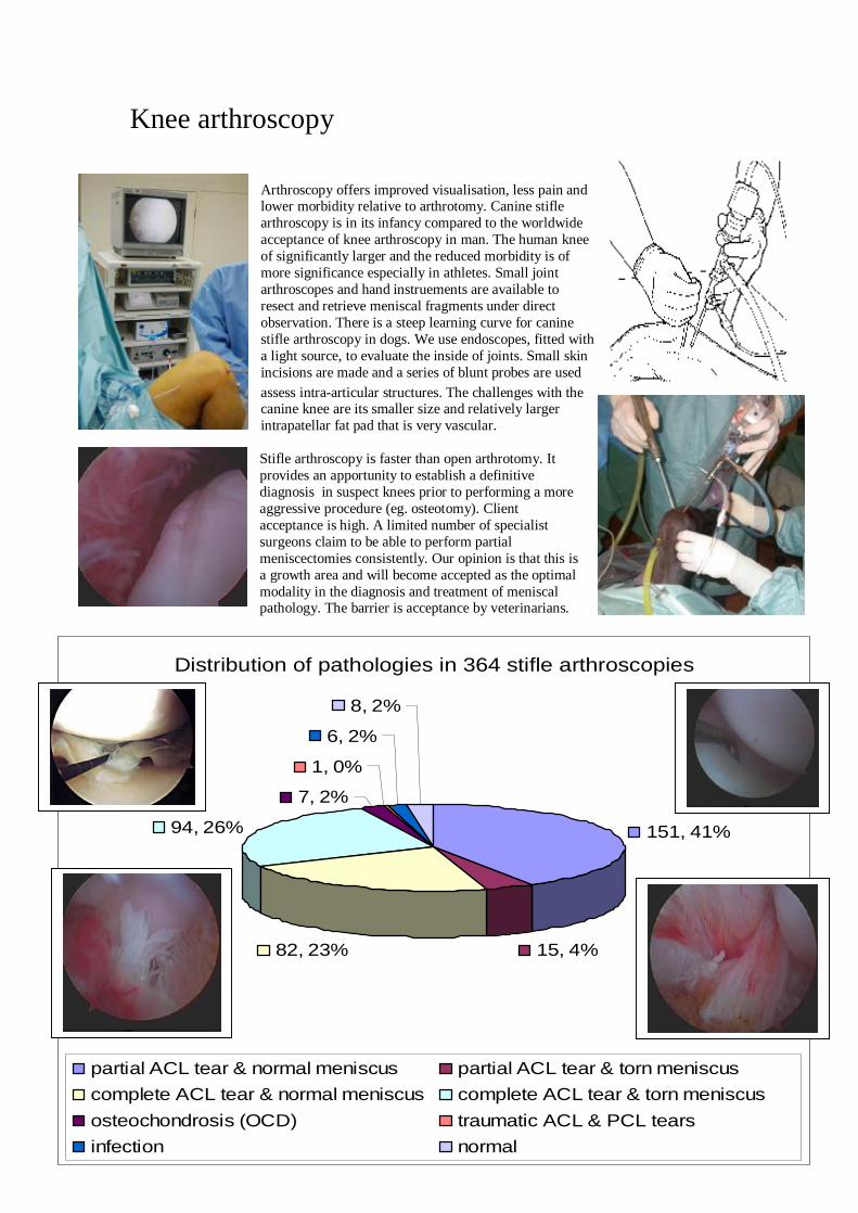
Distribution of pathologies in 364 stifle arthroscopies
151, 41%
15, 4%82, 23%
94, 26%
7, 2%
1, 0%
6, 2%
8, 2%
partial ACL tear & normal meniscus partial ACL tear & torn meniscuscomplete ACL tear & normal meniscus complete ACL tear & torn meniscus osteochondrosis (OCD) traumatic ACL & PCL tearsinfection normal
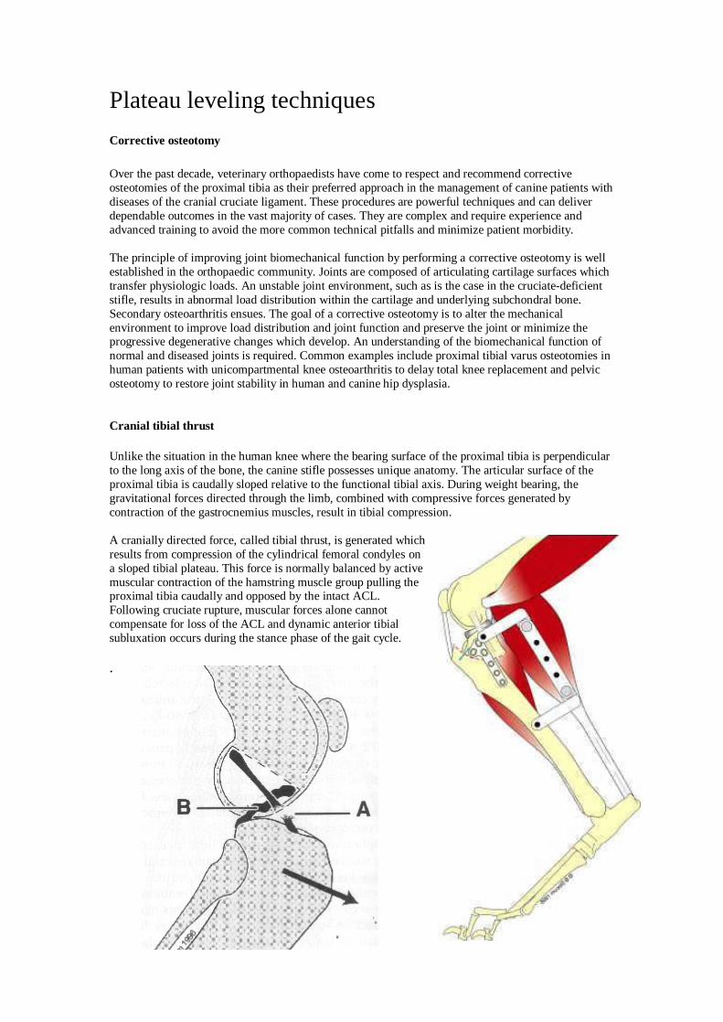
Plateau leveling techniques Corrective osteotomy Over the past decade, veterinary orthopaedists have come to respect and recommend corrective osteotomies of the proximal tibia as their preferred approach in the management of canine patients with diseases of the cranial cruciate ligament. These procedures are powerful techniques and can deliver dependable outcomes in the vast majority of cases. They are complex and require experience and advanced training to avoid the more common technical pitfalls and minimize patient morbidity. The principle of improving joint biomechanical function by performing a corrective osteotomy is well established in the orthopaedic community. Joints are composed of articulating cartilage surfaces which transfer physiologic loads. An unstable joint environment, such as is the case in the cruciate-deficient stifle, results in abnormal load distribution within the cartilage and underlying subchondral bone. Secondary osteoarthritis ensues. The goal of a corrective osteotomy is to alter the mechanical environment to improve load distribution and joint function and preserve the joint or minimize the progressive degenerative changes which develop. An understanding of the biomechanical function of normal and diseased joints is required. Common examples include proximal tibial varus osteotomies in human patients with unicompartmental knee osteoarthritis to delay total knee replacement and pelvic osteotomy to restore joint stability in human and canine hip dysplasia. Cranial tibial thrust Unlike the situation in the human knee where the bearing surface of the proximal tibia is perpendicular to the long axis of the bone, the canine stifle possesses unique anatomy. The articular surface of the proximal tibia is caudally sloped relative to the functional tibial axis. During weight bearing, the gravitational forces directed through the limb, combined with compressive forces generated by contraction of the gastrocnemius muscles, result in tibial compression. A cranially directed force, called tibial thrust, is generated which results from compression of the cylindrical femoral condyles on a sloped tibial plateau. This force is normally balanced by active muscular contraction of the hamstring muscle group pulling the proximal tibia caudally and opposed by the intact ACL. Following cruciate rupture, muscular forces alone cannot compensate for loss of the ACL and dynamic anterior tibial subluxation occurs during the stance phase of the gait cycle. .
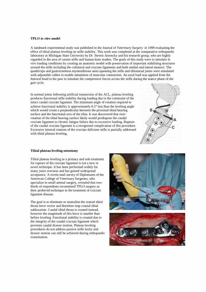
TPLO in vitro model A landmark experimental study was published in the Journal of Veterinary Surgery in 1999 evaluating the effect of tibial plateau leveling on stifle stability. This work was completed at the comparative orthopaedic laboratory at Michigan State University by Dr. Steven Arnoscky and his research group, who are highly regarded in the area of canine stifle and human knee studies. The goals of this study were to simulate in vivo loading conditions by creating an anatomic model with preservation of important stabilising structures around the stifle including the collateral and cruciate ligaments and both medial and lateral menisci. The quadriceps and gastrocnemius myotendinous units spanning the stifle and tibiotarsal joints were simulated with adjustable cables to enable simulation of muscular contraction. An axial load was applied from the femoral head to the paw to simulate the compressive forces across the stifle during the stance phase of the gait cycle. In normal joints following artificial transection of the ACL, plateau leveling produces functional stifle stability during loading due to the constraint of the intact caudal cruciate ligament. The minimum angle of rotation required to achieve functional stability is approximately 6.5° less than the leveling angle which would create a perpendicular between the proximal tibial bearing surface and the functional axis of the tibia. It was discovered that over-rotation of the tibial bearing surface likely would predispose the caudal cruciate ligament to chronic fatigue failure due to excessive loading. Rupture of the caudal cruciate ligament is a recognised complication of this procedure. Excessive internal rotation of the cruciate deficient stifle is partially addressed with tibial plateau leveling. Tibial plateau leveling osteotomy Tibial plateau leveling as a primary and sole treatment for rupture of the cruciate ligament is not a new or novel technique. It has been performed widely for many years overseas and has gained widespread acceptance. A recent mail survey of Diplomates of the American College of Veterinary Surgeons, who specialize in small animal surgery, revealed that two-thirds of respondents recommend TPLO surgery as their preferred technique in the treatment of cruciate ligament disease. The goal is to eliminate or neutralise the cranial tibial thrust force vector and therefore stop cranial tibial subluxation. Caudal tibial thrust is created instead, however the magnitude of this force is smaller than before leveling. Functional stability is created due to the integrity of the caudal cruciate ligament which prevents caudal drawer motion. Plateau leveling procedures do not address passive stifle laxity and drawer motion can still be achieved during orthopaedic examination.
The most popular and researched technique in achieving a leveled proximal tibial bearing surface is to perform a TPLO using a circular or curved osteotomy in the proximal metaphysis as described by the late Dr. Barclay Slocum. A custom compression plate is applied to the medial aspect of the tibia.
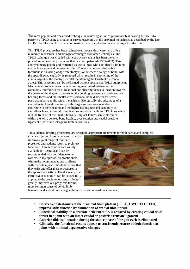
This TPLO procedure has been refined over thousands of cases and offers numerous mechanical and biologic advantages over other techniques. The TPLO technique was clouded with controversy as this has been the only procedure in veterinary medicine that has been patented (1983-2003). This annoyed many people and restricted its use to those who completed a training course in Oregon and became certified. The most common alternative technique is a closing wedge osteotomy (CWO) where a wedge of bone, with the apex directed caudally, is removed which results in shortening of the cranial aspect of the diaphysis whilst maintaining the length of the caudal aspect. This procedure can be performed without specialised TPLO equipment. Mechanical disadvantages include no fragment interdigitation at the osteotomy interface to resist rotational and shearing forces, a location towards the center of the diaphysis increasing the bending moment arm and resultant bending forces and the smaller cross-sectional bone diameter for screw purchase relative to the wider metaphysis. Biologically, the advantage of a curved metaphyseal osteotomy is the larger surface area available to contribute to bone healing and the higher healing rate and capability of cancellous bone. Potential complications associated with the TPLO procedure include fracture of the tibial tuberosity, implant failure, screw placement within the joint, delayed bone healing, over-rotation and caudal cruciate ligament rupture and iatrogenic limb deformities. Tibial plateau leveling procedures are accepted, appropriate treatments for both partial and complete cruciate injuries. Muscle bulk consistently improves, joint range of motion is preserved and patients return to preinjury function. These techniques are widely available in Australia and can be recommended with confidence to pet owners. In my opinion, all practitioners who make recommendations to clients with cruciate injuries should be aware that they exist and offer these procedures in the appropriate setting. The discovery that corrective osteotomies can be successfully applied to the cruciate-deficient stifle has greatly improved our prognosis for the most common cause of pelvic limb lameness and should both intrigue the scientist and reward the clinician.
• Corrective osteotomies of the proximal tibial plateau (TPLO, CWO, TTO, TTA) improve stifle function by elimination of cranial tibial thrust
• Functional stability, in a cruciate-deficient stifle, is restored by creating caudal tibial thrust in a joint with an intact caudal or posterior cruciate ligament
• Anterior tibial subluxation during the stance phase of the gait cycle is eliminated • Clinically, the functional results appear to consistently restore athletic function in
joints with minimal degenerative changes
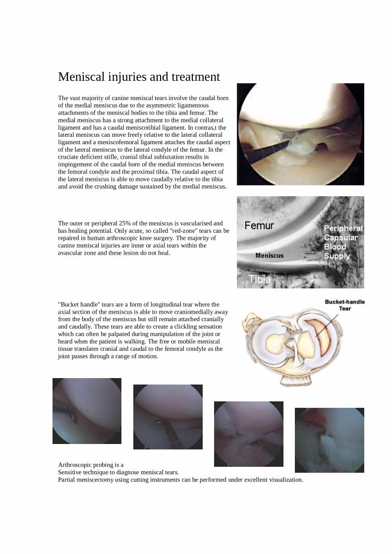
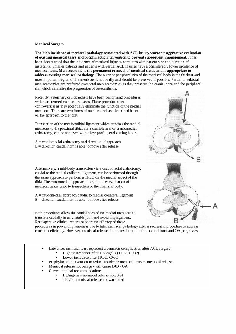
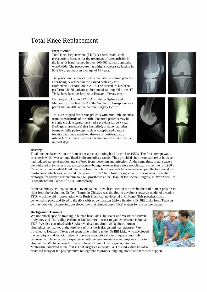
Meniscal injuries and treatment The vast majority of canine meniscal tears involve the caudal horn of the medial meniscus due to the asymmetric ligamentous attachments of the meniscal bodies to the tibia and femur. The medial meniscus has a strong attachment to the medial collateral ligament and has a caudal meniscotibial ligament. In contras,t the lateral meniscus can move freely relative to the lateral collateral ligament and a meniscofemoral ligament attaches the caudal aspect of the lateral meniscus to the lateral condyle of the femur. In the cruciate deficient stifle, cranial tibial subluxation results in impingement of the caudal horn of the medial meniscus between the femoral condyle and the proximal tibia. The caudal aspect of the lateral meniscus is able to move caudally relative to the tibia and avoid the crushing damage sustained by the medial meniscus. The outer or peripheral 25% of the meniscus is vascularised and has healing potential. Only acute, so called "red-zone" tears can be repaired in human arthroscopic knee surgery. The majority of canine meniscal injuries are inner or axial tears within the avascular zone and these lesion do not heal. "Bucket handle" tears are a form of longitudinal tear where the axial section of the meniscus is able to move craniomedially away from the body of the meniscus but still remain attached cranially and caudally. These tears are able to create a clickling sensation which can often be palpated during manipulation of the joint or heard when the patient is walking. The free or mobile meniscal tissue translates cranial and caudal to the femoral condyle as the joint passes through a range of motion.
Arthroscopic probing is a Sensitive technique to diagnose meniscal tears. Partial meniscectomy using cutting instruments can be performed under excellent visualization.
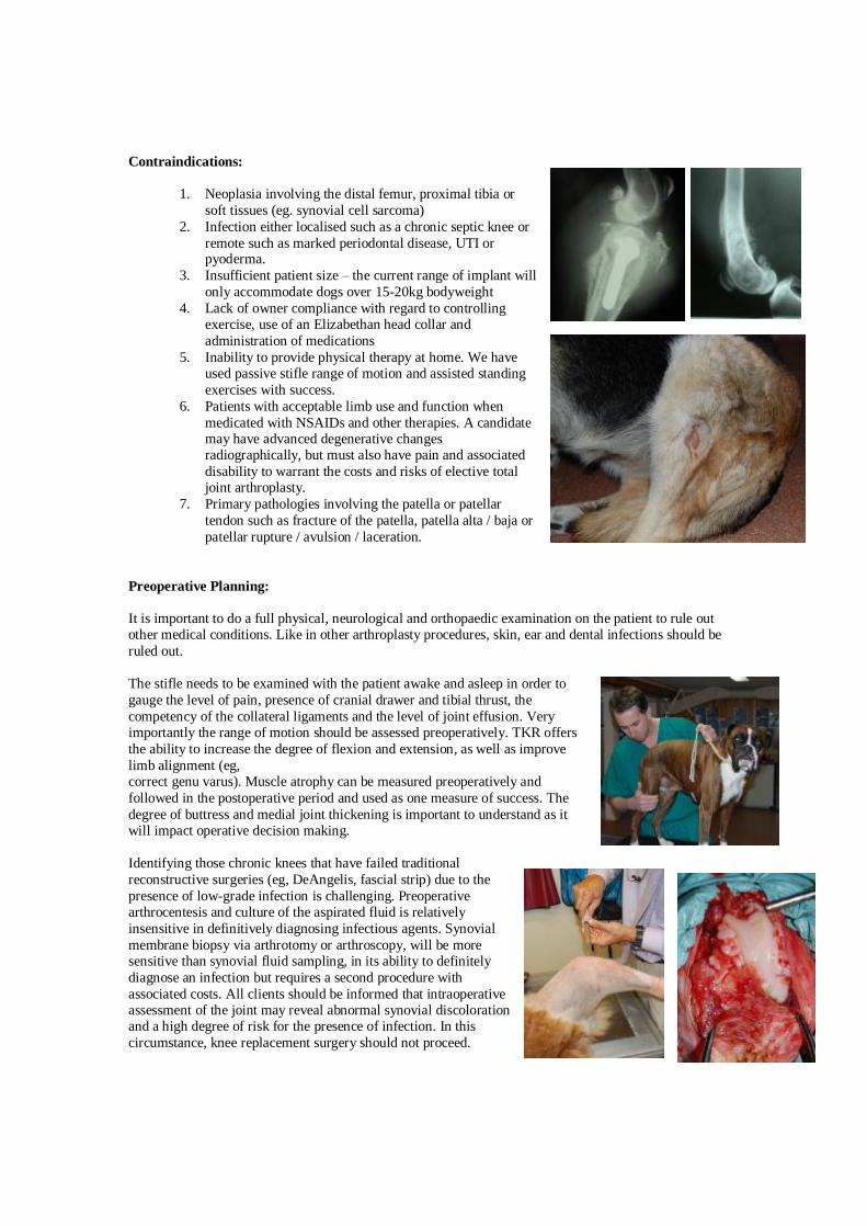
Meniscal Surgery The high incidence of meniscal pathology associated with ACL injury warrants aggressive evaluation of existing meniscal tears and prophylactic intervention to prevent subsequent impingement. It has been documented that the incidence of meniscal injuries correlates with patient size and duration of instability. Smaller patients and patients with partial ACL injuries have a considerably lower incidence of meniscal tears. Meniscectomy is the permanent removal of meniscal tissue and is appropriate to address existing meniscal pathology. The outer or peripheral rim of the meniscal body is the thickest and most important region of the meniscus functionally and should be preserved if possible. Partial or subtotal meniscectomies are preferred over total meniscectomies as they preserve the cranial horn and the peripheral rim which minimise the progression of osteoarthritis. Recently, veterinary orthopaedists have been performing procedures which are termed meniscal releases. These procedures are controversial as they potentially eliminate the function of the medial meniscus. There are two forms of meniscal release described based on the approach to the joint. Transection of the meniscotibial ligament which attaches the medial meniscus to the proximal tibia, via a craniolateral or craniomedial arthrotomy, can be achieved with a low profile, end-cutting blade. A = craniomedial arthrotomy and direction of approach B = direction caudal horn is able to move after release Alternatively, a mid-body transection via a caudomedial arthrotomy, caudal to the medial collateral ligament, can be performed through the same approach to perform a TPLO on the medial aspect of the tibia. The caudomedial approach does not offer evaluation of meniscal tissue prior to transection of the meniscal body. A = caudomedial approach caudal to medial collateral ligament B = direction caudal horn is able to move after release Both procedures allow the caudal horn of the medial meniscus to translate caudally in an unstable joint and avoid impingement. Retrospective clinical reports support the efficacy of these procedures in preventing lameness due to later meniscal pathology after a successful procedure to address cruciate deficiency. However, meniscal release eliminates function of the caudal horn and OA progresses.
• Late onset meniscal tears represent a common complication after ACL surgery: • Highest incidence after DeAngelis (TTA? TTO?) • Lower incidence after TPLO, CWO
• Prophylactic intervention to reduce incidence meniscal tears = meniscal release: • Meniscal release not benign - will cause DJD / OA • Current clinical recommendations:
• DeAngelis – meniscal release accepted • TPLO – meniscal release not warranted
Total Knee Replacement
Introduction: Total Knee Replacement (TKR) is a well established procedure in humans for the treatment of osteoarthritis in the knee. It is performed in over 600,000 patients annually world wide. The procedure has a high success rate lasting in 90-95% of patients an average of 15 years. The procedure is now clinically available in canine patients after being developed in the United States by the Biomedtrix Corporation in 2007. The procedure has been performed in 30 patients at the time of writing. Of these, 17 TKRs have been performed in Houston, Texas, one in
Birmingham, UK and 12 in Australia in Sydney and Melbourne. The first TKR in the Southern Hemisphere was performed in 2008 at the Animal Surgery Centre. TKR is designed for canine patients with hindlimb lameness from osteoarthritis of the stifle. Potential patients may be chronic cruciate cases, have had a previous surgery (e.g. DeAngelis procedure) that has failed, or have had other forms of stifle pathology such as complicated patella luxation, immune mediated disease or post-traumatic osteoarthritis. Early results show the procedure is effective in most dogs.
History: Total knee replacement in the human has a history dating back to the late 1950s. The first attempt was a prosthesis which was a hinge fixed in the medullary canals. They provided short term pain relief however had reduced range of motion and suffered from loosening and infection. At the same time, metal spacers were trialled in joints in order to stop bone rubbing, however these were not clinically effective. In 1968 a Canadian surgeon called Frank Gunston from Sir John Charnley’s hip centre developed the first metal on plastic knee which was cemented into place. In 1972 John Insall designed a prosthesis which was the prototype for today’s current human TKR prostheses at the Hospital for Special Surgery, in New York. He is considered the Father of Knee Arthroplasty. In the veterinary setting, canine and ovine patients have been used in the development of human prostheses right from the beginning. Dr Tom Turner in Chicago was the first to develop a research model of a canine TKR which he did in association with Rush Presbyterian Hospital in Chicago. This prosthesis was cemented in place and fixed to the tibia with screw fixation (delete fixation). Dr Bill Liska from Texas in conjunction with Biomedtrix developed the first clinical based TKR system for the canine patient. Background Training: We undertook specific training in human hospitals (The Mater and Westmead Private in Sydney and The Valley Private in Melbourne) in order to gain experience in human TKR. We also consulted with Stryker Medical and Smith & Nephew, human biomedical companies at the forefront of prosthesis design and manufacture. We travelled to Houston, Texas and spent time training under Dr Bill Liska who developed the technique in dogs. Our introduction was to practice the technique on multiple cadavers which helped gain experience with the instrumentation and implants prior to clinical use. We have been fortunate to have a human knee surgeon, based in Melbourne, involved in the first 4 TKR surgeries in Australia. This individual has also reviewed many of the postoperative radiographs to provide ongoing advice and technical support.

Patient Selection: 1. Failed DeAngelis procedure:
The first patient group that should be considered for a TKR is dogs that have undergone a form of anterior cruciate ligament (ACL) repair that has failed to return the dog to a satisfactory (good) level of soundness. The most common failure is seen in dogs that have a lateral fabella to tibial crest suture or DeAngelis procedure. In theses cases, there is frequently recurrent instability because the fishing line has either avulsed off the fabella or broken. There is frequently advanced osteoarthritis and sometimes a medial meniscal tear will be present. It is essential to ensure there are no signs of joint or leader line infection actively present.
1. Chronic Cruciate patients: TKR is not designed to replace existing cruciate repair techniques e.g. TPLO, when the disease is diagnosed early. Cruciate ligament disease in most patients, especially in larger patients, is a degenerative condition. As the ligament weakens and progressively fails, the joint is undergoing concurrent degeneration and develops osteoarthritis quite rapidly. If instability (cranial drawer or tibial thrust) is the primary problem then stabilisation via DeAngelis or TPLO is still warranted. If instability and significant osteoarthritis are present, then a TKR should be considered as a treatment option. In these cases stabilisation will not relieve the pain associated with OA.
2. Complex Medial Patella Luxation: Most cases of MPL still need to be dealt with traditional techniques such as sulcoplasty, tibial tuberosity transposition, soft tissue tightening procedures and occasional femoral varus osteotomy. In cases of deformation of the articular stifle structures, TKR can be used successfully.
3. Stifle Osteochondrosis: Distal femoral OCD is most commonly seen in large or giant breed dogs and presents as a juvenile lameness that can be quite severe. Initial management involves arthroscopic or open removal of the flap and forage/curettage of the subchondral bed. Most dogs develop significant osteoarthritis at a later date and may have associated lameness. In these cases a TKR can be used later in the life as a resurfacing procedure.
4. Trauma In dogs that have had previous stifle trauma and fractures, it is now feasible to offer a delayed revision surgery and replace the diseased arthritic joint. Such condition may include: malunion of a distal femoral physeal fracture, malunion of a femoral condylar fracture, DJD secondary to a luxated stifle with marked periarticular fibrosis and proximal tibial plateau injuries secondary to trauma.
Contraindications:
1. Neoplasia involving the distal femur, proximal tibia or soft tissues (eg. synovial cell sarcoma)
2. Infection either localised such as a chronic septic knee or remote such as marked periodontal disease, UTI or pyoderma.
3. Insufficient patient size – the current range of implant will only accommodate dogs over 15-20kg bodyweight
4. Lack of owner compliance with regard to controlling exercise, use of an Elizabethan head collar and administration of medications
5. Inability to provide physical therapy at home. We have used passive stifle range of motion and assisted standing exercises with success.
6. Patients with acceptable limb use and function when medicated with NSAIDs and other therapies. A candidate may have advanced degenerative changes radiographically, but must also have pain and associated disability to warrant the costs and risks of elective total joint arthroplasty.
7. Primary pathologies involving the patella or patellar tendon such as fracture of the patella, patella alta / baja or patellar rupture / avulsion / laceration.
Preoperative Planning: It is important to do a full physical, neurological and orthopaedic examination on the patient to rule out other medical conditions. Like in other arthroplasty procedures, skin, ear and dental infections should be ruled out. The stifle needs to be examined with the patient awake and asleep in order to gauge the level of pain, presence of cranial drawer and tibial thrust, the competency of the collateral ligaments and the level of joint effusion. Very importantly the range of motion should be assessed preoperatively. TKR offers the ability to increase the degree of flexion and extension, as well as improve limb alignment (eg, correct genu varus). Muscle atrophy can be measured preoperatively and followed in the postoperative period and used as one measure of success. The degree of buttress and medial joint thickening is important to understand as it will impact operative decision making. Identifying those chronic knees that have failed traditional reconstructive surgeries (eg, DeAngelis, fascial strip) due to the presence of low-grade infection is challenging. Preoperative arthrocentesis and culture of the aspirated fluid is relatively insensitive in definitively diagnosing infectious agents. Synovial membrane biopsy via arthrotomy or arthroscopy, will be more sensitive than synovial fluid sampling, in its ability to definitely diagnose an infection but requires a second procedure with associated costs. All clients should be informed that intraoperative assessment of the joint may reveal abnormal synovial discoloration and a high degree of risk for the presence of infection. In this circumstance, knee replacement surgery should not proceed.
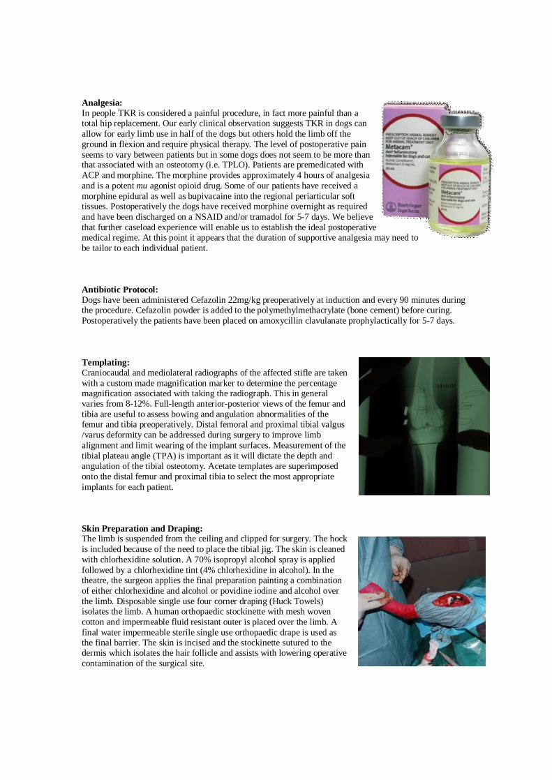
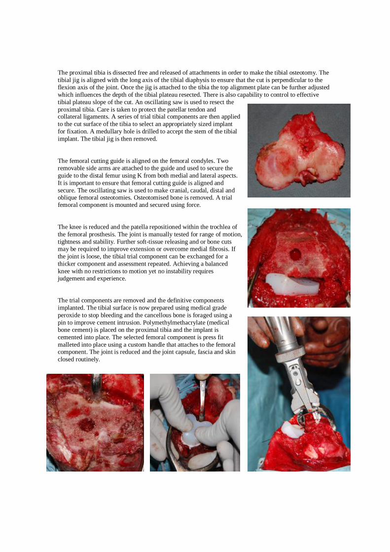
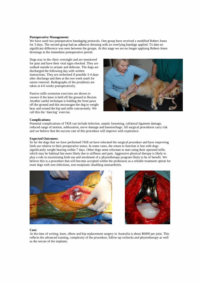
Analgesia: In people TKR is considered a painful procedure, in fact more painful than a total hip replacement. Our early clinical observation suggests TKR in dogs can allow for early limb use in half of the dogs but others hold the limb off the ground in flexion and require physical therapy. The level of postoperative pain seems to vary between patients but in some dogs does not seem to be more than that associated with an osteotomy (i.e. TPLO). Patients are premedicated with ACP and morphine. The morphine provides approximately 4 hours of analgesia and is a potent mu agonist opioid drug. Some of our patients have received a morphine epidural as well as bupivacaine into the regional periarticular soft tissues. Postoperatively the dogs have received morphine overnight as required and have been discharged on a NSAID and/or tramadol for 5-7 days. We believe that further caseload experience will enable us to establish the ideal postoperative medical regime. At this point it appears that the duration of supportive analgesia may need to be tailor to each individual patient. Antibiotic Protocol: Dogs have been administered Cefazolin 22mg/kg preoperatively at induction and every 90 minutes during the procedure. Cefazolin powder is added to the polymethylmethacrylate (bone cement) before curing. Postoperatively the patients have been placed on amoxycillin clavulanate prophylactically for 5-7 days. Templating: Craniocaudal and mediolateral radiographs of the affected stifle are taken with a custom made magnification marker to determine the percentage magnification associated with taking the radiograph. This in general varies from 8-12%. Full-length anterior-posterior views of the femur and tibia are useful to assess bowing and angulation abnormalities of the femur and tibia preoperatively. Distal femoral and proximal tibial valgus /varus deformity can be addressed during surgery to improve limb alignment and limit wearing of the implant surfaces. Measurement of the tibial plateau angle (TPA) is important as it will dictate the depth and angulation of the tibial osteotomy. Acetate templates are superimposed onto the distal femur and proximal tibia to select the most appropriate implants for each patient. Skin Preparation and Draping: The limb is suspended from the ceiling and clipped for surgery. The hock is included because of the need to place the tibial jig. The skin is cleaned with chlorhexidine solution. A 70% isopropyl alcohol spray is applied followed by a chlorhexidine tint (4% chlorhexidine in alcohol). In the theatre, the surgeon applies the final preparation painting a combination of either chlorhexidine and alcohol or povidine iodine and alcohol over the limb. Disposable single use four corner draping (Huck Towels) isolates the limb. A human orthopaedic stockinette with mesh woven cotton and impermeable fluid resistant outer is placed over the limb. A final water impermeable sterile single use orthopaedic drape is used as the final barrier. The skin is incised and the stockinette sutured to the dermis which isolates the hair follicle and assists with lowering operative contamination of the surgical site.

Components: The femoral component is constructed of cobalt chromium and is bead blasted on the undersurface. It is bicondylar and mimics the anatomy of the distal femur. The femoral sulcus is long extending dorsally. There is a post on the back of the implant with a cruciform shape to provide additional rotational security. The femoral component is designed to be cementless. Initially friction with the underlying cancellous bone provides short term security. Longer term osteointergration occurs with bone ingrowth. There are four surfaces of contact with the distal femoral bone. The cranial and caudal surfaces are offset by a predetermined angle of 10 degrees. This provides a locking mechanism via a taper fit. There are seven different sizes of femoral component. They are designed to fit animals from roughly 15-20 up to the largest of dogs. The tibial component is constructed of ultra high molecular weight polyethylene. There are five sizes each having three thickness (in two millimetre increments). There is no ramp or post caudally for caudal cruciate stability. There again is a post / peg on the undersurface of the tibial component to provide stabilization. The undersurface of the implant is recessed in order to allow enhanced cement contact. The weight bearing surface is concave and has been designed to mimic the concavity and abaxial support of the meniscii. All femoral components are compatible and can be used with all of the different tibial components. This modularity offers the surgeon freedom to chose the femoral and tibial implant sizes independently. Trial components for each size of implant are available to use operatively. A tibial jig is used to create the proximal tibial cut. The jig is designed to make a cut at 6 degrees to the long axis of the tibia and creates a tibial plateau angle of 6 degrees. The normal TPA is approximately 24 degrees. By reducing or levelling the slope to 6 degrees, in effect a TPLO is performed. This is important in providing stability for the TKR prostheses. The tibial jig is attached to the distal tibia, tibial crest and proximal tibia using multiple K wires. The jig is removed following osteotomy of the proximal tibia. The femoral jig is designed to perform four osteotomies that match the undersurface of the femoral prosthesis. The jig is held in contact with the distal aspect of the femoral condyles and is attached to the anterior and lateral surfaces of the distal femur using K-wires. A custom saw blade is used to perform the four sequential osteotomies through a series of slots in the femoral jig. Stryker cordless battery driven System 4 reciprocating saws are used and Stryker 4200 cordless battery driven drill with reamers, wire collets and Jacob’s chuck attachments are used as power equipment. The methodology of the femoral and tibial cuts and use of alignment jigs is similar to the commonly performed human knee replacement techniques. Operative Technique: Patients are placed in dorsal recumbency with a slight tilt in the surgical table (head higher). A traditional lateral parapatellar approach is made for stifle arthrotomy. It should be noted a medial approach can also be used, and we have used this in one case. The long digital extensor tendon is released from the fossa and tagged for later re-attachment to the joint capsule. The joint capsule is incised proximally to it full extent to achieve maximal joint exposure. The bursa of the patellar tendon insertion on the tibia is opened. Infrapatellar fat pad, cranial and caudal cruciate ligaments as well as both medial and lateral menisci are excised en bloc. Hohmann retractors place the tibia into drawer.
The proximal tibia is dissected free and released of attachments in order to make the tibial osteotomy. The tibial jig is aligned with the long axis of the tibial diaphysis to ensure that the cut is perpendicular to the flexion axis of the joint. Once the jig is attached to the tibia the top alignment plate can be further adjusted which influences the depth of the tibial plateau resected. There is also capability to control to effective tibial plateau slope of the cut. An oscillating saw is used to resect the proximal tibia. Care is taken to protect the patellar tendon and collateral ligaments. A series of trial tibial components are then applied to the cut surface of the tibia to select an appropriately sized implant for fixation. A medullary hole is drilled to accept the stem of the tibial implant. The tibial jig is then removed. The femoral cutting guide is aligned on the femoral condyles. Two removable side arms are attached to the guide and used to secure the guide to the distal femur using K from both medial and lateral aspects. It is important to ensure that femoral cutting guide is aligned and secure. The oscillating saw is used to make cranial, caudal, distal and oblique femoral osteotomies. Osteotomised bone is removed. A trial femoral component is mounted and secured using force. The knee is reduced and the patella repositioned within the trochlea of the femoral prosthesis. The joint is manually tested for range of motion, tightness and stability. Further soft-tissue releasing and or bone cuts may be required to improve extension or overcome medial fibrosis. If the joint is loose, the tibial trial component can be exchanged for a thicker component and assessment repeated. Achieving a balanced knee with no restrictions to motion yet no instability requires judgement and experience. The trial components are removed and the definitive components implanted. The tibial surface is now prepared using medical grade peroxide to stop bleeding and the cancellous bone is foraged using a pin to improve cement intrusion. Polymethylmethacrylate (medical bone cement) is placed on the proximal tibia and the implant is cemented into place. The selected femoral component is press fit malleted into place using a custom handle that attaches to the femoral component. The joint is reduced and the joint capsule, fascia and skin closed routinely.
Postoperative Management: We have used two postoperative bandaging protocols. One group have received a modified Robert Jones for 3 days. The second group had an adhesive dressing with no overlying bandage applied. To date no significant difference was seen between the groups. At this stage we are no longer applying Robert-Jones dressings in the immediate postoperative period. Dogs stay in the clinic overnight and are monitored for pain and have their vital signs checked. They are walked outside to urinate and defecate. The dogs are discharged the following day with written instructions. They are rechecked if possible 3-4 days after discharge and then at the two week mark for suture removal. Radiographs of the prosthesis are taken at 4-6 weeks postoperatively. Passive stifle extension exercises are shown to owners if the knee is held off the ground in flexion. Another useful technique is holding the front paws off the ground and this encourages the dog to weight bear and extend the hip and stifle concurrently. We call this the ‘dancing’ exercise. Complications: Potential complications of TKR can include infection, aseptic loosening, collateral ligament damage, reduced range of motion, subluxation, nerve damage and haemorrhage. All surgical procedures carry risk and we believe that the success rate of this procedure will improve with experience. Expected Outcomes: So far the dogs that we have performed TKR on have tolerated the surgical procedure and have improving limb use relative to their preoperative status. In some cases, the return to function is fast with dogs significantly weight bearing within 7 days. Other dogs seem reluctant to start using their operated stifle, which may be habitual but more likely due to stiffness and pain. Aggressive physical therapy is likely to play a role in maximising limb use and enrolment of a physiotherapy program likely to be of benefit. We believe this is a procedure that will become accepted within the profession as a reliable treatment option for most dogs with non-infectious, non-neoplastic disabling osteoarthritis.
Cost: At the time of writing, knee, elbow and hip replacement surgery in Australia is about $6000 per joint. This reflects the advanced training, complexity of the procedure, follow-up rechecks and physiotherapy as well as the oncost of the implants.