Embed Size (px)
Citation preview

4
CT Scanning in Archaeology
Stephen Hughes Department of Physics, Queensland University of Technology, Brisbane, Queensland
Australia
1. Introduction
In this chapter we will review the use of x-ray computed tomography (CT) scanning in the field of archaeology. The story will be told in roughly chronological order, starting with the first reported use of a CT scanner in the field of archaeology and then look at some some possibilities for the future. Since the introduction of the x-ray CT scanner in the 1970’s the quality of the images has steadily improved enabling the role of the CT scanner to expand into the field of archaeology. In the context of this chapter, archaeology will be deemed to include the study of ancient human remains and artefacts but exclude remains from pre-history, which normally comes under the heading of palaeontology. (It would perhaps be appropriate to note that CT scanners have been successfully applied in the study of fossils). CT scans have mostly been used to study mummies but have also been used to examine other archaeological artefacts such as clay tablets, scrolls, pottery, bronze statues and swords.
2. Use of plain x-rays in archaeology
Before reviewing the use of CT scanning in the field of archaeology it would perhaps be useful to briefly review the role of conventional plain x-rays in archaeology to set the scene. X-rays were first used to look inside a mummy in 1898, just three years after the discovery of x-rays by Wilhelm Roentgen in 1895. In the same year, a scientist by the name of Konig x-rayed a mummified child and cat in Frankfurt, Germany, followed by Thurstan Holland in Liverpool, England, who x-rayed a mummified bird. In 1898, the famous archaeologist, Sir Flinders Petrie used x-rays to examine Egyptian mummies, followed by Elliot Smith and Howard Carter in 1904, and two French journalists, C. Leleux and M. Gouineau, in 1926. During the 1960’s a British radiologist of the name of Gray (Dawson and Gray, 1968) carried out the most comprehensive x-ray examination of Egyptian mummies. Gray studied 133 mummies and found that osteoarthritis of the spinal column was common and that 30% of all the mummies had lines of arrested growth, most likely caused by periods of famine. Many post-mortem fractures and dislocations were also found. On plain x-rays, post-mortem fractures can be distinguished from ante-mortem fractures as post-mortem fractures show no evidence of healing. The presence of post-mortem fractures in mummies suggests that occasionally embalmers could be rough, which was likely to happen when there was a backlog of bodies stacked on top of each other waiting to be mummified, which might occur for example in an epidemic.
www.intechopen.com

Computed Tomography – Special Applications 58
Gray found that dental disease and attrition were very common, hardly surprising in view of the unavoidably sandy diet of the Egyptians. However, he found no evidence of bone cancer, tuberculosis, syphilis, leprosy or rickets. The dry atmosphere of Egypt would tend to inhibit the tuberculosis bacillus, which survives best in damp conditions. The abundance of sunlight, essential for the conversion of vitamin D from the inactive to active state, may have been a large contributing factor in the absence of rickets. Osteoarthritis is part of the ageing process, but can occur in the spine by middle age as a result of hard labour, which suggests a large fraction of the Egyptian population was engaged in hard labour – hardly surprising since prior the Industrial Revolution the majority of the populations of all societies worked in agriculture.
3. Physical autopsies
Another subject we should touch on before discussing the use of CT in archaeology is the use of autopsies. The word autopsy is usually applied to the technique of examining a dead body to determine the cause of death. In some cases autopsies have been used to determine the cause of the death of a mummified person several millennia after death. However, in archaeology the term “autopsy” is used in a wider sense and includes determining the type and quality of the mummification process and ascertaining the age of the body at death and wear and tear on the body that occurred during life. Various research groups around the world have carried out autopsies on mummies, most notably at Manchester University in the UK (David et al, 1979, 1992) and Pennsylvania University in America (Cockburn et al, 1975). In most cases, autopsies involve the physical destruction of a mummy in the sense that the body is unwrapped and broken open. In view of this, the highest quality mummies tend not to be subjected to this form of examination. Autopsies are sometimes used to examine the contents of burial urns, also destructive in the sense that it would be extremely difficult, if not impossible, to reassemble the contents of the urn exactly as before the examination. CT scans make it possible to perform virtual autopsies on mummies and burial urns without disturbing internal structure.
4. A brief history of the CT scanner
The first clinical x-ray CT scanner entered operation in the early 1970’s (Beckmann 2006) and was a quantum leap in diagnostic imaging. The CT scanner was the first time that 3D images of the body could be produced, initially just the head, and then the rest of the body as the aperture of later generations of scanners increased. Another factor vital in the introduction of the CT scanner in clinical medicine was the development of the mini computer. In essence, an x-ray CT scanner comprises an x-ray source, similar to that in an ordinary x-ray machine, and a group of x-ray detectors opposite the x-ray source arranged in an arc. A fan-shaped x-ray beam shines through the patient or archaeological artefact and is absorbed differently depending on the material between the x-ray source and detector. The greater the density of material the greater the attenuation of the x-ray beam. (For more information on the workings of an x-ray CT scanner please see Michael (2001)). Generally, the x-ray source and detectors move on a ring so that each detector is in the same position relative to the source. In first generation of scanners, the x-ray source rotated around a stationary patient and the x-ray absorption pattern recorded by the detectors used to generate a 2D cross-
www.intechopen.com
CT Scanning in Archaeology 59
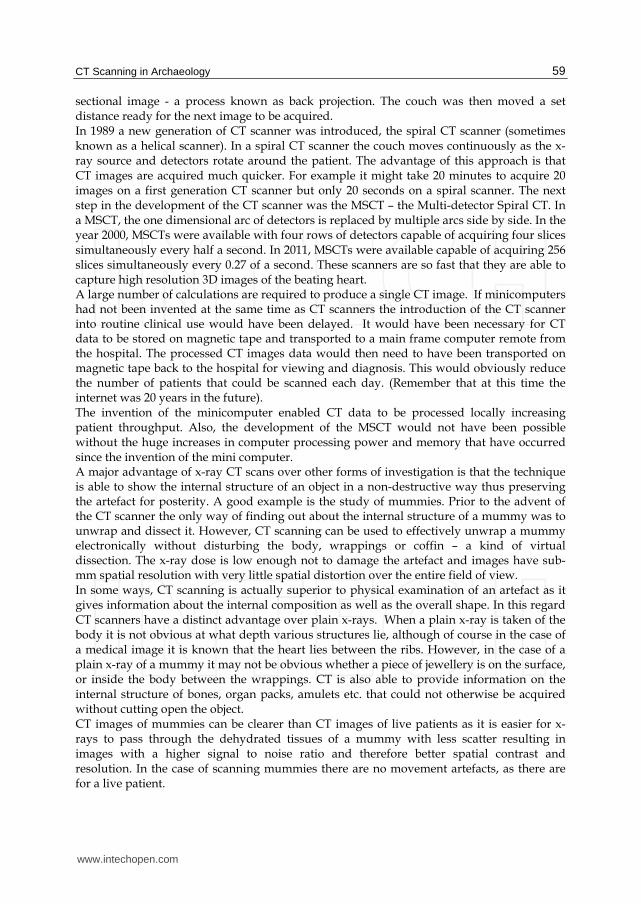
sectional image - a process known as back projection. The couch was then moved a set distance ready for the next image to be acquired. In 1989 a new generation of CT scanner was introduced, the spiral CT scanner (sometimes known as a helical scanner). In a spiral CT scanner the couch moves continuously as the x-ray source and detectors rotate around the patient. The advantage of this approach is that CT images are acquired much quicker. For example it might take 20 minutes to acquire 20 images on a first generation CT scanner but only 20 seconds on a spiral scanner. The next step in the development of the CT scanner was the MSCT – the Multi-detector Spiral CT. In a MSCT, the one dimensional arc of detectors is replaced by multiple arcs side by side. In the year 2000, MSCTs were available with four rows of detectors capable of acquiring four slices simultaneously every half a second. In 2011, MSCTs were available capable of acquiring 256 slices simultaneously every 0.27 of a second. These scanners are so fast that they are able to capture high resolution 3D images of the beating heart. A large number of calculations are required to produce a single CT image. If minicomputers had not been invented at the same time as CT scanners the introduction of the CT scanner into routine clinical use would have been delayed. It would have been necessary for CT data to be stored on magnetic tape and transported to a main frame computer remote from the hospital. The processed CT images data would then need to have been transported on magnetic tape back to the hospital for viewing and diagnosis. This would obviously reduce the number of patients that could be scanned each day. (Remember that at this time the internet was 20 years in the future). The invention of the minicomputer enabled CT data to be processed locally increasing patient throughput. Also, the development of the MSCT would not have been possible without the huge increases in computer processing power and memory that have occurred since the invention of the mini computer. A major advantage of x-ray CT scans over other forms of investigation is that the technique is able to show the internal structure of an object in a non-destructive way thus preserving the artefact for posterity. A good example is the study of mummies. Prior to the advent of the CT scanner the only way of finding out about the internal structure of a mummy was to unwrap and dissect it. However, CT scanning can be used to effectively unwrap a mummy electronically without disturbing the body, wrappings or coffin – a kind of virtual dissection. The x-ray dose is low enough not to damage the artefact and images have sub-mm spatial resolution with very little spatial distortion over the entire field of view. In some ways, CT scanning is actually superior to physical examination of an artefact as it gives information about the internal composition as well as the overall shape. In this regard CT scanners have a distinct advantage over plain x-rays. When a plain x-ray is taken of the body it is not obvious at what depth various structures lie, although of course in the case of a medical image it is known that the heart lies between the ribs. However, in the case of a plain x-ray of a mummy it may not be obvious whether a piece of jewellery is on the surface, or inside the body between the wrappings. CT is also able to provide information on the internal structure of bones, organ packs, amulets etc. that could not otherwise be acquired without cutting open the object. CT images of mummies can be clearer than CT images of live patients as it is easier for x-rays to pass through the dehydrated tissues of a mummy with less scatter resulting in images with a higher signal to noise ratio and therefore better spatial contrast and resolution. In the case of scanning mummies there are no movement artefacts, as there are for a live patient.
www.intechopen.com
Computed Tomography – Special Applications 60
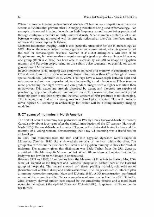
When it comes to imaging archaeological artefacts CT has no real competition as there are serious difficulties that prevent other 3D imaging modalities being used in archaeology. For example, ultrasound imaging depends on high frequency sound waves being propagated through contiguous material of fairly uniform density. Since mummies contain a lot of air between wrappings, ultrasound will be strongly reflected at linen/air interfaces making ultrasound images impossible to form. Magnetic Resonance Imaging (MRI) is also generally unsuitable for use in archaeology as MRI relies on the scanned object having significant moisture content, which is generally not the case for archaeological artefacts. Notman et al (1986) attempted a MR scan of an Egyptian mummy but were unable to acquire enough signal to produce an image. However, one group (Rühli et al 2007) has been able to successfully use MR to image an Egyptian mummy and Peruvian corpse using an ultra short pulse sequence not possible on earlier generations of MR scanners. In 2009 terahertz (THz) imaging was performed on parts of a mummy in conjunction with CT and was found to provide more soft tissue information than CT, although at lower spatial resolution (Ohrstrom et al, 2009). THz rays have a wavelength between light and microwaves and so have properties midway between light and microwaves. THz waves are more penetrating than light waves and can produce images with a higher resolution than microwaves. THz waves are strongly absorbed by water, and therefore are capable of penetrating deep into dehydrated mummified tissue. THz waves are also non-ionizing and therefore safer to use than x-rays and the small amount of heat produced is not damaging. THz imaging may find an increasing role in archaeological imaging. THz will probably never replace CT scanning in archaeology but rather will be a complimentary imaging modality.
5. CT scans of mummies in North America
The first CT scan of a mummy was performed in 1977 by Derek Harwood-Nash in Toronto, Canada only about four years after the clinical introduction of the CT scanner (Harwood-Nash, 1979). Harwood-Nash performed a CT scan on the desiccated brain of a boy and the mummy of a young woman, demonstrating that x-ray CT scanning was a useful tool in archaeology. In 1983, four mummies from the 18th and 25th Egyptian dynasties were x-rayed in Minnesota (Notman 1986). Scans showed the remains of the heart within the chest. The group also carried out the first ever MRI scan of an Egyptian mummy to check for residual moisture. The mummy given this distinction was Lady Tashat from the 25th dynasty, a resident of the Minneapolis Museum of Art. What little moisture still retained within the body was too low for an MR image to be produced. Between 1983 and 1987, 15 mummies from the Museum of Fine Arts in Boston, MA, USA were CT scanned at the Brigham and Womens’ Hospital in Boston (part of the Harvard group of hospitals). The images showed soft tissue packing material, schmorl’s nodes (herniations of vertebral discs) and aortic calcification. The images assisted curators to plan a mummy restoration program (Marx and D’Auria 1986). A 3D reconstruction performed on one of the mummies called Tabes, a songstress of Amun who lived in c.950 BC in the 22nd dynasty, showed sunken eyes caused by the embalming process and a metal heart scarab in the region of the xiphoid (Marx and D’Auria 1988). It appears that Tabes died in her thirties.
www.intechopen.com

CT Scanning in Archaeology 61
A 21st dynasty male and female mummy on display in the Albany Institute of History and Art, Albany, New York, which were originally bought from the Cairo museum in 1909, underwent a series of CT scans and plain X-rays (Wagle 1994). The X-rays showed that the female mummy was given a two-component foot prosthesis by the embalmers, an indication of the importance the Egyptians placed on the body being complete in order for the soul to exist in the afterlife. In 2001, Cessarini et al performed CT scans on 13 mummies from the Egyptian Museum in Torino, Italy using a multi-dector CT scanner. An advantage of using this type of scanner is that whole body can be scanned quickly in a single session without the need to re-position the body between scans and register different sets of images. The group used a computer to perform virtual endoscopy on the mummies, i.e. the computer was used to display views from inside the body as if an actual endoscope had been placed inside the body. A group at the Orthopaedic University Hospital Balgrist, Zurich, performed a CT guided biopsy on the mummy of an Egyptian child from the Museum für Völkerkunde in Burgdorf, Switzerland (Rühli 2002). The mummy had been previously x-rayed in the 1920s and researchers had concluded that the child had died from spinal tuberculosis. However, a sample of lumbar vertebral bone showed that the bone had disintegrated after death and therefore TB was not the cause of death. In 2008, Chan et al published a paper describing a CT study of a female mummy from the Egyptian city of Akhmim (about 470 km south of Cairo on the east bank of the Nile) dating from the Ptolemaic period (305 – 200 BC). The mummy is in the collection of the Academy of Natural Sciences in Philadelphia, Pennsylvania. The mummy was x-rayed at the Hahnemann University Hospital, Philadelphia using a GE Medical Systems 16-detector row helical CT scanner. The main purpose of the study was to determine the social status of the mummy on the basis of the embalming technique used. The CT scans showed that the quality of embalming was good, although the body showed signs of decomposition prior to embalming. There was no obvious physical cause of death. Scans showed that the brain had been removed through the nose and only one of the four molars had erupted indicating the mummy was probably 17 – 25 years old at death. The CT scans showed an unusual dislocation of two of the two cervical vertebrae at the top of the spinal column (C1 and C2). The authors suggest that this dislocation occurred as a result of the body lying in the river Nile after death. According to Herodotus, if the people of a town found someone dead in the Nile they embalmed them as if they were of high social status. Therefore it is possible that the young girl was found floating in the river near Akhmim and given a top quality embalming even although she was unknown to the inhabitants of the city. An interesting CT study was performed on 22 mummies from the Egyptian National Museum of Antiquities in Cairo in February 2009 (Allam et al, 2009). The mummies were dated as being from 1981 BC – AD 334. Seven out of eight mummies who were over 45 years old when they died showed evidence of arteriosclerosis, whereas only two out of eight mummies younger than 45 showed evidence of ateriosclerosis. This study demonstrates that in ancient Egypt people suffered from arteriosclerosis and therefore arteriosclerosis is not exclusively a disease of modern society related to diet and sedentary life style. Potentially, this finding could lead to new approaches to finding a cure for arteriosclerosis.
6. The Ice Man
The Ice Man (Otzi) is probably the most famous mummy of recent times. Otzi’s head and upper torso were found protruding out of the ice in the Austrian Alps by German hikers in
www.intechopen.com

Computed Tomography – Special Applications 62
September 1991. There was no evidence of decay, which suggests that Otzi had only recently been uncovered. If the body had been regularly thawed and refrozen in the intervening millennia significant decay would be expected. In view of the fact that Otzi was discovered in September, i.e. the end of summer in the northern hemisphere this may have been an early sign of global warming. Otzi was transported to the anatomy department of the University of Innsbruck in Austria for extensive analysis that included taking plain x-rays and CT scans. An initial set of CT scans were acquired in September 1991 (Nedden 1994) and three sets of spiral CT scans acquired over the following 10 years (Murphy et al 2003). In total 2,190 CT scans of Otzi were acquired. Artefacts recovered from the ground where Otzi was discovered and carbon 14 dating indicate that Otzi lived about 5,300 years ago. The CT scans revealed an arrowhead lodged in Otzi’s shoulder, which was probably the cause of death, and evidence of calcification was discovered in the region of the carotid artery and abdominal aorta indicating the presence of arteriosclerosis (hardening of the arteries). His brain, although generally well preserved, was shrunken during the natural freeze-drying mummification process. Subsequently it was discovered that Otzi had in fact been found just inside the Italian border and so was later moved from Austria to the South Tyolean Archaeological Museum in Italy1.
7. CT scanning of an Egyptian mummy: A case study
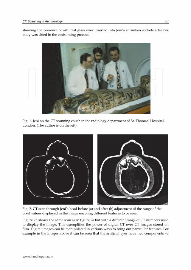
A case study of the use of CT scanning a mummy will now be presented including some examples of the x-ray CT images and 3D reconstructions. The study was performed at the end of 1991 and beginning of 1992 by the author of this chapter and associates at St. Thomas’ Hospital (STH), London and the Department of Egyptian Antiquities of the British Museum (BM), London (Hughes 2010). It is interesting to note that this appears to be the first time an Egyptian mummy was CT scanned outside North America. The mummy in question was an Egyptian priestess/temple singer who lived at Karnak (modern day Luxor) during the 22nd dynasty ((c. 945-715 BC). The mummies name is Tjenmutengebtiu but will be contracted to Jeni in this chapter. Jeni was selected for scanning by the staff of the BM, as the art/science of embalming is considered to have peaked in 22nd dynasty. The purpose in performing the scan was to find out more about Jeni than had been discovered from plain x-rays. Some questions were: where were amulets placed on the body, how was the brain removed from the skull? The Egyptians embalmed their dead in order to preserve the body over a long period of time. Jeni was scanned about 2,700 years after she died and open arteries can still be seen in her legs and the remains of her spinal cord, confirming that her embalmers were experts in their field. Jeni was brought over to STH from the BM on five occasions at night when the scanner was not being used for patients. Software developed to produce 3D images of the head from CT scans for planning brain surgery was used to produce 3D images of Jeni. The head and neck were scanned with 2mm slices, the teeth with 1mm slices and the rest of the body with 4 mm slices. In each case a 512 × 512 matrix was used. The 2D CT images, and 3D surface reconstructions, demonstrate many features of the embalming techniques and funerary customs of the 22nd dynasty. Figure 1 shows Jeni on the CT couch and figure 2a shows a CT image of Jeni’s head 1 http://www.bolzano.net/english/museum-archaeology.html
www.intechopen.com
CT Scanning in Archaeology 63
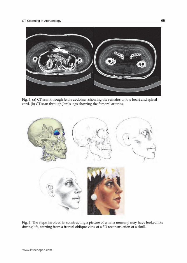
showing the presence of artificial glass eyes inserted into Jeni’s shrunken sockets after her body was dried in the embalming process.
Fig. 1. Jeni on the CT scanning couch in the radiology department of St. Thomas’ Hospital, London. (The author is on the left).
Fig. 2. CT scan through Jeni’s head before (a) and after (b) adjustment of the range of the pixel values displayed in the image enabling different features to be seen.
Figure 2b shows the same scan as in figure 2a but with a different range of CT numbers used to display the image. This exemplifies the power of digital CT over CT images stored on film. Digital images can be manipulated in various ways to bring out particular features. For example in the images above it can be seen that the artificial eyes have two components –a
www.intechopen.com

Computed Tomography – Special Applications 64
denser outer eye, and a less dense inner eye and Jeni’s head appears to be empty. Both versions of the image show that the nasal septum and ethmoid bone have been broken. However, when the image is adjusted (a process known as windowing) it is apparent that Jeni’s head is not empty but is filled with some kind of low density material, most probably linen that appears to converge on the nasal cavity. A 3D reconstruction of the lower half of the head confirms that linen passes through from the cranium into the nasal cavity indicating that the embalmers inserted the linen through the nose into the cranium.
Fig. 3. 3D reconstruction of the lower half of Jeni’s head showing the linen (in false colour) converging on the nasal cavity.
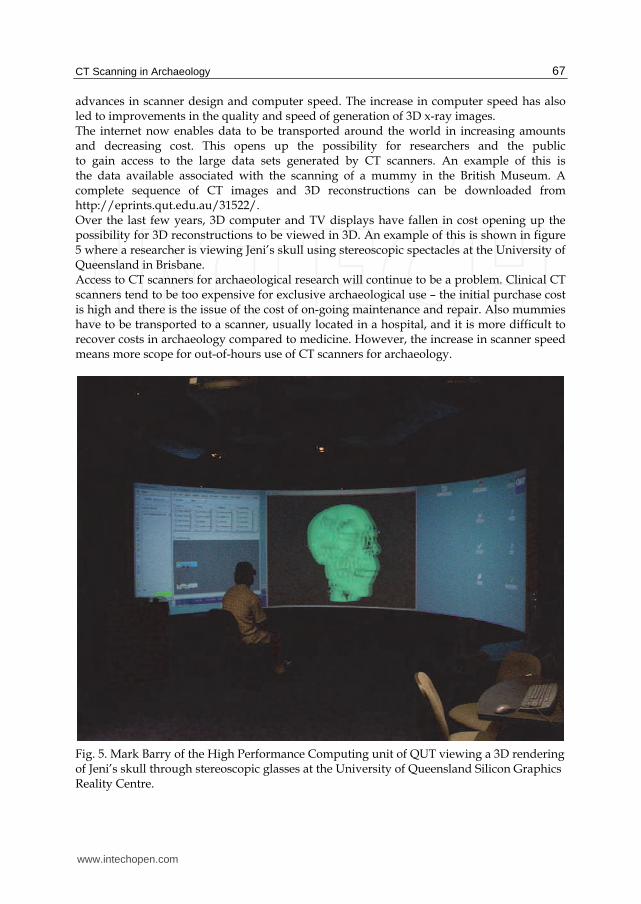
Sometimes the embalmers extracted the brain through the foramen magnum which is the hole in the base of the skull through which the spinal cord enters the cranium. A 3D reconstruction of the linen inside Jeni’s head reveals that the brain was extracted through the nose rather than through the foramen magnum. The reconstruction shows cloth protruding from the nasal cavities into the otherwise empty cranial cavity. This would not have happened if the cloth was pushed through the foramen magnum. This is a good example of the advantage of CT over plain x-rays. The remains of the heart can be seen as well as four organ packs corresponding to the mummified and repackaged lungs, intestines, stomach and liver. Each of the organ packs encloses a wax figurine representing of one of the four sons of Horus. The teeth are in good condition with little signs of wear, which, in view of the gritty diet of the Egyptians, indicates that Jeni was probably between 19 and 23 years old when she died. A young age of death is also suggested by analysis of the shape of the molar teeth. Neither the plain x-rays nor CT scans reveal an obvious cause of death. The plain x-rays reveal that three of Jeni’s back ribs on the right side, as well as the right side of the pelvis have been fractured, possibly indicating a fall or some kind of collision with an animal or chariot as the cause of death. An image of 3D skull was used by an artist to draw a picture of what the mummy may have looked like during life (figure 4). Morphological analysis of the skull revealed that Jeni was most likely an Egyptian female (Hughes, 2005). Other researchers have also used CT scans to reconstruct faces (Hill 1993, Manly 2002).
www.intechopen.com
CT Scanning in Archaeology 65
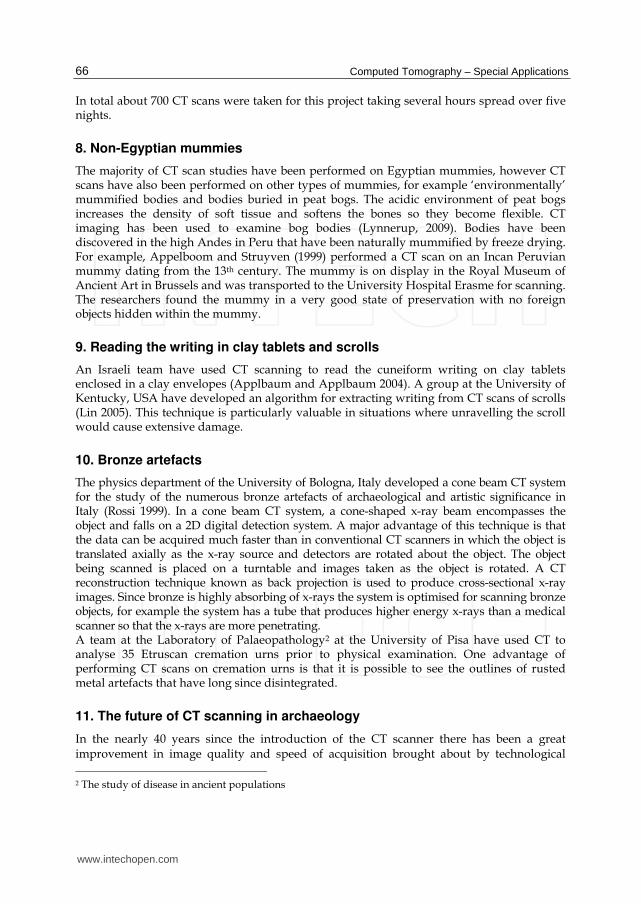
Fig. 3. (a) CT scan through Jeni’s abdomen showing the remains on the heart and spinal cord. (b) CT scan through Jeni’s legs showing the femoral arteries.
Fig. 4. The steps involved in constructing a picture of what a mummy may have looked like during life, starting from a frontal oblique view of a 3D reconstruction of a skull.
www.intechopen.com
Computed Tomography – Special Applications 66
In total about 700 CT scans were taken for this project taking several hours spread over five nights.
8. Non-Egyptian mummies
The majority of CT scan studies have been performed on Egyptian mummies, however CT scans have also been performed on other types of mummies, for example ‘environmentally’ mummified bodies and bodies buried in peat bogs. The acidic environment of peat bogs increases the density of soft tissue and softens the bones so they become flexible. CT imaging has been used to examine bog bodies (Lynnerup, 2009). Bodies have been discovered in the high Andes in Peru that have been naturally mummified by freeze drying. For example, Appelboom and Struyven (1999) performed a CT scan on an Incan Peruvian mummy dating from the 13th century. The mummy is on display in the Royal Museum of Ancient Art in Brussels and was transported to the University Hospital Erasme for scanning. The researchers found the mummy in a very good state of preservation with no foreign objects hidden within the mummy.
9. Reading the writing in clay tablets and scrolls
An Israeli team have used CT scanning to read the cuneiform writing on clay tablets enclosed in a clay envelopes (Applbaum and Applbaum 2004). A group at the University of Kentucky, USA have developed an algorithm for extracting writing from CT scans of scrolls (Lin 2005). This technique is particularly valuable in situations where unravelling the scroll would cause extensive damage.
10. Bronze artefacts
The physics department of the University of Bologna, Italy developed a cone beam CT system for the study of the numerous bronze artefacts of archaeological and artistic significance in Italy (Rossi 1999). In a cone beam CT system, a cone-shaped x-ray beam encompasses the object and falls on a 2D digital detection system. A major advantage of this technique is that the data can be acquired much faster than in conventional CT scanners in which the object is translated axially as the x-ray source and detectors are rotated about the object. The object being scanned is placed on a turntable and images taken as the object is rotated. A CT reconstruction technique known as back projection is used to produce cross-sectional x-ray images. Since bronze is highly absorbing of x-rays the system is optimised for scanning bronze objects, for example the system has a tube that produces higher energy x-rays than a medical scanner so that the x-rays are more penetrating. A team at the Laboratory of Palaeopathology2 at the University of Pisa have used CT to analyse 35 Etruscan cremation urns prior to physical examination. One advantage of performing CT scans on cremation urns is that it is possible to see the outlines of rusted metal artefacts that have long since disintegrated.
11. The future of CT scanning in archaeology
In the nearly 40 years since the introduction of the CT scanner there has been a great improvement in image quality and speed of acquisition brought about by technological 2 The study of disease in ancient populations
www.intechopen.com
CT Scanning in Archaeology 67
advances in scanner design and computer speed. The increase in computer speed has also led to improvements in the quality and speed of generation of 3D x-ray images. The internet now enables data to be transported around the world in increasing amounts and decreasing cost. This opens up the possibility for researchers and the public to gain access to the large data sets generated by CT scanners. An example of this is the data available associated with the scanning of a mummy in the British Museum. A complete sequence of CT images and 3D reconstructions can be downloaded from http://eprints.qut.edu.au/31522/. Over the last few years, 3D computer and TV displays have fallen in cost opening up the possibility for 3D reconstructions to be viewed in 3D. An example of this is shown in figure 5 where a researcher is viewing Jeni’s skull using stereoscopic spectacles at the University of Queensland in Brisbane. Access to CT scanners for archaeological research will continue to be a problem. Clinical CT scanners tend to be too expensive for exclusive archaeological use – the initial purchase cost is high and there is the issue of the cost of on-going maintenance and repair. Also mummies have to be transported to a scanner, usually located in a hospital, and it is more difficult to recover costs in archaeology compared to medicine. However, the increase in scanner speed means more scope for out-of-hours use of CT scanners for archaeology.
Fig. 5. Mark Barry of the High Performance Computing unit of QUT viewing a 3D rendering of Jeni’s skull through stereoscopic glasses at the University of Queensland Silicon Graphics Reality Centre.
www.intechopen.com
Computed Tomography – Special Applications 68
A major issue in the world at the moment is security of archaeological artefacts, i.e. the danger of artefacts being lost to theft, fire, earthquake, tsunami or war. An example of this occurred February 2011 when two mummies were destroyed in the Cairo Museum during the anti-government protests3. X-ray CT scans combined with photography are the best way of ‘saving’ archaeological artefacts for posterity. Such a scheme would be expensive and could run into several tens of millions of dollars but the cost would be a tiny fraction of the amount of money spent on the wars leading to the destruction of artefacts. One possible scenario would be for money to be obtained to perform scans of the highest quality currently possible on most of the significant archaeological artefacts in the world – mummies, clay tablets, scrolls, pottery, statues etc - and make the data publicly available. Transportable CT scanners now exist – these are scanners that are housed in a truck and can be taken to remote locations to scan patients. One of these could be parked in the grounds of a museum to improve access to artefacts. The data would then be available for researchers to perform ‘digital autopsies’. At the same time of scanning, a high resolution digital camera attached to a robot could take photographs of the object from all directions and map the photos onto the 3D surface of the artefact generated from the 3D reconstructions. The data could be deposited in a Virtual Museum – maybe in a way similar the Virtual Observatory project in astronomy (http://en.wikipedia.org/wiki/Virtual_Observatory). An advantage of this approach is that artefacts preserved in digital form will still be available in cases where the body has decayed.
12. References
Allam, A.H; Thompspon, R.C; Wann, L. S; Miyamoto, M.I; Thomas, G.S 2009 Computed tomographic assessment of atherosclerosis in ancient Egyptian mummies, Journal of the American Medical Association 18:2091-2094.
Applbaum, N; and Y. H. Applbaum, Y,H The use of medical computed tomography (ct) imaging in the study of ceramic and clay artifacts from the ancient near
east. In X-rays and Archaeology. Kluwer, 2004. Appelboom, T; Struyven, J 1999 Medical imaging of the Peruvian mummy Rascar Capac,
The Lancet 354:2153-2155. Beckmann, E. C. 2006 CT scanning the early days. The British Journal of Radiology, 79:5-8
doi:10.1259/bjr/29444122 Cesarani, F; Martina, M.C; Farraris, A; Grilletto, R; Boano, R; Marochetti; E.F; Donadoni,
A.M; Gandini, G 2003 Whole-body three-dimensional multidetector CT of 13 Egyptian human mummies, American Journal of Roentgenology 180:597-606.
Chan, S.S; Elias, J.P; Hysell, M.E; Hallowell, M.J. 2008 CT of a Ptolemaic period mummy from the ancient Egyptian city of Akhmim, RadioGraphics 28:2023-2032, doi: 10.1148/rg.287085039
Cockburn, A; Barraco, R, A; Reyman, T, A; Peck, W, H. 1975. Autopsy of an Egyptian mummy, Science 187:1155-60.
David, A.R., ed., 1979. The Mystery of the Mummies, The Manchester Mummy Project, Manchester.
3 http://www.telegraph.co.uk/news/worldnews/africaandindianocean/egypt/8291526/Egypt-crisis-
Looters-destroy-mummies-in-Cairo-museum.html
www.intechopen.com
CT Scanning in Archaeology 69
David, A.R. and Tapp, E., eds., 1992. The Mummy’s Tale, Michael O’Mara Books Ltd, London.
Dawson, W.R. and Gray, P.H.K., 1968. Catalogue of Egyptian antiquities in the British Museum: 1 mummies and human remains, British Museum Press, London.
Harwood-Nash, D.F.C, 1979. Computed tomography of ancient Egyptian mummies, Journal of Computer Assisted Tomography 3:768-773.
Hill, B. et al., 1993. Facial reconstruction of a 3500-year-old Egyptian mummy using axial computed tomography Journal of Audiovisual Media in Medicine 16:11-3
Hughes, S.W. 2010 Unwrapping an ancient Egyptian mummy using x-rays, Physics Education 45:235-242, doi:10.1088/0031-9120/45/3/002
Hughes, S.W; Wright, R; Barry, M 2005 Virtual reconstruction and morphological analysis of the cranium of an ancient Egyptian mummy. Physics Education 28:122-127
Lin, Y (2005). "Opaque document imaging: Building images of inaccessible texts". Computer Vision (ICCV), International Conference on , 1, p. 662
Lynnerup, N (2009) Medical imaging of mummies and bog bodies – a mini-review, Gerontology 56:441-448 doi:10.1159/000266031
Manly, B; Eremin, K; Shortland, A; Wilkinson, C 2002 The facial reconstruction of an ancient Egyptian queen, Journal of Audiovisual Media in Medicine 25:155-159 doi: 10.1080/0140511021000051144
Marx, M; D’Auria, S.H 1986 CT examination of eleven Egyptian mummies, Radiographics 6:321-330.
Marx, M; D’Auria, S.D. 1988 Three dimensional CT reconstruction of an ancient Egyptian mummy. American Journal of Radiology 150:147-149.
Michael, G. 2001 X-ray computed tomography, Physics Education 36:442-451, doi: 10.1088/0031-9120/36/6/301 (http://eprints.qut.edu.au/31012).
Murphy, William A., Jr.; zur Nedden, Dieter; Gostner, Paul; Knapp, Rudolf; Recheis, Wolfgang; Seidler, Horst (24 January 2003), "The Iceman: Discovery and imaging", Radiology (Oak Brook, Il.: Radiology) 226 (3): 614–629, doi:10.1148/radiol.2263020338, issn = 0033-8419, PMID 12601185. On-line pre-publication version.
Nedden, D; Knapp, R; Wicke, K; Judmaier, W; Murphy, WA, Seidler, H; Platzer, W; Skull of a 5,300 year old mummy: reproduction and investigation with CT guided stereolithography, Radiology 193:269-272, 1994.
Notman, 1986 Modern imaging and endoscopic biopsy techniques in Egyptian mummies, American Journal of Roentgenology 146:93-96.
Öhrström, L; Bitzer, A; Walther, M; Rühl, F. J. 2009 Technical note: terahertz imaging of ancient mummies and bone, American Journal of Physical Anthropology 142:497-500, doi: 10.1002/ajpa.21292
Rossi, M; Casali, F; Chirco, P; Morigi, M.P; Nava, E; Querzola, E; Zanarini, M 1999 X-ray 3D computed tomography of bronze archaeological samples IEEE Transactions on Nuclear Science 46:897-903. doi: 10.1109/23.790700
Rühli, F.J; von Waldburg, H; Nielles-Vallespin, S. Böni, T; Speier, P 2007 Clinical magnetic resonance imaging of ancient dry human mummies without rehydration
Journal of the American Medical Association 298: 2618 – 2620. Rühli, F.J; Hodler, J; Böni, T 2002 Technical Note: CT-guided biopsy: a new diagnostic
method for the paleopathological research. American Journal of Physical Anthropology 117:272-275, doi: 10.1002/ajpa.20003
www.intechopen.com

Computed Tomography – Special Applications 70
Wagle, W.A. 1976 Toe prosthesis in an Egyptian human mummy, American Journal of Roentgenology, 164:999.
www.intechopen.com
Computed Tomography - Special ApplicationsEdited by Dr. Luca Saba
ISBN 978-953-307-723-9Hard cover, 318 pagesPublisher InTechPublished online 21, November, 2011Published in print edition November, 2011
InTech EuropeUniversity Campus STeP Ri Slavka Krautzeka 83/A 51000 Rijeka, Croatia Phone: +385 (51) 770 447 Fax: +385 (51) 686 166www.intechopen.com
InTech ChinaUnit 405, Office Block, Hotel Equatorial Shanghai No.65, Yan An Road (West), Shanghai, 200040, China
Phone: +86-21-62489820 Fax: +86-21-62489821
CT has evolved into an indispensable imaging method in clinical routine. The first generation of CT scannersdeveloped in the 1970s and numerous innovations have improved the utility and application field of the CT,such as the introduction of helical systems that allowed the development of the "volumetric CT" concept.Recently interesting technical, anthropomorphic, forensic and archeological as well as paleontologicalapplications of computed tomography have been developed. These applications further strengthen the methodas a generic diagnostic tool for non destructive material testing and three dimensional visualization beyond itsmedical use.
How to referenceIn order to correctly reference this scholarly work, feel free to copy and paste the following:
Stephen Hughes (2011). CT Scanning in Archaeology, Computed Tomography - Special Applications, Dr. LucaSaba (Ed.), ISBN: 978-953-307-723-9, InTech, Available from: http://www.intechopen.com/books/computed-tomography-special-applications/ct-scanning-in-archaeology