Embed Size (px)
Citation preview
HAL Id: hal-02432425https://hal.uca.fr/hal-02432425
Submitted on 8 Jan 2020
HAL is a multi-disciplinary open accessarchive for the deposit and dissemination of sci-entific research documents, whether they are pub-lished or not. The documents may come fromteaching and research institutions in France orabroad, or from public or private research centers.
L’archive ouverte pluridisciplinaire HAL, estdestinée au dépôt et à la diffusion de documentsscientifiques de niveau recherche, publiés ou non,émanant des établissements d’enseignement et derecherche français ou étrangers, des laboratoirespublics ou privés.
Curative Treatment of Candidiasis by the LiveBiotherapeutic Microorganism Lactobacillus rhamnosusLcr35 in the Invertebrate Model Caenorhabditis elegans:
First Mechanistic InsightsCyril Poupet, Philippe Veisseire, Muriel Bonnet, Olivier Camares, Marylise
Gachinat, Caroline Dausset, Christophe Chassard, Adrien Nivoliez, StéphanieBornes
To cite this version:Cyril Poupet, Philippe Veisseire, Muriel Bonnet, Olivier Camares, Marylise Gachinat, et al.. CurativeTreatment of Candidiasis by the Live Biotherapeutic Microorganism Lactobacillus rhamnosus Lcr35 inthe Invertebrate Model Caenorhabditis elegans: First Mechanistic Insights. Microorganisms, MDPI,2020, 8 (1), �10.3390/microorganisms8010034�. �hal-02432425�
microorganisms
Article
Curative Treatment of Candidiasis by the LiveBiotherapeutic Microorganism Lactobacillusrhamnosus Lcr35® in the Invertebrate ModelCaenorhabditis elegans: First Mechanistic Insights
Cyril Poupet 1,* , Philippe Veisseire 1, Muriel Bonnet 1, Olivier Camarès 1, Marylise Gachinat 1,Caroline Dausset 2, Christophe Chassard 1, Adrien Nivoliez 2 and Stéphanie Bornes 1
1 Université Clermont Auvergne, INRAE, VetAgro Sup, UMRF, 15000 Aurillac, France;[email protected] (P.V.); [email protected] (M.B.); [email protected] (O.C.);[email protected] (M.G.); [email protected] (C.C.); [email protected] (S.B.)
2 Biose Industrie, 24 avenue Georges Pompidou, 15000 Aurillac, France; [email protected] (C.D.);[email protected] (A.N.)
* Correspondence: [email protected]; Tel.: +33-(0)4-43-79-11-29
Received: 7 November 2019; Accepted: 20 December 2019; Published: 23 December 2019 �����������������
Abstract: The resistance of Candida albicans to conventional drug treatments, as well as the recurrencephenomena due to dysbiosis caused by antifungal treatments, have highlighted the need to implementnew therapeutic methodologies. The antifungal potential of live biotherapeutic products (LBP) hasalready been demonstrated using preclinical models (cell cultures, laboratory animals). Understandingtheir mechanisms of action is strategic for the development of new therapeutics for humans. In thisstudy, we investigated the curative anti-C. albicans properties of Lactobacillus rhamnosus Lcr35® usingthe in vitro Caco-2 cell and the in vivo Caenorhabditis elegans models. We showed that Lcr35® doesinhibit neither the growth (p = 0.603) nor the biofilm formation (p = 0.869) of C. albicans in vitro.Lcr35® protects the animal from the fungal infection (+225% of survival, p < 2 × 10–16) even if theyeast is detectable in its intestine. In contrast, the Lcr35® cell-free supernatant does not appear to haveany antipathogenic effect. At the mechanistic level, the DAF-16/Forkhead Box O transcription factoris activated by Lcr35® and genes of the p38 MAP Kinase signaling pathway and genes involved inthe antifungal response are upregulated in presence of Lcr35® after C. albicans infection. These resultssuggest that the LBM strain acts by stimulating its host via DAF-16 and the p38 MAPK pathway.
Keywords: Lactobacillus rhamnosus Lcr35®; Candida albicans; Caenorhabditis elegans; curative treatment;immune response
1. Introduction
Despite being a minority in relation to bacterial infections, fungal infections are a growing publichealth problem. The list of opportunistic fungi causing serious, life-threatening fungal diseasesincreases almost every year and contains species belonging to Candida, Aspergillus, Cryptococcus,Geotrichum, Trichosporon, Rhodotorula, or Saccharomyces genera [1,2].
Yeasts of the genus Candida are frequent colonizers of the skin and mucous membranes of animalsand dissemination in nature is widespread. Only a few of the more than 150 described species are regularlyfound as infectious agents in humans including C. albicans, in over 50% of cases [2]. The term candidiasiscomprises several categories of infections: systemic or invasive Candida infections/diseases, such ascandidaemia as well as superficial Candida infections [3]. Beyond the systemic manifestations, C. albicansis also the cause of infection of the gastrointestinal and vaginal tracts (vulvovaginal candidiasis) [4,5]. Thecandidiasis and fungal infections in general are becoming more difficult to treat, mainly because of the
Microorganisms 2020, 8, 34; doi:10.3390/microorganisms8010034 www.mdpi.com/journal/microorganisms
Microorganisms 2020, 8, 34 2 of 17
emergence of antimicrobial multiresistances [6]. This is particularly associated with a decrease in thequality of life (state of health) of patients and a very strong increase in associated medical costs [7].
The concept of anti-infective or antifungal stewardship (AFS) may be defined as an ongoing healthcareeffort to optimize antimicrobial use in order to improve patient outcomes, ensure cost-effective therapyand limit antimicrobial resistance [8]. Concepts may not only include the appropriate use of antimicrobialsby selecting the proper drug, dosage, duration, route of administration and finally costs related to patient’smanagement. An understanding of the pharmacokinetics and pharmacodynamics (PK/PD) of these drugshas been demonstrated to be important to optimize drug choice and dosing regimen [2].
One of the alternatives to the use of traditional antifungals could be the establishment of a therapywith Live Biotherapeutic Products (LBP). A LBP, as defined by the Center for Biologics Evaluationand Research (CBER), is “a biological product that: (1) contains live organisms, such as bacteria; (2) isapplicable to the prevention, treatment, or cure of a disease or condition of human beings; and (3) is nota vaccine” [9]. Described in the literature for many years, they have the status of Generally Regardedas Safe (GRAS) in the United States of America and Qualified Presumption of Safety (QPS) accordingto European Food Safety Authority (EFSA) because of the absence of adverse effects when consumed inhumans [10]. The interest in using Live Biotherapeutic Microorganisms lies in two characteristics. Theyinitially allow the inhibition and elimination of the pathogen(s) and in a second time, the restoration ofa healthy commensal microbiota.
During in vitro investigations, Nivoliez et al. [11] demonstrated the ability of Lactobacillusrhamnosus Lcr35® to kill the pathogenic yeast Candida albicans and also Gardnerella vaginalis, frequentlyencountered during bacterial vaginosis and some intestinal pathogens as Escherichia coli (EPEC, ETEC)and Shigella flexnerii. Moreover, beyond the anti-pathogenic properties, its weak persistence onthe mucous membranes linked to its transient residency on mucous membranes is favorable to theresilience of the commensal microbiota. Up to now, we know little about the molecular mechanismsunderlying these anti-pathogen properties. Deciphering these mechanisms therefore requires theuse of cellular and animal models having enough homology with humans. This type of approach,implemented during our previous work [12], highlighted the preventive protective capacities ofthe LBM on intestinal Caco-2 cells and the invertebrate Caenorhabditis elegans, with respectively aninhibition of the growth of the yeast and an optimal survival of the host. So far, most studies havebeen conducted in contexts of fungal infection prevention. These have provided answers about themechanisms of action allowing an LBM to prevent the installation of a pathogen with, for instance,the reduction of C. albicans hyphae formation [12–16]. However, few studies showing the efficacyof LBM against candidiasis have been conducted and no “curative” study relative to C. albicans wasconducted using the nematode C. elegans [12,16]. The only curative study was carried out by Sharmaand colleagues against infections with E. coli pathogenic strains [17]. In addition, it is important tonote that patients rarely take preventive treatment instead of a curative one. The study presented hereis the first in vitro and in vivo use of an LBM to curatively treat candidiasis.
With this aim in view, we proposed two experimental models to study the host-microorganismsand microorganism-microorganism interactions. In this context, we wanted to evaluate the effect ofLactobacillus rhamnosus Lcr35® strain to cure a fungal infection due to C. albicans. In order to overcome theexperimental limits of the Caco-2, an enterocyte-like cell line in vitro model, we conducted the mechanisticstudy on C. elegans, an in vivo model relevant for interaction and mechanistic investigations [12]. Theworm survival and gene expression, in response to the pathogen and/or the LBM, were evaluated.
2. Results
2.1. Anti C. albicans Effects of L. rhamnosus Lcr35® on Caco-2 Cell Monolayer
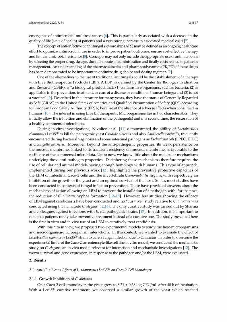
2.1.1. Growth Inhibition of C. albicansOn a Caco-2 cells monolayer, the yeast grew to 8.31 ± 0.38 log CFU/mL after 48 h of incubation.
With a Lcr35® curative treatment, we observed a similar growth of the yeast which reached
Microorganisms 2020, 8, 34 3 of 17
7.96 ± 0.18 log CFU/mL (Figure 1). Thus, no significant inhibition (after 24 h, p = 0.902; after 48 h,p = 0.603) of yeast growth was noted in the presence of the LBM in vitro and in curative condition.
Microorganisms 2019, 7, x FOR PEER REVIEW 3 of 17
On a Caco-2 cells monolayer, the yeast grew to 8.31 ± 0.38 log CFU/mL after 48 h of incubation. With a Lcr35® curative treatment, we observed a similar growth of the yeast which reached 7.96 ± 0.18 log CFU/mL (Figure 1). Thus, no significant inhibition (after 24 h, p = 0.902; after 48 h, p = 0.603) of yeast growth was noted in the presence of the LBM in vitro and in curative condition.
Figure 1. Evolution of the concentration of C. albicans in the presence or not of Lcr35® onto Caco-2 cells monolayers. Results are expressed as log10 CFU/mL of yeasts alone (controls) and in co-incubation with Lcr35® (mean ± standard deviation). Comparison between conditions with and without Lcr35® was performed using a two-way ANOVA followed by a Fisher’s Least Significant Difference (LSD) post hoc test (n.s.: not significant).
2.1.2. Inhibition of C. albicans Biofilm Formation
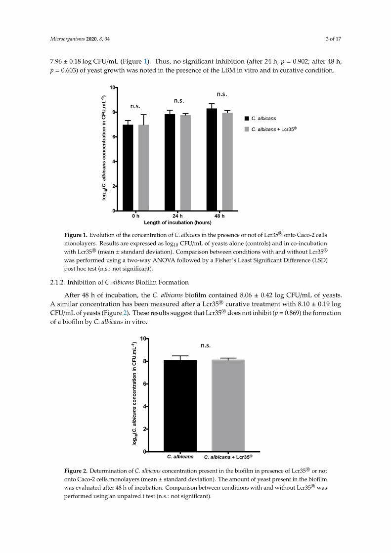
After 48 h of incubation, the C. albicans biofilm contained 8.06 ± 0.42 log CFU/mL of yeasts. A similar concentration has been measured after a Lcr35® curative treatment with 8.10 ± 0.19 log CFU/mL of yeasts (Figure 2). These results suggest that Lcr35® does not inhibit (p = 0.869) the formation of a biofilm by C. albicans in vitro.
Figure 1. Evolution of the concentration of C. albicans in the presence or not of Lcr35® onto Caco-2 cellsmonolayers. Results are expressed as log10 CFU/mL of yeasts alone (controls) and in co-incubationwith Lcr35® (mean ± standard deviation). Comparison between conditions with and without Lcr35®
was performed using a two-way ANOVA followed by a Fisher’s Least Significant Difference (LSD)post hoc test (n.s.: not significant).
2.1.2. Inhibition of C. albicans Biofilm Formation
After 48 h of incubation, the C. albicans biofilm contained 8.06 ± 0.42 log CFU/mL of yeasts.A similar concentration has been measured after a Lcr35® curative treatment with 8.10 ± 0.19 logCFU/mL of yeasts (Figure 2). These results suggest that Lcr35® does not inhibit (p = 0.869) the formationof a biofilm by C. albicans in vitro.
Microorganisms 2019, 7, x FOR PEER REVIEW 3 of 17
On a Caco-2 cells monolayer, the yeast grew to 8.31 ± 0.38 log CFU/mL after 48 h of incubation. With a Lcr35® curative treatment, we observed a similar growth of the yeast which reached 7.96 ± 0.18 log CFU/mL (Figure 1). Thus, no significant inhibition (after 24 h, p = 0.902; after 48 h, p = 0.603) of yeast growth was noted in the presence of the LBM in vitro and in curative condition.
Figure 1. Evolution of the concentration of C. albicans in the presence or not of Lcr35® onto Caco-2 cells monolayers. Results are expressed as log10 CFU/mL of yeasts alone (controls) and in co-incubation with Lcr35® (mean ± standard deviation). Comparison between conditions with and without Lcr35® was performed using a two-way ANOVA followed by a Fisher’s Least Significant Difference (LSD) post hoc test (n.s.: not significant).
2.1.2. Inhibition of C. albicans Biofilm Formation
After 48 h of incubation, the C. albicans biofilm contained 8.06 ± 0.42 log CFU/mL of yeasts. A similar concentration has been measured after a Lcr35® curative treatment with 8.10 ± 0.19 log CFU/mL of yeasts (Figure 2). These results suggest that Lcr35® does not inhibit (p = 0.869) the formation of a biofilm by C. albicans in vitro.
Figure 2. Determination of C. albicans concentration present in the biofilm in presence of Lcr35® or notonto Caco-2 cells monolayers (mean ± standard deviation). The amount of yeast present in the biofilmwas evaluated after 48 h of incubation. Comparison between conditions with and without Lcr35® wasperformed using an unpaired t test (n.s.: not significant).
Microorganisms 2020, 8, 34 4 of 17
2.2. Effect of L. rhamnosus Lcr35® Curative Treatment on Candidiasis
2.2.1. Effect of L. rhamnosus Lcr35® on C. elegans Survival after C. albicans Exposure
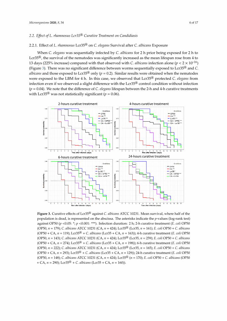
When C. elegans was sequentially infected by C. albicans for 2 h prior being exposed for 2 h toLcr35®, the survival of the nematodes was significantly increased as the mean lifespan rose from 4 to13 days (225% increase) compared with that observed with C. albicans infection alone (p < 2 × 10–16)(Figure 3). There was no significant difference between worms sequentially exposed to Lcr35® and C.albicans and those exposed to Lcr35® only (p = 0.2). Similar results were obtained when the nematodeswere exposed to the LBM for 4 h. In this case, we observed that Lcr35® protected C. elegans frominfection even if we observed a slight difference with the Lcr35® control condition without infection(p = 0.04). We note that the difference of C. elegans lifespan between the 2-h and 4-h curative treatmentswith Lcr35® was not statistically significant (p = 0.06).
Microorganisms 2019, 7, x FOR PEER REVIEW 4 of 17
Figure 2. Determination of C. albicans concentration present in the biofilm in presence of Lcr35® or not onto Caco-2 cells monolayers (mean ± standard deviation). The amount of yeast present in the biofilm was evaluated after 48 h of incubation. Comparison between conditions with and without Lcr35® was performed using an unpaired t test (n.s.: not significant).
2.2. Effect of L. rhamnosus Lcr35® Curative Treatment on Candidiasis
2.2.1. Effect of L. rhamnosus Lcr35® on C. elegans Survival after C. albicans Exposure
When C. elegans was sequentially infected by C. albicans for 2 h prior being exposed for 2 h to Lcr35®, the survival of the nematodes was significantly increased as the mean lifespan rose from 4 to 13 days (225% increase) compared with that observed with C. albicans infection alone (p < 2 × 10–16) (Figure 3). There was no significant difference between worms sequentially exposed to Lcr35® and C. albicans and those exposed to Lcr35® only (p = 0.2). Similar results were obtained when the nematodes were exposed to the LBM for 4 h. In this case, we observed that Lcr35® protected C. elegans from infection even if we observed a slight difference with the Lcr35® control condition without infection (p = 0.04). We note that the difference of C. elegans lifespan between the 2-h and 4-h curative treatments with Lcr35® was not statistically significant (p = 0.06).
Figure 3. Curative effects of Lcr35® against C. albicans ATCC 10231. Mean survival, where half of the population is dead, is represented on the abscissa. The asterisks indicate the p-values (log-rank test) against OP50 (p <0.05: *; p <0.001: ***). Infection duration: 2 h; 2-h curative treatment (E. coli OP50 (OP50, n = 179); C. albicans ATCC 10231 (CA, n = 424); Lcr35® (Lcr35, n = 161); E. coli OP50 + C. albicans (OP50 + CA, n = 119); Lcr35® + C. albicans (Lcr35 + CA, n = 163)); 4-h curative treatment (E. coli OP50 (OP50, n = 143); C. albicans ATCC 10231 (CA, n = 424); Lcr35® (Lcr35, n = 259); E. coli OP50 + C. albicans (OP50 + CA, n = 274); Lcr35® + C. albicans (Lcr35 + CA, n = 198)); 6-h curative treatment (E. coli OP50 (OP50, n = 222); C. albicans ATCC 10231 (CA, n = 424); Lcr35® (Lcr35, n = 165); E. coli OP50 + C. albicans (OP50 + CA, n = 293); Lcr35® + C. albicans (Lcr35 + CA, n = 129)); 24-h curative treatment (E. coli OP50
Figure 3. Curative effects of Lcr35® against C. albicans ATCC 10231. Mean survival, where half of thepopulation is dead, is represented on the abscissa. The asterisks indicate the p-values (log-rank test)against OP50 (p <0.05: *; p <0.001: ***). Infection duration: 2 h; 2-h curative treatment (E. coli OP50(OP50, n = 179); C. albicans ATCC 10231 (CA, n = 424); Lcr35® (Lcr35, n = 161); E. coli OP50 + C. albicans(OP50 + CA, n = 119); Lcr35® + C. albicans (Lcr35 + CA, n = 163)); 4-h curative treatment (E. coli OP50(OP50, n = 143); C. albicans ATCC 10231 (CA, n = 424); Lcr35® (Lcr35, n = 259); E. coli OP50 + C. albicans(OP50 + CA, n = 274); Lcr35® + C. albicans (Lcr35 + CA, n = 198)); 6-h curative treatment (E. coli OP50(OP50, n = 222); C. albicans ATCC 10231 (CA, n = 424); Lcr35® (Lcr35, n = 165); E. coli OP50 + C. albicans(OP50 + CA, n = 293); Lcr35® + C. albicans (Lcr35 + CA, n = 129)); 24-h curative treatment (E. coli OP50(OP50, n = 148); C. albicans ATCC 10231 (CA, n = 424); Lcr35® (n = 170); E. coli OP50 + C. albicans (OP50+ CA, n = 290); Lcr35® + C. albicans (Lcr35 + CA, n = 160)).
Microorganisms 2020, 8, 34 5 of 17
For longer Lcr35® treatment times (6 and 24 h), we observed a significant decrease of C. elegansmean survival on Lcr35® alone (condition 6 h: p = 3 × 10⁻⁴, condition 24 h: p < 2 × 10–16) or in thepresence of both Lcr35® and C. albicans (condition 6 h: p = 5 × 10⁻⁶, condition 24 h: p < 2 × 10–16)compared to the treatment of 4 h. Taken together, the results showed that the 2-h and the 4-h LBMtreatments were the most protective against C. albicans infection.
2.2.2. Influence of L. rhamnosus Lcr35® on C. albicans Presence in the Worm Gut
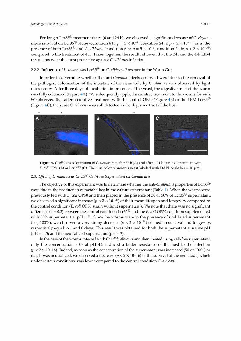
In order to determine whether the anti-Candida effects observed were due to the removal ofthe pathogen, colonization of the intestine of the nematode by C. albicans was observed by lightmicroscopy. After three days of incubation in presence of the yeast, the digestive tract of the wormwas fully colonized (Figure 4A). We subsequently applied a curative treatment to the worms for 24 h.We observed that after a curative treatment with the control OP50 (Figure 4B) or the LBM Lcr35®
(Figure 4C), the yeast C. albicans was still detected in the digestive tract of the host.
Microorganisms 2019, 7, x FOR PEER REVIEW 5 of 17
(OP50, n = 148); C. albicans ATCC 10231 (CA, n = 424); Lcr35® (n = 170); E. coli OP50 + C. albicans (OP50 + CA, n = 290); Lcr35® + C. albicans (Lcr35 + CA, n = 160)).
For longer Lcr35® treatment times (6 and 24 h), we observed a significant decrease of C. elegans mean survival on Lcr35® alone (condition 6 h: p = 3 × 10⁻⁴, condition 24 h: p < 2 × 10–16) or in the presence of both Lcr35® and C. albicans (condition 6 h: p = 5 × 10⁻⁶, condition 24 h: p < 2 × 10–16) compared to the treatment of 4 h. Taken together, the results showed that the 2-h and the 4-h LBM treatments were the most protective against C. albicans infection.
2.2.2. Influence of L. rhamnosus Lcr35® on C. albicans Presence in the Worm Gut
In order to determine whether the anti-Candida effects observed were due to the removal of the pathogen, colonization of the intestine of the nematode by C. albicans was observed by light microscopy. After three days of incubation in presence of the yeast, the digestive tract of the worm was fully colonized (Figure 4A). We subsequently applied a curative treatment to the worms for 24 h. We observed that after a curative treatment with the control OP50 (Figure 4B) or the LBM Lcr35® (Figure 4C), the yeast C. albicans was still detected in the digestive tract of the host.
Figure 4. C. albicans colonization of C. elegans gut after 72 h (A) and after a 24-h-curative treatment with E. coli OP50 (B) or Lcr35® (C). The blue color represents yeast labeled with DAPI. Scale bar = 10 μm.
2.3. Effect of L. rhamnosus Lcr35® Cell-Free Supernatant on Candidiasis
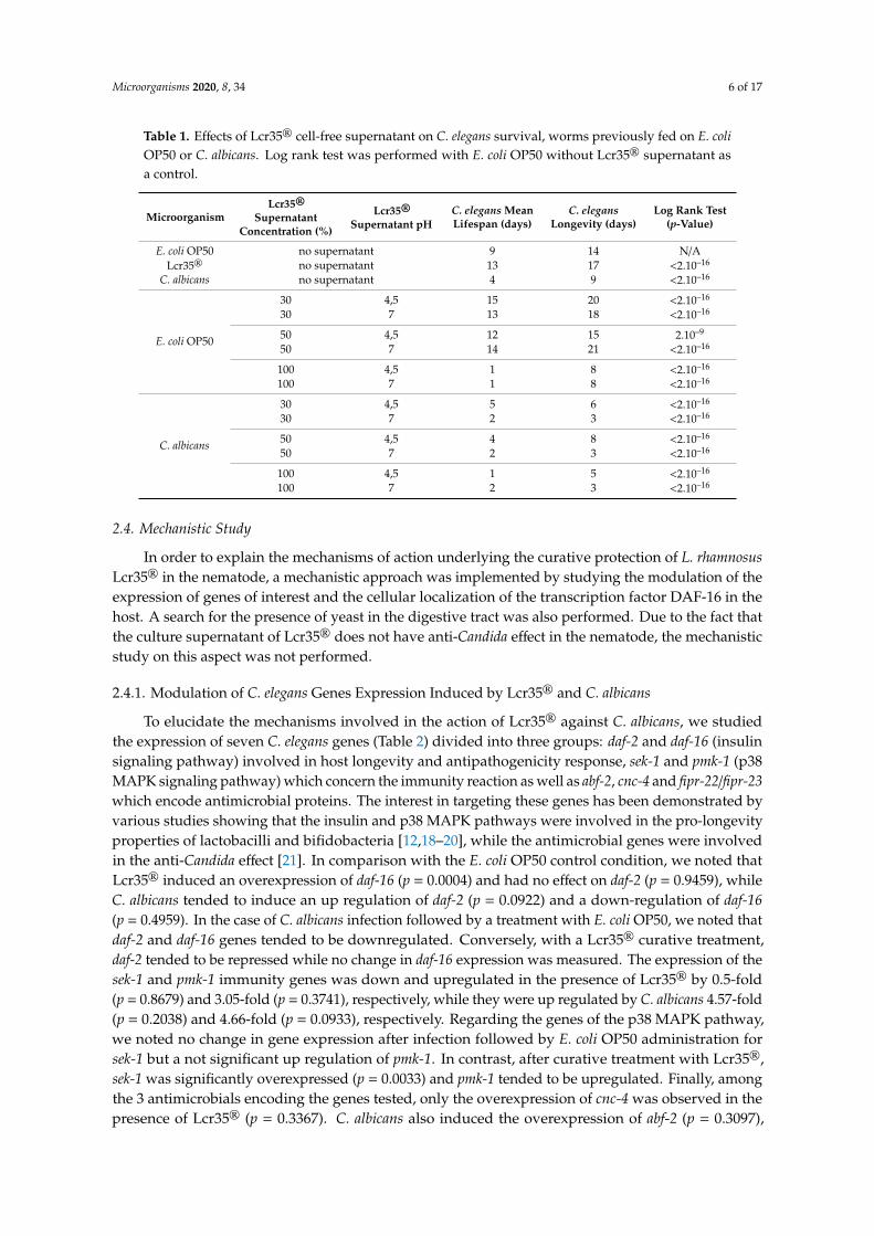
The objective of this experiment was to determine whether the anti-C. albicans properties of Lcr35® were due to the production of metabolites in the culture supernatant (Table 1). When the worms were previously fed with E. coli OP50 and then placed in the presence of 30 or 50% of Lcr35® supernatant, we observed a significant increase (p < 2 × 10–16) of their mean lifespan and longevity compared to the control condition (E. coli OP50 strain without supernatant). We note that there was no significant difference (p = 0.2) between the control condition Lcr35® and the E. coli OP50 condition supplemented with 30% supernatant at pH = 7. Since the worms were in the presence of undiluted supernatant (i.e., 100%), we observed a very strong decrease (p < 2 × 10–16) of median survival and longevity, respectively equal to 1 and 8 days. This result was obtained for both the supernatant at native pH (pH = 4.5) and the neutralized supernatant (pH = 7).
In the case of the worms infected with Candida albicans and then treated using cell-free supernatant, only the concentration 30% at pH 4.5 induced a better resistance of the host to the infection (p < 2 × 10–16). Indeed, as soon as the concentration of the supernatant was increased (50 or 100%) or its pH was neutralized, we observed a decrease (p < 2 × 10–16) of the survival of the nematode, which under certain conditions, was lower compared to the control condition C. albicans.
Table 1. Effects of Lcr35® cell-free supernatant on C. elegans survival, worms previously fed on E. coli OP50 or C. albicans. Log rank test was performed with E. coli OP50 without Lcr35® supernatant as a control.
Figure 4. C. albicans colonization of C. elegans gut after 72 h (A) and after a 24-h-curative treatment withE. coli OP50 (B) or Lcr35® (C). The blue color represents yeast labeled with DAPI. Scale bar = 10 µm.
2.3. Effect of L. rhamnosus Lcr35® Cell-Free Supernatant on Candidiasis
The objective of this experiment was to determine whether the anti-C. albicans properties of Lcr35®
were due to the production of metabolites in the culture supernatant (Table 1). When the worms werepreviously fed with E. coli OP50 and then placed in the presence of 30 or 50% of Lcr35® supernatant,we observed a significant increase (p < 2 × 10–16) of their mean lifespan and longevity compared tothe control condition (E. coli OP50 strain without supernatant). We note that there was no significantdifference (p = 0.2) between the control condition Lcr35® and the E. coli OP50 condition supplementedwith 30% supernatant at pH = 7. Since the worms were in the presence of undiluted supernatant(i.e., 100%), we observed a very strong decrease (p < 2 × 10–16) of median survival and longevity,respectively equal to 1 and 8 days. This result was obtained for both the supernatant at native pH(pH = 4.5) and the neutralized supernatant (pH = 7).
In the case of the worms infected with Candida albicans and then treated using cell-free supernatant,only the concentration 30% at pH 4.5 induced a better resistance of the host to the infection(p < 2 × 10–16). Indeed, as soon as the concentration of the supernatant was increased (50 or 100%) orits pH was neutralized, we observed a decrease (p < 2 × 10–16) of the survival of the nematode, whichunder certain conditions, was lower compared to the control condition C. albicans.
Microorganisms 2020, 8, 34 6 of 17
Table 1. Effects of Lcr35® cell-free supernatant on C. elegans survival, worms previously fed on E. coliOP50 or C. albicans. Log rank test was performed with E. coli OP50 without Lcr35® supernatant asa control.
MicroorganismLcr35®
SupernatantConcentration (%)
Lcr35®
Supernatant pHC. elegans MeanLifespan (days)
C. elegansLongevity (days)
Log Rank Test(p-Value)
E. coli OP50 no supernatant 9 14 N/ALcr35® no supernatant 13 17 <2.10–16
C. albicans no supernatant 4 9 <2.10–16
E. coli OP50
30 4,5 15 20 <2.10–16
30 7 13 18 <2.10–16
50 4,5 12 15 2.10–9
50 7 14 21 <2.10–16
100 4,5 1 8 <2.10–16
100 7 1 8 <2.10–16
C. albicans
30 4,5 5 6 <2.10–16
30 7 2 3 <2.10–16
50 4,5 4 8 <2.10–16
50 7 2 3 <2.10–16
100 4,5 1 5 <2.10–16
100 7 2 3 <2.10–16
2.4. Mechanistic Study
In order to explain the mechanisms of action underlying the curative protection of L. rhamnosusLcr35® in the nematode, a mechanistic approach was implemented by studying the modulation of theexpression of genes of interest and the cellular localization of the transcription factor DAF-16 in thehost. A search for the presence of yeast in the digestive tract was also performed. Due to the fact thatthe culture supernatant of Lcr35® does not have anti-Candida effect in the nematode, the mechanisticstudy on this aspect was not performed.
2.4.1. Modulation of C. elegans Genes Expression Induced by Lcr35® and C. albicans
To elucidate the mechanisms involved in the action of Lcr35® against C. albicans, we studiedthe expression of seven C. elegans genes (Table 2) divided into three groups: daf-2 and daf-16 (insulinsignaling pathway) involved in host longevity and antipathogenicity response, sek-1 and pmk-1 (p38MAPK signaling pathway) which concern the immunity reaction as well as abf-2, cnc-4 and fipr-22/fipr-23which encode antimicrobial proteins. The interest in targeting these genes has been demonstrated byvarious studies showing that the insulin and p38 MAPK pathways were involved in the pro-longevityproperties of lactobacilli and bifidobacteria [12,18–20], while the antimicrobial genes were involvedin the anti-Candida effect [21]. In comparison with the E. coli OP50 control condition, we noted thatLcr35® induced an overexpression of daf-16 (p = 0.0004) and had no effect on daf-2 (p = 0.9459), whileC. albicans tended to induce an up regulation of daf-2 (p = 0.0922) and a down-regulation of daf-16(p = 0.4959). In the case of C. albicans infection followed by a treatment with E. coli OP50, we noted thatdaf-2 and daf-16 genes tended to be downregulated. Conversely, with a Lcr35® curative treatment,daf-2 tended to be repressed while no change in daf-16 expression was measured. The expression of thesek-1 and pmk-1 immunity genes was down and upregulated in the presence of Lcr35® by 0.5-fold(p = 0.8679) and 3.05-fold (p = 0.3741), respectively, while they were up regulated by C. albicans 4.57-fold(p = 0.2038) and 4.66-fold (p = 0.0933), respectively. Regarding the genes of the p38 MAPK pathway,we noted no change in gene expression after infection followed by E. coli OP50 administration forsek-1 but a not significant up regulation of pmk-1. In contrast, after curative treatment with Lcr35®,sek-1 was significantly overexpressed (p = 0.0033) and pmk-1 tended to be upregulated. Finally, amongthe 3 antimicrobials encoding the genes tested, only the overexpression of cnc-4 was observed in thepresence of Lcr35® (p = 0.3367). C. albicans also induced the overexpression of abf-2 (p = 0.3097),
Microorganisms 2020, 8, 34 7 of 17
cnc-4 (p = 0.0722), and fipr-22/fipr-23 (p = 0.0736). cnc-4 tended to be overexpressed and fipr-22/fipr-23tended to be repressed when the worms were treated with E. coli OP50. On the other hand, abf-2 andfipr-22/fipr-23 were significantly overexpressed (p = 0.0043 and p = 0.0209) after a treatment with Lcr35®.
Table 2. Relative expression of C. elegans genes of interest in presence of Lcr35® and C. albicans in pureor in sequential cultures in comparison with the control condition E. coli OP50 (alone). Genes wereconsidered differentially expressed when the p-value was lower than 0.05 (*), 0.01 (**), or 0.001 (***)according to Fisher’s LSD test, and simultaneously when the expression change was of at least 2 timesor 0.5 times.
Genes of Interest
Insulin SignalingPathway
p38 MAPKSignaling Pathway Antimicrobials
Conditions daf -2 daf-16 sek-1 pmk-1 abf-2 cnc-4 fipr-22/fipr-23
E. coli OP50 1 1 1 1 1 1 1
Lcr35® 0.97 5.84 *** 0.50 3.05 0.90 8.48 0.89
C. albicans 1.87 0.31 4.57 4.66 3.58 14.31 3.45
C. albicans + E. coli OP50 0.52 0.41 1.01 2.57 0.95 7.04 0.26
C. albicans + Lcr35® 0.61 1.47 11.24 ** 2.42 8.00 ** 1.31 4.29 *
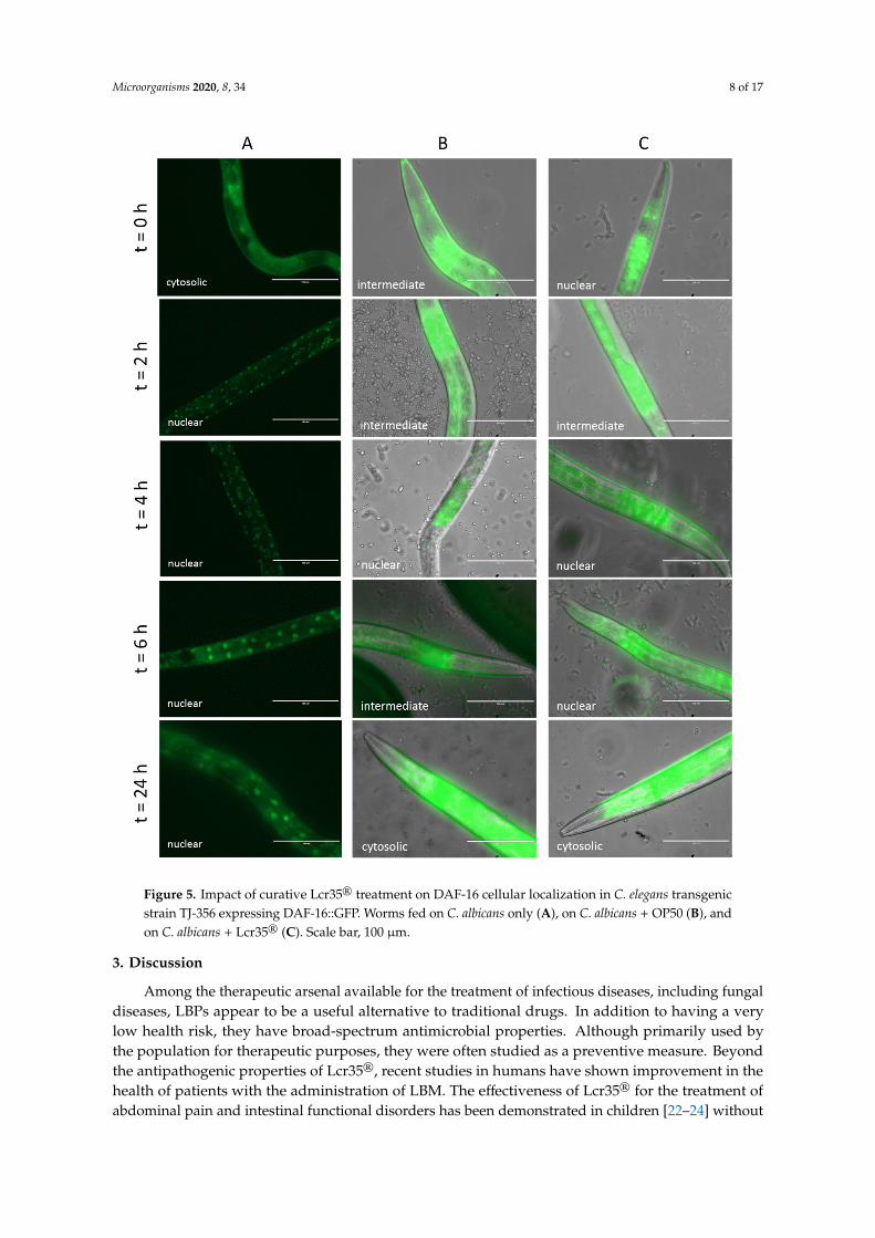
2.4.2. Effect of Lcr35® Curative Treatment on DAF-16 Nuclear Translocation
We investigated the effect of curative treatment on the cellular localization of DAF-16 over timeafter infection by C. albicans. With our control condition (C. albicans only), the transcription factorDAF-16 was quickly translocated into the nuclei after only 2 h of incubation (Figure 5A). When theworms were infected by the yeast prior being fed with OP50, we observed that DAF-16 was mainlylocated intermediately between the cytoplasm and the nuclei during the first hours of treatment andthen cytoplasmic after 24 h (Figure 5B). After a curative treatment with Lcr35®, the transcription factorappeared more translocated to the nuclei during the first part of the treatment before also getting backto a cytoplasmic localization (Figure 5C).
Microorganisms 2020, 8, 34 8 of 17Microorganisms 2019, 7, x FOR PEER REVIEW 8 of 17
Figure 5. Impact of curative Lcr35® treatment on DAF-16 cellular localization in C. elegans transgenic strain TJ-356 expressing DAF-16::GFP. Worms fed on C. albicans only (A), on C. albicans + OP50 (B), and on C. albicans + Lcr35® (C). Scale bar, 100 μm.
3. Discussion
Among the therapeutic arsenal available for the treatment of infectious diseases, including fungal diseases, LBPs appear to be a useful alternative to traditional drugs. In addition to having a
Figure 5. Impact of curative Lcr35® treatment on DAF-16 cellular localization in C. elegans transgenicstrain TJ-356 expressing DAF-16::GFP. Worms fed on C. albicans only (A), on C. albicans + OP50 (B), andon C. albicans + Lcr35® (C). Scale bar, 100 µm.
3. Discussion
Among the therapeutic arsenal available for the treatment of infectious diseases, including fungaldiseases, LBPs appear to be a useful alternative to traditional drugs. In addition to having a verylow health risk, they have broad-spectrum antimicrobial properties. Although primarily used bythe population for therapeutic purposes, they were often studied as a preventive measure. Beyondthe antipathogenic properties of Lcr35®, recent studies in humans have shown improvement in thehealth of patients with the administration of LBM. The effectiveness of Lcr35® for the treatment ofabdominal pain and intestinal functional disorders has been demonstrated in children [22–24] without
Microorganisms 2020, 8, 34 9 of 17
however highlighting the molecular mechanisms. Recently, Dausset and colleagues demonstrated inwomen the safety and well-tolerated characteristics of a new galenic form based on Lcr35®, promotingthe growth of endogenous vaginal Lactobacillus, in order to prevent an eventual dysbiosis [25]. Ina previous study [12], we demonstrated that a preventive administration of Lcr35® strain had twoin vitro effects: inhibition of C. albicans growth and biofilm formation on a Caco-2 cells monolayer.Anti-C. albicans properties targeting in particular the adhesion of the pathogen to the epithelial cellshave been attributed to exopolysaccharides (EPS) by various studies [26,27]. We then hypothesized thatLcr35® may synthesize antimicrobial molecules including EPS. However, in these conditions, whereLcr35® is used as a curative treatment, it no longer allows the inhibition of the pathogen on Caco-2 cellsmonolayers. We have performed here experiments with sequential cultures (C. albicans then nativeLcr35®) on intestinal cells while Nivoliez et al. was interested in co-cultures (C. albicans and industriallyformulated Lcr35®) on vaginal cells. The experimental conditions (culture medium, cells) as well asthe origin of the LBM strain (native strain vs industrially formulated strain) are two parameters to takeinto consideration and which are very probably the cause of the difference observed compared with aprevious study [11]. The mechanisms involved in a preventive context would therefore not be effectivein a curative one, when the pathogen is already present on the cells. Several hypotheses could explainthese results. Antimicrobial molecules that inhibit the growth of yeast may only act on dividing cells.Therefore, when the yeast is on stationary phase or does not divide, it is insensitive to these molecules.With regard to adhesion, we can wonder if C. albicans adheres to epithelial cell surfaces covalently orusing low energy bonds. EPS secreted by Lcr35® will not be able to break covalent bonds becauseof the absence of hydrolytic activity. If they are low energies, the competition for cell receptors maybe in favor of the pathogen. The competition hypothesis between the LBM and the yeast could befurther investigated by limiting the initial amount of yeast in order to favor the LBM. However, to fullyunderstand the LBM mechanisms of action, in vitro approaches are too limited. Moving to an in vivoapproach is mandatory to study the interactions between microorganisms (LBMs and pathogens) andthe host response.
We choose C. elegans as a relevant in vivo model for studying the pathogenicity ofmicroorganisms [21,28–31] and the antimicrobial properties of lactic acid bacteria [32,33]. Unlikein vitro experiments which demonstrated no significant antifungal effects of the LBM, those performedin vivo showed that Lcr35® had interesting curative anti-C. albicans properties in the nematode.Contradictory results can be explained by the experimental conditions. Thus, in vitro, Lcr35® isprobably not in optimal conditions to allow inhibition of the yeast. This reveals the importance of usingcomplementary experimental models (in vitro and in vivo) to overcome their limitations. During ourin vivo tests, Lcr35® allowed a statistically significant increase in the survival of the host, includingafter being contaminated by the yeast. The results obtained are in accordance with those using Lcr35®
in a preventive approach [12]. However, the experimental conditions are different. In a preventiveapproach, Lcr35® has an action on its host before having an antipathogenic activity targeting C. albicans.In a curative approach, the LBM first has an action against yeast and eventually on its host. Sharma etal. recently tested the inhibitory properties of Lactobacillus spp against infection with pathogenic E. colistrains, by co-infection, preventive, and curative approaches. The authors have put forward severalhypotheses that can explain the healing properties: the steric exclusion of the pathogen by the probioticstrains as well as the competition for the cellular receptors because of the shared carbohydrate-bindingspecificities with some entero-pathogens [17]. Our data showed that using supernatants did not allowoptimal protection of the host, confirming the essential presence of the LBM Lcr35® cells to observethe anti-Candida effect. Thus, it is likely that the mechanism of action is based on a mischaracterizeddirect interaction between Lcr35® and the yeast. In the absence of pathogenic yeast, with 30–50%supernatant, we observed a significant increase in mean lifespan and worm longevity. On the otherhand, during a fungal infection, only 30% of supernatant at pH = 4.5 allows an increase in meanlifespan. We hypothesize that there are two different mechanisms, one or more molecule present in theculture supernatant have a pro-longevity action while others appear to have antifungal effect. In their
Microorganisms 2020, 8, 34 10 of 17
work, Dausset et al. demonstrated in vitro that the molecule secreted by Lcr35® and having an antiC. albicans activity had a molecular weight smaller than 10 kDa and was resistant to protease, lipaseand thermal treatments [34]. However, the molecule in question has not been identified precisely. It isconceivable that this molecule is a bacteriocin but it would be interesting to study its antifungal activityin C. elegans. Indeed, a recent study has shown that the bacteriocin EntV secreted by Enterococcus faecalisinhibited the growth of yeast and protected the nematode from infection at the same time [34,35].In addition, another study showed the involvement of extracellular vesicles of L. plantarum in theactivation of the host immune system against vancomycin-resistant enterococcal infection [36].
In our study, we observed that the duration of the Lcr35® treatment influences the curativeanti-C. albicans effect on nematode lifespan, suggesting that the quantity of Lcr35® ingested and/or thetreatment duration may have an impact on the effectiveness of the treatment. It is conceivable thatthere is also a dose effect explaining the results obtained with the culture supernatants. The work ofKomura et al. showed that there was a dose effect linked to the pro-longevity effects of bifidobacteria,in favor of a low Bifidobacterium/E. coli OP50 ratio [37]. A thorough transcriptional study will beinteresting to characterize the dose-dependent effect of LBPs administered. Therefore, we can questionthe relevance of the evaluation of toxicokinetic and pharmacodynamic parameters as is the case fortraditional drugs. Such parameters would make it possible to adapt the prescription of LBPs andimprove their effectiveness.
We demonstrated that Lcr35® induced a transcriptional response in the host by activating thetranscription factor DAF-16 (nuclear translocation), and the activation of the p38 MAPK signallingpathway involved genes, including in the presence of C. albicans. From a mechanistic point of view,several hypotheses can explain the anti-C. albicans properties of Lcr35® in the nematode: a directinteraction between the two microorganisms as well as an immunomodulation of the host by the LBM.We have shown that even after curative treatment with the LBM, the digestive tract of the nematode iscolonized by the pathogen without showing a pathological state. These data are in accordance withDe Barros et al. and suggest that Lcr35® induced repression of virulence factors in C. albicans [16]. InC. elegans, DAF-16 is closely related to mammalian FOXO3a, a transcription factor involved in theinflammatory process [38]. Therefore, nuclear translocation of DAF-16 by Lcr35® can be interpreted asthe establishment of an inflammatory response in the host allowing it to survive an infection. The workof Pukkila-Worley et al. [21] demonstrated that C. albicans induced a fast antifungal response in thehost with the overexpression of antimicrobial genes such as abf-2, cnc-4, cnc-7, fipr-22, and fipr-23. Withthe exception of abf-2, all these genes are under the control of PMK-1, whose inactivation makes thenematode susceptible to infection. In our study, we showed that Lcr35® curative treatment induced anup regulation of most targeted genes (sek-1, pmk-1, fipr-22, and fipr-23), while cnc-4 and daf-16 remainedunchanged compared to the control condition. Based on the data of Pukkila-Worley et al. [21], thepresence of Lcr35® would allow activation of the host’s immune defenses via the highly conserved p38MAPK signaling pathway. Also, it is possible that the bacterium exerts a direct action on the yeast byat least partially inhibiting its virulence, thus limiting the deleterious effects of C. albicans in C. elegans.The use of C. elegans mutants or RNAi could be further considered deciphering with precision thesignalling pathway(s) involved and the regulation mechanisms.
4. Materials and Methods
4.1. Microbial Strains and Growth Conditions
The E. coli OP50 strain was provided by the Caenorhabditis Genetics Center (Minneapolis, MN,USA) and was grown on Lysogeny Broth (LB, Miller’s Modification) (Conda, Madrid, Spain) at 37 ◦Covernight. The L. rhamnosus Lcr35® strain was provided by biose® (Aurillac, France) and was grownin de Man, Rogosa, Sharpe (MRS) broth (bioMérieux, Marcy l’Etoile, France) at 37 ◦C overnight. C.albicans ATCC® 10231™ was grown in yeast peptone glucose (YPG) broth pH 6.5 (per L: 10 g yeastextract (Fisher Scientific, Hampton, NH, USA), 10 g peptone (Conda), 20 g glucose (Sigma, Saint-Louis,
Microorganisms 2020, 8, 34 11 of 17
USA)) at 37 ◦C for 48 h. Microbial suspensions were spun down for 2 min at 1500 rpm (Rotofix32A, Hettich Zentrifugen, Tuttlingen, Germany) and washed with M9 buffer (per L: 3 g KH2PO4, 6 gNa2HPO4, 5 g NaCl, 1 mL 1 M MgSO4) (Sigma) to obtain a final concentration of 100 mg/mL.
4.2. Influence of L. rhamnosus Lcr35® on Growth of C. albicans on Caco-2 Cells Monolayer and onBiofilm Formation
Growth inhibition of C. albicans by the LBM Lcr35® was examined using the human colorectaladenocarcinoma cell line Caco-2 [39]. Caco-2 cells were grown in Dulbecco’s modified Eagle’s minimalessential medium (DMEM, Life Technologie, Villebon-sur-Yvette, France) supplemented with 20%inactivated fetal calf serum (Life Technologie) at 37 ◦C with a 5% CO2 in air atmosphere. For the assays,the cells were seeded at a concentration of 3.5 × 10⁵ cells/well in 24-well plates (Dutscher, Brumath,France) and placed in growth conditions for 24 h. Microbial strains were grown according to Nivoliezet al. [11]. After growth, cell culture medium was removed and replaced by 1 mL of DMEM and 250 µLof C. albicans culture (107 CFU/mL) in each well and incubated for 24 h. Lcr35® (250 µL of culture(108 CFU/mL)) was added in each well and incubated for 48 h. The inhibition of C. albicans by Lcr35®
was evaluated during (24 h) and after co-incubation (48 h). One hundred microliters of suspensionwere taken from each of the wells and the number of viable bacteria and/or yeasts was determined byplating serial dilutions of the suspensions onto MRS or Sabouraud agar plates. For the measurementof C. albicans biofilm formation, after incubation for 48 h, the wells were washed twice with 0.5 mL ofPBS and cells harvested with 1 mL of trypsin at 37 ◦C. As for the inhibition assay, the number of viablebacteria or/and yeasts were determined by plating serial dilutions of the suspensions onto MRS orSabouraud agar plates. The plates were incubated at 37 ◦C for 72 h (MRS) or 48 h (Sabouraud). Eachassay, performed three times independently, contained two technical replicates.
4.3. C. elegans Maintenance
C. elegans N2 (wild-type) and TJ356 (daf-16p::daf-16a/b::GFP + rol-6(su1006)) strains were acquiredfrom the Caenorhabditis Genetics Center. The nematodes were grown and maintained at 20 ◦C onnematode growth medium (NGM) (per L: 3 g NaCl (Sigma); 2.5 g peptone (Conda); 17 g agar (BiokarDiagnostics, Beauvais, France); 5 mg cholesterol (Sigma); 1 mM CaCl2 (Sigma); 1 mM MgSO4 (Sigma),25 mL 1 M potassium phosphate (Sigma) buffer at pH 6) plates supplemented with yeast extract (4 g/L)(NGMY) and seeded with E. coli OP50 [40]. For all experiments, wild-type C. elegans N2 was usedexcept for the study of the localization of DAF-16 (TJ356 strain).
4.4. C. elegans Synchronization
To avoid variations in results due to age differences, a worm synchronous population was required.Gravid worms were washed off using M9 buffer and spun down for 2 min at 1500 rpm. Five millilitresof worm bleach (2.5 mL of M9 buffer, 1.5 mL of bleach, 1 mL of 5 M sodium hydroxide) were addedto the pellet and vigorously shaken until adult worm body disruption. The action of worm bleachwas stopped by adding 20 mL of M9 buffer. The egg suspension was then spun down for 2 min at1500 rpm and washed twice with 20 mL of M9 buffer. Eggs were allowed to hatch under slow agitationat 25 ◦C for 24 h in approximately 20 mL of M9 buffer. L1 larvae were then transferred onto NGMYplates seeded with E. coli OP50 until they reached the L4/young adult stage.
4.5. Effects of L. rhamnosus Lcr35® on Candidiasis in C. elegans
Sequential feeding with Lcr35® and C. albicans was induced in C. elegans in all experiments (curativeassays). As control groups, a monotypic contamination was induced in C. elegans by inoculating withonly C. albicans, Lcr35® or E. coli OP50.
Microorganisms 2020, 8, 34 12 of 17
4.5.1. Preparation of Plates Containing LBM or Pathogen Yeasts
One hundred microliters of Lcr35® or E. coli OP50 suspension (100 mg/mL) were spread onNGMY + 0.12 mM FUdR (Sigma) plates (in order to prevent C. elegans progeny) and incubated at 37 ◦Covernight. Concerning C. albicans strain, 100 µL of suspension were spread on Brain Heart InfusionBHI (Biokar Diagnostics) + 0.12 mM FUdR plates and incubated at 37 ◦C overnight.
4.5.2. Survival Assay: Curative Treatment
The survival assay was performed according to Poupet et al. [12], with some modifications.During a curative treatment, young L4 adult worms were placed on plates containing C. albicans for 2 hat 20 ◦C. Next, worms were washed with M9 buffer to remove yeasts prior being placed on Lcr35®, at20 ◦C for different times (2, 4, 6, and 24 h). Infected nematodes were washed off plates using M9 bufferprior to be transferred into a 6-well microtiter plate (approximately 50 worms per well) containing2 mL of BHI/M9 (20%/80%) + 0.12 mM FUdR liquid assay medium per well and incubated at 20 ◦C.For the control groups (i.e., C. albicans + E. coli OP50, E. coli OP50 alone, Lcr35® alone and C. albicansalone), worms were treated in the same way. Nematodes were observed daily and were considereddead when they did not respond to a gentle mechanical stimulation. This assay was performed asthree independent experiments containing three wells per condition.
4.6. Study of L. rhamnosus Lcr35® Cell-Free Supernatant on Candidiasis in C. elegans
The potential protective effect of Lcr35® cell-free supernatant was investigated. For this, a cultureof Lcr35® was carried out in MRS broth and incubated for 24 h at 37 ◦C. The broth was centrifuged for15 min at 14,000 rpm (Rotofix 32A, Hettich Zentrifugen). The supernatant was then sterilized using a0.22 µm filter. As before, L4/young adult worms were placed on plates containing C. albicans for 2 h at20 ◦C. Next, worms were washed with M9 buffer to remove yeasts prior being transferred into a 6-wellmicrotiter plate (about 50 worms per well) containing 2 mL of BHI/M9 (20%/80%) liquid assay mediumsupplemented by Lcr35® cell-free supernatant (30%, 50%, 100% at pH = 4.5 or pH = 7) and 0.12 mMFUdR per well and incubated at 20 ◦C. Regarding the supernatant at pH = 7, it was neutralized with a5N sodium hydroxide solution (Panreac, Barcelona, Spain). For the control groups (i.e., E. coli OP50 +
Lcr35® cell-free supernatant), worms were treated in the same way. Nematodes were observed dailyand were considered dead when they did not respond to a gentle mechanical stimulation. This assaywas performed as three independent experiments containing three wells per condition.
4.7. Visualization of C. albicans in C. elegans Intestine
In order to study the presence of the pathogen C. albicans in the worm gut, a fluorescent stainingof the yeast was performed. The yeast was stained with DAPI (Thermo Scientific, Karlsruhe, Germany)according to the manufacturer’s instructions. A fresh culture of C. albicans was performed in YPGbroth as described in Section 4.1, 10 µL of DAPI at 300 nM was added to 1 mL of C. albicans suspensionand incubated at room temperature in the dark for 15 min. The unbound dye was removed bycentrifugation (14,000 rpm for 5 min at 4 ◦C) (Beckman J2-MC Centrifuge, Beckman Coulter, Brea, USA)and washed with 1 mL of M9 buffer. Subsequently, the nematodes were fed on labelled-C. albicans onBHI plates for 72 h and then with E. coli OP50 or Lcr35® on NGMY plates for 24 h. The nematodeswere then visualized using a fluorescence microscope at 100×magnification (Evos FL, Invitrogen).
4.8. RNA Isolation and Real-Time Quantitative PCR
Approximately 10,000 worms were harvested from NGMY plates with M9 buffer. Total RNA wasextracted by adding 500 µL of TRIzol reagent (Ambion by Life Technologies, Carlsbad, USA). Wormswere disrupted using a Precellys (Bertin Instruments, Montigny-le-Bretonneux, France) and glass beads(PowerBead Tubes Glass 0.1 mm, Mo Bio Laboratories, USA). Beads were removed by centrifugationat 14,000 rpm for 1 min (Eppendorf® 5415D, Hamburg, Germany), and 100 µL of chloroform were
Microorganisms 2020, 8, 34 13 of 17
added to the supernatant. Tubes were vortexed for 30 s and incubated at room temperature for 3 min.The phenolic phase was removed by centrifugation at 12,000 rpm for 15 min at 4 ◦C. The aqueousphase was treated with chloroform as previously described. RNA was precipitated by adding 250 µLof isopropanol for 4 min at room temperature and spun down at 12,000 rpm for 10 min (4 ◦C). Thesupernatant was discarded, and the pellet was washed with 1 mL of 70% ethanol. The supernatantwas discarded after centrifugation at 14,000 rpm for 5 min (4 ◦C), and the pellet was dissolved in 20 µLof RNase-free water. RNA was reverse-transcribed using a High-Capacity cDNA Archive kit (AppliedBiosystems, Foster City, USA) according to the manufacturer’s instructions. For real-time qPCR assay,each tube contained 2.5 µL of cDNA, 6.25 µL of Rotor-Gene SYBR Green Mix (Qiagen GmbH, Hilden,Germany), 1.25 µL of 10 µM primers (reported in Table 3) (Eurogentec, Seraing, Belgium), and 1.25 µLof water. All samples were run in triplicate. Rotor-Gene Q Series Software (Qiagen GmbH) was usedfor the analysis. The quantification of gene-of-interest expression (EGOI) was performed accordingto the following formula [41] taking into account the efficiency of the PCR for each primer pair andnormalizing the expression of the gene of interest by two reference genes: cdc-42 and Y45F10D.4.
EGOI =(GOI efficiency)∆CtGOI√
(cdc− 42 efficiency)∆Ctcdc−42 × (Y45F10D.4 efficiency)∆CtY45F10D.5
(1)
The worms fed with E. coli OP50 were used as control conditions for the gene expression calculation.
Table 3. Targeted C. elegans genes primers for qPCR analysis.
Gene Name Gene Type Forward Primer (5′–3′) Reverse Primer (5′–3′) Reference
cdc-42 housekeeping ATCCACAGACCGACGTGTTT GTCTTTGAGCAATGATGCGA [42]Y45F10D.4 housekeeping CGAGAACCCGCGAAATGTCGGA CGGTTGCCAGGGAAGATGAGGC [43]
daf-2 GOI AAAAGATTTGGCTGGTCAGAGA TTTCAGTACAAATGAGATTGTCAGC [44]daf-16 GOI TTCAATGCAAGGAGCATTTG AGCTGGAGAAACACGAGACG [44]sek-1 GOI GCCGATGGAAAGTGGTTTTA TAAACGGCATCGCCAATAAT [44]
pmk-1 GOI CCGACTCCACGAGAAGGATA AGCGAGTACATTCAGCAGCA [44]abf-2 GOI TCGTCCGTTCCCTTTTCCTT CCTCTCTTAATAAGAGCACC [12]
fipr-22/fipr-23 GOI CCCAATCCAGTATGAAGTTG ATTTCAGTCTTCACACCGGA [12]cnc-4 GOI ATGCTTCGCTACATTCTCGT TTACTTTCCAATGAGCATTC [12]cnc-7 GOI TTTTGTTGGCTCTGGTGGCA ATGAGTCCAGGACGGTACAT [12]
4.9. DAF-16 Nuclear Localization
DAF-16 nuclear localization was followed as described by others [45] using a transgenic TJ-356worm strain constitutively expressing the DAF-16 transcription factor fused to GFP (DAF-16::GFP).Once adults, worms were exposed to C. albicans for 2, 4, 6, and 24 h at 20 ◦C. A curative approachwas conducted: worms were put in the presence of C. albicans for 2 h and after with E. coli OP50 orLcr35® for 4 h at 20 ◦C. The nematodes were subsequently imaged 2, 4, 6, and 24 h after infection. Thetranslocation of DAF-16::GFP was scored by the observation of the presence of GFP accumulation inthe C. elegans cell nuclei, using a fluorescence microscope at 40×magnification (Evos FL, Invitrogen).
4.10. Statistical Analysis
Data are expressed as the mean ± standard deviation. The C. elegans survival assay was examinedby using the Kaplan-Meier method, and differences were determined by using the log-rank test withR software version 3.6.1 [46], and the survival [47] and survminer [48] packages. For C. albicansgrowth inhibition and C. elegans gene expressions, differences between conditions were determinedby a two-way ANOVA followed by a Fisher’s Least Significant Difference (LSD) post hoc test usingGraphPad Prism version 8.2.0 for macOS (GraphPad Software, La Jolla, California, USA). For C.albicans biofilm formation, differences between conditions were determined by an unpaired t test usingGraphPad Prism version 8.2.0 for macOS. Each experiment was performed in three different replicates.A difference with p-value ≤ 0.05 was considered as significant.
Microorganisms 2020, 8, 34 14 of 17
5. Conclusions
This study showed the effectiveness of the LBM Lcr35® in a curative context. Although it didnot allow the reduction of the fungal growth in vitro in our experimental conditions, it had in vivoantifungal capabilities with a protection of the C. albicans infected worm C. elegans. Our data suggestthat a Lcr35® treatment tends to activate C. elegans immune response (overexpression genes of the p38MAPK pathway and encoding for antimicrobials). An exhaustive transcriptomic study would allow abetter understanding of the interactions between C. elegans, C. albicans and Lcr35®. Once the molecularmechanisms well characterized, it would be of interest to evaluate the possibility to extrapolate toother strains of Lactobacillus spp. and Candida spp. and to include the C. elegans approaches in the usualtests allowing the identification of new live biotherapeutic microorganisms.
Author Contributions: C.P., M.G., and O.C. designed the protocols and scientific approaches. C.P., P.V., M.G.,and O.C. carried out the experiments with help from M.B., and S.B. C.P. wrote the manuscript. P.V., M.B., C.D.,C.C., and S.B. provided critical feedback. A.N. and S.B. supervised the project. All authors have read and agreedto the published version of the manuscript.
Funding: This work was supported by the European funds FEDER and the Auvergne-Rhône-Alpes Region in theform of grants to C.P. as well as biose® as part of the doctoral thesis of C.P. The funder biose® provided supportin the form of salaries for authors C.D. and A.N.
Acknowledgments: This work was supported by the European funds FEDER, the Auvergne Rhône Alpes Regionand biose® as part of the doctoral thesis of Cyril Poupet. Some strains were provided by the CGC, which isfunded by NIH Office of Research Infrastructure Programs (P40 OD010440). We thank Jonathan Heuzé and MurielThéret, trainees at the UMRF 0545 (UCA/INRAE/VetAgro Sup) for their involvement in the implementation ofseveral experiments. We thank Claudia Thoral and Haymen Girgis for the careful review and advice provided forthe writing of this article. We thank greatly all those who took part in the writing of this article.
Conflicts of Interest: The funders (the European union, the Auvergne Rhône Alpes and biose® Industrie), had norole in the design of the study; in the collection, analyses, or interpretation of data; in the writing of the manuscript,or in the decision to publish the results.
References
1. Pfaller, M.A.; Diekema, D.J. Epidemiology of Invasive Candidiasis: A Persistent Public Health Problem.Clin. Microbiol. Rev. 2007, 20, 133–163. [CrossRef]
2. Ruhnke, M. Clinical Syndromes: Candida and Candidosis. In Clinically Relevant Mycoses; Presterl, E., Ed.;Springer: Cham, Switzerland, 2019.
3. Pappas, P.G.; Kauffman, C.A.; Andes, D.; Benjamin, D.K., Jr.; Calandra, T.F.; Edwards, J.E., Jr.; Filler, S.G.;Fisher, J.F.; Kullberg, B.J.; Ostrosky-Zeichner, L.; et al. Clinical Practice Guidelines for the Management ofCandidiasis: 2009 Update by the Infectious Diseases Society of America. Clin. Infect. Dis. 2009, 48, 503–535.[CrossRef] [PubMed]
4. Yano, J.; Sobel, J.D.; Nyirjesy, P.; Sobel, R.; Williams, V.L.; Yu, Q.; Noverr, M.C.; Fidel, P.L. Current patientperspectives of vulvovaginal candidiasis: Incidence, symptoms, management and post-treatment outcomes.BMC Womens Health 2019, 19, 48. [CrossRef]
5. Kumamoto, C. Inflammation and gastrointestinal Candida colonization Carol. Curr. Opin. Microbiol. 2011,14, 386–391. [CrossRef] [PubMed]
6. Spettel, K.; Barousch, W.; Makristathis, A.; Zeller, I.; Nehr, M.; Selitsch, B.; Lackner, M.; Rath, P.M.;Steinmann, J.; Willinger, B. Analysis of antifungal resistance genes in Candida albicans and Candida glabratausing next generation sequencing. PLoS ONE 2019, 14, e0210397. [CrossRef] [PubMed]
7. Moran, C.; Grussemeyer, C.A.; Spalding, J.R.; Benjamin, D.K.; Reed, S.D.; Reed, D.S.D. Comparison of costs,length of stay, and mortality associated with Candida glabrata and Candida albicans bloodstream infections.Am. J. Infect. Control 2010, 38, 78–80. [CrossRef] [PubMed]
8. Mac Dougall, C.; Polk, R.E. Antimicrobial Stewardship Programs in Health Care Systems. Clin. Microbiol.Rev. 2005, 18, 638–656. [CrossRef]
9. Dreher-Lesnick, S.M.; Stibitz, S.; Carlson, P.E., Jr. US Regulatory Considerations for Development of LiveBiotherapeutic Products as Drugs. Microbiol. Spectr. 2017, 5. [CrossRef]
Microorganisms 2020, 8, 34 15 of 17
10. O’Toole, P.W.; Marchesi, J.R.; Hill, C. Next-generation probiotics: The spectrum from probiotics to livebiotherapeutics. Nat. Microbiol. 2017, 2, 17057. [CrossRef]
11. Nivoliez, A.; Camares, O.; Paquet-Gachinat, M.; Bornes, S.; Forestier, C.; Veisseire, P. Influence ofmanufacturing processes on in vitro properties of the probiotic strain Lactobacillus rhamnosus Lcr35®.J. Biotechnol. 2012, 160, 236–241. [CrossRef]
12. Poupet, C.; Saraoui, T.; Veisseire, P.; Bonnet, M.; Dausset, C.; Gachinat, M.; Camarès, O.; Chassard, C.;Nivoliez, A.; Bornes, S. Lactobacillus rhamnosus Lcr35 as an effective treatment for preventing Candida albicansinfection in the invertebrate model Caenorhabditis elegans: First mechanistic insights. PLoS ONE 2019, 14,e0216184. [CrossRef] [PubMed]
13. Patrignani, F.; Siroli, L.; Parolin, C.; Serrazanetti, D.I.; Vitali, B.; Lanciotti, R. Use of Lactobacillus crispatus toproduce a probiotic cheese as potential gender food for preventing gynaecological infections. PLoS ONE2019, 14, e0208906. [CrossRef] [PubMed]
14. Romeo, M.G.; Romeo, D.M.; Trovato, L.; Oliveri, S.; Palermo, F.; Cota, F.; Betta, P. Role of probiotics in theprevention of the enteric colonization by Candida in preterm newborns: Incidence of late-onset sepsis andneurological outcome. J. Perinatol. 2011, 31, 63–69. [CrossRef] [PubMed]
15. Kumar, S.; Bansal, A.; Chakrabarti, A.; Singhi, S. Evaluation of Efficacy of Probiotics in Prevention of CandidaColonization in a PICU—A Randomized Controlled Trial*. Crit. Care. Med. 2013, 41, 565–572. [CrossRef][PubMed]
16. De Barros, P.P.; Scorzoni, L.; Ribeiro, F.C.; Fugisaki, L.R.O.; Fuchs, B.B.; Mylonakis, E.; Jorge, A.O.C.;Junqueira, J.C.; Rossoni, R.D. Lactobacillus paracasei 28.4 reduces in vitro hyphae formation of Candida albicansand prevents the filamentation in an experimental model of Caenorhabditis elegans. Microb. Pathog. 2018, 117,80–87. [CrossRef]
17. Sharma, K.; Pooranachithra, M.; Balamurugan, K.; Goel, G. Probiotic mediated colonization resistanceagainst E. coli infection in experimentally challenged Caenorhabditis elegans. Microb. Pathog. 2019, 127, 39–47.[CrossRef]
18. Zhou, M.; Liu, X.; Yu, H.; Yin, X.; Nie, S.-P.; Xie, M.-Y.; Chen, W.; Gong, J. Cell Signaling of Caenorhabditiselegans in Response to Enterotoxigenic Escherichia coli Infection and Lactobacillus zeae Protection. Front.Immunol. 2018, 9, 1745. [CrossRef]
19. Schifano, E.; Zinno, P.; Guantario, B.; Roselli, M.; Marcoccia, S.; Devirgiliis, C.; Uccelletti, D. The FoodborneStrain Lactobacillus fermentum MBC2 Triggers pept-1-Dependent Pro-Longevity Effects in Caenorhabditis elegans.Microorganisms 2019, 7, 45. [CrossRef]
20. Zhao, L.; Zhao, Y.; Liu, R.; Zheng, X.; Zhang, M.; Guo, H.; Zhang, H.; Ren, F. The transcription factor DAF-16is essential for increased longevity in C. elegans Exposed to Bifidobacterium longum BB68. Sci. Rep. 2017,7, 7408. [CrossRef]
21. Pukkila-Worley, R.; Ausubel, F.M.; Mylonakis, E. Candida albicans infection of Caenorhabditis elegans inducesantifungal immune defenses. PLoS Pathog. 2011, 7, e1002074. [CrossRef]
22. Wegh, C.A.M.; Benninga, M.A.; Tabbers, M.M. Effectiveness of Probiotics in Children with FunctionalAbdominal Pain Disorders and Functional Constipation A Systematic Review. J. Clin. Gastroenterol. 2018, 52,S10–S26. [CrossRef] [PubMed]
23. Wojtyniak, K.; Szajewska, H. Systematic Review: Probiotics for Functional Constipation in Children. J. Pediatr.2017, 176, 1155–1162. [CrossRef] [PubMed]
24. Wojtyniak, K.; Horvath, A.; Dziechciarz, P.; Szajewska, H. Lactobacillus casei rhamnosus Lcr35 in theManagement of Functional Constipation in Children: A Randomized Trial. J. Pediatr. 2017, 184, 101–105.e1.[CrossRef] [PubMed]
25. Dausset, C.; Patrier, S.; Gajer, P.; Thoral, C.; Lenglet, Y.; Cardot, J.M.; Judlin, P.; Ravel, J.; Nivoliez, A.Comparative phase I randomized open-label pilot clinical trial of Gynophilus® (Lcr regenerans®) immediaterelease capsules versus slow release muco-adhesive tablets. Eur. J. Clin. Microbiol. Infect. Dis. 2018, 37,1869–1880. [CrossRef] [PubMed]
26. Nowak, A.; Motyl, I.; Slizewska, K.; Libudzisz, Z.; Klewicka, E. Adherence of probiotic bacteria to humancolon epithelial cells and inhibitory effect against enteric pathogens—In vitro study. Int. J. Dairy Technol.2016, 69, 532–539.
Microorganisms 2020, 8, 34 16 of 17
27. Allonsius, C.N.; van den Broek, M.F.L.; De Boeck, I.; Kiekens, S.; Oerlemans, E.F.M.; Kiekens, F.; Foubert, K.;Vandenheuvel, D.; Cos, P.; Delputte, P.; et al. Interplay between Lactobacillus rhamnosus GG and Candida andthe involvement of exopolysaccharides. Microb. Biotechnol. 2017, 10, 1753–1763. [CrossRef]
28. Irazoqui, J.E.; Troemel, E.R.; Feinbaum, R.L.; Luhachack, L.G.; Cezairliyan, B.O.; Ausubel, F.M. Distinctpathogenesis and host responses during infection of C. elegans by P. aeruginosa and S. aureus. PLoS Pathog.2010, 6, e1000982. [CrossRef]
29. Wu, K.; Conly, J.; McClure, J.A.; Elsayed, S.; Louie, T.; Zhang, K. Caenorhabditis elegans as a host model forcommunity-associated methicillin-resistant Staphylococcus aureus. Clin. Microbiol. Infect. 2010, 16, 245–254.[CrossRef]
30. Souza, A.C.R.; Fuchs, B.B.; Alves V de, S.; Jayamani, E.; Colombo, A.L.; Mylonakis, E. Pathogenesis of theCandida parapsilosis complex in the model host Caenorhabditis elegans. Genes 2018, 9, 401. [CrossRef]
31. Pukkila-Worley, R.; Peleg, A.Y.; Tampakakis, E.; Mylonakis, E. Candida albicans hyphal formation andvirulence assessed using a Caenorhabditis elegans infection model. Eukaryot. Cell 2009, 8, 1750–1758. [CrossRef]
32. Park, M.R.; Ryu, S.; Maburutse, B.E.; Oh, N.S.; Kim, S.H.; Oh, S.; Jeong, S.Y.; Jeong, D.Y.; Oh, S.; Kim, Y. ProbioticLactobacillus fermentum strain JDFM216 stimulates the longevity and immune response of Caenorhabditiselegans through a nuclear hormone receptor. Sci. Rep. 2018, 8, 7441. [CrossRef] [PubMed]
33. Kim, Y.; Mylonakis, E. Caenorhabditis elegans immune conditioning with the probiotic bacterium Lactobacillusacidophilus strain ncfm enhances gram-positive immune responses. Infect. Immun. 2012, 80, 2500–2508.[CrossRef] [PubMed]
34. Dausset, C. Détermination des mécanismes d’inhibition de souches de lactobacilles. In Influence du procédéindustriel sur leurs caractéristiques probiotiques; AgroParisTech: Jouy-en-Josas, France, 2015.
35. Graham, C.E.; Cruz, M.R.; Garsin, D.A.; Lorenz, M.C. Enterococcus faecalis bacteriocin EntV inhibits hyphalmorphogenesis, biofilm formation, and virulence of Candida albicans. Proc. Natl. Acad. Sci. USA 2017, 114,4507–4512. [CrossRef] [PubMed]
36. Li, M.; Lee, K.; Hsu, M.; Nau, G.; Mylonakis, E.; Ramratnam, B. Lactobacillus-derived extracellular vesiclesenhance host immune responses against vancomycin-resistant enterococci. BMC Microbiol. 2017, 17, 66.[CrossRef] [PubMed]
37. Komura, T.; Ikeda, T.; Yasui, C.; Saeki, S.; Nishikawa, Y. Mechanism underlying prolongevity induced bybifidobacteria in Caenorhabditis elegans. Biogerontology 2013, 14, 73–87. [CrossRef]
38. Singh, V.; Aballay, A. Regulation of DAF-16-mediated Innate Immunity in Caenorhabditis elegans. J. Biol.Chem. 2009, 284, 35580–35587. [CrossRef]
39. Pinto, M.; Robineleon, S.; Appay, M.D.; Kedinger, M.; Triadou, N.; Dussaulx, E.; Lacroix, B.; Simonassmann, P.;Haffen, K.; Fogh, J.; et al. Enterocyte-like differentiation and polarization of the human colon carcinoma cellline Caco-2 in culture. Biol. Cell 1983, 47, 323–330.
40. Brenner, S. The genetics of Caenorhabditis elegans. Genetics 1974, 77, 71–94.41. Hellemans, J.; Mortier, G.; De Paepe, A.; Speleman, F.; Vandesompele, J. qBase relative quantification
framework and software for management and automated analysis of real-time quantitative PCR data.Genome. Biol. 2007, 8, R19. [CrossRef]
42. Semple, J.I.; Garcia-Verdugo, R.; Lehner, B. Rapid selection of transgenic C. elegans using antibiotic resistance.Nat. Methods. 2010, 7, 725–727. [CrossRef]
43. Hoogewijs, D.; Houthoofd, K.; Matthijssens, F.; Vandesompele, J.; Vanfleteren, J.R. Selection and validationof a set of reliable reference genes for quantitative sod gene expression analysis in C. elegans. BMC Mol. Biol.2008, 9, 9. [CrossRef] [PubMed]
44. Nakagawa, H.; Shiozaki, T.; Kobatake, E.; Hosoya, T.; Moriya, T.; Sakai, F.; Taru, H.; Miyazaki, T. Effects andmechanisms of prolongevity induced by Lactobacillus gasseri SBT2055 in Caenorhabditis elegans. Aging Cell2016, 15, 227–236. [CrossRef] [PubMed]
45. Fatima, S.; Haque, R.; Jadiya, P.; Shamsuzzama Kumar, L.; Nazir, A. Ida-1, the Caenorhabditis elegans orthologueof mammalian diabetes autoantigen IA-2, potentially acts as a common modulator between Parkinson’sdisease and diabetes: Role of Daf-2/Daf-16 insulin like signalling pathway. PLoS ONE 2014, 9, e113986.[CrossRef] [PubMed]
46. R Core Team. R: A Language and Environment for Statistical Computing; R Foundation for Statistical Computing:Vienna, Austria, 2018; Available online: https://www.r-project.org/ (accessed on 10 September 2019).
Microorganisms 2020, 8, 34 17 of 17
47. Therneau, T.M. A Package for Survival Analysis in R. 2015. Available online: https://github.com/therneau/
survival (accessed on 10 September 2019).48. Kassambara, A.; Kosinski, M. Survminer: Drawing Survival Curves using “ggplot2.” 2017. Available online:
http://www.sthda.com/english/rpkgs/survminer/ (accessed on 10 September 2019).
© 2019 by the authors. Licensee MDPI, Basel, Switzerland. This article is an open accessarticle distributed under the terms and conditions of the Creative Commons Attribution(CC BY) license (http://creativecommons.org/licenses/by/4.0/).