Embed Size (px)
Citation preview

CXCR4–CCR5: A couple modulating T cell functionsRita Lucia Contento*†‡, Barbara Molon‡§, Cedric Boularan¶�, Tullio Pozzan*†**, Santos Manes††, Stefano Marullo¶�,and Antonella Viola*,**‡‡
*Venetian Institute of Molecular Medicine, via Orus 2, 35129 Padua, Italy; †Department of Biomedical Sciences, University of Padua, Viale G. Colombo 3,35121 Padua, Italy; §Istituto Oncologico Veneto, Istituto Di Ricovero e Cura a Carattere Scientifico, Via Gattamelata 64, 35128 Padua, Italy; ¶Institut Cochin,Universite Paris Descartes, Centre National de la Recherche Scientifique, Unite� Mixte de Recherche 8104, 75014 Paris, France; �Institut National de la Santeet de la Recherche Medicale, Unite 567, 75014 Paris, France; ††Department of Immunology and Oncology, Centro Nacional de Biotecnologia, ConsejoSuperior de Investigaciones Cientıficas, 28049 Madrid, Spain; and ‡‡Istituto Clinico Humanitas, Istituto Di Ricovero e Cura a Carattere Scientifico, ViaManzoni 56, 20089 Rozzano, Milan, Italy
Contributed by Tullio Pozzan, May 2, 2008 (sent for review November 23, 2007)
Chemokines and their receptors direct leukocyte migration amongblood, lymph and tissues. Evidence has recently accumulated that,besides their chemotactic functions, chemokine receptors arehighly versatile players that fine tune immune responses. Duringhuman T cell activation by antigen-presenting cells, the chemokinereceptors CCR5 and CXCR4 are recruited into the immunologicalsynapse, where they deliver costimulatory signals. However, themolecular mechanisms allowing signaling versatility of chemokinereceptors are unknown. Here, we describe the functional interac-tion between CXCR4 and CCR5 to exert specific biological functionsand modulate T lymphocyte responses. We demonstrate thatsimultaneous expression and cooperation between CCR5 andCXCR4 are required for chemokine-induced T cell costimulation atthe immunological synapse. In addition, we provide evidence for aphysical association of the two receptors in a signaling complexthat activates distinct T cell functions. We suggest that cooperationbetween receptors represents one key strategy for the functionalplasticity of chemokines.
chemokine receptors � heterodimerization � T cell costimulation
The immune system is able to mount an immune responseagainst antigens present in the body at very low concentra-
tions and, at the same time, to discriminate precisely between aninfectious stimulus and a noninfectious one. During T cellactivation, this sensitivity and specificity are achieved by mech-anisms of sustained interactions with antigen-presenting cells(APCs) as well as by tunable activation thresholds and signalmodulation (1). Thus, in addition to the interaction between theT cell receptor (TCR) and its ligand, T cell activation dependson accessory signals delivered by costimulatory molecules. CD28is one of the most important costimulatory receptors for T cellpriming in lymph nodes, but the costimulatory signals foreffector T cells in the inflammatory microenvironment are lessdefined.
Chemokines are small cytokines with selective chemoattrac-tant properties coordinating tissue homeostasis and inf lam-mation. Besides their chemotactic functions, chemokines areinvolved in several biological and physiopathological pro-cesses. Thus, deregulated expression of chemokines and theirreceptors is involved in the development of autoimmunity,chronic inf lammation, immunodeficiency and cancer (2, 3).The broad range of activities displayed by chemokines is theconsequence of multiple signaling pathways induced by che-mokine receptors—seven-transmembrane molecules coupledto heterotrimeric G proteins (4).
In T lymphocytes, the chemokine receptor CXCR4 is con-stitutively expressed and regulates T cell migration alonggradients of the chemokine CXCL12. In contrast, CCR5 isexpressed in activated T cells only and directs their migrationalong CCL3, CCL4 and CCL5 gradients. CXCR4 and CCR5receptors are involved in several pathological processes, in-cluding autoimmunity, cancer, and HIV infection (3). Werecently demonstrated that during T cell stimulation CCR5
and CXCR4 are recruited to and accumulate at the immuno-logical synapse (IS) by a mechanism requiring chemokinesecretion by APCs and chemokine-receptor signaling througha G�i-independent pathway (5). Recruitment of chemokinereceptors into the IS results in stronger T cell–APC attraction,reduction of T cell responsiveness to chemotactic gradients,and in higher levels of T cell proliferation and IFN-� produc-tion (5).
To understand the basis of the signaling and functionalversatility of CXCR4 and CCR5, we performed a study aimed atidentifying the requirements for chemokine-induced costimula-tion. Here, we show that CXCR4 and CCR5 are co-recruitedinto the T cell IS and that cooperation between the two receptorsis required for chemokine-mediated T cell costimulation. Ourdata suggest that CXCR4 and CCR5 may hetero-oligomerize toallow signaling versatility in T lymphocytes.
ResultsCXCR4 and CCR5 Are Co-Recruited into the IS. We had previouslyshown that CCR5- and CXCR4-specific antagonists inhibited re-cruitment of the specific receptor into the T cell IS (5). In JurkatT cells that endogenously express both CXCR4 and CCR5, in whicheither GFP-CCR5 or CXCR4-GFP were transfected [see support-ing information (SI) Fig. S1 for expression levels], we found thatpreincubation of cells with either the chemokine CXCL12(CXCR4-ligand) or CCL5 (CCR5-ligand), or with the antagonistAMD3100 (CXCR4-specific) or TAK779 (CCR5-specific) inhib-ited recruitment of both chemokine receptors into the IS (Fig. 1A).These data suggest that CXCR4 and CCR5 are likely co-recruitedinto the synapse because the recruitment of each receptor isinhibited by the ligand/antagonist of the other receptor. In supportof the data showing that CXCR4 needs CCR5 to be recruited intothe IS, we found that CXCR4 did not accumulate at the IS ofperipheral blood (PB) resting T cells (Fig. 1B), which do not expressCCR5 (3). In contrast, pretreatment of T cells with TAK779 orAMD3100 did not affect T cell migration toward CXCL12 orCCL5, respectively (Fig. S2). Collectively, our data suggest thatCXCR4 and CCR5 must cooperate to be recruited to the IS but notfor their chemotactic functions.
To understand if there is a physical interaction betweenCXCR4 and CCR5 at the IS, we performed immunoprecipita-tion experiments in Jurkat T cells stably expressing GFP-CCR5(5). In these experimental conditions, the endogenous expres-
Author contributions: T.P., S. Marullo, and A.V. designed research; R.L.C., B.M., C.B., and S.Manes performed research; S. Manes contributed new reagents/analytic tools; R.L.C., B.M.,and C.B. analyzed data; and S. Marullo and A.V. wrote the paper.
The authors declare no conflict of interest.
‡R.L.C. and B.M. contributed equally to this work.
**To whom correspondence may be addressed. E-mail: [email protected] [email protected].
This article contains supporting information online at www.pnas.org/cgi/content/full/0804286105/DCSupplemental.
© 2008 by The National Academy of Sciences of the USA
www.pnas.org�cgi�doi�10.1073�pnas.0804286105 PNAS � July 22, 2008 � vol. 105 � no. 29 � 10101–10106
IMM
UN
OLO
GY
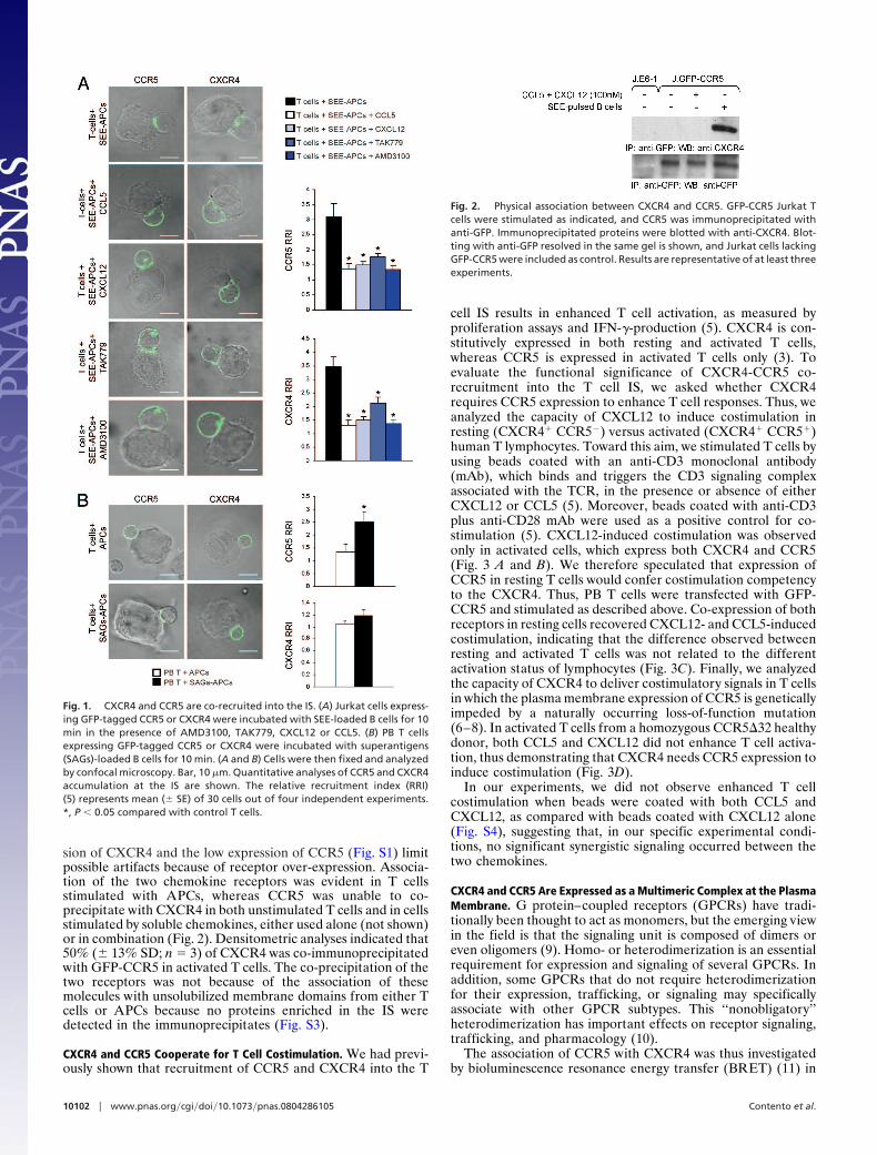
sion of CXCR4 and the low expression of CCR5 (Fig. S1) limitpossible artifacts because of receptor over-expression. Associa-tion of the two chemokine receptors was evident in T cellsstimulated with APCs, whereas CCR5 was unable to co-precipitate with CXCR4 in both unstimulated T cells and in cellsstimulated by soluble chemokines, either used alone (not shown)or in combination (Fig. 2). Densitometric analyses indicated that50% (� 13% SD; n � 3) of CXCR4 was co-immunoprecipitatedwith GFP-CCR5 in activated T cells. The co-precipitation of thetwo receptors was not because of the association of thesemolecules with unsolubilized membrane domains from either Tcells or APCs because no proteins enriched in the IS weredetected in the immunoprecipitates (Fig. S3).
CXCR4 and CCR5 Cooperate for T Cell Costimulation. We had previ-ously shown that recruitment of CCR5 and CXCR4 into the T
cell IS results in enhanced T cell activation, as measured byproliferation assays and IFN-�-production (5). CXCR4 is con-stitutively expressed in both resting and activated T cells,whereas CCR5 is expressed in activated T cells only (3). Toevaluate the functional significance of CXCR4-CCR5 co-recruitment into the T cell IS, we asked whether CXCR4requires CCR5 expression to enhance T cell responses. Thus, weanalyzed the capacity of CXCL12 to induce costimulation inresting (CXCR4� CCR5�) versus activated (CXCR4� CCR5�)human T lymphocytes. Toward this aim, we stimulated T cells byusing beads coated with an anti-CD3 monoclonal antibody(mAb), which binds and triggers the CD3 signaling complexassociated with the TCR, in the presence or absence of eitherCXCL12 or CCL5 (5). Moreover, beads coated with anti-CD3plus anti-CD28 mAb were used as a positive control for co-stimulation (5). CXCL12-induced costimulation was observedonly in activated cells, which express both CXCR4 and CCR5(Fig. 3 A and B). We therefore speculated that expression ofCCR5 in resting T cells would confer costimulation competencyto the CXCR4. Thus, PB T cells were transfected with GFP-CCR5 and stimulated as described above. Co-expression of bothreceptors in resting cells recovered CXCL12- and CCL5-inducedcostimulation, indicating that the difference observed betweenresting and activated T cells was not related to the differentactivation status of lymphocytes (Fig. 3C). Finally, we analyzedthe capacity of CXCR4 to deliver costimulatory signals in T cellsin which the plasma membrane expression of CCR5 is geneticallyimpeded by a naturally occurring loss-of-function mutation(6–8). In activated T cells from a homozygous CCR5�32 healthydonor, both CCL5 and CXCL12 did not enhance T cell activa-tion, thus demonstrating that CXCR4 needs CCR5 expression toinduce costimulation (Fig. 3D).
In our experiments, we did not observe enhanced T cellcostimulation when beads were coated with both CCL5 andCXCL12, as compared with beads coated with CXCL12 alone(Fig. S4), suggesting that, in our specific experimental condi-tions, no significant synergistic signaling occurred between thetwo chemokines.
CXCR4 and CCR5 Are Expressed as a Multimeric Complex at the PlasmaMembrane. G protein–coupled receptors (GPCRs) have tradi-tionally been thought to act as monomers, but the emerging viewin the field is that the signaling unit is composed of dimers oreven oligomers (9). Homo- or heterodimerization is an essentialrequirement for expression and signaling of several GPCRs. Inaddition, some GPCRs that do not require heterodimerizationfor their expression, trafficking, or signaling may specificallyassociate with other GPCR subtypes. This ‘‘nonobligatory’’heterodimerization has important effects on receptor signaling,trafficking, and pharmacology (10).
The association of CCR5 with CXCR4 was thus investigatedby bioluminescence resonance energy transfer (BRET) (11) in
Fig. 1. CXCR4 and CCR5 are co-recruited into the IS. (A) Jurkat cells express-ing GFP-tagged CCR5 or CXCR4 were incubated with SEE-loaded B cells for 10min in the presence of AMD3100, TAK779, CXCL12 or CCL5. (B) PB T cellsexpressing GFP-tagged CCR5 or CXCR4 were incubated with superantigens(SAGs)-loaded B cells for 10 min. (A and B) Cells were then fixed and analyzedby confocal microscopy. Bar, 10 �m. Quantitative analyses of CCR5 and CXCR4accumulation at the IS are shown. The relative recruitment index (RRI)(5) represents mean (� SE) of 30 cells out of four independent experiments.*, P � 0.05 compared with control T cells.
Fig. 2. Physical association between CXCR4 and CCR5. GFP-CCR5 Jurkat Tcells were stimulated as indicated, and CCR5 was immunoprecipitated withanti-GFP. Immunoprecipitated proteins were blotted with anti-CXCR4. Blot-ting with anti-GFP resolved in the same gel is shown, and Jurkat cells lackingGFP-CCR5 were included as control. Results are representative of at least threeexperiments.
10102 � www.pnas.org�cgi�doi�10.1073�pnas.0804286105 Contento et al.

intact Jurkat cells expressing receptors fused to either Renillaluciferase (Rluc), the BRET donor, or the yellow variant ofEGFP (YFP), the BRET acceptor (Fig. 4A). In saturationexperiments, conducted with CCR5-Rluc as the BRET donor,increasing concentration of both CCR5-YFP and CXCR4-YFPresulted in hyperbolic curves, characterized by similar BRET50values (0.71 � 0.18 and 0.39 � 0.11). These data are consistentwith a similar propensity of CCR5 to constitutively self-associateor interact with CXCR4 in Jurkat cells. Similar results were alsoobserved in HEK-293 cells (not shown). Control experimentswere conducted by using the angiotensin receptor AT1AR fusedto YFP, as the BRET acceptor. Weaker BRET signals wereobserved with a much higher BRET50 value (6.45 � 1.04),indicative of a significantly lower propensity of CCR5 to asso-ciate with this receptor. BRET experiments between CCR5-Rluc and CCR5-YFP or CCR5-Rluc and CXCR4-YFP, con-ducted in parallel in Jurkat cells preincubated withStaphylococcal Enterotoxin E (SEE)-pulsed or control APCs,yielded similar BRET values (Fig. 4B), indicating that theconstitutive proximity of BRET partners was not enhancedfurther during their translocation to the IS.
We speculated that if CXCR4 and CCR5 formed a signalingcomplex in T cells, both CXCR4 and CCR5 expression would bedown-regulated in T cells stimulated with either CCL5 orCXCL12. CCR5�CXCR4� Jurkat T cells were stimulated with
CCL5 or CXCL12 for 90 min, and plasma membrane expressionof CCR5 (Fig. 5A) and CXCR4 (Fig. 5B) was analyzed atdifferent time points. Both stimuli induced down-modulation ofCCR5 and CXCR4 expression at the plasma membrane. Asexpected, CXCL12 did not induce down-regulation of CCR5 ina CXCR4� cell line (Fig. S5) used as control (Fig. 5C).
DiscussionOur results are compatible with both a heterodimerization model(i.e., a dimer comprising one protomer each of CCR5 and CXCR4)or a hetero-oligomerization model (i.e., a dimer of CCR5 associatedwith a dimer of CXCR4) for the association of CXCR4 and CCR5.A formal distinction between the two hypotheses goes beyond theaim of our study and is very complicated because of severaltechnical issues. For example, although computational models haveidentified residues involved in receptor heterodimerization, thesame residues seem to be required for homodimerization of thereceptors, thus excluding the possibility of using specific mutants toaddress this question. Nevertheless, some of our data fit better withthe hetero-oligomerization model. Although previous studies failedto detect CXCR4-CCR5 heterodimers (12, 13), BRET saturationexperiments, which allow a clear discrimination between specificand bystander BRET signals (14), indicated the same apparentpropensity (15) of CCR5-YFP and CXCR4-YFP to associate withCCR5-Rluc in HEK-293 and Jurkat T cells, a result that is com-
Fig. 3. CXCL12-induced costimulation requires CCR5 expression. IFN-� production in resting (A), activated (B), CCR5-transfected resting (C), or CCR5�32activated (D) human PB T cells stimulated with beads coated with anti-CD3 mAb in the presence or absence of anti-CD28 mAb, CXCL12, or CCL5. For each celltype, the expression of CCR5, as analyzed by flow cytometry, is indicated (the empty histograms represent the isotype control). Results are representative of atleast three experiments. Bars with different letters are significantly different from each other (Student-Newman-Keuls test, P � 0.05). Mean amounts of IFN-�produced by single living cells upon anti-CD3 stimulation were: 1.2 fg/ml (A), 18 fg/ml (B), 1.1 fg/ml (C) and 15 fg/ml (D). The percentage of living cells after 48 hof stimulation was: �100% in A and B, 10% in C, and 20% in D. FI � Fold of induction over unstimulated cells.
Fig. 4. Constitutive association between CXCR4 and CCR5. (A) BRET saturation curves obtained by measuring BRET in Jurkat T cells expressing fixed quantitiesof BRET donor (CCR5-Rluc) and increasing amounts of BRET acceptors (indicated C-terminally YFP-tagged GPCR constructs). Relative amounts of BRET acceptorare expressed as the ratio between the fluorescence of the acceptor over the luciferase activity of the donor. YFP° corresponds to background fluorescence incells expressing the BRET donor alone. BRET-ratio values were from 18 individual transfections grouped as a function of the amount of BRET acceptor. (B) Thetransfer of energy between CCR5-Rluc and CXCR4-YFP was initiated by the addition of coelenterazine h, and the BRET ratio was monitored in real time in liveJurkat cells incubated with SEE-pulsed APCs (closed squares) or unpulsed APCs (closed triangles). The data shown represent the mean � SD. of triplicates in anexperiment representative of two independent experiments at constant (YFP-YFP°)/(Rluc-RLuc°) values (between 0.5 and 2).
Contento et al. PNAS � July 22, 2008 � vol. 105 � no. 29 � 10103
IMM
UN
OLO
GY

patible with both models. However, in a previous study, we failedto displace the BRET signal between CCR5-Rluc and CCR5-YFPwith unlabeled CXCR4, whereas unlabeled CCR5 did inhibit theassociation of two CCR5 protomers in a concentration-dependentmanner (13). Interestingly, a previous study proposed a role forCD4 in promoting CXCR4-CCR5 interactions (16). Although wedetected CXCR4-CCR5 complexes in CD4� HEK cells by usingBRET, we cannot exclude the possibility that CD4 may modulatethe interaction between the two receptors in lymphocytes. Furtherstudies using CD8� T cells will address this question.
Association between the two chemokine receptors was alsoconfirmed by the co-modulation experiment, showing thatCXCR4-specific stimuli induced down-modulation of CCR5expression, and vice versa. When stimulated by their specificagonists, CXCR4 and CCR5 showed different kinetics of down-modulation, likely explained by different fates of the two che-mokine receptors upon internalization, and suggesting thatoligomeric complexes formed between the two receptors may besensitive to endosomal pH. Indeed, upon stimulation CXCR4 isthought to be sorted to a degradative pathway (17), whereasCCR5 recycles at the plasma membrane (18). Interestingly, whenthe CCR5-CXCR4 heterodimer was internalized by a CCR5ligand, CXCR4 showed kinetics of down-modulation similar tothat of CCR5 upon its internalization, suggesting that whenpassively internalized by binding to CCR5, CXCR4 is nottargeted to a degradative pathway.
The biological significance of the CXCR4-CCR5 interactionis provided by the functional data showing that CXCR4requires CCR5 for its recruitment into the IS and costimula-tory functions. These data indicate that a putative multimericcomplex formed by the two receptors has distinctive signalingand biological properties, which argues against the possibilityof mere aggregation of CXCR4 and CCR5 homodimers in the
same membrane domains. The observation that BRET signalsremained unchanged upon receptor translocation to the IS,whereas the association of CCR5 with CXCR4 appearedmarkedly enhanced in co-immunoprecipitation experimentsconducted in Jurkat cells incubated with SEE-pulsed APCs,suggests that the two chemokine receptors are already in closeproximity in resting cells and that, once in the IS, the complexis further stabilized by additional interactions. Thus, the highlysensitive BRET technique can detect CXCR4-CCR5 com-plexes even in resting cells, whereas the less sensitive biochem-ical approach (19) can only detect the chemokine receptorcomplexes stabilized at the IS.
It was recently suggested that soluble CXCL12 inducesCXCR4 association with the TCR and that CXCL12 enhancesexpression of IL-2 and IL-10 in T cells stimulated with immo-bilized monoclonal anti-CD3 (20). In our experiments, CXCR4was not recruited into the IS of CCR5� resting T cells withCXCL12-secreting APCs (Fig. 1B), suggesting that, in a physi-ological context of antigen and chemokine presentation, CXCR4does not substantially associate with the TCR. Moreover, wecould not detect costimulation induced by CXCL12 in restingCCR5� T cells (Fig. 3A). In addition, we found that, whencompared to anti-CD3 stimulation, CXCL12 plus anti-CD3 mAbinduced higher T cell responses in terms of IFN-�, whereas IL-2production was not affected (Fig. S6). These different resultsmay be explained by the different stimulation protocol used.Indeed, plate-bound anti-CD3 mAb delivers a very strong signalto T cells, and thus soluble chemokines are probably workingdifferently than membrane-bound ones. Interestingly, it wasrecently demonstrated that dendritic cells bind chemokines attheir plasma membrane (21). The CXCR4-CCR5 in cis costimu-lation reported here may therefore be different from chemokinereceptor in trans costimulation, as described for CCR7 (21). Inagreement with these data, we have demonstrated that CCR7 isnot recruited into the IS formed between T cells and APCsproducing CCR7 ligands (5).
Clearly, the critical questions are when CXCR4-CCR5 coop-eration occurs and how CXCR4-CCR5 signaling influences Tcell responses in vivo. Although CXCR4 is constitutively ex-pressed in T cells, CCR5 expression is restricted to activated Tcells. It would be therefore possible to conclude that chemokine-mediated costimulation occurs in the periphery, likely in in-f lamed tissues, and does not influence T cell priming in lymphnodes. It was recently demonstrated, however, that inflammationleads to CCR5 expression by naıve CD8� T cells, permitting theirrecruitment to sites of CD4� T cell interaction with dendriticcells, where CCL3 and CCL4 are produced (22). This chemo-kine-driven cell clustering appears to be fundamental for devel-opment of proper long-term CD8� T cell memory, and thus it istempting to speculate that the expression of CCR5 is not onlyimportant for CD8 cell recruitment but also for costimulatorysignals required for efficient T cell priming. As an example, theexpression of CCR5 is crucial for control of infection by WestNile virus, a re-emerging pathogen capable of causing humanfatal encephalitis (23); hence, again we speculate that bothCCR5-mediated recruitment and the key role of CCR5 in T cellcostimulation (5) might explain the higher incidence of thisdisease in CCR5�32 individuals.
On one hand, CXCR4-CCR5 signaling may be beneficial forfighting pathogens, but on the other hand it may amplify T cellresponses in chronic inflammation. The CCR5�32 polymor-phism was found to be a genetic marker inversely related to theseverity of rheumatoid arthritis (24), and several lines of evi-dence indicate that both receptors play key roles in autoimmu-nity (3). Although chemokines and their receptors representideal therapeutic targets in autoimmunity, they are also impli-cated in homeostatic cell trafficking (3). Our data suggest thepossibility of uncoupling migration and in situ activation by
Fig. 5. Co-modulation of CXCR4 and CCR5. CCR5�CXCR4� Jurkat T cells (Aand B) or CCR5�CXCR4� A7 cells (C) were stimulated with CCL5 or CXCL12 for90 min, and plasma membrane expression of CCR5 (A and C) and CXCR4 (B) wasanalyzed at different time points. The graphs show the mean fluorescenceintensity (MFI) for treated cells as a proportion of the MFI for untreated cellsat the indicated times. Data points represent mean (� SE) of triplicates.
10104 � www.pnas.org�cgi�doi�10.1073�pnas.0804286105 Contento et al.

designing specific antagonists that would not impair immunesystem homeostasis but would inhibit activation of autoreactiveT cells.
CXCR4 and CCR5 are coreceptors for HIV entry in humancells. Almost all cases of HIV-1 transmission involve strains thatuse CCR5 for entry (R5 viruses); however, in up to 50% ofinfected people after five years, on average, viruses that are ableto use CXCR4 become predominant (R5X4 or X4 viruses). Ourstudy may explain recent ‘‘paradoxical findings’’, as defined bythe authors of the study, showing that CCR5 ligands protectneurons from HIV/gp120 and CXCL12 toxicity (25), becauseCCR5 ligands might indeed cross-compete with CXCR4 ligandsand prevent their neurotoxic effects (9).
In conclusion, we have demonstrated a previously undescribedand functional cooperation between CXCR4 and CCR5 tomodulate T lymphocyte responses. These results identify amolecular mechanism pivotal to chemokine-receptor signalingversatility, and a chemokine–receptor couple that may representa good target for pharmacological research.
Materials and MethodsCell Culture, Constructs, and Transfections. The Jurkat T cell line J.E6–1, JurkatE6–1 cells expressing GFP-CCR5 (5) and EBV-B 221 cell lines were cultured inRPMI medium 1640 (Gibco) supplemented with 10% FCS, 2 mM L-glutamine,100 units/ml penicillin, and 100 �g/ml streptomycin. The melanoma cell line A7expressing GFP-CCR5 (26) was cultured in D-MEM medium (Gibco) supple-mented with 10% FCS, 2 mM L-glutamine, 100 units/ml penicillin, 100 �g/mlstreptomycin, and 0.5 mg/ml G418. Human PB CD4� T cells were sorted bynegative selection by using RosetteSep kit (StemCell Technologies). Bloodfrom a CCR5�32 donor was kindly provided by Christophe Combadiere (Uni-versite Pierre et Marie Curie Paris 6, Hopital Pitie-Salpetriere, Paris, France).
The GFP-CCR5 and CXCR4-GFP (5) and the CCR5-Rluc, CCR5-YFP, CXCR4-YFPand AT1AR-YFP (13) constructs already have been described.
Human PB T cells were transiently transfected with GFP-CCR5 or withCXCR4-GFP by using an electroporation system (Amaxa Biosystems) accordingto manufacturer’s guidelines and were used for experiments 24 h later. Jurkatcells were transiently transfected with CXCR4-GFP by using a Bio-Rad electro-poration system as described (27).
Flow Cytometry. The expression of CCR5 and CXCR4 on Jurkat, A7 and PB T cellswas assessed by flow cytometry analysis (FACSCalibur or FACS Canto; BectonDickinson) by using the commercial anti-human CCR5 mAb (R&D, clone:45531; or R&D, clone: CTC5) and the commercial anti-CXCR4 mAb (BD PharM-ingen, clone: 12G5; or R&D, clone: 44717). Data were processed by usingCELLQUEST (Becton Dickinson).
Immunofluorescence Confocal Microscopy. For experiments with human PB Tcells, B cells were suspended at 107 per milliliter and incubated alone or with1 �g/ml of bacterial superantigens [Staphylococcal Enterotoxin A, Staphylo-coccal Enterotoxin B, Staphylococcal Enterotoxin E (SEE); Toxin Technology] at37°C for 2 h, mixing every 20 min. For experiments with Jurkat cells, B cellswere loaded with 1 �g/ml SEE.
In some experiments, Jurkat cells expressing GPF-CCR5 or CXCR4-GFP weretreated with 10 �g/ml AMD3100 (Sigma) at room temperature for 15 min or with5 �M TAK-779 (NIH AIDS Research and Reference Reagent Program) at 37°C for15 min or with 100 nM CXCL12 or CCL5 (Peprotech) at 37°C for 15 min. After theTreatment T Cells Were Incubated with Equal Numbers of B Cells (37°C, 15 min).Conjugates were fixed with 4% paraformaldehyde, adhered to microscope slidescoated with 0.05 mg/ml poly-L-lysine, washed and mounted in 2.5% 1,4-diazobicyclo[2.2.2]octane (DABCO, Fluka) in 90% glycerol/10% PBS. Confocalmicroscopy was performed with a Leica confocal microscope TCS SP5 (Leica) usinglaser excitation at 488 nm. Images were analyzed by using Adobe Photoshop 7.0and NIH-Image J programs. A minimum of 20 cells (or 20 conjugates) was exam-inedquantitatively foreachexperiment.Thefluorescencepatternsreported inallof the figures are representative of at least 90% of the cells.
Fluorescence Quantification. To quantify GFP-CCR5 or CXCR4-GFP recruitmentat the IS, boxes were drawn at the immune synapse, at the cell membrane notin contact with the APC, and at a background area outside the cell. Conjugatesin which the plasma membrane and Golgi fluorescence could not be clearlydistinguished were not included in the analysis. The relative recruitment indexwas calculated as indicated: [mean fluorescence intensity (MFI) at synapse �
background] / [MFI at regions not in contact with APC � background]. Quan-titative analysis of MFI was performed with the Image J program.
Immunoprecipitation and Western Blotting. Jurkat cells (107 cells) expressingGFP-CCR5 were stimulated (or not) for 15 min at 37°C with 107 SEE-pulsed Bcells. Cells were then lysed in 1% Brij 96 V (Fluka) buffer (20 mM Tris-HCl pH7.5, 150 mM NaCl, 1 mM MgCl2, 1 mM EGTA), containing 10 �g/ml aprotinin,10 �g/ml leupeptin, 20 mM NaF, 1 mM Pefabloc-SC, 1 mM Na3VO4 and 10 mMNa4P2O7. Postnuclear lysates were precleared for 30 min at 4°C with proteinG-Sepharose (Amersham Pharmacia Biotech Inc.) and then incubated for 2 hwith anti-GFP rabbit polyclonal antibodies (Clontech) preadsorbed to proteinG. Immunoprecipitates were washed twice in 1% Brij 96 V, twice in 0.05% Brij96 V lysis buffer, and boiled in SDS-PAGE sample buffer before electrophoresison 10% SDS-polyacrylamide gels. After protein transfer, nitrocellulose mem-branes were blotted with anti-CXCR4 antibody (28), anti-GFP polyclonal an-tibodies (Clontech), anti-B7.1 (R&D) and anti-LAT (Upstate biotechnology).
In some experiments GFP-CCR5-expressing Jurkat cells were serum-starvedfor 4 h and then stimulated for 15 min with soluble chemokines, and GFP-CCR5was immunoprecipitated as above. For control experiments, GFP was immu-noprecipitated as above from 107 Jurkat cells not expressing GFP-CCR5.
Densitometric analyses were performed on a Image Master VDS-CL densi-tometer by using volume analysis of Image MasterTM Total Lab software(Amersham Biosciences). All densitometric values obtained were calculatedfrom nonsaturated signals. The percentage of CXCR4 coimmunoprecipitatedwith GFP-CCR5 was calculated as: [volume of immunoprecipitated CXCR4band / volume of cell total lysate CXCR4 band] / [volume of immunoprecipi-tated GFP band / volume of cell total lysate GFP band] %.
T Cell Activation and ELISA. PB CD4� T cells were stimulated with 2 �g/mlphytohemagglutinin (PHA) (Sigma) in the presence of 400 units/ml IL-2(Chemicon) and feeders for 10 days. Beads (Polybead carboxylate 4.5-�mmicrospheres, Polysciences, Inc.) were coated with 1 �g/ml anti-CD3 (OKT3clone) alone or in combination with 10 nM recombinant human CCL5(PeproTech), or with 10 nM recombinant human CXCL12 (PeproTech) or 1�g/ml anti-CD28 (clone CD28.1, PharMingen).
Activated PB CD4� T cells (either from normal or from homozygousCCR5�32 donors) or resting PB CD4� T cells (either CCR5� or expressingGFP-CCR5) were plated with beads at a 1:2 ratio, in a 96-well, U-bottom cultureplate (FALCON). After 48 h, supernatants were collected and IFN-� concen-trations were measured by using standard commercially available ELISA kits(Pierce Endogen) according to the manufacturer’s instructions. Because trans-fected T cells have a high mortality and CCR5�32 T cells were frozen andshipped before the experiment, for each cell type we calculated the percent-age of living cells by flow cytometry before plating, and every 24 h by TrypanBlue exclusion.
Supernatants from three different experiments were also analyzed bySearchLight human TH1/TH2 cytokine array 1 (Pierce Biotechnology). For themultiplex arrays, in each separate experiment, duplicates of three dilutions ofeach sample were used.
BRET Saturation Assays. Jurkat cells (5 106) expressing the SV40 T-antigen(JTAg cells) were electroporated with 0.5 �g of the DNA construct encodingBRET donor (CCR5-Rluc) and increasing amounts (0.5–10 �g) of the BRETacceptor plasmid (CCR5-YFP, CXCR4-YFP or AT1AR-YFP). Electroporationwas performed in a Gene Pulser II electroporator at 950 �F and 250 V ina Gene-Pulse cuvette (Bio-Rad, Hercules, CA, USA). Total transfectedDNA was maintained constant by using appropriate amounts of pcDNA3(Invitrogen). At 24 h after transfection, the luciferase substrate, coelen-terazine (Molecular Probes), was added at a final concentration of 5 �M to1 105 cells. Luminescence and fluorescence were measured simulta-neously by using the MithrasTM fluorescence-luminescence detector(Berthold). Cells expressing BRET donors alone were used to determinebackground. Filter sets were 485 � 10 nm for luciferase emission and 530 �12.5 nm for YFP emission. BRET ratios were calculated as described (29).
CXCR4-CCR5 Co-Modulation. Experiments were performed as described (30). Inbrief, Jurkat cells or A7 cells (2 107) stably expressing GFP-CCR5 by retroviralexpression were incubated in basal medium containing 25 nM CXCL12 (Pep-rotech) or 50 nM CCL5 (Peprotech) at 37°C. Cells were analyzed by flowcytometry after staining with anti-CXCR4 (BD PharMingen, clone: 12G5) oranti-CCR5 (R&D, clone: CTC5) mAb.
Chemotaxis Assay. Jurkat cells stably expressing GFP-CCR5 and endogenousCXCR4 were treated with 10 �M AMD3100 (Sigma) or 100 nM TAK779 (NIHAIDS Research and Reference Reagent Program) at 37°C for 30 min. Cells were
Contento et al. PNAS � July 22, 2008 � vol. 105 � no. 29 � 10105
IMM
UN
OLO
GY

then seeded in the upper chamber of a Transwell plate (CORNING) in serum-free medium. The lower chambers were filled with serum-free medium aloneor serum-free medium containing CXCL12 (25 nM) or CCL5 (25 nM). After 2 hat 37°C, the number of T cells that migrated into the lower chamber wasestimated by flow cytometry on a FACSCalibur.
Statistical Analysis. All data are representative of at least three differentexperiments. Values are expressed as mean � SE or SD. Statistical analysis wasperformed by using Student’s t test (Microsoft Office) or, where indicated, byANOVA followed by the nonparametric Student-Newman-Keuls test for mul-tiple comparisons.
ACKNOWLEDGMENTS. We thank Anna Cabrelle, Denis Bison, Sonia Jimenez-Baranda, Felix Ortego and Achille Anselmo for technical help. We are gratefulto Christophe Combadiere for providing CCR5�32 peripheral blood cells andto Sergio Pantano, Marc Parmentier, Frances Lund, Mario Mellado and GiorgioTrinchieri for critical discussions. This work was supported by grants from theItalian Association for Cancer Research (AIRC), the MIUR-PRIN, Telethon,Alleanza contro il cancro to A.V., the Sidaction and Fondation de France to S.Marullo, and the European Community (INNOCHEM, LSHB-CT-2005–518167)and MEC (SAF2005–00241) to S. Manes. The following reagent was obtainedthrough the NIH AIDS Research and Reference Reagent Program, Division ofAIDS, NIAID, NIH: TAK-779, (cat. No. 4983).
1. Lanzavecchia A, Lezzi G, Viola A (1999) From TCR engagement to T cell activation: Akinetic view of T cell behavior. Cell 96:1–4.
2. Gerard C, Rollins BJ (2001) Chemokines and disease. Nat Immunol 2:108–115.3. Viola A, Luster AD (2008) Chemokines and Their Receptors: Drug Targets in Immunity
and Inflammation. Annu Rev Pharmacol Toxicol 48:171–197.4. Viola A, Contento RL, Molon B (2006) T cells and their partners: The chemokine dating
agency. Trends Immunol 27:421–427.5. Molon B, Gri G, Bettella M, Gomez-Mouton C, Lanzavecchia A, et al. (2005) T cell
costimulation by chemokine receptors. Nat Immunol 6:465–471.6. Benkirane M, Jin DY, Chun RF, Koup RA, Jeang KT (1997) Mechanism of transdominant
inhibition of CCR5-mediated HIV-1 infection by ccr5delta32. J Biol Chem 272:30603–30606.
7. Mellado M, Rodriguez-Frade JM, Vila-Coro AJ, Fernandez S, Martin de Ana A, et al.(2001) Chemokine receptor homo- or heterodimerization activates distinct signalingpathways. EMBO J 20:2497–2507.
8. Liu R, Paxton WA, Choe S, Ceradini D, Martin SR, et al. (1996) Homozygous defect inHIV-1 coreceptor accounts for resistance of some multiply-exposed individuals to HIV-1infection. Cell 86:367–377.
9. Springael JY, Urizar E, Parmentier M (2005) Dimerization of chemokine receptors andits functional consequences. Cytokine Growth Factor Rev 16:611–623.
10. Bulenger S, Marullo S, Bouvier M (2005) Emerging role of homo- and heterodimeriza-tion in G-protein-coupled receptor biosynthesis and maturation. Trends Pharmacol Sci26:131–137.
11. Boute N, Jockers R, Issad T (2002) The use of resonance energy transfer in high-throughput screening: BRET versus FRET. Trends Pharmacol Sci 23:351–354.
12. Babcock GJ, Farzan M, Sodroski J (2003) Ligand-independent dimerization of CXCR4,a principal HIV-1 coreceptor. J Biol Chem 278:3378–3385.
13. Issafras H, Angers S, Bulenger S, Blanpain C, Parmentier M, et al. (2002) Constitutiveagonist-independent CCR5 oligomerization and antibody-mediated clustering occur-ring at physiological levels of receptors. J Biol Chem 277:34666–34673.
14. Marullo S, Bouvier M (2007) Resonance energy transfer approaches in molecularpharmacology and beyond. Trends Pharmacol Sci 28:362–365.
15. Issad T, Jockers R (2006) Bioluminescence resonance energy transfer to monitor pro-tein-protein interactions. Methods Mol Biol 332:195–209.
16. Wang J, Alvarez R, Roderiquez G, Guan E, Norcross MA (2004) Constitutive associationof cell surface CCR5 and CXCR4 in the presence of CD4. J Cell Biochem 93:753–760.
17. Marchese A, Benovic JL (2001) Agonist-promoted ubiquitination of the G protein-coupled receptor CXCR4 mediates lysosomal sorting. J Biol Chem 276:45509–45512.
18. Signoret N, Pelchen-Matthews A, Mack M, Proudfoot AE, Marsh M (2000) Endocytosisand recycling of the HIV coreceptor CCR5. J Cell Biol 151:1281–1294.
19. Hernanz-Falcon P, Rodriguez-Frade JM, Serrano A, Juan D, del Sol A, et al. (2004)Identification of amino acid residues crucial for chemokine receptor dimerization. NatImmunol 5:216–223.
20. Kumar A, Humphreys TD, Kremer KN, Bramati PS, Bradfield L, et al. (2006) CXCR4physically associates with the T cell receptor to signal in T cells. Immunity 25:213–224.
21. Friedman RS, Jacobelli J, Krummel MF (2006) Surface-bound chemokines capture andprime T cells for synapse formation. Nat Immunol 7:1101–1108.
22. Castellino F, Huang AY, Altan-Bonnet G, Stoll S, Scheinecker C, et al. (2006) Chemokinesenhance immunity by guiding naive CD8� T cells to sites of CD4� T cell-dendritic cellinteraction. Nature 440:890–895.
23. Glass WG, Lim JK, Cholera R, Pletnev AG, Gao JL, et al. (2005) Chemokine receptor CCR5promotes leukocyte trafficking to the brain and survival in West Nile virus infection. JExp Med 202:1087–1098.
24. Zapico I, Coto E, Rodriguez A, Alvarez C, Torre JC, et al. (2000) CCR5 (chemokinereceptor-5) DNA-polymorphism influences the severity of rheumatoid arthritis. GenesImmun 1:288–289.
25. Kaul M, Ma Q, Medders KE, Desai MK, Lipton SA (2007) HIV-1 coreceptors CCR5 andCXCR4 both mediate neuronal cell death but CCR5 paradoxically can also contribute toprotection. Cell Death Differ 14:296–305.
26. Jimenez-Baranda S, Gomez-Mouton C, Rojas A, Martinez-Prats L, Mira E, et al. (2007)Filamin-A regulates actin-dependent clustering of HIV receptors. Nat Cell Biol 9:838–846.
27. Gri G, Molon B, Manes S, Pozzan T, Viola A (2004) The inner side of T cell lipid rafts.Immunol Lett 94:247–252.
28. Vila-Coro AJ, Rodriguez-Frade JM, Martin De Ana A, Moreno-Ortiz MC, Martinez AC,et al. (1999) The chemokine SDF-1alpha triggers CXCR4 receptor dimerization andactivates the JAK/STAT pathway. Faseb J 13:1699–1710.
29. Storez H, Scott MG, Issafras H, Burtey A, Benmerah A, et al. (2005) Homo- andhetero-oligomerization of beta-arrestins in living cells. J Biol Chem 280:40210–40215.
30. Signoret N, Rosenkilde MM, Klasse PJ, Schwartz TW, Malim MH, et al. (1998) Differ-ential regulation of CXCR4 and CCR5 endocytosis. J Cell Sci 111 (Pt 18):2819–2830.
10106 � www.pnas.org�cgi�doi�10.1073�pnas.0804286105 Contento et al.
![Journal of Falkenhagen et al, J Antivir Antiretrovir 213 ... · CCR5 gene via Zinc finger nucleases [4], cleavage of CCR5 mRNA by multimeric ribozymes [5], inhibition of CCR5 mRNA](https://img.pdfslide.net/doc/110x75/5fd3f8f670db7b30b42beea9/journal-of-falkenhagen-et-al-j-antivir-antiretrovir-213-ccr5-gene-via-zinc.jpg)