Embed Size (px)
Citation preview
Ceylon Journal of Science (Bio. Sci.) 40 (2): 71-88, 2011
Cyanobacteria: Pioneers of Planet Earth
S. A. Kulasooriya
Emeritus Professor of Botany, University of Peradeniya, Peradeniya, Sri Lanka and Visiting Professor
Institute of Fundamental Studies, Kandy, Sri Lanka.
ABSTRACT
Cyanobacteria are among the earliest of inhabitants of Planet Earth and their existence can be traced back
to 3.8 billion years. Their oxygenic photosynthesis led to the gradual conversion of the Earth‟s atmosphere
from an anaerobic to an aerobic one. This change enabled the advent of aerobic organisms that eventually
underwent rapid evolution and became the dominant, highly diverse members of the current global
biodiversity. Cyanobacteria are ubiquitous in their distribution and are found in all the latitudes from
Arctic and Anatarctic regions to the Tropical deserts perhaps reflecting their pioneering habitation of the
primitive earth. They are also unique in their ability to simultaneously perform oxygenic photosynthesis
and oxygen labile nitrogen fixation. Through these processes they make significant contributions to the
Carbon and Nitrogen bio-geochemical cycles, particularly in the deep oceans. The ability of these
organisms to fix N2 either independently or in symbiosis with other organisms not only contributes to
natural ecosystems but is applied in certain countries particularly for rice cultivation. Their ability to grow
in highly polluted environments is also used in the treatment of sewage and industrial effluents.
Cyanobacteria are the most efficient among all living organisms in the harvesting of solar energy and are
currently looked at as highly attractive candidates for biofuel production. A few species are being used for
the production of highly nutritive food supplements. On the negative side, some cyanobacteria form
massive growths called „blooms‟ in water bodies and many of them produce toxins harmful to fish,
digastric animals and are suspected to be responsible for certain human ailments. Having reviewed most of
these aspects of cyanobacteria, it is concluded that knowledge on these little known organisms would be
invaluable not only for students, scientists and environmentalists but also for industrialists and policy
makers.
Key words: cyanobacteria, blue-green algae, algal toxins, biofertilizers, biofuels
INTRODUCTION
Cyanobacteria, also known as blue-green algae
include a highly diverse group of prokaryotic
microorganisms exhibiting oxygenic
photosynthesis. Oxygen released by this process
gradually changed the original reducing
atmosphere of the primitive earth to an oxidizing
one (Olsen, 2006) triggering off a dramatic
evolution of global biodiversity. The
chloroplasts of eukaryotic algae and higher
plants have originated from endosymbiotic
relationships with cyanobacteria (Martin and
Kowallik, 1999 and Raven and Allen, 2003) and
this event in the early evolution of life has
stimulated the advent of oxygen tolerant flora
and fauna capable of aerobic respiration, a
highly efficient mechanism of energy utilization.
The rapid development of such organisms
resulted in the predominance of oxygenic and
aerobic species diversity on earth.
Cyanobacteria are genetically highly diverse;
they occupy a broad range of habitats across all
latitudes perhaps demonstrating the abilities of
their pioneering ancestors as the earliest
inhabitants of Earth. They are not only
widespread in freshwater, marine and terrestrial
ecosystems but also found in extreme habitats
such as hot springs, hypersaline localities,
freezing environments and arid deserts (Fogg et
al., 1973). They often live in association with
other organisms forming microbial mats,
biofilms and benthic communities and such
associations are the predominant and sometimes
the only life forms found in certain extreme
habitats. The fossilized stromatolites believed to
have been formed some 3.4 billion years ago
(bya) are also microbial communities with
cyanobacteria as their autotrophic partners.
Diverse species having different ecological
demands exhibit differential adaptations to the
conditions at their source locality. The massive
communities of Prochlorococcus in the
oligotrophic deep oceans that live in close
association with cyanophages enabling rapid
lateral transfer of genomic material opens up __________________________________________ Author‟s email: [email protected]
S. A. Kulasooriya 72
new vistas on biological adaptation and
evolution (Johnson et al., 2006). The endolithic
Chroococcidiopsis living as the only inhabitant
in the extremely inhospitable habitat of the arid
Atacama Desert provides a new tool for
Astrobiology (Friedmann, 1982 and Wierzchos
et al., 2006).
Sequencing of 16S rRNA while confirming
the existence of several morphologically uniform
and well-defined traditional genera, has also
placed similar morphotypes in distant positions
in phylogenetic trees. Molecular data therefore
provide basic criteria for cyanobacterial
taxonomy, but to construct a comprehensive
phylogenetic system of cyanobacteria a
combination with knowledge on their
morphology, physiology and biochemistry is
essential (Komarek, 2006).
The ability of certain cyanobacteria to fix
atmospheric nitrogen make them unique in their
ability to independently secure their carbon and
nitrogen requirements (Kulasooriya, 2008).
Some of these organisms and symbiotic systems
like Azolla are used as biofertilizers, particularly
in rice production (Venkataraman, 1972 and
Azolla Utilization, 1987). They are also applied
in oxidation ponds and sewage and sludge
treatment plants (Lincoln et al., 1996). Recently
a few species have been investigated for biofuel
production because their ability to convert solar
energy has been found to be the most efficient
among all living organisms. Furthermore, their
simple genomic structure has enabled the
production of biofuel secreting strains through
genetic engineering (Lane, 2010). Certain
cyanobacteria form blooms in eutrophied water
bodies and most of such bloom forming strains
produce cyanotoxins that are harmful and
sometimes lethal to animals and humans
(Carmichael, 1994 and Carmichael et al., 2001).
A few species are utilized for the production of
highly nutritious food supplements
(Kulshreshtha et al., 2008)
This paper reviews the current information
on these aspects of cyanobacteria, the roles they
may have played in the origin and early
evolution of life on earth and their current
impacts on global biodiversity.
Role in the oxygenation of the primitive
earth’s atmosphere
The Planet Earth is believed to have cooled
down, solidified and formed oceans and
terrestrial habitats some 4.8 to 4.5 (bya). During
the next billion years chemical or abiotic
evolutionary processes in the primordial soup of
the oceans are believed to have given rise to
primitive entities capable of self replication.
Various scientists have reported on these
processes which have resulted in the formation
of amino acids (monomers and proteinoid
polymers) that integrated to form aggregates
such as protobionts and microspheres with semi-
permeable membranes. Ribose-nucleic acid
(RNA) is believed to have been the original
hereditary molecule that grew, split and was
passed onto the progeny. These have given rise
to the earliest life forms on earth which are
believed to be prokaryotic organisms, today
represented by the Archea, Eubacteria and the
Cyanobacteria. Citing evidence from the Isua
super-crustal belt in Western Greenland and
similar formations in Akilia Island, Mojzsis et
al. (1996) proposed life to have existed 3.8 bya.
They have also estimated the time that would be
taken for a 100 Kbp genome of a primitive
heterotroph to develop onto a 7500 Kbp gene of
a filamentous cyanobacterium to be 7 million
years.
It is most likely that the original cells were
heterotrophic utilizing plenty of organic
molecules available in the primordial soup
together with those produced by other cells. As
these food supplies gradually became limiting
some of the cells would have developed
strategies to use the readily available solar
energy by anaerobic photosynthesis. These
archaic processes are still retained by a few
pigmented bacterial species like Chlorobium and
Rhodospirillum which utilizes energy from the
sun to reduce CO2 and form organic compounds
deriving electrons from substrates such as H2S
and FeS exhibiting non-oxygenic
photosynthesis.
In a comprehensive mini review Olson
(2006) has presented possible pathways on the
early evolution of photosynthesis primarily
based upon fossil evidence during the Archeon
Era. According to him the earliest reductant for
CO2 fixation (even by primitive cyanobacteria)
would have been H2 some 3.8 bya. Fossilized
filamentous mats found in the Buck Reef Cherts
of South Africa and Evaporites, Stromatolites
and micro-fossils observed in the Warrawoona
Megasequence in Australia have been attributed
to the H2S driven photosynthesis between 3.5 to
3.4 bya and subsequently Proteobacteria and
Protocyanobacteria have utilized Fe2+
ions as
reductants around 3.0 bya. Evidence of retention
of such archaic processes can be observed in
certain present day species of cyanobacteria
which exhibit light driven CO2 fixation through
cyclic photophosphorylation under anaerobic
conditions using electron donors such as H2S,
thiosulphate, or even molecular H2. Citing
evidence from microfossils, stromatolites and
Cyanobacteria 73
chemical biomarkers Olson (2006) suggested
that cyanobacteria containing chlorophyll-a
capable of O2 evolving photosynthesis appeared
around 2.8 bya, but this process did not have a
significant impact on the composition of the
atmospheric gases for another 5 million years.
The O2 released to the atmosphere became
bound to limestone, iron and other minerals as
evidenced from the iron-oxide rich geologic
strata observed from those periods. It is also
believed that such oxidative reactions of
minerals contributed to the „greening of oceans‟.
Once most of the minerals got oxidized free O2
gradually began to accumulate in the
atmosphere. Intense solar rays bombarding the
earth converted some of the O2 to O3 which
collected in the upper part of the atmosphere to
build up the ozone layer that absorbs the
mutagenic UV rays and protects all life forms
from unsustainable levels of mutation. Such a
build up of a protective O3 layer would also have
enabled the evolution and migration of life forms
from the oceans to land.
Towards the end of the Archaeozoic era and
beginning of the Proterozoic era the earth began
to cool and this led to the reduction of the major
greenhouse gases of the atmosphere e.g. water
vapour, CO2 and methane. This cooling also
reduced the hygrometric capacity of air and
water vapour condensed resulting in continuous
torrential rain. These changes of the physical
environment led to the proliferation of
cyanobacteria in the marine phytoplankton
which fixed and stored part of the carbon in the
sea and contributed to the reduction of CO2 in
the atmosphere. Release of O2 to the atmosphere
due to oxygenic photosynthesis by cyanobacteria
would also have contributed to the reduction of
atmospheric methane through oxidation.
Meanwhile the chloroplasts that are today so
common in eukaryotic algae and green plants
evolved through processes of endosymbioses
between cyanobacterial and heterotrophic
eukaryotic ancestors. This endosymbiotic
hypothesis was originally put forward by
Mereschkowsky (1905). Though originally taken
with skepticism subsequent electron
microscopic, biochemical and molecular
biological studies gave credence to this
hypothesis and later Martin & Kowallik (1999)
translated this paper into English and provided
evidence in support of it by molecular analysis
of the chloroplast genome of Arabidopsis
thaliana and comparing it with those of Nostoc
punctiforme, Prochlorococcus marinus and
Synechocystis sp. PCC 6803 (Martin et al.,
2002). Raven and Allen (2003) confirmed these
evidences and stated that the complete genome
sequences of cyanobacteria and of the higher
plant Arabidopsis thaliana leave no doubt that
the green plant chloroplast originated through
endosymbiosis with a cyanobacterium.
The oxygenic photosynthesis of
cyanobacteria and chloroplast containing
eukaryotes would have accelerated the
oxygenation of the atmosphere and this would
have had significant impacts on the existing life
forms of that time. Oxygen would have been
toxic to most of the primitive organisms which
were predominantly anaerobic and a large
number of them would have died in what was
called the „oxygen catastrophe‟. As this process
of oxygenation had been very slow and gradual
through several millions of years, it enabled the
evolution of resistant forms some of which even
developed mechanisms to utilize O2 to secure
energy from food through aerobic respiration.
This metabolic process was far more efficient
than the anaerobic processes of fermentation and
the aerobes on earth developed at a dramatic
pace driving the anaerobes to near extinction.
Today aerobes are the predominant living forms
among both flora and fauna thriving in an
atmosphere containing 21% of oxygen.
Distribution and associations with other
organisms
Cyanobacteria are ubiquitous in their global
distribution. They occupy a broad range of
habitats across all latitudes, widespread in
freshwater, marine and terrestrial ecosystems,
also found in extreme habitats such as hot
springs, hypersaline localities, freezing
environments and arid deserts. Frequently they
are the pioneer invaders (often as lichens), of
exposed habitats such as bare rocks, recently
piled up soil or bare land after natural disasters,
exposed walls and partly constructed buildings
and similar substrates which offer little or no
nutrients. They are also common inhabitants of
polluted water bodies, drains and garbage dumps
which are generally inhospitable to most other
organisms. The versatile abilities of these
autotrophic prokaryotes are perhaps a reflection
of their pioneering ancestry which at that time
dominated the inhospitable primitive earth.
Initial colonization of habitats by cyanobacteria
if left undisturbed has the potential to develop
through progressive autotrophic successions to
reach even the final stage of a climax forest
under favorable environmental conditions. Such
important roles played by these simple
prokaryotes justify referring to them as Pioneers
of Planet Earth.
Among the planktonic cyanobacteria, the
ocean dwelling Prochlorococcus marinus
S. A. Kulasooriya 74
occupies a unique position. Reported as the
tiniest photosynthetic organisms and the most
populous autotrophic prokaryotes in the oceans,
they are found among the phytoplankton of the
oligotrophic deep ocean habitats in dense
populations sometimes around 105 cells per ml
of water. The vast extents of these populations in
the oligotrophic deep oceans make a significant
contribution to primary production and C-
sequestration that ameliorates global warming.
Research studies led by Professor Sally W.
Chisholm of the Massachusetts Institute of
Technology, Boston, USA, have shown the
important role played by the massive
populations of these minute organisms in carbon
sequestration in the vast expanses of the deep
open ocean ecosystems. When we realize that
over 70% of the earth‟s surface is covered by
oceans it is easy to comprehend the significance
of the contribution made by them to global C-
sequestration. Another astounding finding of the
Massachusetts research team is the close
association Prochlorococcus cells are having
with numerous viruses that facilitate rapid lateral
gene transfers among them. With these new
findings they were able to get a clear picture of
gene diversity and gene flow among these
organisms and postulate that such lateral gene
transfers endow upon them an intrinsic ability to
rapidly adapt to environmental variables such as
temperature, predators, light and nutrient
changes which these organisms constantly
encounter in the deep ocean ecosystems. The
team‟s studies have shown that closely related
Prochlorococcus strains display an array of
physiological differences regulated by the
genetic diversity of the constituent cells that
provide an extraordinary stability to the overall
community which internally adjusts itself to ever
changing environmental conditions. These novel
observations perhaps warrant the introduction of
new terminology to such ecosystems. What is
seen here are homogenous populations of
Prochlorococcus marinus that are
“heterogenomic”. This dynamic hetero-
genomicity due to the continuous lateral gene
transfer through closely associated viral vectors
enable the homogenous cyanobacterial
community to sustain itself in the ever changing
environment of the deep ocean. Johnson et al.
(2006) have categorized these heterogenomic
groups as ecotypes and presented a
comprehensive report on their niche distribution
in ocean scale environmental gradients.
Populations of Procholorococcus have been
categorized into two broad groups. The High
light tolerant group found on the surface of the
Equatorial Pacific, down to 5 m in the
Mediterranean Sea, 50 m in the Arabian Sea, 35
m in the Gulf Stream and 90 m in the Sargasso
Sea. The Low light tolerant group was observed
down to 10 m in the North Atlantic, 83 m in the
Equatorial Pacific, 120 m in the Sargasso sea
and 135 m in the Gulf Stream. Quoting the
works of Sally W. Chisholm of the MIT and
Robert J. Olsen of the Woods Hole
Oceanographic Institute, Nadis (2003) compares
this tiny cyanobacterium to the archaic ancestors
that originally developed oxygenic
photosynthesis on Planet Earth and claims that
they account for 50% of the total oceanic
photosynthesis. Prochlorococcus is also unique
among all cyanobacteria as the only genus to
possess chlorophyll-b in addition to chlorophyll-
a, resembling green algae. In fact this feature led
to some debate over its taxonomic position, but
all the prokaryotic features that it shares with
other cyanobacteria have eventually kept it
within this taxonomic group.
Another deep ocean dwelling
cyanobacterium of ecological importance is
Trichosdesmium, a filamentous non-
heterocystous cyanobacterium which fixes
atmospheric N2. How this organism has
reconciled the O2 sensitive N2-fixation while
carrying out oxygenic photosynthesis is still an
enigma and this will be discussed later under the
section on N2-fixation in cyanobacteria. Marine
plankton species of cyanobacteria are not as
common as freshwater species but
Trichodesmium is arguably one of the
commonest among them. T.erythraenum is more
frequently encountered than other species like
T.theibautii, T.hilderbrandi and T.rubescens.
Drouet (1968) in his revision of the Family
Oscillatoriaceae has grouped all these species as
Oscillatoria erythraea but this did not receive
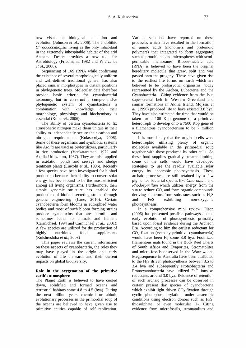
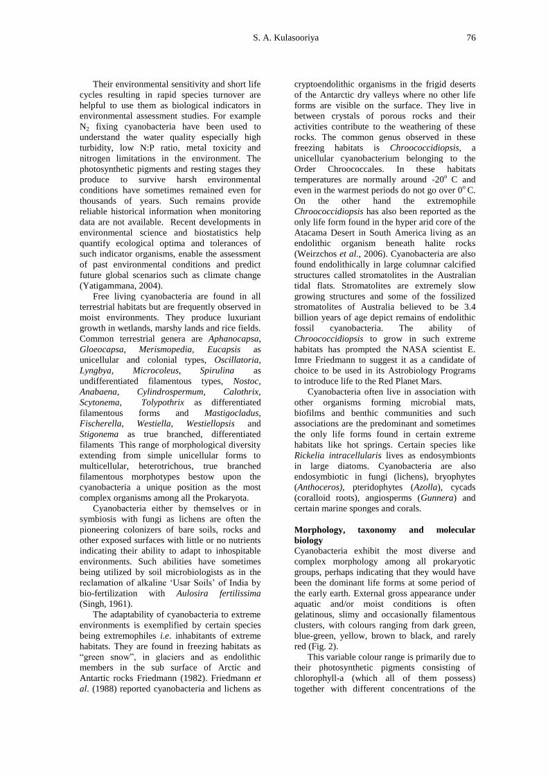
universal acceptance. Trichodesmium exists in
nature as bundles of filaments visible to the
naked eye (Fig. 1). Usually this cynaobacterium
is red in colour due to the high content of the
accessory red pigment phyco-erythrin but they
have been reported to have a range of colours
from grey through yellow, green, and purple
(Fogg et al., 1973).
It is believed by some that the “Red Sea” got
its name due to thick blooms of T.erythraenum
that frequently formed in it. Trichodesmium
occurs in tropical oceans where the surface
temperature is above 25oC and salinity near
3.5%. It normally occurs as long windrows from
a few feet to several miles in length. Wood
(1965) has reported a massive bloom of nearly
52,000 km2 (almost 80% of the area of Sri
Lanka). Carpenter and Romans (1991) present
Trichodesmium as the most important primary

Cyanobacteria 75
producer in the North Atlantic Ocean (ca 165
mgC/m2/day) which also introduces the largest
fraction of new nitrogen to the euphotic zone
through fixation (ca 30 mgN/m2/day). Capone et
al. (1997) confirmed this report and illustrates in
a map its global distribution in all the major
oceans along the tropical belt showing its
extension to the sub-tropical waters just above
and just below the latitudes of 26.5oN and 26.5
oS
respectively. It is however claimed that its
activities are confined to water temperatures
above 20oC. Karl et al. (1997) presenting results
of seven years of time series observations in the
North Pacific Ocean Gyre, claimed that N2
fixation by cyanobacteria accounted for half of
the new production and demanded a
reassessment of previous bio-geochemical
nutrient cycling budgets of one of the world‟s
largest biomes.
Another marine planktic cyanobacterium is
Richelia intracellularis which lives as an
endosymbiont within some large diatoms that
are frequently encountered among the
phytoplankton of the oligotrophic oceans
(Carpenter and Romans, 1991). Although not as
common as eukaryotic algae, cyanobacteria are
found in the marine intertidal and littoral
ecosystems, the common species being Lyngbya
majuscula, Microcoleus ethnoplasts, (a common
mat former), Spirulina sp. (a highly nutritious
cyanobacterium) and several species of
Oscillatoria. They are also found in association
with marine coralline algae and marine lime
stones e.g. Hyella stella and Scytonema
endolithicum.
Nevertheless, the greatest abundance and
diversity of cyanobacteria are encountered in
freshwaters where they are far more common
than their marine counterparts. Several
unicellular, colonial and filamentous species
have been recorded both as plankton, and
benthic micro flora. They are invariably present
together with other photosynthetic organisms in
mesotrophic waters, the common genera being
Microcystis, Synechococcus, Anacystis,
Gloeocapsa, Agmenellum (syn. Merismopedia)
as unicellular and colonial types and
Oscillatoria, Lyngbya, Spirulina, Ananbaena,
Aphanizomenon, Nostoc, Cylindrospermopsis,
Planktothrix, Calothrix, Rivularia and
Gleoetrichia as filamentous types. With
pollution and nutrient loading such water bodies
become eutrophic and very often certain
cyanobacteria that prefer such environments
outgrow their counterparts producing explosive
growths to form „algal blooms‟. The common
bloom forming genera are Microcystis,
Cylindrospermopsis, Anabaena and
Aphanizomenon. Some times these blooms could
be massive. The author once had the experience
of measuring such a bloom in 1991 that formed
adjacent to the Kotmale Hydro-Power Reservoir
Dam which was 1.5 m deep and extended to
more than 2000 m2
(unpublished personal
observation). They foul the water and most of
them produce „algal toxins‟ that are often lethal
to fish and digastric mammals, cause diseases,
irritations, allergies and also contribute to liver
and kidney ailments among humans. (A more
detailed analysis is included in the section on
„toxigenic cyanobacteria‟). In the aquatic
ecosystems cyanobacteria play a crucial role as
oxygenic primary producers as well as nitrogen
fixers.
Figure 1. Colonies of Trichodesmium erythraenum. (Source: Reproduced from Fogg et al.,1973)
S. A. Kulasooriya 76
Their environmental sensitivity and short life
cycles resulting in rapid species turnover are
helpful to use them as biological indicators in
environmental assessment studies. For example
N2 fixing cyanobacteria have been used to
understand the water quality especially high
turbidity, low N:P ratio, metal toxicity and
nitrogen limitations in the environment. The
photosynthetic pigments and resting stages they
produce to survive harsh environmental
conditions have sometimes remained even for
thousands of years. Such remains provide
reliable historical information when monitoring
data are not available. Recent developments in
environmental science and biostatistics help
quantify ecological optima and tolerances of
such indicator organisms, enable the assessment
of past environmental conditions and predict
future global scenarios such as climate change
(Yatigammana, 2004).
Free living cyanobacteria are found in all
terrestrial habitats but are frequently observed in
moist environments. They produce luxuriant
growth in wetlands, marshy lands and rice fields.
Common terrestrial genera are Aphanocapsa,
Gloeocapsa, Merismopedia, Eucapsis as
unicellular and colonial types, Oscillatoria,
Lyngbya, Microcoleus, Spirulina as
undifferentiated filamentous types, Nostoc,
Anabaena, Cylindrospermum, Calothrix,
Scytonema, Tolypothrix as differentiated
filamentous forms and Mastigocladus,
Fischerella, Westiella, Westiellopsis and
Stigonema as true branched, differentiated
filaments This range of morphological diversity
extending from simple unicellular forms to
multicellular, heterotrichous, true branched
filamentous morphotypes bestow upon the
cyanobacteria a unique position as the most
complex organisms among all the Prokaryota.
Cyanobacteria either by themselves or in
symbiosis with fungi as lichens are often the
pioneering colonizers of bare soils, rocks and
other exposed surfaces with little or no nutrients
indicating their ability to adapt to inhospitable
environments. Such abilities have sometimes
being utilized by soil microbiologists as in the
reclamation of alkaline „Usar Soils‟ of India by
bio-fertilization with Aulosira fertilissima
(Singh, 1961).
The adaptability of cyanobacteria to extreme
environments is exemplified by certain species
being extremophiles i.e. inhabitants of extreme
habitats. They are found in freezing habitats as
“green snow”, in glaciers and as endolithic
members in the sub surface of Arctic and
Antartic rocks Friedmann (1982). Friedmann et
al. (1988) reported cyanobacteria and lichens as
cryptoendolithic organisms in the frigid deserts
of the Antarctic dry valleys where no other life
forms are visible on the surface. They live in
between crystals of porous rocks and their
activities contribute to the weathering of these
rocks. The common genus observed in these
freezing habitats is Chroococcidiopsis, a
unicellular cyanobacterium belonging to the
Order Chroococcales. In these habitats
temperatures are normally around -20o
C and
even in the warmest periods do not go over 0o
C.
On the other hand the extremophile
Chroococcidiopsis has also been reported as the
only life form found in the hyper arid core of the
Atacama Desert in South America living as an
endolithic organism beneath halite rocks
(Weirzchos et al., 2006). Cyanobacteria are also
found endolithically in large columnar calcified
structures called stromatolites in the Australian
tidal flats. Stromatolites are extremely slow
growing structures and some of the fossilized
stromatolites of Australia believed to be 3.4
billion years of age depict remains of endolithic
fossil cyanobacteria. The ability of
Chroococcidiopsis to grow in such extreme
habitats has prompted the NASA scientist E.
Imre Friedmann to suggest it as a candidate of
choice to be used in its Astrobiology Programs
to introduce life to the Red Planet Mars.
Cyanobacteria often live in association with
other organisms forming microbial mats,
biofilms and benthic communities and such
associations are the predominant and sometimes
the only life forms found in certain extreme
habitats like hot springs. Certain species like
Rickelia intracellularis lives as endosymbionts
in large diatoms. Cyanobacteria are also
endosymbiotic in fungi (lichens), bryophytes
(Anthoceros), pteridophytes (Azolla), cycads
(coralloid roots), angiosperms (Gunnera) and
certain marine sponges and corals.
Morphology, taxonomy and molecular
biology
Cyanobacteria exhibit the most diverse and
complex morphology among all prokaryotic
groups, perhaps indicating that they would have
been the dominant life forms at some period of
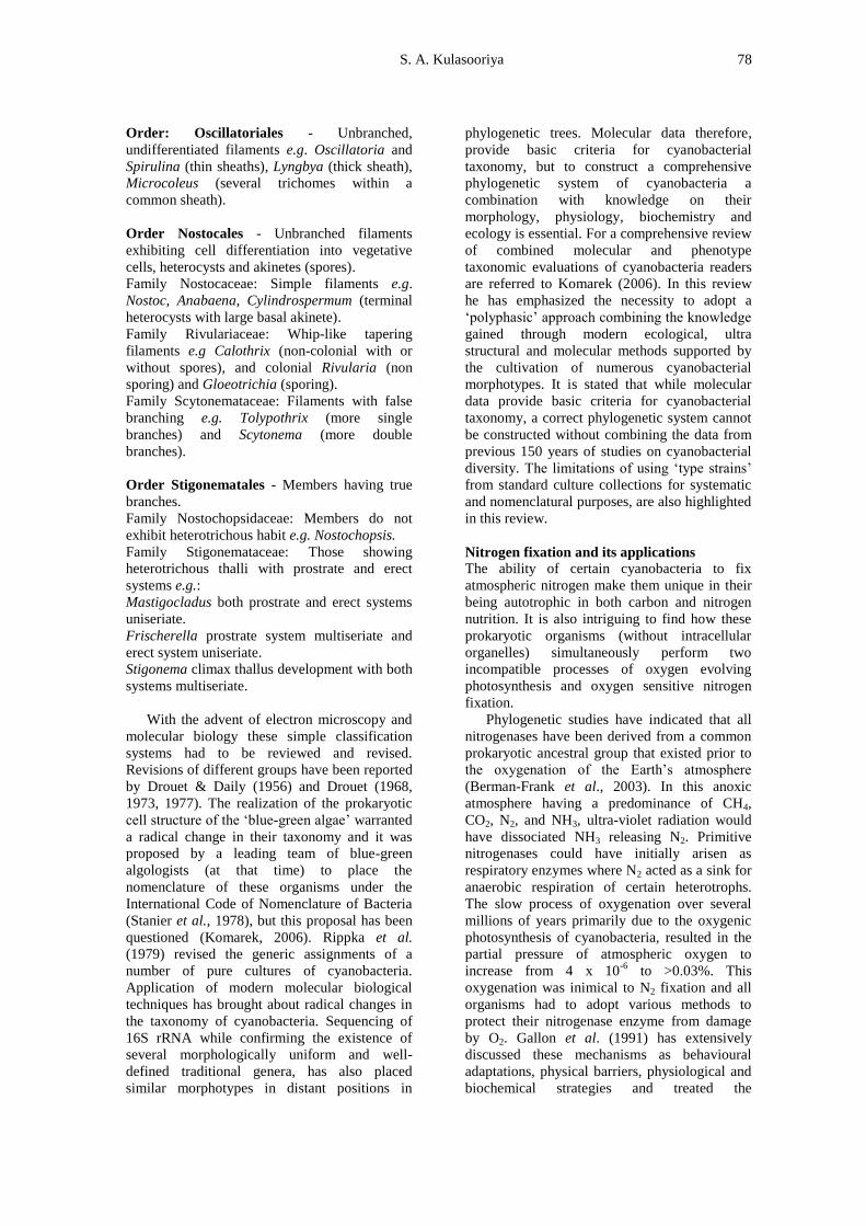
the early earth. External gross appearance under
aquatic and/or moist conditions is often
gelatinous, slimy and occasionally filamentous
clusters, with colours ranging from dark green,
blue-green, yellow, brown to black, and rarely
red (Fig. 2).
This variable colour range is primarily due to
their photosynthetic pigments consisting of
chlorophyll-a (which all of them possess)
together with different concentrations of the

Cyanobacteria 77
accessory phyco-biliprotein pigments,
phycocyanin (blue) and phycoerythrin (red). The
final external colour of a specimen is largely
dependent upon the concentrations of these
pigments. Characteristically all cyanobacteria
have thin or thick gelatinous sheaths outside
their cell walls, their thickness and sometimes
their colour contribute to the final appearance of
the organism.
Although cyanobacteria perform oxygenic
photosynthesis just like eukaryotic green algae
and higher plants, they do not store food in the
form of starch. Their principal storage product is
cyanophycean starch and glycogen together with
specialized intracellular storage compounds like
lipids, protein containing cyanophycean granules
and polyphosphate bodies. Some cyanobacteria
particularly the plankton species produce
intracellular „gas vacoules‟. These are quite
different from typical membrane bound vacoules
of eukaryotic cells and are actually groups of gas
filled vesicles that collapse under sudden shock
and pressure. These vesicles enhance buoyancy
and this helps such species to move up and down
a water column and occupy a depth that provides
the best micro-niche in an aquatic environment.
The somatic diversity of cyanobacteria
extends from unicellular forms, colonial
assemblages, unbranched filaments to filaments
with false and true branching. As these
organisms do not show sexual reproduction their
taxonomy has been entirely based upon
morphology until the mid 20th
century (Geitler,
1932, Fritsch, 1942 and Desikachary, 1959). A
sample of a brief classification based on
morphological diversity is given below:
Group: Algae (Eukaryotic)
Division: Chlorophyta (Green algae)
Class Cyanophyceae or Myxophyceae (Blue-
Green algae)
Order Chroococcales – unicellular and colonial
members.
Unicells e.g. Anacystis, Synechococcus and
Chroococcus,
Loose colonies e.g. Microcystis,
Compact colonies e.g. Gloeocapsa,
Colonies of definite shape e.g. flat plates
Merismopedia (syn. Agmenellum) and cuboidal
colonies e.g. Eucapsis.
Order Chaemosiphonales - A small group that
grows attached and produces a type of asexual
exospores e.g. Chaemosiphon and Dermocarpa.
Figure 2. External appearance of some cyanobacteria collected from paddy fields in Sri Lanka.
S. A. Kulasooriya 78
Order: Oscillatoriales - Unbranched,
undifferentiated filaments e.g. Oscillatoria and
Spirulina (thin sheaths), Lyngbya (thick sheath),
Microcoleus (several trichomes within a
common sheath).
Order Nostocales - Unbranched filaments
exhibiting cell differentiation into vegetative
cells, heterocysts and akinetes (spores).
Family Nostocaceae: Simple filaments e.g.
Nostoc, Anabaena, Cylindrospermum (terminal
heterocysts with large basal akinete).
Family Rivulariaceae: Whip-like tapering
filaments e.g Calothrix (non-colonial with or
without spores), and colonial Rivularia (non
sporing) and Gloeotrichia (sporing).
Family Scytonemataceae: Filaments with false
branching e.g. Tolypothrix (more single
branches) and Scytonema (more double
branches).
Order Stigonematales - Members having true
branches.
Family Nostochopsidaceae: Members do not
exhibit heterotrichous habit e.g. Nostochopsis.
Family Stigonemataceae: Those showing
heterotrichous thalli with prostrate and erect
systems e.g.:
Mastigocladus both prostrate and erect systems
uniseriate.
Frischerella prostrate system multiseriate and
erect system uniseriate.
Stigonema climax thallus development with both
systems multiseriate.
With the advent of electron microscopy and
molecular biology these simple classification
systems had to be reviewed and revised.
Revisions of different groups have been reported
by Drouet & Daily (1956) and Drouet (1968,
1973, 1977). The realization of the prokaryotic
cell structure of the „blue-green algae‟ warranted
a radical change in their taxonomy and it was
proposed by a leading team of blue-green
algologists (at that time) to place the
nomenclature of these organisms under the
International Code of Nomenclature of Bacteria
(Stanier et al., 1978), but this proposal has been
questioned (Komarek, 2006). Rippka et al.
(1979) revised the generic assignments of a
number of pure cultures of cyanobacteria.
Application of modern molecular biological
techniques has brought about radical changes in
the taxonomy of cyanobacteria. Sequencing of
16S rRNA while confirming the existence of
several morphologically uniform and well-
defined traditional genera, has also placed
similar morphotypes in distant positions in
phylogenetic trees. Molecular data therefore,
provide basic criteria for cyanobacterial
taxonomy, but to construct a comprehensive
phylogenetic system of cyanobacteria a
combination with knowledge on their
morphology, physiology, biochemistry and
ecology is essential. For a comprehensive review
of combined molecular and phenotype
taxonomic evaluations of cyanobacteria readers
are referred to Komarek (2006). In this review
he has emphasized the necessity to adopt a
„polyphasic‟ approach combining the knowledge
gained through modern ecological, ultra
structural and molecular methods supported by
the cultivation of numerous cyanobacterial
morphotypes. It is stated that while molecular
data provide basic criteria for cyanobacterial
taxonomy, a correct phylogenetic system cannot
be constructed without combining the data from
previous 150 years of studies on cyanobacterial
diversity. The limitations of using „type strains‟
from standard culture collections for systematic
and nomenclatural purposes, are also highlighted
in this review.
Nitrogen fixation and its applications
The ability of certain cyanobacteria to fix
atmospheric nitrogen make them unique in their
being autotrophic in both carbon and nitrogen
nutrition. It is also intriguing to find how these
prokaryotic organisms (without intracellular
organelles) simultaneously perform two
incompatible processes of oxygen evolving
photosynthesis and oxygen sensitive nitrogen
fixation.
Phylogenetic studies have indicated that all
nitrogenases have been derived from a common
prokaryotic ancestral group that existed prior to
the oxygenation of the Earth‟s atmosphere
(Berman-Frank et al., 2003). In this anoxic
atmosphere having a predominance of CH4,
CO2, N2, and NH3, ultra-violet radiation would
have dissociated NH3 releasing N2. Primitive
nitrogenases could have initially arisen as
respiratory enzymes where N2 acted as a sink for
anaerobic respiration of certain heterotrophs.
The slow process of oxygenation over several
millions of years primarily due to the oxygenic
photosynthesis of cyanobacteria, resulted in the
partial pressure of atmospheric oxygen to
increase from 4 x 10⁻⁶
to >0.03%. This
oxygenation was inimical to N2 fixation and all
organisms had to adopt various methods to
protect their nitrogenase enzyme from damage
by O2. Gallon et al. (1991) has extensively
discussed these mechanisms as behavioural
adaptations, physical barriers, physiological and
biochemical strategies and treated the
Cyanobacteria 79
cyanobacteria as a special group and these have
been discussed by Kulasooriya (2008).
Major adaptations among cyanobacteria are
structural changes among their cells. The vast
majority of cyanobacteria that exhibit aerobic
nitrogen fixation possess a type of specialized
cell called heterocyst. Compared to vegetative
cells heterocysts are generally larger in size,
lighter in colour, have thicker cell walls and
have thickenings called polar nodules at the
points of attachment to adjacent cells.
Kulasooriya et al. (1972) reported on the
differentiation of heterocysts in Anabaena
cylindrica and its coincidence with the
restoration of nitrogenase activity. When
cultured in the laboratory in media with
combined nitrogen (particularly NH4 ions), the
filaments did not produce heterocysts and they
lost their nitrogenase activity. When such
induced „non-heterocystous‟ filaments were
transferred to N-free media, pro-heterocysts
reformed within 24 hours. Once these pro-
heterocysts matured into heterocysts within the
next 24 hours (which could be detected by the
formation of polar nodules), nitrogenase activity
was restored. It was also found that the cellular
C:N ratio was critical for heterocyst formation as
well as expression of nitrogenase activity. A
mature heterocyst has a thick envelope that
consists of an outer polysaccharide fibrous layer,
a middle homogenous layer and an inner
glycolipid laminated layer (Lang and Fay, 1971).
This cell envelope has been shown to be
critically important for nitrogen fixation and
limit the ingress of O2 into the heterocysts
(Murry et al., 1984 and Murry and Wolk, 1989).
Isolated heterocysts showed higher levels of
respiration and their absorption spectrum
coincided more closely than that of the
vegetative cells, with the action spectrum of
nitrogenase activity of whole filaments (Fay and
Walsby, 1966). Isolated heterocysts contained
very little chlorophyll-a and were devoid of
phycocyanin and phycoerythrin pigments which
are associated with the O2 evolving
photosystem-II of photosynthesis (Thomas,
1970). Using 14
C labeled CO2 Wolk (1970)
showed that heterocysts do not fix carbon and
Fay and Kulasooriya (1972) demonstrated the
prevalence of reducing conditions in heterocysts
by the reduction of Triphenyl Tetrazolium
Chloride. Eventually Wolk and Wojciuch (1971)
demonstrated photo-reduction of acetylene and
(Janaki and Wolk, 1982) showed the presence of
nitrogenase in isolated heterocysts. Wolk (1982)
reviewed and summarized all the available
evidences and concluded that the main function
of the heterocyst is nitrogen fixation.
Meanwhile reports came in on the detection
of nitrogenase activity in certain unicellular as
well as non-heterocystous, filamentous species
of cyanobacteria. The first report of a unicellular
species was that of Wyatt and Silvey (1969) who
demonstrated nitrogenase activity in Gloeocapsa
(syn.Gloeothece and Cyanothece). These were
followed by reports on other unicellular species
such as Synechococcus and Aphanothece (Mitsui
et al., 1986), and filamentous non-heterocystous
forms like Microcoleus chnoplastes (Pearson et
al., 1981) and Oscillatoria sp. (Gallon et al.,
1991). In all these cases it has been found that
there was a temporal separation of the two
incompatible processes of oxygenic
photosynthesis and oxygen sensitive nitrogen
fixation along the growth cycle of these
organisms. The other reported physiological and
biochemical strategies adopted by these
organisms have been reviewed in Kulasooriya
(2008).
The most enigmatic situation was
encountered with the large marine blooms of the
non-heterocystous, filamentous cyanobacterium
Trichodesmium erythraenum which showed very
high nitrogenase activity and it was the main
contributor to the nitrogen budget of the deep
oceans (Karl et al., 1997). Two other species T.
theibautii and T. aureum have also being
identified as nitrogen fixing, non-heterocystous,
marine cyanobacteria. Certain studies have
demonstrated the presence of some specialized
cells called diazocytes in these organisms and it
has been suggested that the nitrogenase enzyme
is confined to these cells (Jansen et al., 1994,
Fredrickson and Bergman, 1995 & 1997, Lin et
al., 1998 and Mulholland and Capone, 2000).
Nitrogen fixation in Trichodesmium peaked
around midday and varied inversely with
photosynthetic O2 evolution. It has also been
suggested that N2 fixation and photosynthesis in
Trichodesmium is regulated by circadian
rhythms (Chen et al., 1998). Molecular studies
on natural populations of T. theibautii have
shown that nitrogenase is synthesized early
morning and nifH mRNA is highest before dawn
(Wyman et al., 1996). This enzyme remained
active until noon when it reached a peak, then
declined in the afternoon and got degraded
during the night (Capone et al., 1990 and Zer et
al., 1993). Twelve nif genes cloned and
sequenced from Trichodesmium are basically
similar to those of other cyanobacteria, but show
a closer resemblance to „vegetative cell
nitrogenase genes‟ induced in Anabaena
variabilis under micro-aerobic conditions. Many
other non-heterocystous cyanobacteria such as
species of Oscillatoria, Lyngbya, Microcoleus
S. A. Kulasooriya 80
and Plectonema boryanum have been induced to
fix nitrogen under micro-aerobic conditions.
Among other strategies, aggregation of
unicellular and non-heterocystous cyanobacteria
to form algal mats, colonies, biofilms among
themselves and with other microorganisms have
been reported to exhibit low levels of
nitrogenase activity in marine Synechococcus
and Trichodesmium and terrestrial and
freshwater Oscillatoria, Lyngbya, Microcoleus
and Plectonema. Physical barriers such as thick
mucilaginous sheaths and capsules have enabled
expression of nitrogenase activity in Gleoetheca
(syn. Gloeocapsa and Cyanothece). As a
biochemical protective mechanism nitrogenase
activity has been shown to be regulated by ADP-
ribosylation of the Fe-protein (the more O2
sensitive component of the nitrogenase enzyme
complex). Such adaptations originally
demonstrated in photosynthetic bacteria have
been found to be widespread among
cyanobacteria such as Anabaena CA, Anabaena
variabilis, Aphanizomenon flos-aqaue,
Oscillatoria limosa, Trichodesmium, Gloeotheca
and Microcoleus chthnoplasts as reviewed by
Kulasooriya (2008). Continuous synthesis of
nitrogenase enzyme has also been demonstrated
in certain taxa like Anabaena CA, Anabaena
variabilis, Anabaena flos-aquae and Gloeothece,
but this strategy appears to be of limited
distribution.
All these evidences support the idea that
although heterocysts are specialized cells for N2
fixation in cyanobacteria the potential to fix N2
is present even in the vegetative cells but this is
expressed only under reduced oxygen
concentrations. These observations have
important ecological implications and it is quite
likely that the subterranean layers of thick mats
of non-heterocystous cyanobacteria may be
fixing nitrogen particularly under low light
conditions or in the dark. This idea is also
compatible with the evolution of the process of
N2-fixation from an archaic reducing Earth‟s
atmosphere through a micro-aerobic atmosphere
to the oxygenated atmosphere of present times
(Gallon et al., 1991 and Berman-Frank et al.,
2003). In symbiotic associations it is always the
cyanobacterial partners that fix nitrogen. They
provide nitrogen to the ecosystem in which they
live and often the rates of fixation by these
systems are higher than those of free-living
cyanobacteria, perhaps due to the additional
nutrition and micro-aerobic conditions provided
by the host. Occasionally cyanobacteria have
been reported to be epiphytic on rice plants
(Roger et al., 1981) and weeds in wetland rice
fields (Kulasooriya et al.,1981a) and even
endophytic in the leaf sheaths of deepwater rice
(Kulasooriya et al., 1981b) and the nitrogen they
fix has been shown to be available to the
associated host plants (Watanabe et al.1982).
Free living nitrogen fixing cyanobacteria and
the aquatic fern Azolla (which has nitrogen
fixing Anabaena azollae as an endosymbiont),
have been used as biofertilizers in rice
cultivation. Venkataraman (1972) provides a
comprehensive account of the large scale
production of cyanobacterial bio-fertilizers in
India. Cyanobacteia are initially isolated from
rice fields, purified and screened for rapid
growth and high N2-fixation. Selected isolates
are then semi-mass cultured in laboratory media
and these are used as inoculants for soil based
outdoor mass cultures in open ponds or large
trays under non-sterile conditions. A sprinkle of
P fertilizer, lime and pesticides are usually added
to ensure uninhibited rapid growth. After
sufficient growth is obtained the cultures are
allowed to sun dry and the soil based algal flakes
are packed and distributed among farmers to be
used as bio-fertilizers. This low cost technology
quite suitable for resource poor rural farmers has
been successful only in a few areas in India
where edaphic conditions are favourable for the
growth of cyanobacteria. Reports from certain
areas of Myannmar, China and Egypt have also
recorded some successes. Roger and
Kulasooriya (1980) reviewed most of the
literature on the use of cyanobacterial
biofertilizers and concluded that their N-input
potential ranged from 0 to 80 Kg/ha/season with
an average of 25 Kg/ha. Attempts to adopt this
technology in Sri Lanka was successful only up
to the stage of pot experiments and most of the
cyanobacterial fertilizers added to the rice fields
could not overcome consumption by the rice
field micro-fauna and competition by indigenous
micro-flora and failed to colonize the fields
(Kulasooriya and Hirimburegama, 1989).
Compared to free living cyanobacteria, the
application of Azolla in rice cultivation has been
more successful and widely used particularly in
Vietnam, China and certain parts of Philippines.
The status of this technology has been well
reviewed at a workshop held in Fuzhou in Fujian
Province, China and published by the
International Rice Research Institute, Philippines
in 1987 as „Azolla Utilization‟. Even in Sri
Lanka this technology appeared to be more
feasible than free living cyanobacterial
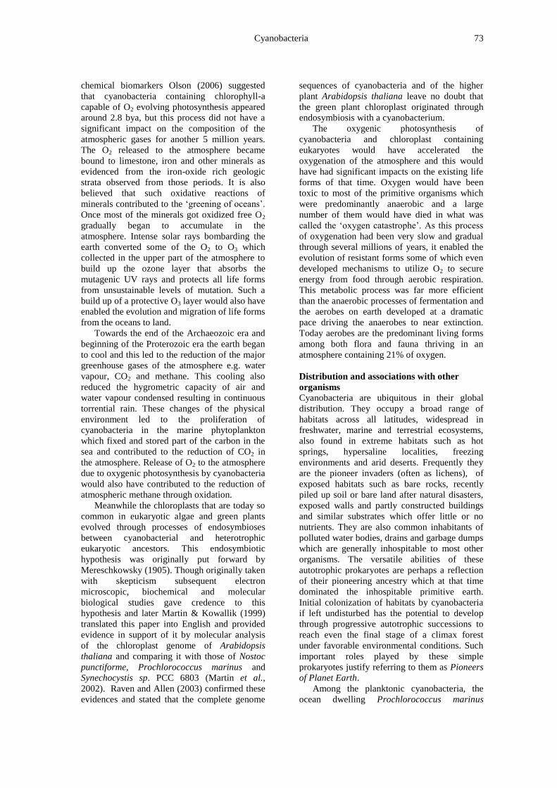
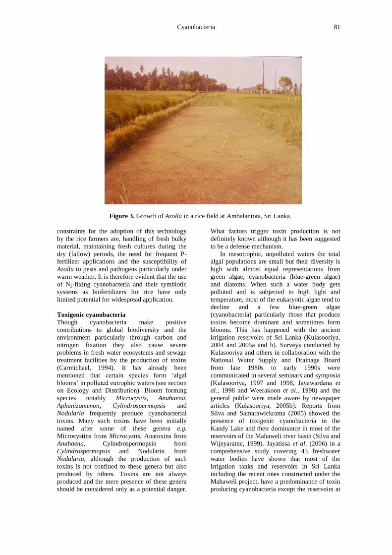
biofertlizers. Azolla grew well in rice fields at
Ambalantota (Fig. 3), Bombuwela, and
Peradeniya and showed a potential to replace an
equivalence of 55 to 85 KgN/ha of chemical N-
fertilizer (Kulasooriya et al., 1987). The major
Cyanobacteria 81
Figure 3. Growth of Azolla in a rice field at Ambalantota, Sri Lanka.
constraints for the adoption of this technology
by the rice farmers are, handling of fresh bulky
material, maintaining fresh cultures during the
dry (fallow) periods, the need for frequent P-
fertilizer applications and the susceptibility of
Azolla to pests and pathogens particularly under
warm weather. It is therefore evident that the use
of N2-fixing cyanobacteria and their symbiotic
systems as biofertilizers for rice have only
limited potential for widespread application.
Toxigenic cyanobacteria
Though cyanobacteria make positive
contributions to global biodiversity and the
environment particularly through carbon and
nitrogen fixation they also cause severe
problems in fresh water ecosystems and sewage
treatment facilities by the production of toxins
(Carmichael, 1994). It has already been
mentioned that certain species form „algal
blooms‟ in polluted eutrophic waters (see section
on Ecology and Distribution). Bloom forming
species notably Microcystis, Anabaena,
Aphanizomenon, Cylindrospermopsis and
Nodularia frequently produce cyanobacterial
toxins. Many such toxins have been initially
named after some of these genera e.g.
Microcystins from Microcystis, Anatoxins from
Anabaena, Cylindrospermopsin from
Cylindrospermopsis and Nodularin from
Nodularia, although the production of such
toxins is not confined to these genera but also
produced by others. Toxins are not always
produced and the mere presence of these genera
should be considered only as a potential danger.
What factors trigger toxin production is not
definitely known although it has been suggested
to be a defense mechanism.
In mesotrophic, unpolluted waters the total
algal populations are small but their diversity is
high with almost equal representations from
green algae, cyanobacteria (blue-green algae)
and diatoms. When such a water body gets
polluted and is subjected to high light and
temperature, most of the eukaryotic algae tend to
decline and a few blue-green algae
(cyanobacteria) particularly those that produce
toxins become dominant and sometimes form
blooms. This has happened with the ancient
irrigation reservoirs of Sri Lanka (Kulasooriya,
2004 and 2005a and b). Surveys conducted by
Kulasooriya and others in collaboration with the
National Water Supply and Drainage Board
from late 1980s to early 1990s were
communicated in several seminars and symposia
(Kulasooriya, 1997 and 1998, Jayawardana et
al., 1998 and Weerakoon et al., 1998) and the
general public were made aware by newspaper
articles (Kulasooriya, 2005b). Reports from
Silva and Samarawickrama (2005) showed the
presence of toxigenic cyanobacteria in the
Kandy Lake and their dominance in most of the
reservoirs of the Mahaweli river basin (Silva and
Wijeyaratne, 1999). Jayatissa et al. (2006) in a
comprehensive study covering 43 freshwater
water bodies have shown that most of the
irrigation tanks and reservoirs in Sri Lanka
including the recent ones constructed under the
Mahaweli project, have a predominance of toxin
producing cyanobacteria except the reservoirs at
S. A. Kulasooriya 82
Kalatuwawa and Labugama in which the
catchment areas are under strict nature reserves.
This is an indication that if pollution is
minimized the occurrence of toxin producing
cyanobacteria can be reduced. How such
conditions can be established and sustained in
irrigation tanks (reservoirs) would always
remain a dilemma. Jayatissa et al. (2006) also
reported for the first time the presence of
microcystin LR toxin in some of the
cyanobacterial samples collected in Sri Lanka.
More recent studies have confirmed these results
and it is now quite evident that a large number of
inland freshwater bodies of Sri Lanka carry
heavy populations of toxigenic cyanobacteria
with Cylindrospermopsis being the dominant
genus in a majority of them (Perera et al., 2011a,
Perera et al., 2011b, Kulasooriya and Padmasiri,
unpublished). The Institute of Fundamental
Studies, Sri Lanka, now possesses facilities to
detect and quantify cyanobacterial toxins in
water samples. A number of samples collected
from reservoirs and irrigation tanks from the dry
zones of North Central and Wayamba Provinces
have shown the presence of such toxins
sometimes well over the levels permitted by
WHO standards (Dr. D. N. Magana-Arachchi,
IFS, personal communication). It is evident from
these results that immediate steps should be
taken to ameliorate the predominance of
toxigenic cyanobacteria in water bodies of Sri
Lanka, commencing from those used as sources
for water supply schemes. It is also imperative to
conduct well formulated scientific studies to
verify the claims that toxigenic cyanobacteria
are responsible for the prevalence and rapid
increase of chronic kidney diseases of unknown
etiology in certain areas of Sri Lanka.
Some industrial applications
Cyanobacteria are of common occurrence in
waste water treatment plants. In some instances
they have been found to be useful as low cost
converters of odour causing and pollution
creating dairy effluents into micro-algal biomass
using sunlight (Lincoln et al., 1996). They were
found to be very effective in removing NH4 ions
and other nitrogen rich compounds from such
effluents. In other cases toxigenic species have
reduced the efficiency of waste water treatment
plants (Martins et al., 2011). Toxins produced,
particularly microcystins have not been
antibacterial but they have adversely affected the
protozoan populations. Nonetheless, other
secondary metabolites of cyanobacteria have
been found to be anti-bacterial and affected the
decomposition rates of both aerobic and
anaerobic bacteria.
On the other hand, cyanobacteria have been
found to be useful in the bioremediation of
industrial effluents and extraction of heavy
metals. A number of species belonging to the
genera of Synechococcus, Cyanothece,
Oscillatoria, Nostoc and Nodularia isolated
from pharmaceutical and textile industry
effluents were demonstrated to be capable of
biodegradation and biosorption of several heavy
metals (Dubey et al., 2011). Using lyophilized
Synechococcus sp. PCC 7942 immobilized in a
silica polymer, Gardia-Torresday et al. (2011)
demonstrated that this cyanobacterium could
effectively bind Cu2+
, Pb2+
, Ni2+
, Cd2+
, Cr3+
&
Cr4+
and these metals could be recovered by
elution with 0.1M HCl. It has even being
reported that certain cyanobacteria can be useful
in the cleaning of oil spills (Cohen, 2002). He
tested species isolated from cyanobacterial mats
developing in oil-contaminated sabkhas along
the African coasts of the Gulf of Suez and in the
pristine Solar Lake, Sinai. He found that axenic
cultures of the isolated cyanobacteria by
themselves are unable to degrade oil. In
association with sulphate reducing and other
heterotrophic bacteria from the mats, when
exposed to light rapid degradation of oil was
observed. The oxygenic photosynthesis of the
cyanobacteria stimulated the aerobic
decomposition by the associated bacteria. This
shows the potential of producing cyanobacterial
- eubacterial biofilms as efficient microbial
consortia for cleaning of oil spills. Abed (2010)
has reported the commencement of such a
project in Oman.
Recently a few species of cyanobacteria have
been investigated for biofuel production because
their ability to convert solar energy to chemical
energy has been found to be the most efficient
among all living organisms. The efficiency of
solar energy conversion in corn and sugarcane is
1–2%, for eukaryotic algae it is around 5% while
for cyanobacteria it is 10%. This can be
understood if one looks at the photosynthetic
pigments of cyanobacteria. In addition to
chlorophyll-a these organisms contain
phycobilisomes attached to the thylakoid
membranes and these act as antennae in
harvesting light for photosystem II. Among the
pigments contained in the phycobilisomes,
Phycoerythrin absorbs energy between the wave
lengths of 500 – 600 nm, Phycocyanin absorbs
between 550 – 650 nm and Allophycocyanin
absorbs between 600 – 675 nm. Together with
chlorophyll-a, their light absorption capacity will
therefore extend right across the entire spectrum
of visible light (Dr. Celia Smith, Department of
Botany, University of Hawaii at Manoa,

Cyanobacteria 83
http://www. biologie.uni-hamburg.de/ b-online/
librarywebb/ BOT311/ Cyanobacteria).
Furthermore, the simple genomic structure of
these organisms has enabled gene sequencing
and genetic engineering relatively easier.
Professor James C. Liao of the University of
California, Los Angeles, has inserted and spliced
genes from other organisms to increase the CO2
fixing ability of Rubisco enzyme of
Synechococcus elongatus. This transgenic strain
produced isobutyraldehyde gas which could be
recovered and converted to butanol. Prof.
Malcolm R. Brown and Dr. David Nobles of the
University of Texas, Austin have inserted a gene
from Acetobacter xylinum to a cyanobacterium
which could secrete alcohol and sugars. Prof. P.
Fu and Dr. J. Dexter of the University of Hawaii
have transformed Synechocystis strain PC 6803
by inserting pyruvate dehydrogenase and alcohol
dehydrogenase II genes from Zymomonas
mobilis to produce ethanol. The biggest break
through has been reported by a private company
Joule Technologies of Boston, Massachusetts,
which has developed Bioreactors containing
genetically modified cyanobacteria that could
continuously secrete biofuel and ethanol (Lane,
2010). Reports on biofuel production capacities
from different sources are given in Table 1.
Table 1. Production capacities of biofuel from
different sources.
Source Gals/ac* L/ha+
Soybean 50 92
Corn 250 458
Sugarcane 450 825
Oil palm 650 1192
Algae 2000 3668
Joule technologies 15,000 27,512 Source: *Original report, + Converted
The 15,000 gals/ac given by Dr. Dan
Roberts, the principal scientist at Joule
Technologies, eclipses all other claims. (Mail
Online, Science & Technology March 2, 2011).
It is interesting to note that in all these cases
the most productive cyanobacteria have been
unicellular species. This is perhaps due to their
ability to harvest maximum solar energy per cell
because the single cells turn and rotate with
minimum self shading as they move up and
down a bioreactor. Also their simple genomes
permit easy genetic engineering compared to
more complex counterpart resources. This opens
a window of opportunity to conduct fundamental
studies in Sri Lanka that could lead us to a
commercially viable break through in biofuel
production. Most of the other industrial
platforms for biofuel production based upon feed
stocks derived from higher plants require vast
areas of cultivable land. For a small country like
Sri Lanka it is a formidable task to allocate such
extents of land without compromising on other
more important economic uses. Algae based
biofuels therefore appear to be an attractive
option. Already unicellular cyanobacteria
belonging to Synechococcus, Synechocystis,
Chroococcidiopsis and Cyanothece have been
isolated from different localities in Sri Lanka
including brackish water and hot springs and
partially sequenced (Wanigatunga et al., in
preparation). With the commissioning of coal
powered electricity generation systems
sequestering CO2 emissions would be a major
problem and such waste gases could be
harnessed to grow biofuel cyanobacteria. Even
the CO2 emitted by other industries like
breweries and cement production factories can
be used for algal cultivation.
Cyanobacteria as a food supplement
Among the vast array of cyanobacteria, two
species have been used as food supplements of
high nutritive value. They are Spirulina
plantensis (syn. Arthrospira plantensis) and
Aphanizomenon flos-aquae. These species have
come from the traditional food habits of ancient
civilizations of Aztecs, Mayans and Chinese and
have been harvested from natural freshwater
lakes such as Lake Chad in Africa, Upper
Klamath in North America, Lake Texcoco in
Mexico, and Lake Titicaca in Peru, South
America. Currently they are also cultivated in
large open raceway ponds with paddle wheel
stirring under high pH and high temperature to
minimize contamination by human pathogenic
microorganisms. Such commercial production
facilities are today found in the United States,
Thailand, India, Taiwan, China, Pakistan,
Myanmar and Chile. Spriulina is marketed as a
source of complete nutrition which no other
single organism can offer. Besides having a very
high protein content (65 – 71% of dry weight)
with all the essential amino acids, it is claimed to
contain a wide spectrum of prophylactic and
therapeutic nutrients including B-complex
vitamins, minerals, proteins gamma linolenic
acid and super antioxidants such as beta
carotene, Vitamin E, trace elements and a
number of unexplored bioactive compounds
(Kulshreshtha et al., 2008).

S. A. Kulasooriya 84
The other species, Aphanizomenon flos-
aquae as a food supplement (Carmichael et al.,
2000) is wrought with the fear that certain
species of Aphanizomenon produce toxins. It has
been reported that colony forming species of
Aphanizomenon do not produce toxins
(Carmichael, 1994) and based upon 16s rRNA
characteristics certain species have been
misidentified (Li et al., 2003). However, reports
of cylindrospermopsin producing species of
Aphanizomenon flos-aquae have been reported
from Germany (Preussela et al., 2006). Thus
Food and Drug Administration Agencies as well
as commercial enterprises that make
Aphanizomenon based food supplements have a
major responsibility to ensure that their products
originate from non toxigenic species.
However, some of the claims of high
nutritive value of these cyanobacterial products
have been questioned. (Watanabe et al., 1999)
who reported that Spirulina contains a „psuedo-
Vitamin B12‟ stated that it cannot replace the
proper Vitamin B12 requirement, particularly in
complete vegetarian diets. The general
consensus of opinion appears to be that
cyanobacterial based food supplements could
provide a high level of nutrition particularly to
poor sectors of the population who are
malnourished, but they should not be considered
as a replacement diet for animal protein sources
such as fish, eggs, meat and milk.
Recently Spirulina has gained prominence
for its therapeutic value against radiation,
particularly after the disasters caused by the
Chernobyl accident in Belarus, Russia and the
tsunami damage to the Fukushima nuclear power
plant in Japan. Citing several scientific papers
published in the Journal of Applied Phycology
Dr. Amha Belay, Vice President of Earthrise
Farms in Irvine, California (a large scale
Spirulina producing commercial venture) has
posted review articles on the therapeutic value of
this cyanobacterium particularly against effects
of radiation (Belay, 2011).
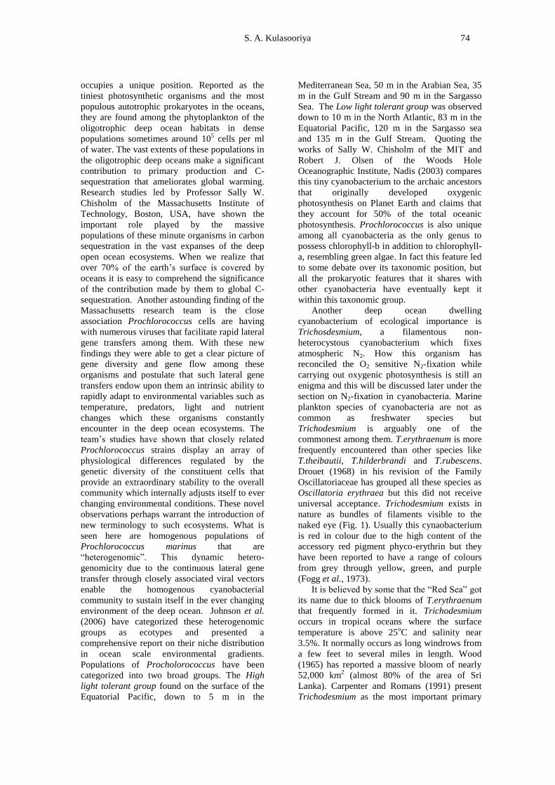
The Institute of Fundamental Studies, Sri
Lanka, has commenced a project in collaboration
with the Mahatma Gandhi Center, Colombo, to
introduce mass culture of Spirulina to villages in
Sri Lanka as a low cost commercial activity to
help alleviate poverty and reduce rural
malnutrition Fig. 4 (a), (b) and (c).
Figure 4. Spirulina cultures at the Institute of Fundamental Studies, Sri Lanka. (a) Open tray culture,
(b) Close up of surface and (c) Microscopic view (x100).
a b
c
Cyanobacteria 85
CONCLUSION
From the foregoing it is evident that although
cyanobacteria are microscopic, prokaryotic
organisms they are ubiquitous in their
distribution and play crucial roles in the
sustenance of many ecosystems. Their
contributions to the global carbon and nitrogen
bio-geochemical cycles are significant. These
organisms also possess features that are useful as
well as harmful to other living organisms
including humans. Some of them appear to be
good candidates for biofertilizer and biofuel
industries and a few species are gaining
recognition as highly nutritive food supplements
as well therapeutic agents. Having knowledge
about these little known microorganisms is
therefore important and useful. If Planet Earth
ever faced a global catastrophe resulting in
colossal destruction of life, it is most likely that
a few cyanobacterial species would survive in
some extreme ecological niches and perhaps
play a pioneering role in re-establishing life
forms in a virtually barren Earth.
REFERENCES
Abed, Raed (2010). Oil bioremediation by
cyanobacterial consortia under extreme
environmental conditions. Oman Daily Sun
(December).
Azolla Utilization (1987). Proceedings of the
Workshop on Azolla Use, Fuzhou, Fujian,
China 1985. The International Rice Research
Institute, Los Banos, Philippines. Pp. 296.
Belay Amha (2011) The potential role of
Spirulina in protection against radiation and
its effects. http://www.iherb.com/i/info/
pic/Spirulina_RadiationA.pdf and http://www.
earthrise.com/pdf/earthrisepdf.
Berman-Frank, I., Lundgren, P. and Fallwoski,
P. (2003). Nitrogen fixation and
photosynthetic oxygen evolution in
Cyanobacteria (Mini review) Research in
Microbiology 154: 157 – 164.
Capone, D. G., Zehr, J. P., Paerl, H. W.,
Bergman, B. and Carpenter, E. J. (1997).
Trichodesmium, a globally significant marine
cyanobacterium. Science 276 (May): 1221-
1229.
Carmichael, W. W. (1994). The toxins of
Cyanobacteria. Scientific American 270 (1):
78-86.
Carmichael, W. W., Li., R. And Pereira, P.
(2000). Harvesting of Aphanizomenon flos-
aquae Ralfs ex Born. & Flah.var. flos-aquae
(cyanobacteria) from Klamath Lake for human
dietary use. J. Appl. Phycol. 12: 585-595.
Carmichael, W. W., Azevedo, S. M., An, J. S.,
Molica, R. J., Jochimsen, E. M., Lau, S.,
Rinehart, K. L., Shaw, G. R. and Eaglesham,
G. K. (2001). Human fatalities from
cyanobacteria: chemical and biological
evidence for cyanotoxins. Environ. Health
Perspective 109 (7): 663 – 668.
Carpenter, E. J. and Romans, K. (1991). Major
role of the cyanobacterium Trichodesmium in
nutrient cycling in the North Atlantic Ocean.
Science 254 (Nov): 1356 – 1358.
Cohen, Yehuda (2002). Bioremediation of oil by
marine microbial mats. International
Microbiology 5: 189 – 193.
Desikachary, T. V. (1959). Cyanophyta. Indian
Council for Agricultural Research, New Delhi:
686p.
Drouet, F. (1968). Revision of the classification
of the Oscillatoriaceae. Acad. Nat. Sci.
Philadelphia, Monograph 15: p. 370.
Drouet, F. (1973). Revision of the Nostocaceae
with cylindrical trichomes. Hafner Press, New
York – London: 292p.
Drouet, F. (1977). Revision of the Nostocaceae
with constricted trichomes. (beihefte Zur Nova
Hedwigia, Heft 57), Publisher: Bisen Singh
Mahendra Pal Singh (ISBN 812110503X and
9788211033): 258p.
Drouet, F and Daily, W. A. (1956). Revision of
the coccoid Myxophyceae. Butler Univ. Bot.
Stud. 12: 1218p.
Dubey, Sanjay Kumar, Dubey, Jaishree, Mehra,
Sandeep, Tiwari, Pradeep and Bishwas A. J.
(2011). Potential use of cyanobacterial species
in bioremediation of industrial effluents.
African Jour. Biotechnol. 10 (7): 1125 – 1132.
Fay, P. and Walsby, A. E. (1966). Metabolic
activities of isolated heterocysts of the blue-
green alga Anabaena cylindrica. Nature 209:
810 – 812.
Fay, P. and Kulasooriya, S. A. (1972).
Tetrazolium reduction and nitrogenase activity
in heterocystous blue-green algae. Arch.
Mikrobiol. 87: 341 – 352.
Fogg, G. E., Stewart, W. D. P., Fay, P. and
Walsby, A. E. (1973). The Blue-Green Algae,
Academic Press, London & New York: 459
Pp.
Friedmann, Imre E. (1982). Endolithic
microorganisms in the Antarctic cold desert.
Science 215 (4536): 1045 – 1053.
Friedmann, Imre E., Hua, Maosen and
Friedmann, Roseli Ocampo (1988).
Cryptoendolithic lichen and cyanobacteria
communities of the Ross Desert, Antarctica.
Polarforschung 58 (2/3): 251 – 259.
S. A. Kulasooriya 86
Fritsch, F. E. (1942). The interrelations and
classification of the Myxophyceae
(Cyanophyceae). New Phytol. 41: 134 – 148.
Gallon, J. R., Hashem, M. A., and Chaplin, A. E.
(1991). Nitrogen fixation by Oscillatoria sp.
under autotrohic and photoheterotrophic
conditions. Jour. Gen. Microbiol. 137: 31- 39.
Gardea-Torresday, J. L., Arenas J. L., Francisco,
N. M. C., Tiemann, K. J. and Webb, R.
(2011). Ability of immobilized cyanobacteria
to remove metal ions from solution and
demonstration of the presence of
metallothionein genes in various strains. Jour.
Hazardous Substance Res. 1: 2 – 18.
Geitler, L. (1932). Cyanophyceae. In:
Kryptogamenflora von Deutschland,
Osterreich und der Schweiz In: L. Rabenhorst
(Ed.), Akademische Verlagsgesellschraft,
Leipzig.14: 32 – 64.
Janaki, S and Wolk, C. P. (1982). Synthesis of
nitrogenase by isolated heterocysts. Biochim.
Biophys. Acta. 696: 187 – 192.
Jayatissa, L.P., Silva, E.I.L., McElhiney, J. and
Lawton, L.A. (2006). Occurrence of toxigenic
cyanobacterial blooms in freshwaters of Sri
Lanka. Systematic and Applied Microbiology
29: 156 – 164.
Jayawardana, B. P. A., Jayalath, J. M. J. C.,
Padmasiri, J. P. D. and Kulasooriya, S. A.
(1998). Occurrence of potential toxin
producing cyanobacteria in reservoirs of Sri
Lanka. In: Symposium on Cyanobacterial
Toxins in Water. Postgraduate Institute of
Science, University of Peradeniya: Pp.7.
(Abstract)
Johnson, I. Zackary, Zinser, Erik R., Coe,
Allison, McNulty, Nathan P. E., Woodward,
Malcolm S. and Chisholm, Sally M. (2006).
Niche partitioning among Prochlorococcus
ecotypes along ocean-scale environmental
gradients. Science 311 (March): 1737 – 1740.
Karl, D., Letelier R., Tupas L., Dore J., Christian
J. and Hebel D. (1997). The Role of Nitrogen
Fixation in Bio-geochemical Cycling in the
Sub-Tropical North Pacific Ocean. Nature
388: 533- 538.
Komarek, J. (2006). Cyanobacterial Taxonomy:
Current Problems and Prospects for the
Inetgration of Traditional and Molecular
Approaches. Algae 21 (4): 349 – 375.
Kulasooriya, S. A. (1997). Cyanobacteria and
their toxins in water: Sri Lanka Country
Report. In: Workshop on Cyanobacteria
(Blue-Green Algae) and their toxins in water.
July 1997, Brisbane, Queensland, Australia.
Kulasooriya, S. A. (1998). Freshwater
cyanobacteria and their relevance to water
supply in Sri Lanka. In: Symposium on
Cyanobacterial toxins in water. Postgraduate
Institute of Science, University of Peradeniya:
8 Pp.
Kulasooriya, S. A. (2004). Ecology of
Freshwater and Terrestrial Cyanobacteria
(blue-green algae) of Sri Lanka. In. National
Workshop on Current Status of Lower Plants
in Sri Lanka. PGRC, Gannoruwa, Ministry of
Environment & Natural Resources, and the
Royal Botanic Gardens.
Kulasooriya, S. A. (2005a). Potential toxin
producing Cyanobacteria (blue-green algae) in
water resources of Sri Lanka. In. Workshop on
Hygiene Promotion & Water Quality
Surveillance for Water Board and Health
Department Officials. PGRC, Gannoruwa,
National Water Supply & Drainage Board.
Kulasooriya, S. A. (2005b). Is the water we
drink safe? Daily News (Monday 1stAugust):
Pp.7 .
Kulasooriya, S. A. (2008). Biological Nitrogen
Fixation: Fundamentals and Utilization.
Science Education Unit, Faculty of Science,
University of Peradeniya. Pp. 143.
Kulasooriya, S. A., Lang, N. J. and Fay, P.
(1972). The heterocysts of blue-green algae.
III Differentiation and nitrogenase activity.
Proc. Roy. Soc. London, B 181: 199 – 209.
Kulasooriya, S. A., Roger, P. A., Barraquio, W.
L. and Watanabe, I. (1981a). Epiphytic
nitrogen fixation on weeds in a rice field
ecosystem. In: Wetselaar, R. Simpson, J. R. &
Rosswall, T. (Eds.) Nitrogen Cycling in South-
East Asian Wet Monsoonal Ecosystems,
Australian Academy of Sciences, Canberra.
Pp 56 – 61.
Kulasooriya, S. A., Roger, P. A., Barraquio, W.
L. and Watanabe, I. (1981b). Epiphytic
nitrogen fixation in deepwater rice. Soil Sci.
Plant Nutr. 27: 19 – 27.
Kulasooriya, S. A., Hirimburegama, W. K. and
Abeysekera, S. W. (1987). Use of Azolla in
Sri Lanka. In: Azolla Utilization, The
International Rice Research Institute,
Philippines Pp. 131 – 140.
Kulasooriya, S. A. and Hirimburegama, W. K.
(1989). Phototrophic nitrogen fixation in
wetland rice. In: Biological Nitrogen Fixation
Associated with Rice Production (Eds) S. K.
Dutta, & C. Sloger, Oxford/IBH Publishers,
New Delhi, Bombay & Calcutta: Pp.191 –
210.
Kulshreshtha, Archana J., Ansih Z., Jarouliya,
Urmila; Bhadauriya, Pratiksha; Prasad, G. B.
K. S. and Bisen, P. S. (2008). Spirulina a
wonder alga full of nutrition. Current
Pharmaceutical Biotechnology 9 (5): 400 –
405.
Cyanobacteria 87
Lane, Jim (2010). Joule wins key patent for
GMO cyanobacteria that creates fuel from
sunlight, CO2 and water. Biofuels Digest 14
(September)
Lang, N. J. and Fay, P. (1971). The heterocysts
of blue-green algae. II Ultrastructure. Proc.
Roy. Soc. London, B 178: 193 -202.
Li, Renhuis, Carmichael, Wayne, W and Pereira,
Paulo (2003). Morphological and 16S rRNA
gene evidence for the re-classification of the
para shellfish toxin producing Aphanizomenon
flos-aquae LMCEYA 31 as Aphanizomenon
issactschenkoi. Jour Applied Phycol. 39: 814
– 818.
Lincoln, E. P., Wilkie, A. C. and French, B. T.
(1996). Cyanobacteria process for renovating
dairy waste water. Biomass & Bioenergy 10
(1): 63 – 68.
Martins, Joana, Peixe, Luisa and Vasconselos,
Vitor M. (2011). Unravelling cyanobacterial
ecology in waste water treatment plants
(WWTP). Microbial Ecology, 62 (2): 241 –
256.
Martin, W. and Kowallik, K. V. (1999).
Annonated English translation of
Mereschkowski‟s 1905 paper. Euro. Jou.r
Phycol. 34 (3): 287 – 295.
Martin, W., Rujan, T., Richly, E., Hamen, A.,
Cornelsen, S., Lin, T., Leister, D., Stoebe, B.,
Hasegawa, M. and Penny, D. (2002).
Evolutionary analysis of Arabidopsis,
cyanobacteria and chloroplast genomes
reveals plastid phylogeny and thousands of
cyanobacterial genes in the nucleus. Proc.
Natn. Acad. Sci., USA 99: 12246 – 12251.
Mereschkowsky, C. (1905). Uber Natur und
Ursprung der Chromatophoren im
Pflanzenreiche. Biol. Centrabl. 25: 593 – 604.
Mitsui, A., Kumazawa, A., Takahashi, A.,
Ikemoto, H., Cao, S. and Arai, T. (1986).
Strategy by which nitrogen-fixing unicellular
cyanobacteria grow photoautotropically.
Nature 323: 720 – 722.
Mojzsis, S. J., Arrhenius, G., McKeegan, K. D.,
Harrison, Nutman A. P. and Friend, C. R. L.
(1996). Evidence for life on Earth before
3,800 million years ago. Nature 384: 55 – 59.
Murry, M. A., Horne, H. J. and Bennemann, J.
R. (1984). Physiological studies of oxygen
protective mechanisms in the heterocysts of
Anabaena cylindrica. Appl. Environ.
Microbiol. 47: 449 – 454.
Murry, M. A. and Wolk, C. P. (1989). Evidence
that the barrier to the penetration of oxygen
into the heterocysts depends upon two layers
of the envelope. Arch. Mikrobiol. 151: 469 –
474.
Nadis, Steve (2003). The cells that rule the seas.
Scientific American (November): 52 - 57.
Olson, John M. ( 2006). Photosynthesis in the
Archean Era. Photosynthesis Research 88:
109–17
Pearson, H. W., Marlin, G. and Howsley, R.
(1981). Physiological studies on in vivo
nitrogenase activity by axenic cultures of the
blue-green alga Microcoleus chthnoplastes.
British Phycol. Jour. 16: 139 Pp.
Perera Buddhika, Yatigammana Sudharma and
Kulasooriya, S. A. (2011a). Diversity of
plankton in some selected reservoirs in Sri
Lanka. Proceedings International Symposium
on Sustainable Use of Water: Challenges
Ahead, Postgraduate Institute of Science,
University of Peradeniya Pp.36 – 37.
Perera, M. B. U., Yatigammana, S. K.,
Kulasooriya, S. A. and Athukorala, N. P.
(2011b). Cyanobacterial species composition
and the limnology of the Padawiya Tank,
during the dry season. Proceedings of
Peradeniya University Sessions. Pp. 149.
Preussel, K., Stuken, A., Wiedner, C., Chorus, I
and Fastner, J. (2006). First report on
cylindrospermopsin producing Aphanizome-
non flos-aqaue isolated from two German
lakes. Toxicon 47 (2): 156 – 162.
Raven, J. A. and Allen, J. F. (2003) Genomics
and Chloroplast evolution. What did
cyanobacteria do for plants? Genome Biology
4: Pp 209.
Rippka, R., Deruelles, J., Waterbury, J. B.,
Herdman, M. and Stanier, R. Y. (1979).
Genetic assignments, strain histories and
properties of pure cultures of cyanobacteria. J.
Gen. Microbiology 111: Pp.161.
Roger, P. A. and Kulasooriya, S. A. (1980).
Blue- Green Algae and Rice. The International
Rice Research Institute, Los Banos,
Philippines Pp.113 .
Roger, P. A., Kulasooriya, S. A., Barraquio, W.
L. and Watanabe, I. (1981). Epiphytic
nitrogen fixation on lowland rice plants. In:
Wetselaar, R. Simpson, J. R. & Rosswall, T.
(Eds.) Nitrogen Cycling in South-East Asian
Wet Monsoonal Ecosystems. Australian
Academy of Sciences, Canberra. 62 – 66.
Silva, E.I.L. and Samaradiwakara, S.R.M.S.
(2005). Limnology of Kandy Lake before the
outbreak of a cyanobacterial bloom in May
1999.III. Phytoplankton composition and
succession. Sri Lanka J. Aquatic Sci.10: 55-
71.
Silva, E. I. L. and Wijeyaratne, M. J. S. (1999).
The occurrence of cyanobacteria in the
reservoirs of the Mahaweli River Basin of Sri
Lanka. Sri Lanka J. Aquatic Sci. 4: 51 – 60.

S. A. Kulasooriya 88
Singh, R. N. (1961). Role of Nitrogen Fixing
Blue-Green Algae. Indian Council for
Agricultural Research, New Delhi: Pp175 .
Smith Dr. Celia, Dept of Botany, University of
Hawaii at Manoa, <http://www.biologie.uni-
hamburg.de/b-
online/librarywebb/BOT311/Cyanobacteria.>
Stanier, R. Y., Sistrom, W. R., Hansen, T. A.,
Whitton, B. A., Castenholz, R. W., Pfennig,
N., Gorlenko, V. N., Kondratieva, E. N.,
Eimhjellen, K. E., Whittenbury, R., Gherna,
R. N. and Truper, H. G. (1978). Proposal to
place the nomenclature of the cyanobacteria
(blue-green algae) under the rules of the
International Code of Nomenclature of
Bacteria. Int. J. Syst. Bacteriol. 28: 335 – 336.
Thomas, J. (1970). Absence of pigments of
photosystem II of photosynthesis in
heterocysts of a blue-green alga. Nature 228:
181 – 183.
Venkataraman, G. S. (1972). Algal Biofertilizers
and Rice Cultivation. Today and Tomorrows
Printers and Publishers, Calcutta: Pp 81.
Wanigatunge
Rasika Pabodani, Magana-
Arachchi Dhammika Nayoma,
Chandrasekharan Naduwiladath, V.
and
Kulasooriya, S. A. (2011). Genetic diversity of
cultured cyanobacterial isolates from Sri
Lanka (in preparation)
Watanabe, F., Katsura, H., Takenaka, S., Fujita,
T., Abe, K., Tamura, Y., Nakatsuka, T. and
Nakano, Y. (1999). Pseudovitamin B (12) is
the predominant cobamide of an algal health
food, spirulina tablets. J. Agric. Food Chem.
47 (11): 4736–41
Watanabe, I., Ventura W., Cholitkul W., Roger
P. A. and Kulasooriya, S. A. (1982). Potential
of biological nitrogen fixation in deepwater
rice. In: Proc. Deepwater Rice Workshop, The
International Rice Research Institute, Los
Banos, Philippines:.Pp 191 – 200.
Weerakoon, A. A., Jayawardana B. P. A.,
Padmasiri J. P. D. and Kulasooriya, S. A.
(1998). Comparison of algal populations of
Tisawewa and Basawakulama tanks in
Anuradhapura. Symposium on Cyanobacterial
Toxins in Water. Postgraduate Institute of
Science, University of Peradeniya: Pp.11.
Weirzchos, Jacek., Ascaso, Carmen and
Mackay, Christopher P. (2006). Endolithic
cyanobacteria in halite rocks from the hyper
arid core of the Atacama Desert. Astrobiology
6 (3): 415-422.
Wolk, C. P. (1970). Aspects of the development
of a blue-green alga. Annals NY Acad. Sci.
175: 641 – 647.
Wolk, C. P. and Wojciuch, C. (1971). Photo
reduction of acetylene by heterocysts. Planta
(Berlin) 97: 126 – 134.
Wolk, C. P (1982). Heterocysts. In: N. G. Carr
and B. A. Whitton (Eds.), The Biology of
Cyanobacteria. University of California,
Berkeley, Press. Pp. 359 -386.
Wood, E. J. F. (1965). Marine Microbial
Ecology, Chapman and Hall, London: 385p.
Yatigammana, S. K. (2004). Development and
application of paleoecological approaches to
study the impacts of anthropogenic activities
on reservoirs in Sri Lanka. PhD. Thesis,
Queens University, Kingston, Canada: Pp.
214.
Zehr, J. P., Wyman, M., Miller, V., Duguay, I.
and Capone, D. G. (1993). Modification of the
Fe-protein of nitrogenase in natural
populations of Trichodesmium thiebautii.
Appl. Environ. Microbiol. 59: 669 – 676.