Embed Size (px)
Citation preview
Cyto
Zubai
Tar
ology W
r W. B
Consultant
rik M. E
Nothi
Works
aloch,
t for Veracy
Elsheik
ng to disclo
shop #
MD, P
yte, INC kh, MD
se
#8
PhD:
D:
10/17/2011
1
Controversies and Diagnostic Challenges in Head and Neck Cytopathology
Zubair W. Baloch, MD, PhD Tarik M. Elsheikh, MD
Disclosures
• Zubair W. Baloch, MD, PhD
– Veracyte, INC – Consultant
• Tarik M. Elsheikh, MD
– None
Cystic Lesions of Head and Neck:
Zubair W. Baloch, MD, PhD
10/17/2011
2
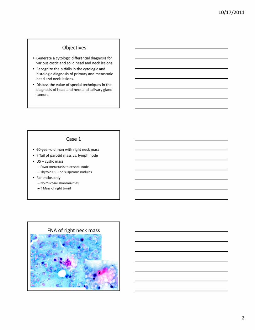
Objectives
• Generate a cytologic differential diagnosis for various cystic and solid head and neck lesions.
• Recognize the pitfalls in the cytologic and histologic diagnosis of primary and metastatic head and neck lesions.
• Discuss the value of special techniques in the diagnosis of head and neck and salivary gland tumors.
Case 1
• 60‐year‐old man with right neck mass
• ? Tail of parotid mass vs. lymph node
• US – cystic mass
– Favor metastasis to cervical node
– Thyroid US – no suspicious nodules
• Panendoscopy
– No mucosal abnormalities
– ? Mass of right tonsil
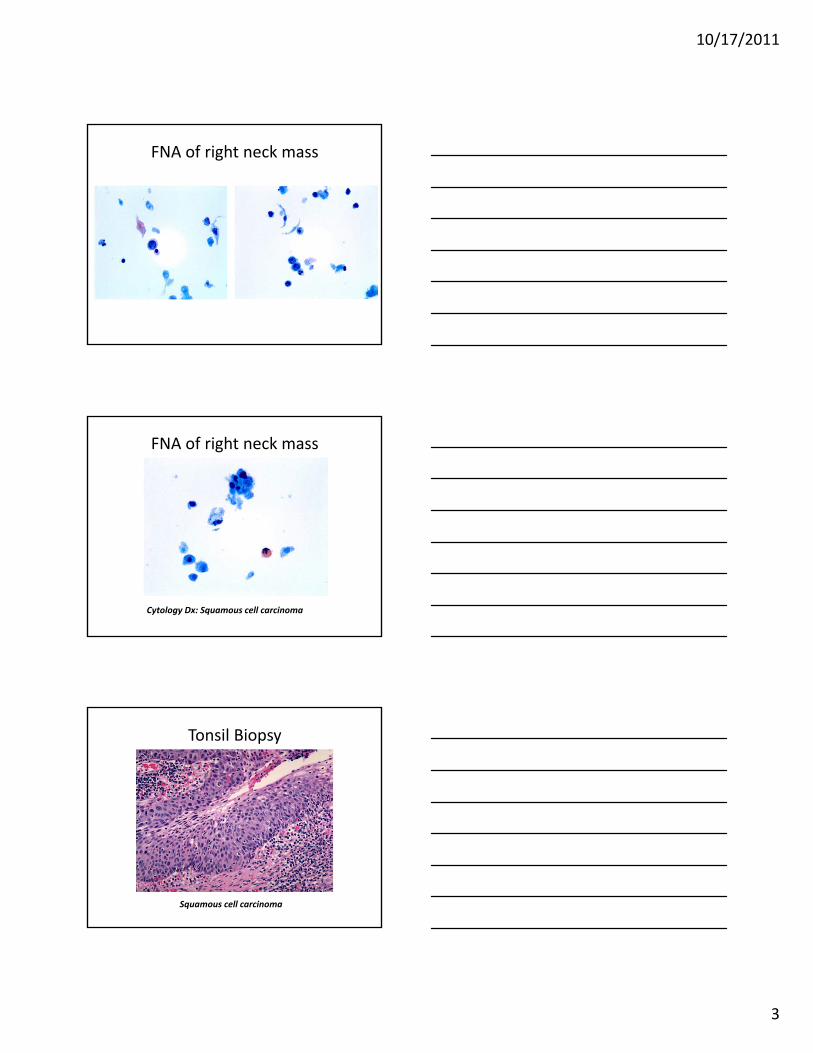
FNA of right neck mass
10/17/2011
3
FNA of right neck mass
FNA of right neck mass
Cytology Dx: Squamous cell carcinoma
Tonsil Biopsy
Squamous cell carcinoma
10/17/2011
4
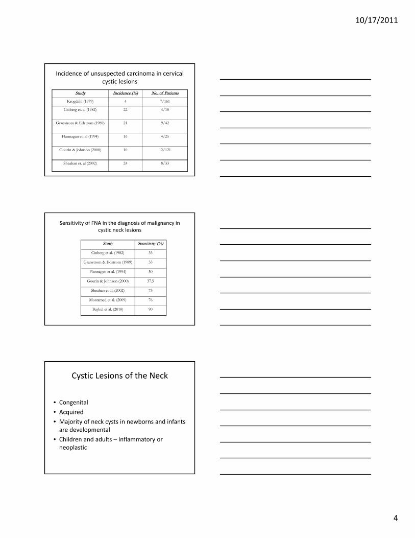
Incidence of unsuspected carcinoma in cervical cystic lesions
Study Incidence (%) No. of Patients
Krogdahl (1979) 4 7/161
Cinberg et. al (1982) 22 4/18
Granstrom & Edstrom (1989) 21 9/42
Flannagan et. al (1994) 16 4/25
Gourin & Johnson (2000) 10 12/121
Sheahan et. al (2002) 24 8/33
Sensitivity of FNA in the diagnosis of malignancy in cystic neck lesions
Study Sensitivity (%)
Cinberg et al. (1982) 33
Granstrom & Edstrom (1989) 33
Flannagan et al. (1994) 50
Gourin & Johnson (2000) 37.5
Sheahan et al. (2002) 73
Moatamed et al. (2009) 76
Baykul et al. (2010) 90
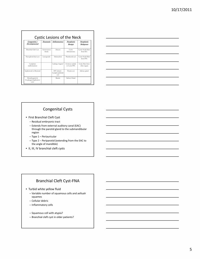
Cystic Lesions of the Neck
• Congenital
• Acquired
• Majority of neck cysts in newborns and infants are developmental
• Children and adults – Inflammatory or neoplastic
10/17/2011
5
Cystic Lesions of the NeckCongenital /
DevelopmentalTraumatic Inflammatory Neoplastic
BenignNeoplasticMalignant
Branchial Cleft Cyst Areriovenous fistula
Abscess Cystic Schwannoma
Cystic Nodal Mets from SCC
Thyroglossal duct cyst Laryngocele Tuberculosis Parathyroid cyst Cystic Nodal Mets from PTC
Lymphatic malformations
Ludwigs Angina* Goitrous nodule or Cystic PTC
Cystic Mets from other organs
Epidermoid or Dermoid HIV related –Lymphoepithelial
cyst
Thymic cyst Salivary gland
Bronchogenic & Esophageal duplication
cysts
Ranula Salivary Gland
Congenital Cysts
• First Branchial Cleft Cyst
– Residual embryonic tract
– Extends from external auditory canal (EAC) through the parotid gland to the submandibular region
– Type 1 – Periauricular
– Type 2 – Periparotid (extending from the EAC to the angle of mandible)
• II, III, IV branchial cleft cysts
Branchial Cleft Cyst‐FNA
• Turbid white yellow fluid
– Variable number of squamous cells and aellualr squames
– Cellular debris
– Inflammatory cells
– Squamous cell with atypia?
– Branchial cleft cyst in older patients?
10/17/2011
6
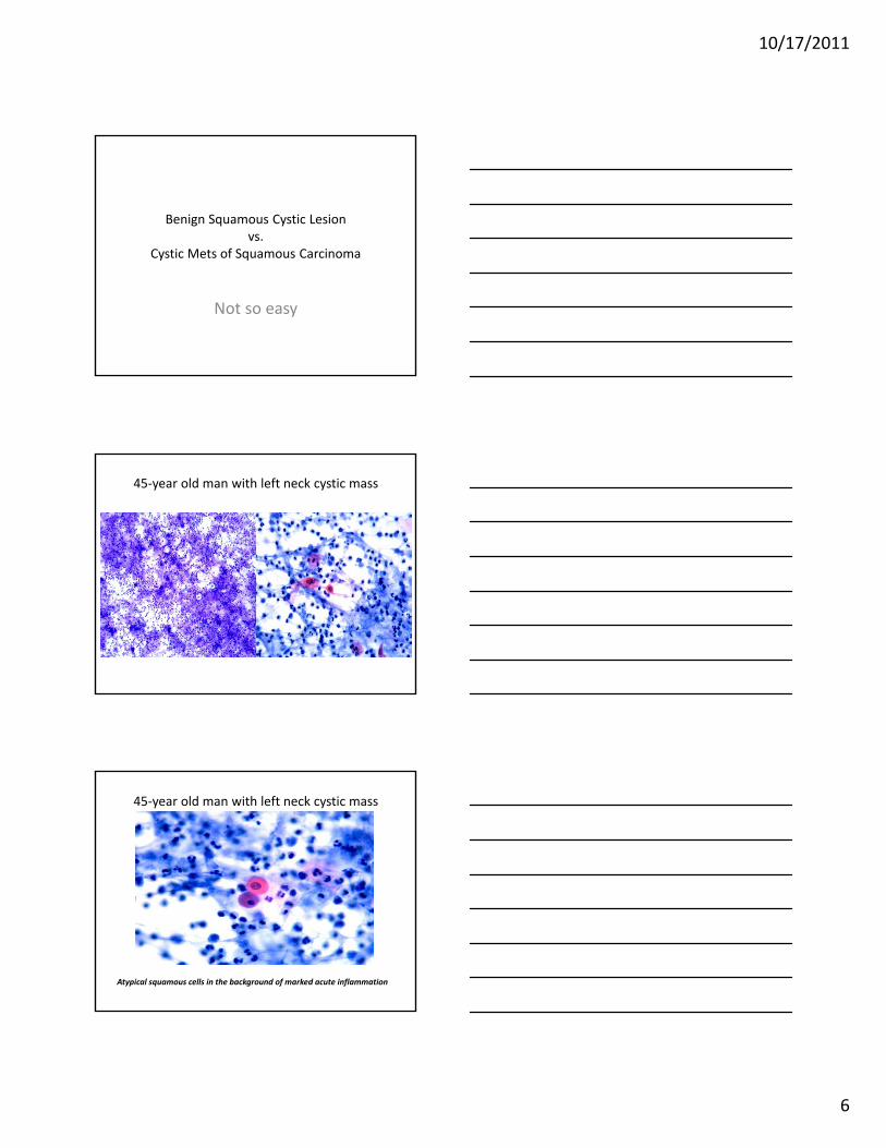
Benign Squamous Cystic Lesion vs.
Cystic Mets of Squamous Carcinoma
Not so easy
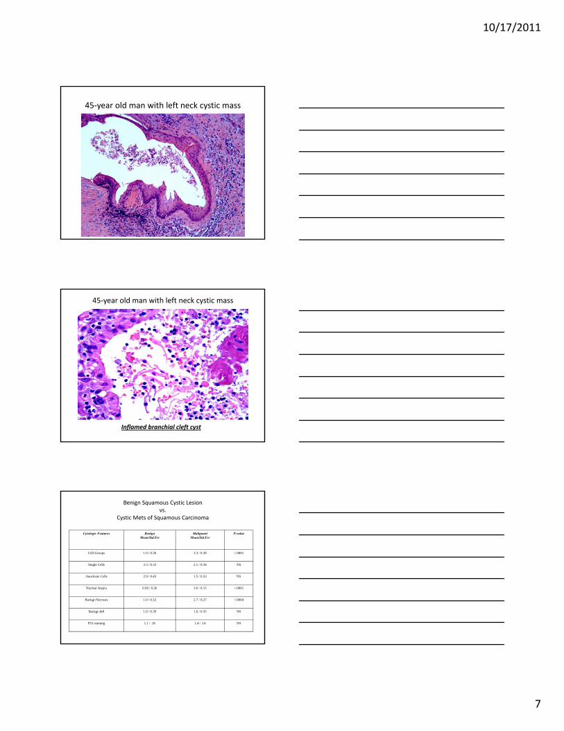
45‐year old man with left neck cystic mass
45‐year old man with left neck cystic mass
Atypical squamous cells in the background of marked acute inflammation
10/17/2011
7
45‐year old man with left neck cystic mass
45‐year old man with left neck cystic mass
Inflamed branchial cleft cyst
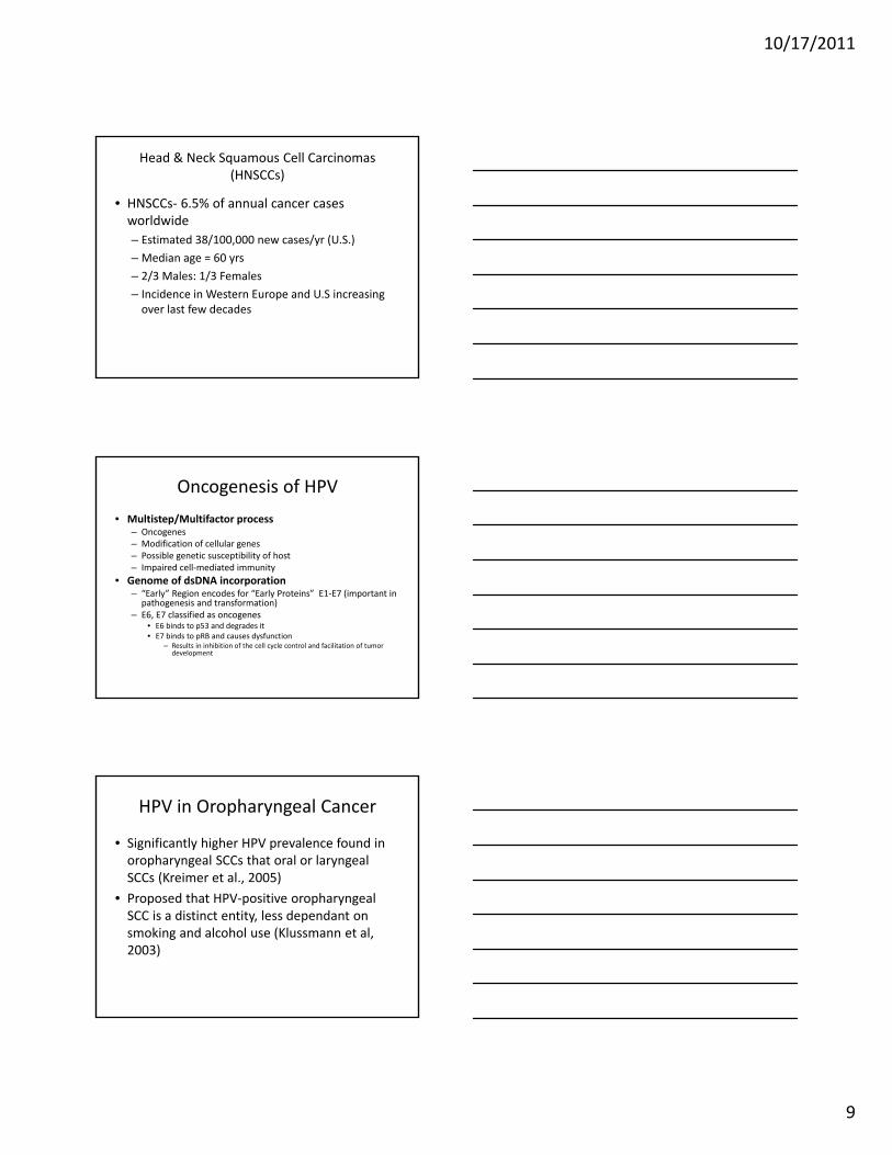
Benign Squamous Cystic Lesion vs.
Cystic Mets of Squamous Carcinoma
Cytologic Features BenignMean/Std.Err
MalignantMean/Std.Err
P-value
Cell-Groups 1.0 / 0.36 3.3 / 0.28 <.0001
Single Cells 3.3 / 0.33 2.1 / 0.34 NS
Anucleate Cells 2.9 / 0.43 1.5 / 0.33 NS
Nuclear Atypia 0.58 / 0.26 3.6 / 0.15 <.0001
Backgr-Necrosis 1.0 / 0.32 2.7 / 0.27 <.0004
Backgr-Infl 1.0 / 0.39 1.6 / 0.35 NS
P53 staining 1.1 / .16 1.4 / .14 NS

10/17/2011
8
Benign Squamous Cystic Lesion vs.
Cystic Mets of Squamous Carcinoma
Inflamed Branchial Cleft Cyst Cystic Mets of Squamous Carcinoma
Benign Squamous Cystic Lesion vs.
Cystic Mets of Squamous Carcinoma
• Clues– Clinical history – could be occult primary
– Inflammation – common in benign• Acute inflammation with keratinizing squamous lesions
– N/C ratio• Maintained in benign lesions
– Nuclear atypia
– Excisional biopsy
– P53, p16
HPV in Squamous Cell Carcinoma
10/17/2011
9
Head & Neck Squamous Cell Carcinomas (HNSCCs)
• HNSCCs‐ 6.5% of annual cancer cases worldwide
– Estimated 38/100,000 new cases/yr (U.S.)
– Median age = 60 yrs
– 2/3 Males: 1/3 Females
– Incidence in Western Europe and U.S increasing over last few decades
Oncogenesis of HPV
• Multistep/Multifactor process– Oncogenes– Modification of cellular genes– Possible genetic susceptibility of host– Impaired cell‐mediated immunity
• Genome of dsDNA incorporation– “Early” Region encodes for “Early Proteins” E1‐E7 (important in
pathogenesis and transformation)– E6, E7 classified as oncogenes
• E6 binds to p53 and degrades it• E7 binds to pRB and causes dysfunction
– Results in inhibition of the cell cycle control and facilitation of tumor development
HPV in Oropharyngeal Cancer
• Significantly higher HPV prevalence found in oropharyngeal SCCs that oral or laryngeal SCCs (Kreimer et al., 2005)
• Proposed that HPV‐positive oropharyngeal SCC is a distinct entity, less dependant on smoking and alcohol use (Klussmann et al, 2003)

10/17/2011
10
HPV in Other Head & Neck Tumors
• Prevalence varies in the literature
– Possibly due to methods of analysis
• 14‐35% by PCR
• 25% by Southern Blot
• 18% by FISH
– Most common HPV locations (other than oropharynx)‐ [Dahlstrand & Dalianis, 2005]
• Tongue Cancer (19‐100%)
• Laryngeal Cancer (10‐50%)
Termine et al. 2008
Subgroup Number of Studies Mean prevalence (%)
HNSCC n.s. 15 24.1
OSCC 47 38.1
ISH based 13 32.9
PCR based 52 34.8
HNSCC n.s.s. ISH based 2 n.c.
HNSCC n.s.s. PCR based 13 20.8
OSCC PCR based 36 39.9
OSCC ISH based 11 29.8
Overall 62 34.5
HPV Status and Prognosis
10/17/2011
11
HPV and Prognosis in Oropharyngeal Cancer
• HPV may be a favorable prognostic factor (Gillison et al, 2000;Mellin et al., 2000)
– Mellin et al., 2000
• 60 pts with tonsillar cancer
– 52% of pts with HPV +ve tumors were disease free after 3 years
– 21% of pts with HPV –ve tumors were disease free after 3 years
• Pts with HPV +ve tumors had significantly increased 5‐year survival rates compared to HPV –ve tumors (53% vs. 31%, p=0.047)
• HPV +ve tumors favorable independent of tumor stage, gender, age or differentiation
HPV and Prognosis in Oropharyngeal Cancer
• 253 head and neck cancer patients– 60 oropharyngeal cancers (mostly tonsil)
• Results:– Disease‐specific survival significantly higher for HPV +ve tumors
– No change in disease‐specific survival for other head and neck cancers
• Multiples studies have shown no change in survival for HPV +ve tumors (except oropharynx)[reviewed by Dahlstrand & Dalia, 2005]
Gillison et al., 2000
How Adequate are Head and Neck Fine‐needle Aspiration Specimens for HPV Molecular Analysis?
Vs.

10/17/2011
12
How Adequate are Head and Neck Fine‐needle Aspiration Specimens for HPV Molecular Analysis?
• 42 specimens in 40 patients
– 37 LN’s & 5 others ites
– On‐site evaluation in 41 (98%)
– Final diagnosis – SCC in all
– 9 cases >80% tumor necrosis
– Adequate DNA for molecular analysis ‐28 (67%)
• 7 (25%) necrotic specimens had adequate for HPV analysis
Thyroglossal Duct Cyst
• Most common congenital neck mass
• Located in mid‐line or paramedian (left side)
• Closely related to hyoid bone– 20% Suprahyoid, 65% infrahyoid & 15% at the level of hyoid bone (Grossman & Yousem 1994)
• Characteristic appearance on US, CT and MRI– Hypoechoic thin walled cyst
– Debris – Hemorrhage or infection
– Solid mass – Carcinoma (95% PTC & 5% SCC)
Thyroglossal Duct Cyst
• Differential diagnosis
– Dermoid cyst
– Necrotic lymphadenopathy
– Cystic goitrous nodule arising from thyroid isthmus
– Thymic cyst
– Branchial cleft cyst – paramedian location
– Cystic hygroma – paramedian location
10/17/2011
13
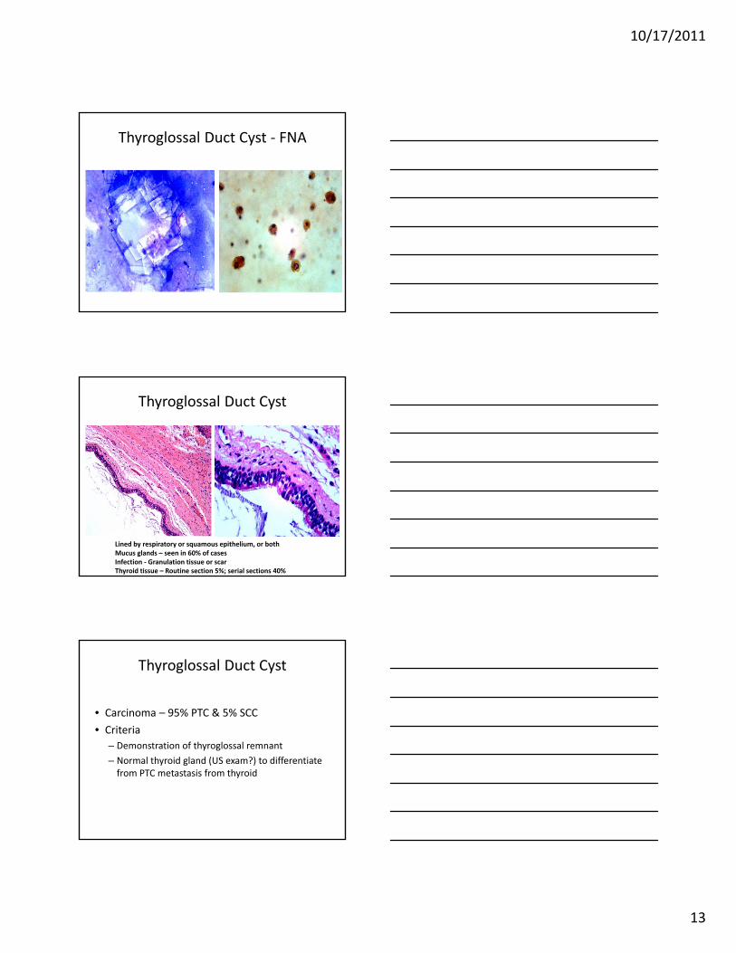
Thyroglossal Duct Cyst ‐ FNA
Thyroglossal Duct Cyst
Lined by respiratory or squamous epithelium, or bothMucus glands – seen in 60% of casesInfection ‐ Granulation tissue or scar Thyroid tissue – Routine section 5%; serial sections 40%
Thyroglossal Duct Cyst
• Carcinoma – 95% PTC & 5% SCC
• Criteria
– Demonstration of thyroglossal remnant
– Normal thyroid gland (US exam?) to differentiate from PTC metastasis from thyroid
10/17/2011
14
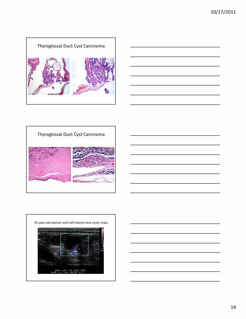
Thyroglossal Duct Cyst Carcinoma
Thyroglossal Duct Cyst Carcinoma
35‐year‐old woman with left lateral neck cystic mass
10/17/2011
15
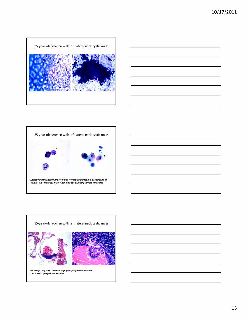
35‐year‐old woman with left lateral neck cystic mass
35‐year‐old woman with left lateral neck cystic mass
Cytology Diagnosis: Lymphocytes and few macrophages in a background of “colloid” type material. Rule‐out metastatic papillary thyroid carcinoma
35‐year‐old woman with left lateral neck cystic mass
Histology Diagnosis: Metastatic papillary thyroid carcinoma; TTF‐1 and Thyroglobulin positive
10/17/2011
16
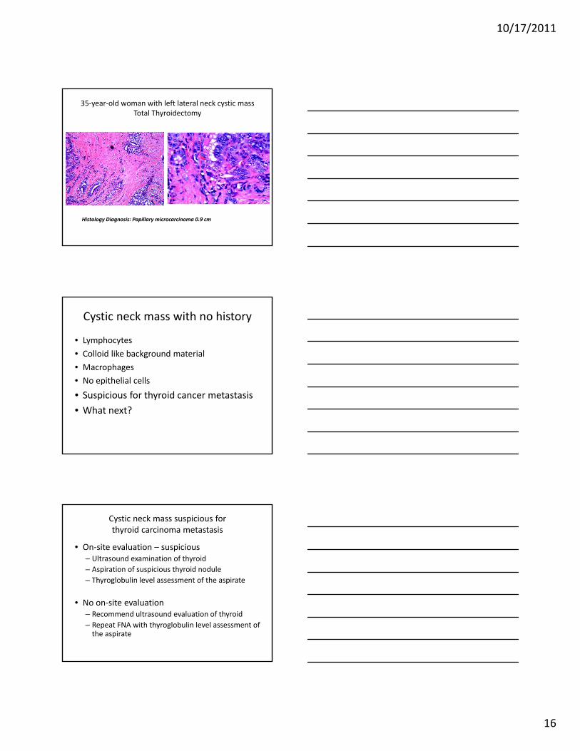
35‐year‐old woman with left lateral neck cystic massTotal Thyroidectomy
Histology Diagnosis: Papillary microcarcinoma 0.9 cm
Cystic neck mass with no history
• Lymphocytes
• Colloid like background material
• Macrophages
• No epithelial cells
• Suspicious for thyroid cancer metastasis
• What next?
Cystic neck mass suspicious forthyroid carcinoma metastasis
• On‐site evaluation – suspicious– Ultrasound examination of thyroid
– Aspiration of suspicious thyroid nodule
– Thyroglobulin level assessment of the aspirate
• No on‐site evaluation– Recommend ultrasound evaluation of thyroid
– Repeat FNA with thyroglobulin level assessment of the aspirate
10/17/2011
17
Thyroglobulin measurement in the lymph node aspirates of patients with PTC
TG Levels ≥10 ng/ml TG Levels ≤10 ng/ml
Cytologic-DX
PTC Other CA No F/U PTC Other CA No F/U
PTC (n=39) 25 0 3 10* 1** 0
NTS (n=35) 5 0 0 0 0 30
NDX(n=23) 3 0 1 0 0 19
ATYP (n=15) 9 0 0 4* 0 1
OTHER (n=3) 0 1*** 0 0 1**** 0
DX = Diagnosis, TG = Thyroglobulin, F/U = surgical pathology follow- up, PTC = Papillary thyroid carcinoma, CA = Carcinoma, NTS = No tumor seen, NDX = Non-diagnostic, ATYP = Atypical/Suspicious, * = includes cases of tall cell variant of papillary carcinoma, ** = metastatic well-differentiated follicular derived carcinoma *** = poorly differentiated carcinoma, **** = carcinoma not otherwise specified.
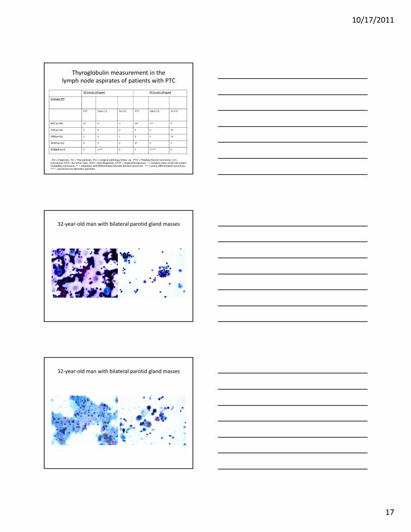
32‐year‐old man with bilateral parotid gland masses
32‐year‐old man with bilateral parotid gland masses
10/17/2011
18
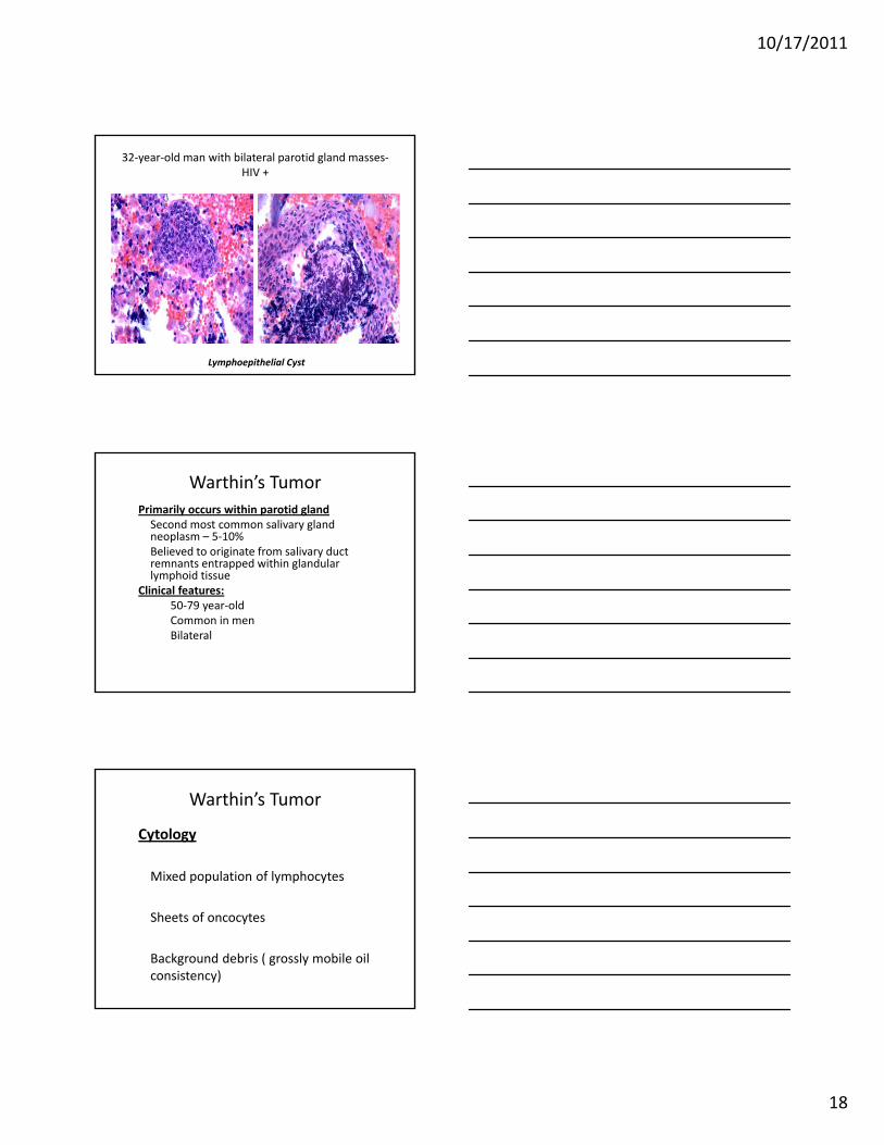
32‐year‐old man with bilateral parotid gland masses‐HIV +
Lymphoepithelial Cyst
Warthin’s Tumor
Primarily occurs within parotid glandSecond most common salivary gland neoplasm – 5‐10%Believed to originate from salivary duct remnants entrapped within glandular lymphoid tissue
Clinical features:50‐79 year‐oldCommon in menBilateral
Warthin’s Tumor
Cytology
Mixed population of lymphocytes
Sheets of oncocytes
Background debris ( grossly mobile oil consistency)
10/17/2011
19
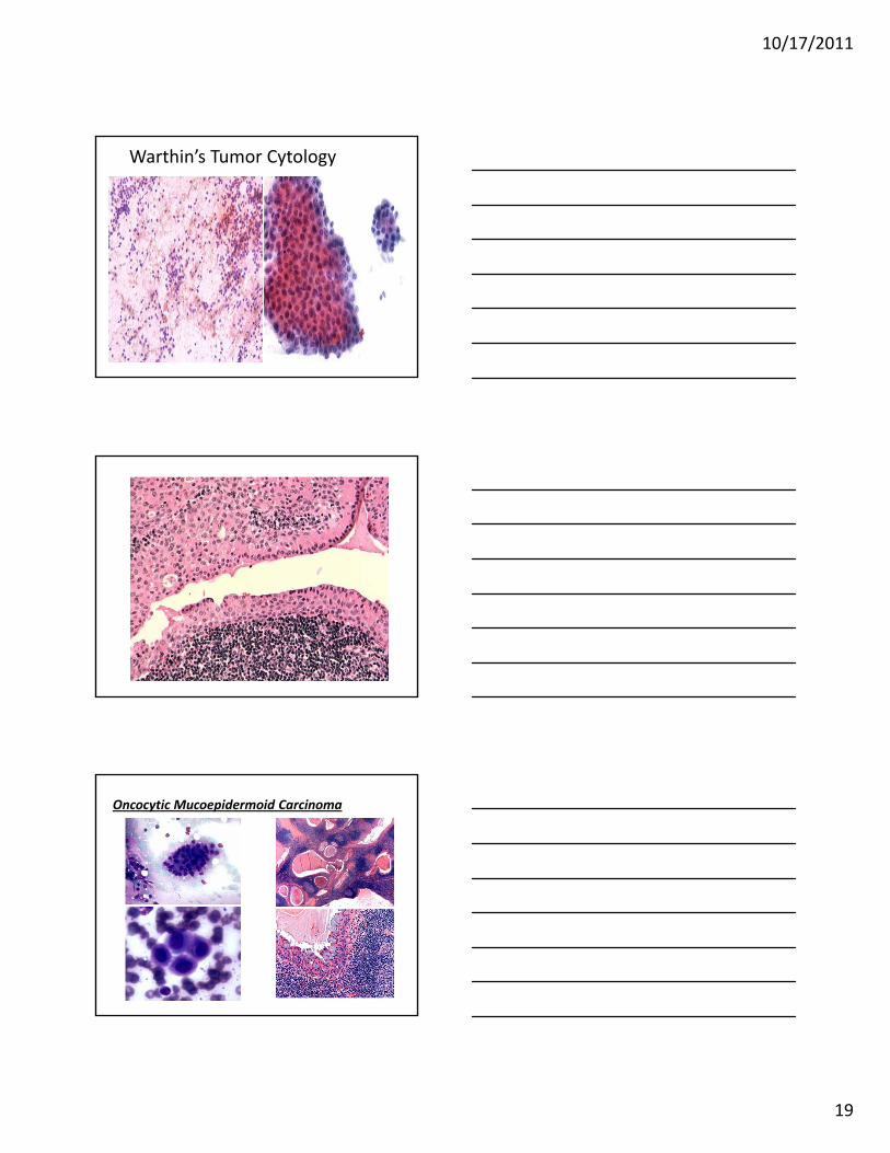
Warthin’s Tumor Cytology
Oncocytic Mucoepidermoid Carcinoma
10/17/2011
20
Overlapping Cytologic Features
• Lymphocytes
–Chai et al (Diag Cytopathol 1997)– 61 cases with prominent lymphoid component
• Warthins 33 cases– Warthins‐31, Benign cyst‐1, SCCA‐1
• Other epithelial malignancies 6 cases– Oncocytoma; Pleomorphic adenoma, ACC
• Lymphomas 12 cases
• Benign 10
Overlapping Cytologic Features
• Lymphocytes– Intraparotid LN
– Lymphoepithelial cyst
– Chronic Sialadenitis
– Warthin’s
– Acinic cell carcinoma
– Mucoepidermoid Carcinoma
– Lymphoma
Approach to cystic neck lesion
Background – Mucoid
• Histiocytes & lymphocytes– Mucus retention cyst
• Salivary gland• Chronic sialadenitis
• Sialolithiasis
• Mucoepidermoid carcinoma
• Myxoid and chondroid fragments – Pleomorphic adenoma – rare
• Atypical cells• Malignancy – Mets vs. primary
10/17/2011
21
Approach to cystic neck lesionBackground – watery proteinaceous fluid
• Lymphocytic infiltrate & few epithelial cells– Lymphoepithelial cyst– Thyroglossal duct cyst – midline location
• Salivary gland– Lymphocytes and oncocytes
• Warthin’s tumor
– Atypical keratinized cells• Squamous cell carcinoma• Metaplasia in benign tumor – history of previous FNA
• Keratinized cells– Atypical – Squamous cell carcinoma– Branchial cleft cyst
Approach to cystic neck lesion
• Benign vs. malignant squamous cystic lesion
– Background – inflammation?
– Necrotic debris
– Cellularity – increased in SCC?
– Abnormal keratinization / Dyskeratosis
– Nuclear atypia
Approach to cystic neck lesion
• Cystic mass suspicious for thyroid cancer
– Recommend ultrasound of thyroid and aspiration of suspicious nodules
– TTF‐1 and thyroglobulin (should do both) if enough cells
– Thyroglobulin assessment of FNA specimen
10/17/2011
22
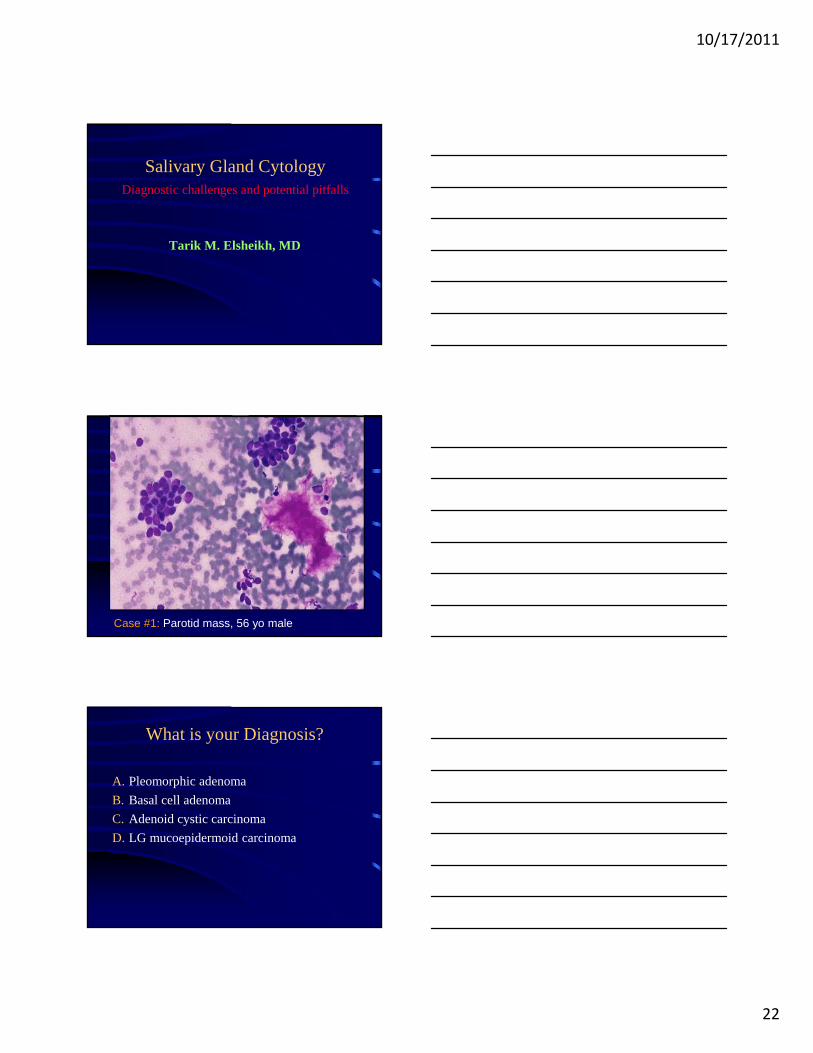
Salivary Gland CytologyDiagnostic challenges and potential pitfalls
Tarik M. Elsheikh, MD
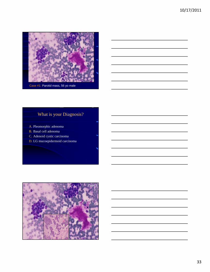
Case #1: Parotid mass, 56 yo male
What is your Diagnosis?
A. Pleomorphic adenoma
B. Basal cell adenoma
C. Adenoid cystic carcinoma
D. LG mucoepidermoid carcinoma
10/17/2011
23
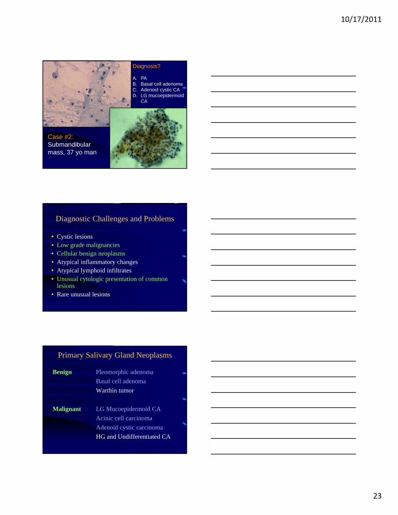
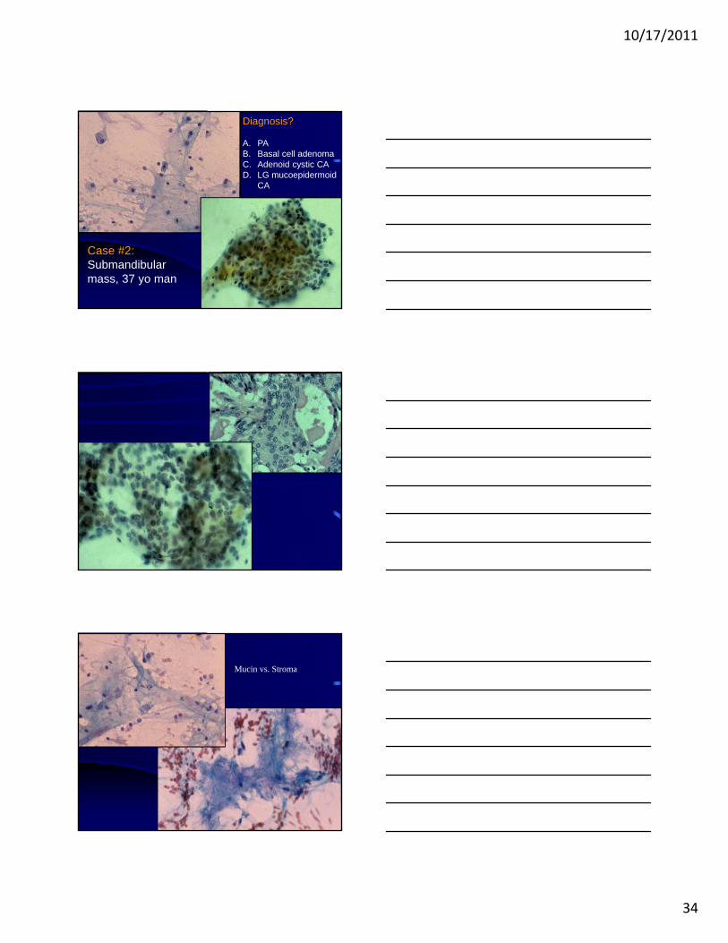
Case #2: Submandibular mass, 37 yo man
Diagnosis?
A. PAB. Basal cell adenomaC. Adenoid cystic CAD. LG mucoepidermoid
CA
Diagnostic Challenges and Problems
• Cystic lesions• Low grade malignancies• Cellular benign neoplasms• Atypical inflammatory changes• Atypical lymphoid infiltrates• Unusual cytologic presentation of common
lesions• Rare unusual lesions
Primary Salivary Gland Neoplasms
Benign Pleomorphic adenoma
Basal cell adenoma
Warthin tumor
Malignant LG Mucoepidermoid CA
Acinic cell carcinoma
Adenoid cystic carcinoma
HG and Undifferentiated CA
10/17/2011
24
Key Cytologic Features
• Low power architectural appearance
• Size of cells and amount of cytoplasm
• Nucleoli
• Character of single cells in background
• Character of background substance
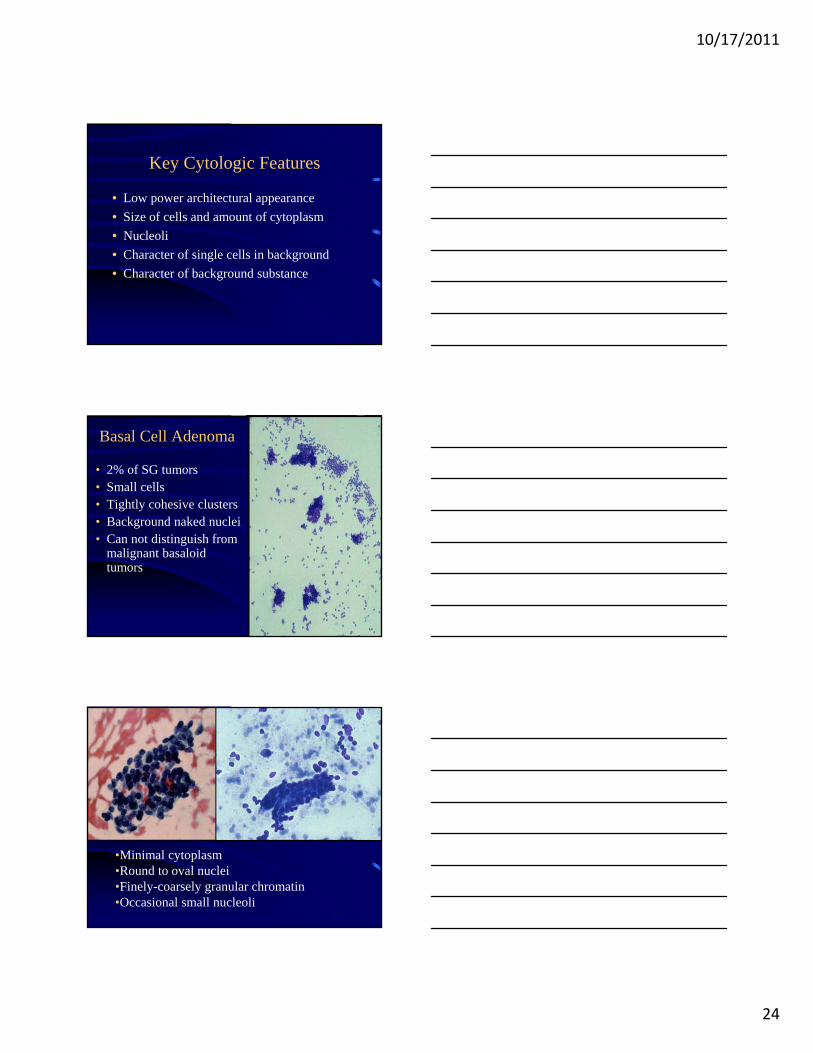
Basal Cell Adenoma
• 2% of SG tumors• Small cells• Tightly cohesive clusters • Background naked nuclei• Can not distinguish from
malignant basaloid tumors
•Minimal cytoplasm •Round to oval nuclei•Finely-coarsely granular chromatin•Occasional small nucleoli
10/17/2011
25
• Amorphous extracellular hyaline material may be seen at periphery of cell clusters.• Not specific for membranous BCA
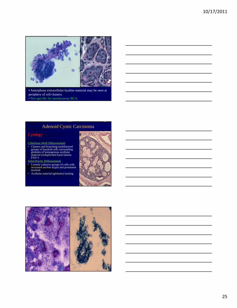
Adenoid Cystic Carcinoma
Cytology
Cribriform (Well Differentiated)• Clusters and branching multilayered
groups of basaloid cells surrounding globules of homogenous acellular material (reduplicated basal lamina PAS+)
Solid (Poorly Differentiated)• Loosely cohesive groups of cells with
increased nuclear atypia and prominent nucleoli
• Acellular material (globules) lacking
10/17/2011
26
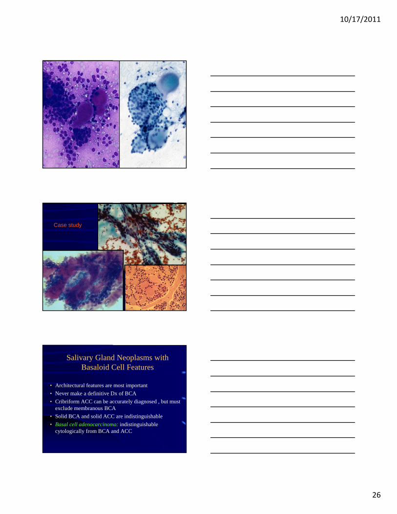
Case study
Salivary Gland Neoplasms with Basaloid Cell Features
• Architectural features are most important
• Never make a definitive Dx of BCA
• Cribriform ACC can be accurately diagnosed , but must exclude membranous BCA
• Solid BCA and solid ACC are indistinguishable
• Basal cell adenocarcinoma: indistinguishable cytologically from BCA and ACC
10/17/2011
27
Sample Cytologic Diagnosis
DX: Cellular neoplasm with basaloid cell features, see comment
Comment: Differential diagnosis includes basal cell adenoma and adenoid cystic carcinoma (Ki 67). Basal cell adenoma is favored (suggested).Histologic confirmation is needed for a definitive diagnosis.
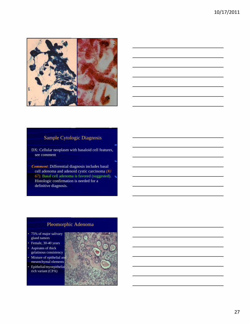
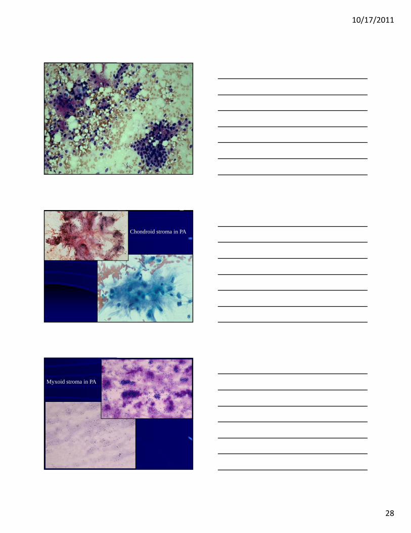
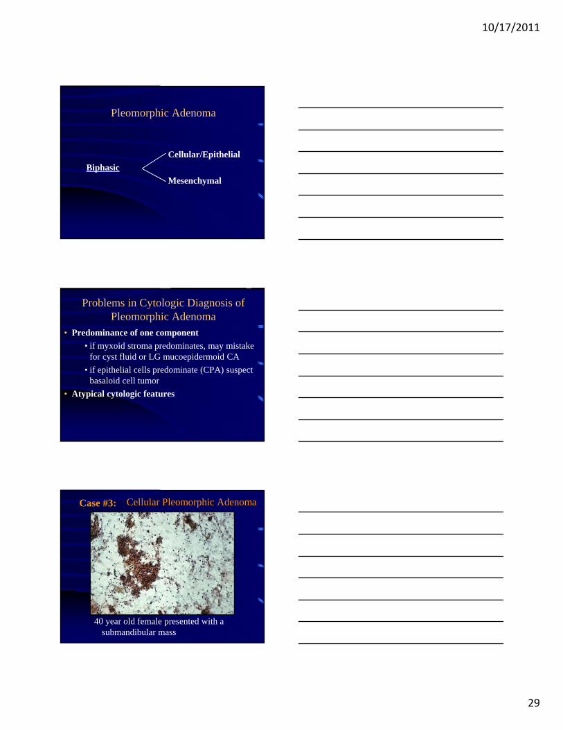
Pleomorphic Adenoma
• 75% of major salivary gland tumors
• Female, 30-40 years
• Aspirates of thick gelatinous consistency
• Mixture of epithelial and mesenchymal elements
• Epithelial/myoepithelial rich variant (CPA)
10/17/2011
28
Chondroid stroma in PA
Myxoid stroma in PA
10/17/2011
29
Pleomorphic Adenoma
Cellular/Epithelial
Biphasic
Mesenchymal
Problems in Cytologic Diagnosis of Pleomorphic Adenoma
• Predominance of one component
• if myxoid stroma predominates, may mistake for cyst fluid or LG mucoepidermoid CA
• if epithelial cells predominate (CPA) suspect basaloid cell tumor
• Atypical cytologic features
Cellular Pleomorphic Adenoma
40 year old female presented with a submandibular mass
Case #3:
10/17/2011
30
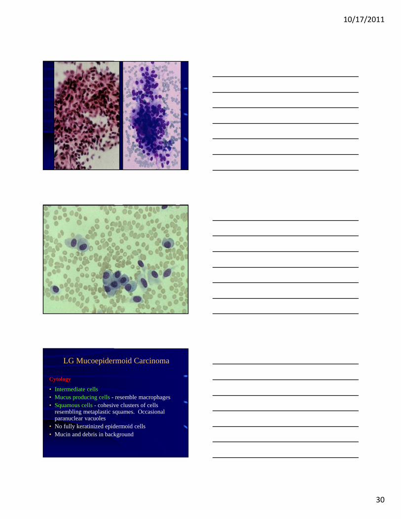
LG Mucoepidermoid Carcinoma
Cytology
• Intermediate cells • Mucus producing cells - resemble macrophages• Squamous cells - cohesive clusters of cells
resembling metaplastic squames. Occasional paranuclear vacuoles
• No fully keratinized epidermoid cells• Mucin and debris in background
10/17/2011
31
10/17/2011
32
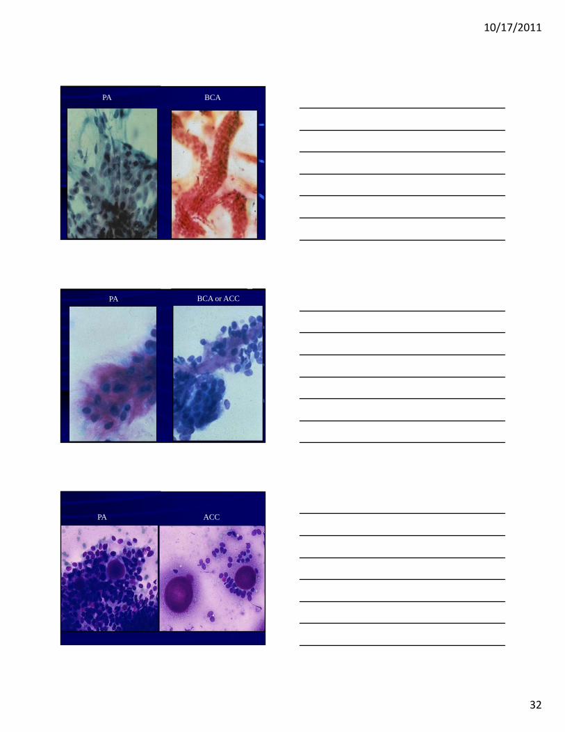
BCAPA
BCA or ACCPA
PA ACC
10/17/2011
33
Case #1: Parotid mass, 56 yo male
What is your Diagnosis?
A. Pleomorphic adenoma
B. Basal cell adenoma
C. Adenoid cystic carcinoma
D. LG mucoepidermoid carcinoma
Case 1
10/17/2011
34
Case #2: Submandibular mass, 37 yo man
Diagnosis?
A. PAB. Basal cell adenomaC. Adenoid cystic CAD. LG mucoepidermoid
CA
Mucin vs. Stroma
10/17/2011
35
The Many Cytologic Faces of Pleomorphic Adenoma
• Basal Cell Adenoma• Adenoid Cystic Carcinoma• Low grade malignancies such as LG
mucoepidermoid CA and acinic cell CA
FNA of Salivary GlandsSummary
• Must exclude PA before making a diagnosis of another neoplasm
• Must exclude ACC and low grade malignancies before making a Dx of PA
• FNA can distinguish in most instances between basaloid neoplasms (ACC, BCA) and PA
FNA of Salivary GlandsSummary 2
• Cellular neoplasm NOS (LG CA vs. B9)
• Familiarity with variable FNA appearances of SG tumors and awareness of potential pitfalls can prevent many false positive and negative diagnoses
• FNA should be interpreted in context of clinical and radiologic findings