Embed Size (px)
Citation preview
Subcutaneous mycoses
ZYCOMYCOSISLECTURE NO (8)
Dalia Kamal Eldien Mohammed
The main subcutaneous fungal infections include:
Mycetoma Chromoblastomycosis Sporotrichosis Lobomycosis Rhinosporidiosis Subcutaneous zygomycosis Subcutaneous phaeohyphomycosis.
introduction
The zygomycetes are a relatively small group in the fungi kingdom and belong to the Phylum Zygomycota.
They include the familiar bread mold Rhizopus stolonifer, which rapidly propagates on the surfaces of breads, fruits, and vegetables.
They are mostly terrestrial in habitat, living in soil or on plants and animals.
Zygomycetes
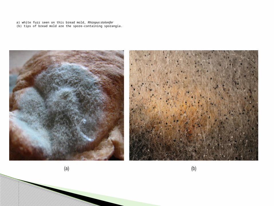
a) white fuzz seen on this bread mold, Rhizopus stolonifer (b) tips of bread mold are the spore-containing sporangia.

Rotting of fruits
The Zygomycetes, a class of fungi with a ubiquitous and worldwide distribution that is characterized by aseptate hyphae, fast growing, saprophytic fungi.
The fungi usually reproduce asexually by producing sporangio spores .
When spores land on a suitable substrate, they germinate and produce a new mycelium.
Sexual reproduction starts when conditions become unfavorable, produce the zygospore
Zygomycetes
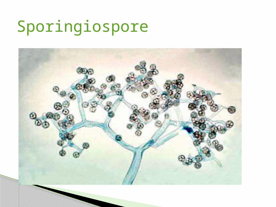
Sporingiospore

The term zygomycosis describes in the broadest sense any infection due to a member of the Zygomycetes.
Zygomycosis is the third most common invasive fungal infection after candidiasis and aspergillosis
invasive fungal infections common in both immunocompetent and immunocompromised individuals
Zygomycosis
This class of fungi encompasses two orders:
Mucorales Entomophthorales
The order Mucorales include several genera Rhizopus Rhizomucor Mucor Absidia involved in rhinocerebral, pulmonary, cutaneous,
gastrointestinal and other less frequent infections in immunocompetent and immunocompromised individuals, and is characterized by a tendency to disseminate
Mucormycosis is the correct term for infections due to fungi of this order.
The term zygomycosis is used to describe any invasive infection due to zygomycetes, although it is frequently used interchangeably with the term mucormycosis
Mucorales
The order Entomophthorales includeConidiobolus Basiodobolus sppSubcutaneous zygomycosis, which is typically
seen in children and adolescents, results from infection with Basidiobolus haptosporus.
It first develops as a localized lesion, usually on the thighs or buttocks, and it spreads slowly to form a hard, painless, non-pitting mass involving the cutaneous and subcutaneous tissues. The mass is shiny, but may later become ulcerated.
Entomophthorales
The symptoms depend on where in the body the fungus is growing.
Mucormycosis most commonly affects the sinuses or lungs.
Symptoms of sinus infections include fever, headache, and sinus pain. Lung infections with the fungus can cause fever and cough.
Skin infections can develop after the fungus enters through a break in the skin caused by surgery, burns, or trauma. A skin infection can look like blisters or ulcers, and the infected tissue may turn black. Other symptoms of a skin infection include fever and tenderness pain, heat, excessive redness, or swelling around a wound.
If the infection is not treated quickly, the fungus can spread throughout the body, and the infection is often fatal.
Symptoms of zygomycosis
Portals of entry of zygomycetes are usually the lungs, skin, and gastrointestinal tract.
These rare yet serious and potentially life -threatening fungal infections, usually affect the face or oropharyngeal(nose/mouth) cavity, lungs, gastrointestinal tract, skin, or less commonly other organ systems.
These types of infections are also common after natural disasters, such as tornadoes
A characteristic property of zygomycetes is their tendency to invade blood vessels and to cause thrombosis—processes that result in subsequent necrosis of involved tissues.
Clinical presentation
Risk factors associated with zygomycosis include: Prolonged neutropenia Use of corticosteroids Solid organ or haematopoietic stem cell
transplantation AIDS Poorly controlled diabetes mellitus Burns& wounds Malnutrition Extremes of age Intravenous drug
Specimen: Skin scrapings from cutaneous lesionssputum and needle biopsies from pulmonary
lesions Nasal discharges, scrapings and aspirates from
Sinuses in patients with rhinocerebral lesions Biopsy tissue from patients with
gastrointestinal and/or disseminated disease.
Laboratory diagnosis:

Direct Microscopy: 10% KOH & Parker ink or Calcofluor mountsTissue sections should be stained with H&E.Results: Examine specimens for broad,
infrequently septate, thin-walled hyphaeAs a rule, a positive direct microscopy,
especially from a sterile site, should be considered significant.
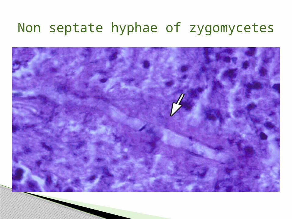
Non septate hyphae of zygomycetes
Culture: Inoculate specimens onto Sabouraud's dextrose
agar. Most zygomycetes are sensitive to
cycloheximide (actidione).Colonial morphology: Look for fast growing, white to grey or
brownish, downy colonies.Serology: some laboratories have developed ELISA tests
for the detection of antibodies to Zygomycetes.
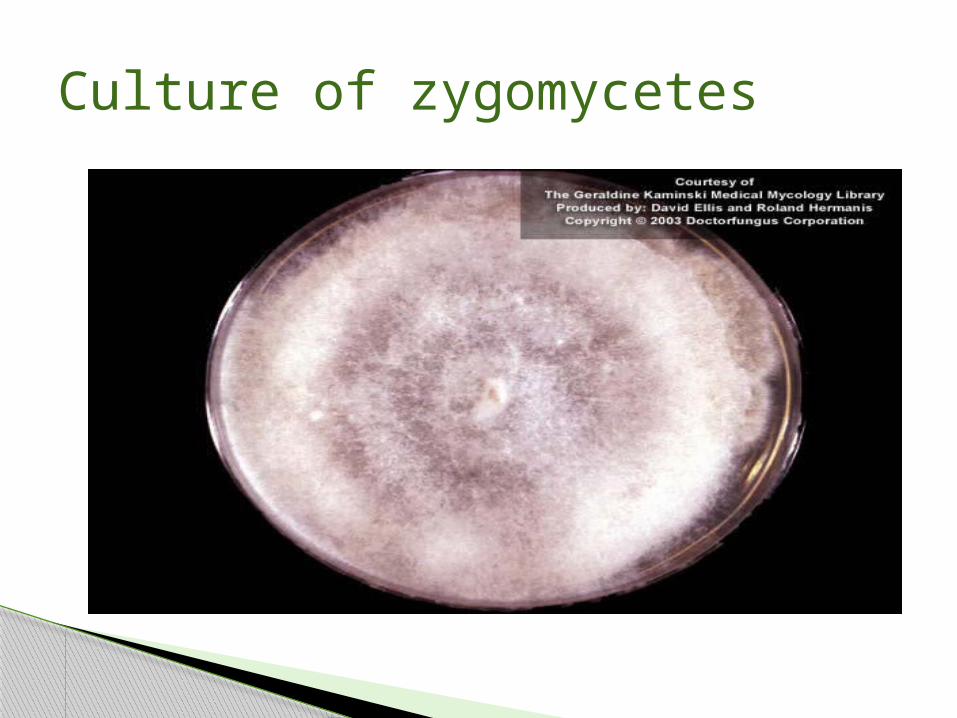
Culture of zygomycetes

Treatment consists of prompt and intensive antifungal drug therapy and surgery to remove the infected tissue.
The prognosis varies vastly depending upon an individual patient's circumstances.
Treatment

Source: Boundless. “Zygomycetes.” Boundless Microbiology. Boundless, 03 Jul. 2014. Retrieved 08 Dec. 2014from
Kwon-Chung KJ and JE Bennett 1992. Medical Mycology Lea & Febiger.
Reference