Embed Size (px)
Citation preview

Journal of Laboratory Physicians / Jan-Jun 2010 / Vol-2 / Issue-144
Disseminated Cutaneous Rhinosporidiosis
Kishan Prasad, S Veena, H S Permi, S Teerthanath, K Padma Shetty, J P Shetty
Department of Pathology, K S Hegde Medical Academy, Mangalore, Karnataka, India
Address for Correspondence: Dr. Veena Hemanth, E-mail: [email protected]
ABSTRACT
Rhinosporidiosis is a granulomatous disorder caused by Rhinosporidium seeberi. It frequently involves the nasopharynx and occasionally affects the skin. We hereby report a case of 46–year-old male who presented with multiple cutaneous plaque-like lesions with nasal obstruction. On examination, multiple cutaneous lesions were found to be present on malar aspect, infraorbital and supraorbital region, right shoulder and over the back near the tip of scapula. Oral cavity revealed a polypoidal lesion, in the base of the tongue. Excision biopsy confirmed the lesions as disseminated cutaneous rhinosporidiosis. The patient was treated with excision and dapsone therapy. After 1 year of therapy, the patient has disease-free survival. We report this rare case of rhinosporidiosis with disseminated cutaneous involvement.
Keywords: Rhinosporidiosis, Rhinosporidium seeberi, disseminated, cutaneous
DOI: 10.4103/0974-2727.66706 www.jlponline.org
INTRODUCTION
R hinosporidiosis is a chronic granulomatous infection caused by Rhinosporidium seeberi is
endemic in India and in Sri Lanka but has also been reported from United States, South America and Iran.[1] Infection is usually caused by contact with fresh water such as swimming pools. Rhinosporidiosis frequently involves the nasopharynx (70%) presenting as painless, friable, polypoidal growth which may hang anteriorly into the nares and posteriorly into the pharynx. Cutaneous dissemination although known, is quite rare.[2] This disease presents to dermatologists and ophthalmologists with the cutaneous, subcutaneous and ocular lesions. They commonly manifest as a sessile or pedunculated vascular polyp in nasal mucosa, nasopharynx and soft palate.[2] The cutaneous lesions without mucosal involvement are extremely rare and they presents as asymptomatic warty growth. We report this rare case of disseminated cutaneous rhinosporidiosis with the pharyngeal lesion.
CASE REPORT
A 46-year- old man presented with multiple cutaneous lesions over the face & back [Figures 1 and 2] with mass in the oral cavity since 1 year. He had nasal obstruction, mouth breathing since 4 years. On
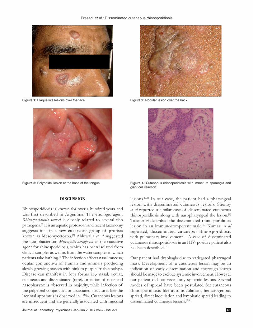
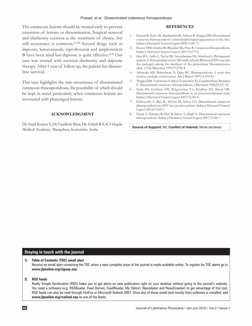
examination, cutaneous lesions were present on malar aspect, infraorbital region, supraorbital region, right shoulder and over the back near the tip of scapula. Lesions were progressively increasing in size and were painless, plaque like with no discharge. Few were nodular lesions with central ulceration. On oral examination, polypoidal lesion was found to be present in the base of tongue which moved with deglutition [Figure 3]. Pharyngeal rhinosporidiosis with disseminated cutaneous involvement was considered in clinical diagnosis. His complete hemogram, blood sugar, liver and renal functions tests were within normal limits. Serology for HIV, HBs Ag and HCV were negative. Excision biopsy from cutaneous lesions and pharyngeal mass was sent for histopathology in 10% formalin. On gross, there were multiple grey white tissue bits; few were polypoidal in nature, larger one measuring 2.5×1×1cms. Cut section showed gelatinous and mucoid areas. Microscopy showed hyperplasic epithelium, with numerous globular cysts of varying sizes representing immature and few mature sporangia in the upper dermis. There was infiltration by chronic inflammatory cells and ruptured sporangia with release of endospores with giant cell reaction [Figure 4]. Final diagnosis of pharyngeal rhinosporidiosis with disseminated cutaneous involvement was made.
Cas
e R
epor
t
Published online: 2020-01-29
Journal of Laboratory Physicians / Jan-Jun 2010 / Vol-2 / Issue-1 45
DISCUSSION
Rhinosporidiosis is known for over a hundred years and was first described in Argentina. The etiologic agent Rhinosporidiosis seeberi is closely related to several fish pathogens.[2] It is an aquatic protozoan and recent taxonomy suggests it is in a new eukaryotic group of protists known as Mesomycetozoa.[3] Ahluwalia et al suggested the cyanobacterium Microcystis aeruginosa as the causative agent for rhinosporidiosis, which has been isolated from clinical samples as well as from the water samples in which patients take bathing.[4] The infection affects nasal mucosa, ocular conjunctiva of human and animals producing slowly growing masses with pink to purple, friable polyps. Disease can manifest in four forms i.e,- nasal, ocular, cutaneous and disseminated (rare). Infection of nose and nasopharynx is observed in majority, while infection of the palpebral conjunctiva or associated structures like the lacrimal apparatus is observed in 15%. Cutaneous lesions are infrequent and are generally associated with mucosal
lesions.[2,5] In our case, the patient had a pharyngeal lesion with disseminated cutaneous lesions. Shenoy et al reported a similar case of disseminated cutaneous rhinosporidiosis along with nasopharyngeal the lesion. [2] Tolat et al described the disseminated rhinosporidiosis lesion in an immunocompetent male.[6] Kumari et al reported, disseminated cutaneous rhinosporidiosis with pulmonary involvement.[1] A case of disseminated cutaneous rhinosporidiosis in an HIV- positive patient also has been described.[7]
Our patient had dysphagia due to variegated pharyngeal mass. Development of a cutaneous lesion may be an indication of early dissemination and thorough search should be made to exclude systemic involvement. However our patient did not reveal any systemic lesions. Several modes of spread have been postulated for cutaneous rhinosporidiosis like autoinoculation, hematogenous spread, direct inoculation and lymphatic spread leading to disseminated cutaneous lesions.[2,8]
Prasad, et al.: Disseminated cutaneous rhinosporidiosis
Figure 1: Plaque like lesions over the face Figure 2: Nodular lesion over the back
Figure 3: Polypoidal lesion at the base of the tongue Figure 4: Cutaneous rhinosporidiosis with immature sporangia and giant cell reaction
Journal of Laboratory Physicians / Jan-Jun 2010 / Vol-2 / Issue-146
The cutaneous lesions should be treated early to prevent extension of lesions or dissemination. Surgical removal and diathermy excision is the treatment of choice, but still recurrence is common.[1,2,8] Several drugs such as dapsone, ketoconazole, ciprofloxacin and amphotericin B have been tried but dapsone is quite effective.[2,6] Our case was treated with excision diathermy and dapsone therapy. After 1 year of follow up, the patient has disease- free survival.
This case highlights the rare occurrence of disseminated cutaneous rhinosporidiosis, the possibility of which should be kept in mind particularly when cutaneous lesions are associated with pharyngeal lesions.
ACKNOWLEDGMENT
Dr. Sunil Kumar Y, Dr.Vaadhish Bhat, Dr. Girish B S, K S Hegde Medical Academy, Mangalore, Karnataka, India.
REFERENCES
1. Kumari R, Nath AK, Rajalakshmi R, Adityan B, Thappa DM. Disseminated cutaneous rhinosporidiosis: varied morphological appearances on the skin. Indian J Dermatol Venerol Leprol 2009;75:68-71.
2. Shenoy MM, Girisha BS, Bhandari SK, Peter R. Cutaneous rhinosporidiosis. Indian J Dermatol Venerol Leprol 2007;73:179-8.
3. Herr RA, Ajello L, Taylor JW, Arsecularatne SN, Mendoza L. Phylogenetic analysis of Rhinosporidium seeberi’s 18S small–subunit Ribosomal DNA groups this pathogen among the members of the protoctistan Mesomycetozoa clade. J Clin Microbiol 1999;37:2750-4.
4. Ahluwalia KB, Maheshwari N, Deka RC. Rhinosporidosis: A study that resolves etiologic controversies. Am J Rhinol 1997;11:479-83.
5. Thappa DM, Venkatesan S, Sirka CS, Jaisankar TJ, Gopalkrishnan, Ratnakar C. Disseminated cutaneous rhinosporidiosis. J Dermatol 1998;25:527-32.
6. Tolat SN, Gokhale NR, Belgaumkar VA, Pradhan SN, Birud NR. Disseminated cutaneous rhinosporidiosis in an immunocompetent male. Indian J Dermatol Venerol Leprol 2007;73:343-5.
7. Padmavathy L, Rao IL, Selvam SS, Sahoo CG. Disseminated cutaneous rhinosporidiosis in a HIV sero-positive patient. Indian J Dermatol Venerol Leprol 2001;67:332-3.
8. Nayak S, Acharjya B, Devi B, Sahoo A, Singh N. Disseminated cutaneous rhinosporidiosis. Indian J Dermatol Venerol Leprol 2007;73:185-7.
Prasad, et al.: Disseminated cutaneous rhinosporidiosis
Source of Support: Nil, Conflict of Interest: None declared.
Staying in touch with the journal
1) Table of Contents (TOC) email alert Receive an email alert containing the TOC when a new complete issue of the journal is made available online. To register for TOC alerts go to
www.jlponline.org/signup.asp.
2) RSS feeds Really Simple Syndication (RSS) helps you to get alerts on new publication right on your desktop without going to the journal’s website.
You need a software (e.g. RSSReader, Feed Demon, FeedReader, My Yahoo!, NewsGator and NewzCrawler) to get advantage of this tool. RSS feeds can also be read through FireFox or Microsoft Outlook 2007. Once any of these small (and mostly free) software is installed, add www.jlponline.org/rssfeed.asp as one of the feeds.


![Lacrimal sac rhinosporidiosis · Rhinosporidiosis is a chronic granulomatous disease affecting the mucous membrane primarily. It is caused by Rhinosporidium seeberi.[1] Previously](https://img.pdfslide.net/doc/110x75/60191b85f83d1c20cd02917f/lacrimal-sac-rhinosporidiosis-rhinosporidiosis-is-a-chronic-granulomatous-disease.jpg)