Embed Size (px)
Citation preview
VOL 5 NO 1 January 2018
PAGE 2
“DOCENDO DECIMUS”
DCMC Emergency Department Radiology Case of the Month
These cases have been removed of identifying information. These cases are intended for peer review and educational purposes only.
Welcome to the DCMC Emergency Department Radiology Case of the Month!
In conjunction with our Pediatric Radiology specialists from ARA, we hope you enjoy these monthly radiological highlights from the case files of the Emergency Department at DCMC. These cases are meant to highlight important chief complaints, cases, and radiology findings that we all encounter every day.
If you enjoy these reviews, we invite you to check out Pediatric Emergency Medicine Fellowship Radiology rounds, which are offered quarterly and are held with the outstanding support of the Pediatric Radiology specialists at Austin Radiologic Association.
If you have and questions or feedback regarding the Case of the Month format, feel free to email Robert Vezzetti, MD at [email protected].
Conference Schedule: January 2018
3rd - 9:00 Envenomations………….……….Drs Schunk & Earp 10:00 Ultrasound - Shock……..………………….Dr Levine
10th - 9:00 Evaluation of the returning traveler…..…Dr Ruttan 10:00 Ophthalmology: Medical……Drs Yee & Schwarz 11:00 Grand Rounds: TBD 12:00 ECG Series..Dr Yee and Guest Peds Cardiologist
17th - 9:00 ENT Emergencies……………………………….TBD 10:00 ED Flow………………………….Drs Iyer & Harrison 12:00 ED Staff Meeting
24th - 9:00 M&M………………………………Drs Ruttan & Gillon 10:00 Board Review: Toxicology………………Dr Remick
31st - 9:00 First Year Fellow Research Presentations
Simulations are held at the Seton CEC.Lectures are held at DCMC Command Rooms 3&4.
Locations subject to change. All are welcome!
This Month: The holidays are always a time of fun, presents, and trauma sustained from playing with presents! This month, let’s ring in the New Year with a young gentleman who presents with back pain after falling from his Christmas present. Will imaging help detect any injury that he may have?
New Year’s Traditions: In Denmark they save all of their unused dishes and
plates until the 31st of December when they affectionately shatter them against the doors of all their friends and family.
VOL 5 NO 1 January 2018
PAGE 2
Case History
Busy night in the ol’ Pediatric Emergency Department, but that’s not unusual for this time of
year. Triage, all three waiting rooms, and almost all of the exam rooms are filled with cough, congestion, and fever patients, except for the room which is attached to the next chart you
pick up in the never-empty chart rack. It’s an 11 year old male who is here with back pain. Apparently he was enjoying his Christmas present a little early this year - a hoverboard (yep,
they’re still around) - and he fell off the device, landing on his back. Since then, he has had upper back pain and is finding it difficult to ambulate because of the pain. He denies
weakness, numbness, neck pain, incontinence, or any other symptoms. He does not appear to
have any other injury. His parents have not given him anything for pain yet. He is otherwise healthy but his parents have been told by his pediatrician that he might have scoliosis and
they are concerned that the fall may have exacerbated this condition.
You examine the child and in doing so note his vital signs - all normal for his age. He is in
mild distress because he states his back is hurting but overall he is well-appearing. you examine his back: there is mild tenderness to the upper and lower back, but the patient can’t
localize exactly where, except to say that it is midline and not lateral. There is no step-off,
deformity, crepitus. He has good range of motion of the trunk but complains of the same pain with flexion and extension at the hips. You see no abrasions or edema. His pelvis is stable. He
has a normal, confocal neurologic examination.
First thing’s first: your quick-thinking and compassionate ED nurse has given the child some
Motrin just prior to your arrival in the room…hopefully this will help with the pain. The bigger issue is whether the child needs imaging or not. He fell on his back but perhaps this is
just a contusion. Does he need imaging and if so, what? Plain films? CT? MRI?
January is named after the Roman god Janus, who was always shown as having two heads. He
looked back to the last year and forward to the new one.
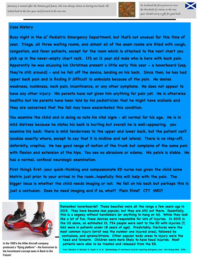
Remember hoverboards? These beauties were all the range a few years ago in 2015. They have become less popular, but they are still out there. Essentially, this is a segway without handlebars (or anything to hang on to). While they look like a lot of fun, these devices were responsible for lots of injuries. In 2015 in the US alone, an estimated 15, 754 people were sent to the ED with injuries (7, 662 were in patients under 18 years of age). Predictably, fractures were the most common injury (wrist was the number one injured area), followed by contusions, and sprains/strains. Other popular body areas to injury were the head and forearm. Children were more likely to have head injuries. Most patients were able to be treated and released from the ED.From: Bandzar S, Bandzar A, Gupta S, et al. Epidemiology of overboard injuries requiring emergency care. Ann Emerg Med. 2016.
In the 1950’s the Hiller Aircraft company produced a “flying platform” - the forerunner to the hoverboard concept seen in Back to the Future!
In Scotland the first person to cross the threshold of a home in the new year should carry a gift for good luck.
VOL 5 NO 1 January 2018
PAGE 2
Imaging Back Injury In Pediatrics
So, when is imaging indicated in back injury in children? That depends on a few things. Most children with back injuries, especially low risk mechanisms of injury, do not require imaging. Indications to perform imaging in the setting of trauma include: vertebral point tenderness, crepitus, step-off, large hematoma, difficulty ambulating, persistent/worsening pain, neurologic symptoms (numbness, tingling, incontinence), high risk mechanism (motor vehicle crashes, for example) or associated injuries (for example, abdominal injuries in an MVC (see the February 2016 Issue - Chance Fractures). Imaging options: 1. Plain Films - most common and easiest modality to use. Plain imaging can detect fractures, spondylolysis, spondylolisthesis, and bony lesions. Most plain imaging is done with 2 views: AP and Lateral. 2. CT - non contrast computed tomography is a great imaging modality and can detect all of the things that plain imaging can; it can also be used to confirm plain imaging findings. CT reconstruction images of the thoracic and lumber spine are often obtained when using CT to evaluate abdominal trauma. However, remember to image gently as CT does use more ionizing radiation than plain imaging. 3.MRI - ah, the elegance of MRI! Consider MRI in the setting of trauma when there are neurologic symptoms, Otherwise, this is not a first line
modality.
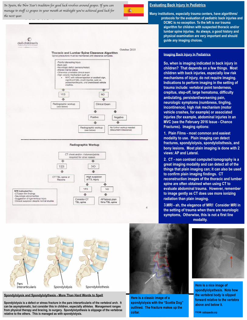
Evaluating Back Injury In Pediatrics
Many institutions, especially trauma centers, have algorithms/protocols for the evaluation of pediatric back injuries and
DCMC is no exception. To the left is our trauma algorithm for children with suspected thoracic and/or lumbar spine injuries. As always, a good history and physical examination are very important and should guide any imaging choices.
Spondylolysis and Spondylolythesis - More Than Hard Words to Spell
Spondylolysis is a defect or stress fracture in the pars interarticularis of the vertebral arch. It can be asymptomatic, but consider this in children, especially athletes. Management ranges from physical therapy and bracing, to surgery. Spondylolysthesis is slippage of the vertebrae relative to the others. This is managed as with spondylolysis.
Here is a classic image of a spondylolysis with the “Scottie Dog” outlined. The fracture makes up the collar.
Here is a nice image of spondlylolysthesis. Note how the vertebral body is slipped forward relative to the vertebra above and below it.
FROM: radiopaedia.org
In Spain, the New Year’s tradition for good luck revolves around grapes. If you can manage to stuff 12 grapes in your mouth at midnight you’ve achieved good luck for the next year.
VOL 5 NO 1 January 2018
PAGE 2
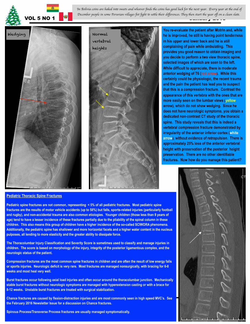
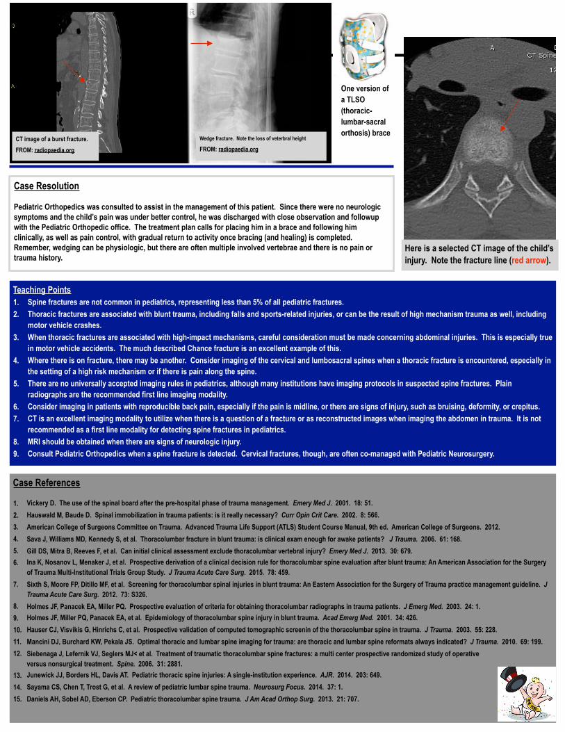
You re-evaluate the patient after Motrin and, while he is improved, he still is having point tenderness in his upper and lower back and he is still complaining of pain while ambulating. This provides you good reason to obtain imaging and you decide to perform a two view thoracic spine, selected images of which are seen to the left. While difficult to appreciate, there is moderate anterior wedging of T6 (red arrow). While this certainly could be physiologic, the recent trauma and the pain the patient has lead you to suspect that this is a compression fracture. Contrast the appearance of this vertebra with the ones that are more easily seen on the lumbar views (yellow arrow), which do not show wedging. Since he does not have neurologic symptoms, you obtain a dedicated non-contrast CT study of the thoracic spine. This study reveals that this is indeed a vertebral compression fracture demonstrated by irregularity of the anterior inferior cortex (white arrow), without evidence of retropulsion. There is approximately 25% loss of the anterior vertebral height with preservation of the posterior height preservation. There are no other identifiable fractures. Now how do you manage this patient?
Pediatric Thoracic Spine Fractures
Pediatric spine fractures are not common, representing < 5% of all pediatric fractures. Most pediatric spine fractures are the results of motor vehicle accidents (up to 58%) but falls, sports-related injuries (particularly football and rugby), and non-accidental trauma are also common etiologies. Younger children (those less than 8 years of age) tend to have a lesser incidence of these fractures partially due to the pliability of the spinal column in these children. This also means this group of children have a higher incidence of the so-called SCIWORA phenomena. Additionally, the pediatric spine has shallower and more horizontal facets and a higher water content in the nucleus pulposes, all lending to more elasticity and the greater ability to dissipate force.
The Thoracolumbar Injury Classification and Severity Score is sometimes used to classify and manage injuries in children. The score is based on morphology of the injury, integrity of the posterior ligamentous complex, and the neurologic status of the patient.
Compression fractures are the most common spine fractures in children and are often the result of low energy falls or sports injuries. Neurologic deficit is very rare. Most fractures are managed nonsurgically, with bracing for 6-8 weeks and most heal very well.
Burst fractures occur following axial load injuries and often occur around the thoracolumbar junction. Mechanically stable burst fractures without neurologic symptoms are managed with hyperextension casting or with a brace for 8-12 weeks. Unstable burst fractures are treated with surgical stabilization.
Chance fractures are caused by flexion-distraction injuries and are most commonly seen in high speed MVC’s. See the February 2016 Newsletter Issue for a discussion on Chance fractures.
Spinous Process/Transverse Process fractures are usually managed symptomatically.
Normal
vertebral
heights
Wedging
In Bolivia coins are baked into sweets and whoever finds the coins has good luck for the next year. Every year at the end of December people in some Peruvian villages fist fight to settle their differences. They then start the year off on a clean slate.
VOL 5 NO 1 January 2018
PAGE 2
Case References
1. Vickery D. The use of the spinal board after the pre-hospital phase of trauma management. Emery Med J. 2001. 18: 51. 2. Hauswald M, Baude D. Spinal immobilization in trauma patients: is it really necessary? Curr Opin Crit Care. 2002. 8: 566. 3. American College of Surgeons Committee on Trauma. Advanced Trauma Life Support (ATLS) Student Course Manual, 9th ed. American College of Surgeons. 2012. 4. Sava J, Williams MD, Kennedy S, et al. Thoracolumbar fracture in blunt trauma: is clinical exam enough for awake patients? J Trauma. 2006. 61: 168. 5. Gill DS, Mitra B, Reeves F, et al. Can initial clinical assessment exclude thoracolumbar vertebral injury? Emery Med J. 2013. 30: 679. 6. Ina K, Nosanov L, Menaker J, et al. Prospective derivation of a clinical decision rule for thoracolumbar spine evaluation after blunt trauma: An American Association for the Surgery
of Trauma Multi-Institutional Trials Group Study. J Trauma Acute Care Surg. 2015. 78: 459. 7. Sixth S, Moore FP, Ditillo MF, et al. Screening for thoracolumbar spinal injuries in blunt trauma: An Eastern Association for the Surgery of Trauma practice management guideline. J
Trauma Acute Care Surg. 2012. 73: S326. 8. Holmes JF, Panacek EA, Miller PQ. Prospective evaluation of criteria for obtaining thoracolumbar radiographs in trauma patients. J Emerg Med. 2003. 24: 1. 9. Holmes JF, Miller PQ, Panacek EA, et al. Epidemiology of thoracolumbar spine injury in blunt trauma. Acad Emerg Med. 2001. 34: 426. 10. Hauser CJ, Visvikis G, Hinrichs C, et al. Prospective validation of computed tomographic screenin of the thoracolumbar spine in trauma. J Trauma. 2003. 55: 228. 11. Mancini DJ, Burchard KW, Pekala JS. Optimal thoracic and lumbar spine imaging for trauma: are thoracic and lumbar spine reformats always indicated? J Trauma. 2010. 69: 199. 12. Siebenaga J, Lefernik VJ, Seglers MJ< et al. Treatment of traumatic thoracolumbar spine fractures: a multi center prospective randomized study of operative
versus nonsurgical treatment. Spine. 2006. 31: 2881. 13. Junewick JJ, Borders HL, Davis AT. Pediatric thoracic spine injuries: A single-institution experience. AJR. 2014. 203: 649. 14. Sayama CS, Chen T, Trost G, et al. A review of pediatric lumbar spine trauma. Neurosurg Focus. 2014. 37: 1. 15. Daniels AH, Sobel AD, Eberson CP. Pediatric thoracolumbar spine trauma. J Am Acad Orthop Surg. 2013. 21: 707.
Teaching Points 1. Spine fractures are not common in pediatrics, representing less than 5% of all pediatric fractures. 2. Thoracic fractures are associated with blunt trauma, including falls and sports-related injuries, or can be the result of high mechanism trauma as well, including
motor vehicle crashes. 3. When thoracic fractures are associated with high-impact mechanisms, careful consideration must be made concerning abdominal injuries. This is especially true
in motor vehicle accidents. The much described Chance fracture is an excellent example of this. 4. Where there is on fracture, there may be another. Consider imaging of the cervical and lumbosacral spines when a thoracic fracture is encountered, especially in
the setting of a high risk mechanism or if there is pain along the spine. 5. There are no universally accepted imaging rules in pediatrics, although many institutions have imaging protocols in suspected spine fractures. Plain
radiographs are the recommended first line imaging modality. 6. Consider imaging in patients with reproducible back pain, especially if the pain is midline, or there are signs of injury, such as bruising, deformity, or crepitus. 7. CT is an excellent imaging modality to utilize when there is a question of a fracture or as reconstructed images when imaging the abdomen in trauma. It is not
recommended as a first line modality for detecting spine fractures in pediatrics. 8. MRI should be obtained when there are signs of neurologic injury. 9. Consult Pediatric Orthopedics when a spine fracture is detected. Cervical fractures, though, are often co-managed with Pediatric Neurosurgery.
Case Resolution
Pediatric Orthopedics was consulted to assist in the management of this patient. Since there were no neurologic symptoms and the child’s pain was under better control, he was discharged with close observation and followup with the Pediatric Orthopedic office. The treatment plan calls for placing him in a brace and following him clinically, as well as pain control, with gradual return to activity once bracing (and healing) is completed. Remember, wedging can be physiologic, but there are often multiple involved vertebrae and there is no pain or trauma history.
Here is a selected CT image of the child’s injury. Note the fracture line (red arrow).
CT image of a burst fracture. FROM: radiopaedia.org
Wedge fracture. Note the loss of veterbral height
FROM: radiopaedia.org
One version of a TLSO (thoracic-lumbar-sacral orthosis) brace

![ORTHOPAEDIC GRAND ROUNDS CONFERENCE 2-27-2019.pdfHand/ Upper Extremity Radiology Conference [1 st] 6:30 – 7:30 A.M. 1512 Duke North Radiology Foot and Ankle MRI Conference [2 nd](https://img.pdfslide.net/doc/110x75/5f0e7c577e708231d43f7a15/orthopaedic-grand-rounds-conference-2-27-2019pdf-hand-upper-extremity-radiology.jpg)