Embed Size (px)
Citation preview
PEDIATRIC DENTISTRY/Copyright © 1987 byThe American Academy of Pediatric Dentist~
Volume 9 Number 3
Dental defects in incontinentia pigmenti: case report
Deborah Ann Himelhoch, DDS, MS
Richard Allen Olsen, DDS
Bonnie June Scott, DDS
Abstract
Incontinentia pigmenti is an uncommon type of ectoder-mal dyspIasia involving abnormalities of the skin, hair, centralnervous system, and teeth. The literature is reviewed and a casewith a variety of dental abnormalities is presented.
Incontinentia pigrnenti (IP) is one of the lesserknown ectodermal dysplasias with fewer than 800 casesreported in the world literature. This genodermatosisaffects mostly females and may involve the skin, hair,nails, eyes, and central nervous system.
Diagnosis usually is made from the clinical presen-tation and occasionally skin biopsy. Characteristicchanges in the dentition of some patients also have beenobserved. Dental abnormalities include hypodontia,pegged incisors and canines, and delayed eruption (But-terworth and Ladda 1981; Freira-Maia and Pinheiro1984). The authors report a case of a 12-year-old femalewith incontinentia pigmenti associated with enamelhypocalcification and hypoplasia of the permanent teeth.
Literature Review
Incontinentia pigmenti is a developmental defectinvolving many structures of ectodermal and mesoder-mal origin (Morgan 1971). Carney (1976), analyzing cases described in the world literature, found the disor-der to be present in all races with a 37:1 female-to-maleratio. Inheritance may be dominant with lethality inmales, but this has been disputed (Curth and Warburton1965; Hecht et al. 1982).
Onset of IP skin lesions usually occurs at or shortlyafter birth and is manifested in a series of stages -- first,vesicubulous or erythematous; second, hypertrophic orverrucous; and third, pigmented (Carney and Carney1970; Ianco et al. 1975).
Most children with IP have other abnormalitiesincluding partial alopecia of the scalp, strabismus, anddental anomalies. Less common manifestations includenail dystrophies, serious ocular abnormalities, epilepsy,
mental retardation, spastic or paralytic disorders, andleukocyte dysfunctions.1 It is important to note, however,that more than 80% of patients have normal or aboveaverage intelligence.
Fewer than half of the reported cases of IP make anymention of the dentition. Gorlin (1960) reviewed 25 caseswith dental abnormalities and reported three new cases.Noted were: hypodontia involving both primary andpermanent dentitions; pegging of incisors, canines andpremolars; and delayed eruption. Of the 306 cases re-viewed by Carney (1976), 64.7% had some major dentalabnormality. Of these, 43.1% had hypodontia, 30.4%malformed or pegged teeth (mostly incisors or canines),and only six had enamel disorders. These six patientswere reported to have multiple caries and "crumbly"teeth.
Most recently, Burgess (1982) reported on hypodon-tia in five children with incontinentia pigmenti, three ofwhom were from the same family. The four older chil-dren had hypodonfia of the permanent dentition. Theinfant and three of the four older children also showedhypodontia of the primary dentition. All five of thechildren had conical teeth and the four older childrenshowed notching of the permanent incisors. Four of thechildren also showed delayed eruption of some teeth.Changes in the quality of the enamel were not reported.
In the present report, a 12-year-old black femalepatient was first seen at the University of Chicago WalterG. Zoller Memorial Dental Clinic at the age of three years.Referral was made by the outpatient pediatrics clinicwhich had been following the patient since age fourmonths. At the time of the initial visit, a diagnosis ofincontinentia pigmenti was established. The patient hadreturned sporadically for dental care and at her lastexamination presented with numerous carious lesionsand other dental problems.
Butterworth and Ladda 1981; Carney and Carney 1970; Carney1976; Ianco et al. 1975; Jensen et al. 1978; Morgan 1971.
236 INCONnXNENTIA PIGMENTI: HIMELHOCH El" AL.
Medical History
The patient was the middle of three children, withone older and one younger brother. The mother denieddifficulty with any of her pregnancies and had no miscar-riages. Both boys are reported to be free of any signs ofsystemic illness or abnormalities. Family history wasnegative for any relative with IP. The mother was adiagnosed epileptic who took anticonvulsant medicationsporadically. It is uncertain whether or not she wastaking phenytoin during her second pregnancy. Themother has fewer than 10 teeth remaining, and reportedthat many teeth were extracted due to "cavities." Shedenied any history of skin lesions and at the time ofexamination had no visible abnormalities of her face,neck, arms, or nails. Oral soft tissues demonstratedevidence of gingivitis and periodontal disease.
The patient was born full term at an uneventfuldelivery. A rash was noted in the first week of life on theinfant's shoulders and neck. The rash initially was de-scribed as bilateral and erythematous. She was sent homeat the age of four days although the rash persisted,gradually resolving in the fifth or sixth month of life. Thechild was left with a "swirled" appearance of her skinwith areas of hypopigmentation where the rash hadoccurred previously.
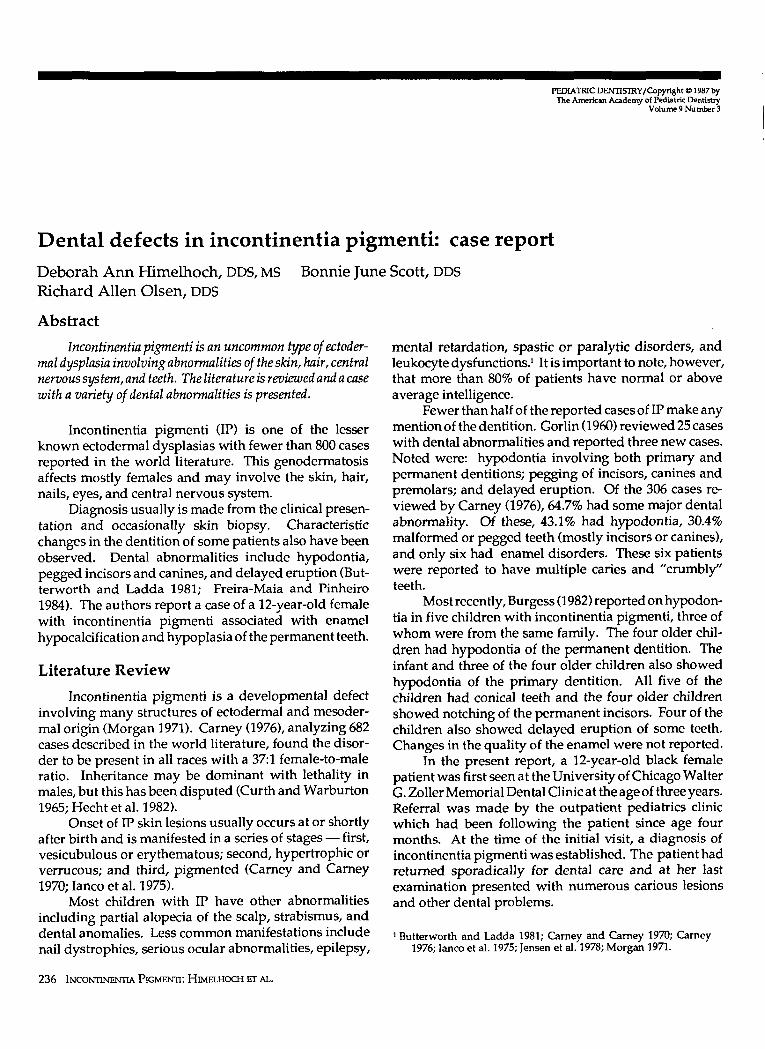
Currently, the patient has numerous darkly pig-mented, slightly elevated lesions of the face extendingonto her neck (Fig 1). She has raised areas of hypopig-mentation on her hands (Fig 2), but no abnormalities ofthe nails. Her hair is of normal color, texture, and pattern.
Coats' disease, an ocular disorder characterized byexudation beneath or in the retina, was noted in both eyesat the age of four years. Esotropia of both eyes waspresent with the right being greater than the left. Thepatient was legally blind and wore corrective glasses.
A submucosal cleft of the palate was noted in in-fancy and the patient is followed by the University ofChicago Craniofacial Anomalies Team. She had a longhistory of chronic otitis media and several cholesteato-mas were removed from both ears. The patient currentlyexhibits normal speech without hypernasality, althoughspeech acquisition was delayed, beginning around agethree years. Speech delays were felt to be due to mentalretardation. The patient is behind her chronological agein both mental and motor development. She attends aspecial school in the Chicago Public School System.
Oral Examination and Dental Treatment
An oral examination, including panoramic, periapi-cal, and bite-wing radiographs was completed. Otherradiographs dating from 1977 were also available. Softtissue examination revealed a bifid uvula and gingivitissecondary to poor oral hygiene. All other soft tissueswere normal and 25 permanent teeth were present.Radiographs revealed an additional six unerupted per-
FIG 1. Note the pigmented streaks and whorls characteristic ofthe third phase of incontinentia pigmenti.
FIG 2. Note the characteristic raised, hypopigmented areas pres-ent on the patient's hands.
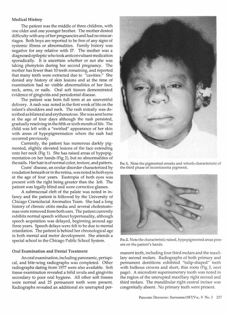
manent teeth, including four third molars and the maxil-lary second molars. Radiographs of both primary andpermanent dentitions exhibited "tulip-shaped" teethwith bulbous crowns and short, thin roots (Fig 3, nextpage). A microdont supernumerary tooth was noted inthe region of the unerupted maxillary right second andthird molars. The mandibular right central incisor wascongenitally absent. No primary teeth were present.
PEDIATRIC DENTISTRY: SEPTEMBERl987/VoL. 9 No. 3 237
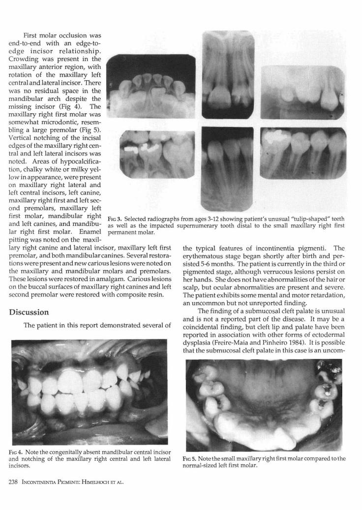
First molar occlusion wasend-to-end with an edge-to-edge incisor relationship.Crowding was present in themaxillary anterior region, withrotation of the maxillary leftcentral and lateral incisor. Therewas no residual space in themandibular arch despite themissing incisor (Fig 4). Themaxillary right first molar wassomewhat microdontic, resem-bling a large premolar (Fig 5).Vertical notching of the incisaledges of the maxillary right cen-tral and left lateral incisors wasnoted. Areas of hypocalcifica-tion, chalky white or milky yel-low in appearance, were presenton maxillary right lateral andleft central incisors, left canine,maxillary right first and left sec-ond premolars, maxillary leftfirst molar, mandibular rightand left canines, and mandibu-lar right first molar. Enamelpitting was noted on the maxil-lary right canine and lateral incisor, maxillary left firstpremolar, and both mandibular canines. Several restora-tions were present and new carious lesions were noted onthe maxillary and mandibular molars and premolars.These lesions were restored in amalgam. Carious lesionson the buccal surfaces of maxillary right canines and leftsecond premolar were restored with composite resin.
Discussion
The patient in this report demonstrated several of
^^HHBHM^te^^h
FIG 3. Selected radiographs from ages 3-12 showing patient's unusual "tulip-shaped" teethas well as the impacted supernumerary tooth distal to the small maxillary right firstpermanent molar.
the typical features of incontinentia pigmenti. Theerythematous stage began shortly after birth and per-sisted 5-6 months. The patient is currently in the third orpigmented stage, although verrucous lesions persist onher hands. She does not have abnormalities of the hair orscalp, but ocular abnormalities are present and severe.The patient exhibits some mental and motor retardation,an uncommon but not unreported finding.
The finding of a submucosal cleft palate is unusualand is not a reported part of the disease. It may be acoincidental finding, but cleft lip and palate have beenreported in association with other forms of ectodermaldysplasia (Freire-Maia and Pinheiro 1984). It is possiblethat the submucosal cleft palate in this case is an uncom-
FIG 4. Note the congenitally absent mandibular central incisorand notching of the maxillary right central and left lateralincisors.
FIG 5. Note the small maxillary right first molar compared to thenormal-sized left first molar.
238 INCONTINENTTA PIGMENTI: HIMELHOCH ET AL.
mon manifestation of the genetic defect that causes in-continentia pigrnenti.
Etiology of incontinentia pigmenti in this case isprobably a sporadic mutation. Although the mother hadlost most of her teeth, she had no evidence of enameldefects or malformations of the remaining teeth. No skinabnormalities were present, nor was there a history of anyabnormalities of ectodermal structures. Finally, no fam-ily members were reported to have incontinentia pig-menti or other dermatological diseases.
The most commonly reported dental anomalies inIP patients are hypodontia and malformed or peggedteeth (usually anteriors). This patient presented withenamel hypoplasia, enamel hypocalcification, a supernu-merary tooth, abnormally formed teeth, and a congeni-tally missing tooth. Most striking, however, was theextensive pitting, hypoplasia, and hypocalcificationpresent on 12 of the 25 erupted teeth. Previously restoredcarious lesions on another 10 teeth may have been due tosimilar enamel defects.
Due to the patient’s youth, conservative alloys andcomposites were chosen to restore carious teeth. Non-carious, unrestored molars and premolars were pro-tected with occlusal sealants. The apparently unaffectedenamel surfaces seemed to etch normally and sealantswere intact several months after placement. However,because of fear that the enamel may be abnormal, carewas taken to achieve mechanical retention for all acid-etch composite restorations. The supernumerary toothwas removed and orthodontic treatment is in progress.The child has received oral hygiene instruction and hasbeen advised to rinse daily with a neutral sodium fluoriderinse. Plaque indices are improving and it is hoped thatthere will be little future dental decay.
At the time of writing Dr. Himelhoch was a clinical assistantprofessor of pediatric dentistry, Dr. Olsen was an assistantprofessor and head of general dentistry, and Dr. Scott was ageneral practice resident, University of Chicago’s Zoller DentalClinic. Dr. Scott also is in private practice :.n Lake Zurich,Illinois. Reprint requests should be sent to: Dr. Deborah A.Himelhoch, 46 Waverley St., 1st Floor, Belmont, MA 02178.
Burgess MC: Incontinentia pigmenti: 6 cases of Bloch-Sulzbergersyndrome. Br Dent J 152:195-96, 1982.
Butterworth T, Ladda RL: Clinical Genodermatology, vol 1. NewYork; Praeger Scientific, 1981 pp 65-70.
Carney RG, Carney RG Jr: Incontinentia pigmenti. Arch Dermatol102:157-62, 1970.
Carney RG: Incontinentia pigmenti: a world statistical analysis.Arch Dermatol 112:535-42, 1976.
Curth HO, Warburton D: The genetics of incontinentia pigmenti.Arch Dermatol 92:229-35, 1965.
Freire-Mala N, Pinheiro M: Ectodermal Dysplasia: A Clinical andGenetic Study. New York; Alan R. Liss Inc, 1984 pp 77-79.
Gorlin RJ, Anderson JA: The characteristic dentition of incontinentiapigmenti. J Pediatr 57:78-83, 1960.
Hecht R, Hecht BK, Austin WJ: Incontinentia pigmenti in ArizonaIndians including transmission from mother to son inconsistentwith the half chromatid mutation model. Clin Genet 21:293-96,1982.
Iancu T, Komlos L, Shabtay F, Elian E, Halbrecht I, Book JA: Inconti-nentia pigmenti. Clin Genet 7:103-10, 1975.
Jessen RT, Van Epps DE, Goodwin JS, Bowerman J: Incontinentiapigmenti: evidence for both neutrophil and lymphocyte dys-function. Arch Dermatol 114:1182-86, 1978.
Morgan JD: Incontinentia pigmenti (Bloch-Sulzberger syndrome):A report of four additional cases. Am J Dis Child 122:294-300,1971.
Hepatitis B traced to dentist
The ninth outbreak of hepatitis B virus (HBV) traceable to a dentist since 1974 resulted in 4 cases of the diseasein a New Hampshire town.
The outbreak was traced to an oral surgeon who had performed tooth extractions, without gloves, 3-5 monthsbefore each patient became ill with the virus. All 4 patients claimed no other risk factors for HBV; all wereserologically confirmed.
While the oral surgeon did not wear gloves, he was careful to scrub his hands between surgical procedures.Following discovery of the outbreak, the doctor, in practice for 25 years, discontinued practicing and sent lettersto all patients informing them of possible exposure and offering free testing for HBV.
The Centers for Disease Control, in its report about this incident, concluded, "Recurrent, avoidable outbreakssuch as this one should prompt dentists and oral surgeons to seek hepatitis B vaccination and to use glovesroutinely when treating patients."
P~h~c IMm~swv: S~r~_~1987/Vou. 9 No. 3 239

![First IKBKG Gene Mutation Study in Serbian Incontinentia ... · Incontinentia pigmenti (IP; Bloch-Sulzberg-er syndrome; MIM 308300) is a rare X-linked dominant genodermatosis [5]](https://img.pdfslide.net/doc/110x75/5f3bedf5651a4c1377610355/first-ikbkg-gene-mutation-study-in-serbian-incontinentia-incontinentia-pigmenti.jpg)


![Tnfa Signaling Through Tnfr2 Protects Skin Against ...eprints.whiterose.ac.uk/81541/1/Tnfa signaling through tnfr2 protects... · genodermatosis incontinentia pigmenti (IP) [17]](https://img.pdfslide.net/doc/110x75/5f3bedf6651a4c137761035c/tnfa-signaling-through-tnfr2-protects-skin-against-signaling-through-tnfr2-protects.jpg)