Embed Size (px)
Citation preview
pubs.acs.org/jmcPublished on Web 11/06/2009r 2009 American Chemical Society
J. Med. Chem. 2010, 53, 325–334 325
DOI: 10.1021/jm901268n
Design, Synthesis, and Biological Evaluations of 2,5-Diaryl-2,3-dihydro-1,3,4-oxadiazoline Analogs of
Combretastatin-A4
Lauren Lee,† Lyda M. Robb,‡ Megan Lee,† Ryan Davis,† Hilary Mackay,† Sameer Chavda,† Balaji Babu,† Erin L. O’Brien,‡
April L. Risinger,‡ Susan L. Mooberry,*,‡ and Moses Lee*,†
†Department of Chemistry and the Division of Natural and Applied Sciences, Hope College, Michigan 49423 and ‡Department of Pharmacology,University of Texas Health Science Center at San Antonio, San Antonio, Texas 78229
Received August 24, 2009
A total of 24 novel 2,5-diaryl-1,3,4-oxadiazoline analogs of combretastatin A-4 (CA-4, 1) were designed,synthesized, and evaluated for biological activities. The compounds represent two structural classes; theType I class has threemethoxygroups on theA ring and theType II class has a singlemethoxygroup on theA ring. Biological evaluations demonstrate that multiple structural features control the biological potency.Four of the compounds, 2-(30-bromophenyl)-5-(300,400,500-trimethoxyphenyl)-2-acetyl-2,3-dihydro-1,3,4-oxadiazoline (9l), 2-(20,50-dimethoxyphenyl)-5-(300-methoxyphenyl)-2-acetyl-2,3-dihydro-1,3,4-oxadia-zoline (10h), 2-(30,40,50-trimethoxyphenyl)-5-(300-methoxyphenyl)-2-acetyl-2,3-dihydro-1,3,4-oxadiazoline(10i), and 2-(30,50-dimethoxyphenyl)-5-(300-methoxyphenyl)-2-acetyl-2,3-dihydro-1,3,4-oxadiazoline (10j),have potent antiproliferative activities against multiple cancer cell lines. Mechanistic studies indicate thatthey retain themicrotubule disrupting effects of compound 1, includingmicrotubule loss, the formation ofaberrant mitotic spindles, and mitotic arrest. Compound 10i inhibits purified tubulin polymerization andcircumvents drug resistance mediated by P-glycoprotein and βIII tubulin expression. The oxadiazolineanalog 10i is a promising lead candidate worthy of further investigation.
Introduction
Aworldwide effort led by theU.S.National Cancer Instituteto identify anticancer agents fromplants led to the identificationof the combretastatins from the African bush willow Combre-tum caffrum.1,2 Combretastatin A-4 (CA-4,a 1, Figure 1) is themost potent of the natural combretastatins and early workshowed it inhibits tubulin polymerization and the proliferationof murine and human cancer cells.3,4 Early reports of theantitumor activity of the more water-soluble prodrug CA-4P(2) againstmurineP388andB16 tumorswerenot encouraging.5
However, detailed mechanistic studies showed that compound1 has specific effects on tumor vasculature, causing rapidvascular shutdown, leading to central tumor necrosis.6,7 Theeffects of prodrug 2 on tumor vasculature have been verified inhuman clinical trials.8,9 While compound 2 causes centralhemorrhagic necrosis in animal tumor models, a rim of viabletumor cells remains, suggesting that the optimal use of 2wouldbe in combination with agents that target the tumor periphery.Compound 2 is currently being evaluated in phase II clinicaltrials for activity in anaplastic thyroid carcinoma, nonsmall celllung cancer, and ovarian cancer in combination with conven-tional cytotoxic drugs including paclitaxel, carboplatin, and theantiangiogenic agent brevacizumab.10 Mechanistically, agent 1rapidly alters HUVEC morphology, causing cell contraction,membrane blebbing, and disruption of endothelial monolayerpermeability.11 These effects are potentiallymediated by aRho/Rho kinase stimulated increase in actin stress fibers. Prodrug 2
has also been shown to disrupt VE-cadherin signaling inHUVECs.12 Most recently, it was shown to rapidly upregulateconnective tissue growth factor (CTGF) in microvascular en-dothelial cells in a cell density dependent manner.13 Upregula-tion of CTGF, a pleotropic endothelial cell growth factor, wasobserved only in subconfluent endothelial cell cultures or inendothelial cells growing as capillary-like tubules. Prodrug 2-mediated activation of the CTGF might explain why thin,unstabilized tumor blood vessels are so susceptible to 1, whileother mature vessels are unaffected. It is not known which ofthese compound 1-mediated signaling events are responsible forthe specific antivascular effects leading to rapid catastrophictumor vascular shutdown.
Significant efforts have been undertaken todevelop analogsof compound 1 that retain the biological actions of the parentmolecule but provide improved pharmacokinetic propertiesandbetterwater solubility.14 In addition toprodrug 2, notableanalogsundergoing clinical evaluations includeAVE8062 (3),the amino acid derivative of AC-7739 (4) and the CA-1 (5)derivativeCA-1P (6;OXI-4503).10,15Ourwork in this areahasyielded several new classes of analogs including pyrazoles16 (7)and pyrazolines (8, Figure 1) of 1.17 These derivatives demon-strate a wide range of potencies. A significant drop in activitywas observed in the pyrazole derivatives as compared to 1.X-ray crystallography studies of a pyrazole analog of com-pound 7 showed that it adopted a planar conformation, asopposed to the nonplanar geometry of compound 1.16 Thestructure of 1 contains two substituted benzene rings, referredto as the A and B rings, which fit into the A and B pocketswithin the colchicines binding site on tubulin. Modelingstudies of 1 within the colchicines binding site of tubulinsuggest that a geometrically planar analog of 1 would not
*To whom correspondence should be addressed. Phone: 616-395-8075 (M.L.); 210-567-4788 (S.L.M.). Fax: 616-395-7923 (M.L.); 210-567-4300 (S.L.M.). E-mail: [email protected] (M.L.); [email protected] (S.L.M.).
326 Journal of Medicinal Chemistry, 2010, Vol. 53, No. 1 Lee et al.
allow optimal interactions with this binding site.18,19 It isreasonable to assume that the pyrazole’s loss of potency canbe attributed to its incompatible conformation within thecolchicines binding site of tubulin. Consistent with this rela-tionship, pyrazolines have a less planar geometry than thepyrazoles and are more potent inhibitors of cell proliferationand antitubulin effects.
A new series of 2,5-diaryl-1,3,4-oxadiazoline analogs weredesigned to retain the geometric features of 1. Compared tothe pyrazolines (8), the oxadiazolinemoiety should provide anoptimal conformational geometry for interaction with thecolchicine binding site while increasing the number of het-eroatoms in the core structure. The net effect is an increase inthe polarity of the molecule, which should enhance watersolubility. Accordingly, 24 specific oxadiazoline analogs weresynthesized and they are divided into two types (Figure 1).Type I compounds (oxadiazoline 9a-l) contain a 3,4,5-tri-methoxyphenyl group on the A ring and Type II compounds(oxadiazoline 10a-l) contain a 4-methoxyphenyl moiety onthe A ring. These analogs were synthesized and evaluated forbiological activities. The most potent compounds were eval-uated in mechanistic studies and attempts have been made tounderstand the structure activity relationships (SAR) amongthese compounds.
Chemistry
Oxadiazoline analogs were synthesized by adapting a pro-cedure reported by Ali, Amer, and Abdel-Rahman.20 As anexample, the reaction of a hydrazide (11) with a benzaldehyde(12) in reflux with water and glacial acetic acid in ethanol for5 h produced the hydrazone intermediate 13 (Scheme 1). Thereactionmixturewas poured into ice water and the precipitatewas collected and dried. When needed, the hydrazones werepurified by recrystallization in aqueous methanol. The yieldsof the hydrazones ranged between 60 and 98%. The chemicalstructures of the hydrazones were confirmed using 400 MHz1HNMRand IR.Theoxadiazolineproductswere synthesizedby refluxing the appropriate hydrazone intermediate in aceticanhydride for 1 h under dry conditions (Scheme 1). Thereaction mixture was poured into ice-water, and the solidproduct was collected and washed with water. Most productswere found to be homogeneous by TLC and 400 MHz 1HNMR analyses, but when needed, heterogeneous productswere readily purified by silica gel column chromatographyusing a methanol/chloroform eluent. Oxadiazoline productyields ranged from 25-70%. The structures of compounds9a-l and 10a-l were confirmed using NMR, IR, massspectrometry, and elemental analysis. The compounds that
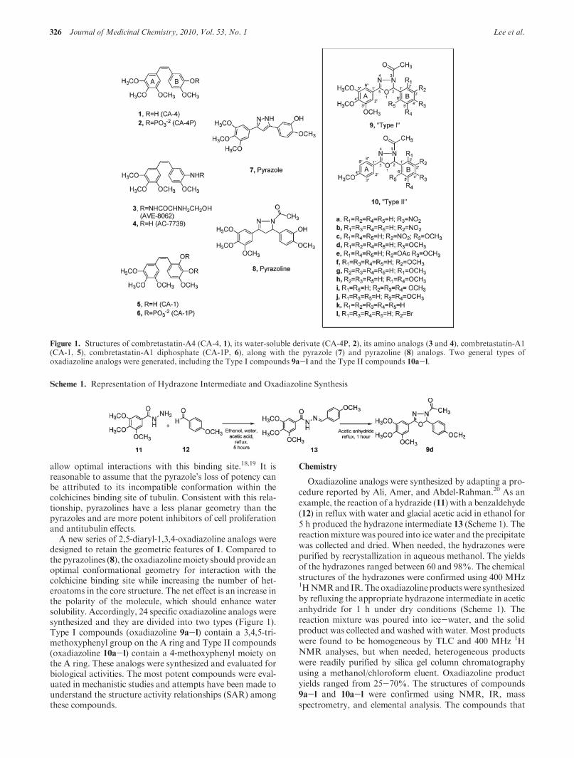
Figure 1. Structures of combretastatin-A4 (CA-4, 1), its water-soluble derivate (CA-4P, 2), its amino analogs (3 and 4), combretastatin-A1(CA-1, 5), combretastatin-A1 diphosphate (CA-1P, 6), along with the pyrazole (7) and pyrazoline (8) analogs. Two general types ofoxadiazoline analogs were generated, including the Type I compounds 9a-l and the Type II compounds 10a-l.
Scheme 1. Representation of Hydrazone Intermediate and Oxadiazoline Synthesis
Article Journal of Medicinal Chemistry, 2010, Vol. 53, No. 1 327
showed a small amount (<5%) of rotational isomers (aboutthe amide bond) were analyzed by reversed phase HPLC(Nova Pak C18 3.6 � 150 mm column using methanol as theeluent). Each of the compounds showed a single peak with aretention time of 2-3 min, confirming the purity of thecompounds.
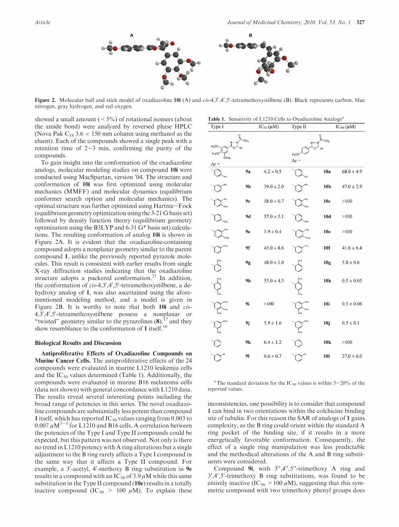
To gain insight into the conformation of the oxadiazolineanalogs, molecular modeling studies on compound 10i wereconducted using MacSpartan, version ’04. The structure andconformation of 10i was first optimized using molecularmechanics (MMFF) and molecular dynamics (equilibriumconformer search option and molecular mechanics). Theoptimal structure was further optimized using Hartree-Fock(equilibriumgeometry optimizationusing the 3-21Gbasis set)followed by density function theory (equilibrium geometryoptimization using the B3LYP and 6-31G* basis set) calcula-tions. The resulting conformation of analog 10i is shown inFigure 2A. It is evident that the oxadiazoline-containingcompound adopts a nonplanar geometry similar to the parentcompound 1, unlike the previously reported pyrazole mole-cules. This result is consistent with earlier results from singleX-ray diffraction studies indicating that the oxadiazolinestructure adopts a puckered conformation.21 In addition,the conformation of cis-4,30,40,50-tetramethoxystilbene, a de-hydroxy analog of 1, was also ascertained using the afore-mentioned modeling method, and a model is given inFigure 2B. It is worthy to note that both 10i and cis-4,30,40,50-tetramethoxystilbene possess a nonplanar or“twisted” geometry similar to the pyrazolines (8),17 and theyshow resemblance to the conformation of 1 itself.16
Biological Results and Discussion
Antiproliferative Effects of Oxadiazoline Compounds on
Murine Cancer Cells. The antiproliferative effects of the 24compounds were evaluated in murine L1210 leukemia cellsand the IC50 values determined (Table 1). Additionally, thecompounds were evaluated in murine B16 melanoma cells(data not shown) with general concordance with L1210 data.The results reveal several interesting points including thebroad range of potencies in this series. The novel oxadiazo-line compounds are substantially less potent than compound1 itself, which has reported IC50 values ranging from 0.003 to0.007 μM3-5 for L1210 and B16 cells. A correlation betweenthe potencies of the Type I and Type II compounds could beexpected, but this patternwas not observed.Not only is thereno trend inL1210 potencywithA ring alterations but a singleadjustment to the B ring rarely affects a Type I compound inthe same way that it affects a Type II compound. Forexample, a 30-acetyl, 40-methoxy B ring substitution in 9e
results in a compoundwith an IC50 of 3.9 μMwhile this samesubstitution in theType II compound (10e) results in a totallyinactive compound (IC50 > 100 μM). To explain these
inconsistencies, one possibility is to consider that compound1 can bind in two orientations within the colchicine bindingsite of tubulin. For this reason the SAR of analogs of 1 gainscomplexity, as the B ring could orient within the standard Aring pocket of the binding site, if it results in a moreenergetically favorable conformation. Consequently, theeffect of a single ring manipulation was less predictableand the methodical alterations of the A and B ring substit-uents were considered.
Compound 9i, with 300,400,500-trimethoxy A ring and30,40,50-trimethoxy B ring substitutions, was found to beentirely inactive (IC50 >100 μM), suggesting that this sym-metric compound with two trimethoxy phenyl groups does
Figure 2. Molecular ball and stick model of oxadiazoline 10i (A) and cis-4,30,40,50-tetramethoxystilbene (B). Black represents carbon, bluenitrogen, gray hydrogen, and red oxygen.
Table 1. Sensitivity of L1210 Cells to Oxadiazoline Analogsa
aThe standard deviation for the IC50 values is within 5-20% of thereported values.
328 Journal of Medicinal Chemistry, 2010, Vol. 53, No. 1 Lee et al.
not interact well within the colchicine binding site. Com-pound 1,with the 300,400,500-trimethoxy A ring is very potent,4
implying the unfavorable nature of the 30,40,50-trimethoxy Bring. At the other extreme, 10d, a Type I compoundwith onlya 400-methoxy on theA ring and a 40-methoxy substitution onthe B ring is also inactive (IC50 > 100 μM). Interestingly,shifting the 40-methoxy to the 30-position (10f) on the Type IIcompound improves potency (IC50 41.0 μM). The methoxygroup in the 20-position gives the highest potency of thesethree positions (10g, IC50 5.8 μM). Together, these datasuggest that a 4-methoxy substituted ring interacts poorlywith one pocket of the binding site. Because of the potency of10i (IC50 0.5 μM), which contains a trisubstituted methoxyring that is predicted to only favorably interact with the Asite, the single methoxy-substituted ring must favorablyinteract with the B site. This indicates that Type II com-pounds might position the 400-methoxy A ring in the B ringpocket of the colchicine binding site on tubulin.
Compounds 10h and 10j have a 20,50-dimethoxy and a30,50-dimethoxy substituted B ring, respectively. These com-pounds are potent, with IC50 values of 500 nM. Compounds9h and 9j have very different potencies (9h, IC50 55.0 μM; 9j,IC50 5.9 μM). The 20,50-dimethoxy B ring on 9h would beexpected to interact poorly with the B ring pocket, whereasthe 30,50-dimethoxy B ring on 9j favorably interacts with thebinding site.
The pattern observed with 10d, 10f, and 10g is notobserved in the Type I compounds; 9d, 9f, and 9g and allhave low potencies with IC50s of 57, 45, and 48 μM,respectively. The 40-methoxy B ring substitution is the leastpotent in both Type I and Type II compounds. In an attemptto improve 9d and 10d, the 40-methoxy substitution wasreplaced with a 40-nitro group (9a, 10a). This resulted in amodest improvement in potency of the Type II compound(10a, IC50 68.0 μM). For the Type I compound, this changeresulted in a dramatic, 13-fold increase in potency (9a IC50
4.2μM).TheType I compound (9a) demonstrates that the 40-nitro group substitution is substantially more favorable thanthe 40-methoxy in the B ring pocket. Shifting the nitro groupto the 30-position, 10a to 10b, results in a slight increase inpotency (10b, IC50 47.0 μM). In the Type I compound,however, this change in the position of the nitro groupdecreased potency 9-fold (9b, IC50 39.0 μM), suggesting thatin the B-ring pocket a p-nitro substitution is more favorablethan an m-nitro substitution. The 30-nitro substitution oncompound 10bmay orient into theA-ring pocket, improvinginteraction and biological potency. This is consistent withthe pattern observed with 10d, 10f, and 10g, which suggests apreferable A-pocket interaction with an ortho or meta sub-stitution, as compared to the para substitution.
Compounds 9c and 10c, with a 30-nitro, 40-methoxyB ring,are less potent than a single 30-nitro substituted B ring.Replacing the 30-nitro with a 30-bromo group produces amore potent Type I compound (9b, IC50 39.0 μM; 9l, IC50
0.6 μM). In the Type II compounds, the 30-nitro a 30-bromogroup (10b, IC50 47.0 μM; 10l, IC50 37 μM) have similaractivities. Compounds 9k and 10k have no substitution onthe B ring, and they have dramatically different potencies(9k, IC50 6.4 μM; 10k, IC50>100 μM). Compound 9k couldfavorably position the nonsubstituted ring in the B-ringpocket, however, the low potency of 10k (>100 μM) isconsistent with the notion that the Type I and Type IIcompounds might orient differently within the colchicinebinding site.
Antiproliferative Effects against MDA-MB-435. In thedevelopment of chemical agents with potential applicationin human anticancer therapeutics, it was important to de-termine the activity of the most potent oxadiazolines inhuman cancer cells. The five most potent inhibitors ofL1210 cell growth were evaluated in our reference humancell line, MDA-MB-435, a cell line originally designated asoriginating from breast but which has been shown defini-tively to be derived from the M14 melanoma cell line.22 Theresults, presented in Table 2, show that there is excellentcorrelation between the L1210 and MDA-MB-435 poten-cies, with 4 of the 5 compounds having submicromolarpotency against human cancer cells. These compounds wereevaluated further in mechanistic assays to determine if theyretain the microtubule disrupting and antimitotic effects ofthe combretastatins.
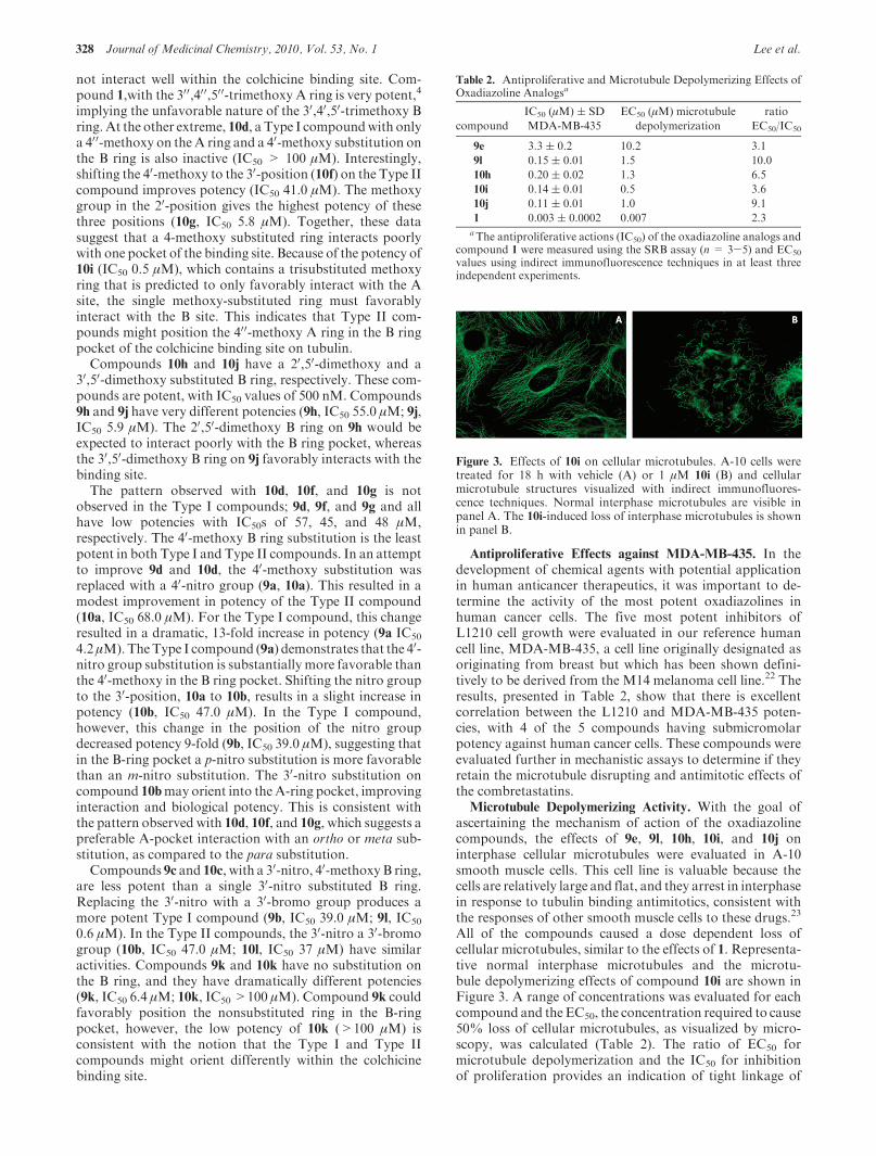
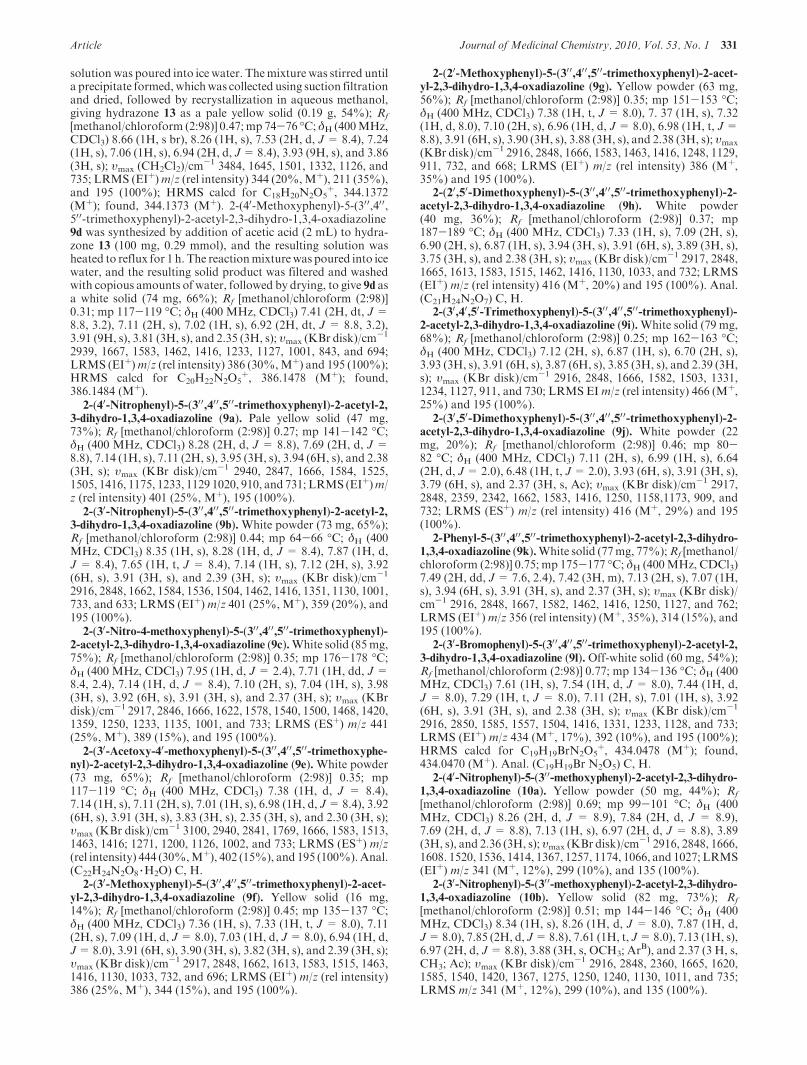
Microtubule Depolymerizing Activity. With the goal ofascertaining the mechanism of action of the oxadiazolinecompounds, the effects of 9e, 9l, 10h, 10i, and 10j oninterphase cellular microtubules were evaluated in A-10smooth muscle cells. This cell line is valuable because thecells are relatively large and flat, and they arrest in interphasein response to tubulin binding antimitotics, consistent withthe responses of other smooth muscle cells to these drugs.23
All of the compounds caused a dose dependent loss ofcellular microtubules, similar to the effects of 1. Representa-tive normal interphase microtubules and the microtu-bule depolymerizing effects of compound 10i are shown inFigure 3. A range of concentrations was evaluated for eachcompound and theEC50, the concentration required to cause50% loss of cellular microtubules, as visualized by micro-scopy, was calculated (Table 2). The ratio of EC50 formicrotubule depolymerization and the IC50 for inhibitionof proliferation provides an indication of tight linkage of
Table 2. Antiproliferative and Microtubule Depolymerizing Effects ofOxadiazoline Analogsa
compound
IC50 (μM) ( SD
MDA-MB-435
EC50 (μM) microtubule
depolymerization
ratio
EC50/IC50
9e 3.3 ( 0.2 10.2 3.1
9l 0.15 ( 0.01 1.5 10.0
10h 0.20 ( 0.02 1.3 6.5
10i 0.14 ( 0.01 0.5 3.6
10j 0.11 ( 0.01 1.0 9.1
1 0.003 ( 0.0002 0.007 2.3aThe antiproliferative actions (IC50) of the oxadiazoline analogs and
compound 1 were measured using the SRB assay (n = 3-5) and EC50
values using indirect immunofluorescence techniques in at least threeindependent experiments.
Figure 3. Effects of 10i on cellular microtubules. A-10 cells weretreated for 18 h with vehicle (A) or 1 μM 10i (B) and cellularmicrotubule structures visualized with indirect immunofluores-cence techniques. Normal interphase microtubules are visible inpanel A. The 10i-induced loss of interphase microtubules is shownin panel B.
Article Journal of Medicinal Chemistry, 2010, Vol. 53, No. 1 329
antiproliferative effects and the compound’s tubulin-depen-dentmechanisms of action.24,25 Compound 10i had a ratio of3.5, 9e a ratio of 3.1, 10h a ratio of 6.5, and 1 a ratio of 2.3(Table 2). The ratios are within the ranges we have seen with2-methoxyestradiol analogs24 and a large subset of polysub-stituted pyrrole compounds.25 Interestingly, the other potentantiproliferative compounds, and 10j and 9l did not demon-strate correspondingly potentmicrotubule depolymerizationeffects, and their ratios of EC50/IC50 ratios were 8.8 and 10,respectively (Table 2). This suggests that these compoundsmight exert cytotoxicity through mechanisms in addition totubulin inhibition as has been shown with similar moleculesthat can bind directly to DNA.26
Interaction of Compound 10i with Purified Bovine Brain
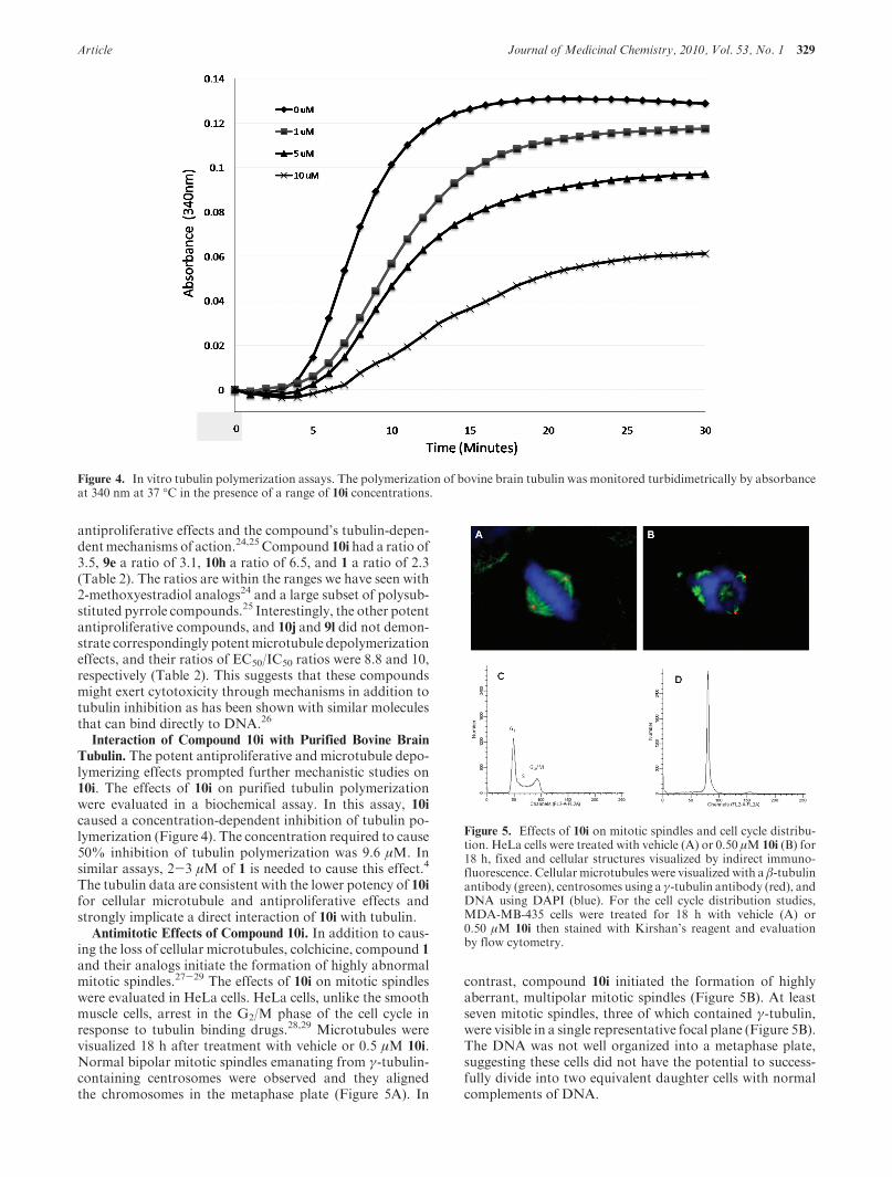
Tubulin. The potent antiproliferative and microtubule depo-lymerizing effects prompted further mechanistic studies on10i. The effects of 10i on purified tubulin polymerizationwere evaluated in a biochemical assay. In this assay, 10icaused a concentration-dependent inhibition of tubulin po-lymerization (Figure 4). The concentration required to cause50% inhibition of tubulin polymerization was 9.6 μM. Insimilar assays, 2-3 μM of 1 is needed to cause this effect.4
The tubulin data are consistent with the lower potency of 10ifor cellular microtubule and antiproliferative effects andstrongly implicate a direct interaction of 10i with tubulin.
Antimitotic Effects of Compound 10i. In addition to caus-ing the loss of cellular microtubules, colchicine, compound 1
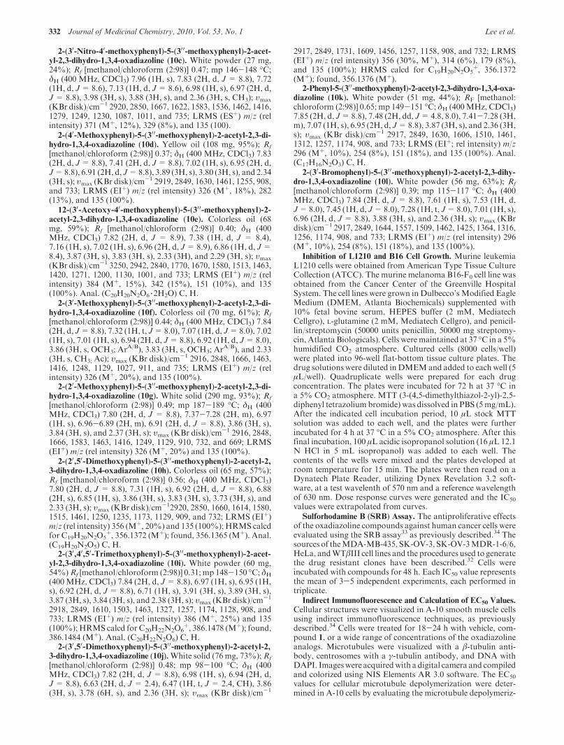
and their analogs initiate the formation of highly abnormalmitotic spindles.27-29 The effects of 10i on mitotic spindleswere evaluated in HeLa cells. HeLa cells, unlike the smoothmuscle cells, arrest in the G2/M phase of the cell cycle inresponse to tubulin binding drugs.28,29 Microtubules werevisualized 18 h after treatment with vehicle or 0.5 μM 10i.Normal bipolar mitotic spindles emanating from γ-tubulin-containing centrosomes were observed and they alignedthe chromosomes in the metaphase plate (Figure 5A). In
contrast, compound 10i initiated the formation of highlyaberrant, multipolar mitotic spindles (Figure 5B). At leastseven mitotic spindles, three of which contained γ-tubulin,were visible in a single representative focal plane (Figure 5B).The DNA was not well organized into a metaphase plate,suggesting these cells did not have the potential to success-fully divide into two equivalent daughter cells with normalcomplements of DNA.
Figure 4. In vitro tubulin polymerization assays. The polymerization of bovine brain tubulin was monitored turbidimetrically by absorbanceat 340 nm at 37 �C in the presence of a range of 10i concentrations.
Figure 5. Effects of 10i on mitotic spindles and cell cycle distribu-tion. HeLa cells were treated with vehicle (A) or 0.50 μM 10i (B) for18 h, fixed and cellular structures visualized by indirect immuno-fluorescence. Cellular microtubules were visualized with a β-tubulinantibody (green), centrosomes using a γ-tubulin antibody (red), andDNA using DAPI (blue). For the cell cycle distribution studies,MDA-MB-435 cells were treated for 18 h with vehicle (A) or0.50 μM 10i then stained with Kirshan’s reagent and evaluationby flow cytometry.
330 Journal of Medicinal Chemistry, 2010, Vol. 53, No. 1 Lee et al.
The cell cycle distribution of cells treated with 10i wasfurther evaluated by flow cytometry to determine whetherthe formation of abnormal mitotic spindles caused cells toaccumulate in the G2/M phase of the cell cycle. MDA-MB-435 cells were treated for 18 h with vehicle or 0.5 μM 10i. Anormal cell cycle distribution for MDA-MB-435 cells wasseen in the vehicle treated cells (Figure 5C). In contrast, in the10i-treated cells complete G2/M accumulation occurred,indicative of mitotic blockade. Paclitaxel caused an identicaleffect (data not shown). These data, combined with thecellular microtubule depolymerizing effects and the abilityof 10i to directly inhibit purified tubulin assembly, providestrong evidence that this oxadiazoline retains the cellularantitubulin mechanisms of action of 1.
Compound 10i can Circumvent Clinically Relevant Me-
chanisms of Drug Resistance. One major limitation of antic-ancer drugs is intrinsic or acquired multidrug resistance.Tumor expression of the ATP-dependent drug efflux pump,P-glycoprotein (Pgp), is associatedwith anticancer treatmentfailure.30 A second clinically relevant mechanism of drugresistance to tubulin-targeted agents, including the taxanesand vinorelbine, is the expression of the βIII isotype oftubulin.31 Drugs that can overcome these mechanisms ofresistance might have advantages in the treatment of drugresistant tumors. The ability of the oxadiazoline 10i toovercome these mechanisms of resistance was tested inisogenic cell line pairs. The sensitivity of the Pgp expressingSK-OV-3-MDR-6-1-6/6 cell line was compared to the sensi-tivity of the parental cell line, SK-OV-3.32 IC50s were mea-sured for each cell line and relative resistance (Rr) valueswere calculated by dividing the IC50 of the geneticallyengineered cell line by the IC50 of the parental cell line. Inthis cell line pair, the Pgp substrate paclitaxel had an Rr of860 and the non-Pgp substrate 2-methoxyestradiol had anRrvalue of 2.6.32 As shown in Table 3, 1 has anRr of 1.8 and 10ihas anRr of 1.3, suggesting that 10i, like 1, is a poor substratefor transport by Pgp. The effect of βIII tubulin expressionwas evaluated using theWTβIII andHeLa cell line pair.32 Inthis cell line pair, paclitaxel, which is susceptible to βIII-mediated resistance, has an Rr of 4.7, while 2-methoxyestra-diol, which is unaffected by βIII expression, has aRr of 1.0.32
The Rr value for compound 10i was 1.2 and a Rr of 1.1 wasmeasured for compound 1 in these cell lines (Table 3). Thesedata suggest that the oxadiazoline 10i retains the ability of 1to overcome drug resistance mediated by the expression ofPgp and the βIII isotype of tubulin. Therefore, these drugswould be expected to have advantages in the treatment ofmultidrug resistant tumors.
Analytical Measurement To Estimate Aqueous Solubility.
One major limitation of the combretastatins compounds 1and 6 (CA-1) is low aqueous solubility; this led to the
synthesis and use of the more water-soluble prodrugs 2 and6. Excellent water solubility would provide a significantadvantage for new analogs of 1. There was a good expecta-tion that the increased number of heteroatoms in the corestructure of the oxadiazoline analogs would facilitate aqu-eous solubility. An analytical assay derived from the shake-flask solubility method was used to evaluate the relativeaqueous solubility of the most potent oxadiazoline analogs.This assay is rapid and highly reproducible, requires onlysmall quantities of material, and provides good, but notperfect, correlation with the shake-flask solubility method.The results (Table 4) show that there was a wide range ofaqueous solubilities in this subset of compounds. Com-pounds 10h and 10i have aqueous solubilities in the samerange as compound 1, while compounds 9e, 9l, and 10j havelower aqueous solubility.
In summary, the synthesis of novel 2,5-diaryl-1,3,4-oxa-diazolines as antitubulin, antimitotic cytotoxins is described.Compound 10i exhibits promising antiproliferative activitiesand a mechanism of action identical to prodrug 2 in vitro.The promising activities of this compound will be investi-gated further with in vivo murine antitumor experiments,which will be reported in due course.
Experimental Section
Solvents and organic reagents were purchased fromAldrich orFisher andwere usedwithout further purification.Melting points(mp) were performed using a Mel-temp instrument, and theresults were uncorrected. Infrared (IR) spectra were recordedwith a Midac M1700 FT-IR instrument as films on KBr discs,unless stated otherwise. ProtonNMR spectra were recorded on aVarian INOVA 400 MHz Fourier transform spectrometer usingan internal deuterium lock. Chemical shifts are quoted in partsper million downfield from tetramethylsilane. High-resolutionmass spectra (HRMS) and low-resolution mass spectra (LRMS)were provided by theMass Spectrometry Laboratory, Universityof South Carolina, Columbia, SC. Elemental analyses wereperformed by Midwest Microlab, LLC, Indianapolis, IN. Reac-tions were monitored using thin-layer chromatography (TLC)using commercially available precoated plates (Merck Kieselgel60 F254 silica). Visualization was achieved with UV light at254 nm or I2 vapor staining. In addition to NMR and elementalanalysis,HPLCanalysis was used to determine the purity (>95%)of the compounds. Compounds were dissolved in methanol(1.5 mL). A reversed phase Nova Pak C18 3.9 � 150 mm columnattached to a Perkin-Elmer Series 200 pump coupled to a HitachiUV-vis detector was used. Each sample was injected at avolume of 5 μL, the eluent was methanol and the flow rate was0.5 mL/min.
Representative Experimental Procedure for the Synthesis of
Oxadiazoline 9d via Hydrazone Intermediate 13. To a stirredsolution of hydrazide 11 (226 mg, 1 mmol) and p-anisaldehyde(0.12 mL, 1 mmol) in ethanol (25 mL), water (5 mL) was addedfollowed by dropwise addition of glacial acetic acid (0.2 mL).The resulting mixture was refluxed for 5 h, after which the
Table 3. Sensitivity of Cell Lines Expressing Pgp and βIII Tubulin
cell line IC50 (μM) ( SD 1 IC50 (μM) ( SD 10i
SK-OV-3 0.0033 ( 0.0002 0.34 ( 0.01
SK-OV-3-MDR 1-6/6a 0.0035 ( 0.0005 0.45 ( 0.01
Rrc 1.8 1.3
HeLa 0.0029 ( 0.0003 0.23 ( 0.03
WTβIIIb 0.0033 ( 0.0008 0.27 ( 0.02
Rrc 1.1 1.2a SK-OV-3-MDR 1-6/6 cells were generated from SK-OV-3 by
adenoviral expression of MDR-1. bHeLa cells were transfected withthe βIII tubulin isotype to generate the WTβIII cells. cThe Rr valueswere calculated by dividing the IC50 of the genetically engineered line bythe IC50 of the parental cell line.
Table 4. Relative Aqueous Solubility of Oxadiazoline Analogsa
compound aqueous solubility (μM)
9e 112
9l 66
10h 259
10i 257
10j 94
1 350aThe relative aqueous solubilities were measured using a protocol
based on Millipore’s MultiScreen aqueous solubility filter plate assay.Maximal solubility in this assay is 500 μM.
Article Journal of Medicinal Chemistry, 2010, Vol. 53, No. 1 331
solutionwas poured into ice water. Themixture was stirred untila precipitate formed,whichwas collected using suction filtrationand dried, followed by recrystallization in aqueous methanol,giving hydrazone 13 as a pale yellow solid (0.19 g, 54%); Rf
[methanol/chloroform (2:98)] 0.47;mp 74-76 �C; δH (400MHz,CDCl3) 8.66 (1H, s br), 8.26 (1H, s), 7.53 (2H, d, J= 8.4), 7.24(1H, s), 7.06 (1H, s), 6.94 (2H, d, J=8.4), 3.93 (9H, s), and 3.86(3H, s); υmax (CH2Cl2)/cm
-1 3484, 1645, 1501, 1332, 1126, and735; LRMS (EIþ)m/z (rel intensity) 344 (20%,Mþ), 211 (35%),and 195 (100%); HRMS calcd for C18H20N2O5
þ, 344.1372(Mþ); found, 344.1373 (Mþ). 2-(40-Methoxyphenyl)-5-(30 0,40 0,50 0-trimethoxyphenyl)-2-acetyl-2,3-dihydro-1,3,4-oxadiazoline9d was synthesized by addition of acetic acid (2 mL) to hydra-zone 13 (100 mg, 0.29 mmol), and the resulting solution washeated to reflux for 1 h. The reactionmixturewas poured into icewater, and the resulting solid product was filtered and washedwith copious amounts of water, followed by drying, to give 9d asa white solid (74 mg, 66%); Rf [methanol/chloroform (2:98)]0.31; mp 117-119 �C; δH (400 MHz, CDCl3) 7.41 (2H, dt, J =8.8, 3.2), 7.11 (2H, s), 7.02 (1H, s), 6.92 (2H, dt, J = 8.8, 3.2),3.91 (9H, s), 3.81 (3H, s), and 2.35 (3H, s); υmax (KBr disk)/cm-1
2939, 1667, 1583, 1462, 1416, 1233, 1127, 1001, 843, and 694;LRMS (EIþ)m/z (rel intensity) 386 (30%,Mþ) and 195 (100%);HRMS calcd for C20H22N2O5
þ, 386.1478 (Mþ); found,386.1484 (Mþ).
2-(40-Nitrophenyl)-5-(300,40 0,50 0-trimethoxyphenyl)-2-acetyl-2,3-dihydro-1,3,4-oxadiazoline (9a). Pale yellow solid (47 mg,73%); Rf [methanol/chloroform (2:98)] 0.27; mp 141-142 �C;δH (400 MHz, CDCl3) 8.28 (2H, d, J = 8.8), 7.69 (2H, d, J =8.8), 7.14 (1H, s), 7.11 (2H, s), 3.95 (3H, s), 3.94 (6H, s), and 2.38(3H, s); υmax (KBr disk)/cm-1 2940, 2847, 1666, 1584, 1525,1505, 1416, 1175, 1233, 1129 1020, 910, and 731; LRMS (EIþ)m/z (rel intensity) 401 (25%, Mþ), 195 (100%).
2-(30-Nitrophenyl)-5-(300,40 0,50 0-trimethoxyphenyl)-2-acetyl-2,3-dihydro-1,3,4-oxadiazoline (9b).White powder (73 mg, 65%);Rf [methanol/chloroform (2:98)] 0.44; mp 64-66 �C; δH (400MHz, CDCl3) 8.35 (1H, s), 8.28 (1H, d, J = 8.4), 7.87 (1H, d,J = 8.4), 7.65 (1H, t, J = 8.4), 7.14 (1H, s), 7.12 (2H, s), 3.92(6H, s), 3.91 (3H, s), and 2.39 (3H, s); υmax (KBr disk)/cm-1
2916, 2848, 1662, 1584, 1536, 1504, 1462, 1416, 1351, 1130, 1001,733, and 633; LRMS (EIþ)m/z 401 (25%,Mþ), 359 (20%), and195 (100%).
2-(30-Nitro-4-methoxyphenyl)-5-(30 0,40 0,50 0-trimethoxyphenyl)-2-acetyl-2,3-dihydro-1,3,4-oxadiazoline (9c).White solid (85mg,75%); Rf [methanol/chloroform (2:98)] 0.35; mp 176-178 �C;δH (400 MHz, CDCl3) 7.95 (1H, d, J= 2.4), 7.71 (1H, dd, J=8.4, 2.4), 7.14 (1H, d, J = 8.4), 7.10 (2H, s), 7.04 (1H, s), 3.98(3H, s), 3.92 (6H, s), 3.91 (3H, s), and 2.37 (3H, s); υmax (KBrdisk)/cm-1 2917, 2846, 1666, 1622, 1578, 1540, 1500, 1468, 1420,1359, 1250, 1233, 1135, 1001, and 733; LRMS (ESþ) m/z 441(25%, Mþ), 389 (15%), and 195 (100%).
2-(30-Acetoxy-40-methoxyphenyl)-5-(300,40 0,50 0-trimethoxyphe-
nyl)-2-acetyl-2,3-dihydro-1,3,4-oxadiazoline (9e).White powder(73 mg, 65%); Rf [methanol/chloroform (2:98)] 0.35; mp117-119 �C; δH (400 MHz, CDCl3) 7.38 (1H, d, J = 8.4),7.14 (1H, s), 7.11 (2H, s), 7.01 (1H, s), 6.98 (1H, d, J=8.4), 3.92(6H, s), 3.91 (3H, s), 3.83 (3H, s), 2.35 (3H, s), and 2.30 (3H, s);υmax (KBr disk)/cm-1 3100, 2940, 2841, 1769, 1666, 1583, 1513,1463, 1416; 1271, 1200, 1126, 1002, and 733; LRMS (ESþ) m/z(rel intensity) 444 (30%,Mþ), 402 (15%), and 195 (100%).Anal.(C22H24N2O8 3H2O) C, H.
2-(30-Methoxyphenyl)-5-(30 0,40 0,500-trimethoxyphenyl)-2-acet-yl-2,3-dihydro-1,3,4-oxadiazoline (9f). Yellow solid (16 mg,14%); Rf [methanol/chloroform (2:98)] 0.45; mp 135-137 �C;δH (400 MHz, CDCl3) 7.36 (1H, s), 7.33 (1H, t, J = 8.0), 7.11(2H, s), 7.09 (1H, d, J=8.0), 7.03 (1H, d, J=8.0), 6.94 (1H, d,J=8.0), 3.91 (6H, s), 3.90 (3H, s), 3.82 (3H, s), and 2.39 (3H, s);υmax (KBr disk)/cm-1 2917, 2848, 1662, 1613, 1583, 1515, 1463,1416, 1130, 1033, 732, and 696; LRMS (EIþ) m/z (rel intensity)386 (25%, Mþ), 344 (15%), and 195 (100%).
2-(20-Methoxyphenyl)-5-(30 0,40 0,50 0-trimethoxyphenyl)-2-acet-yl-2,3-dihydro-1,3,4-oxadiazoline (9g). Yellow powder (63 mg,56%); Rf [methanol/chloroform (2:98)] 0.35; mp 151-153 �C;δH (400 MHz, CDCl3) 7.38 (1H, t, J = 8.0), 7. 37 (1H, s), 7.32(1H, d, 8.0), 7.10 (2H, s), 6.96 (1H, d, J= 8.0), 6.98 (1H, t, J=8.8), 3.91 (6H, s), 3.90 (3H, s), 3.88 (3H, s), and 2.38 (3H, s); υmax
(KBr disk)/cm-1 2916, 2848, 1666, 1583, 1463, 1416, 1248, 1129,911, 732, and 668; LRMS (EIþ) m/z (rel intensity) 386 (Mþ,35%) and 195 (100%).
2-(20,50-Dimethoxyphenyl)-5-(300,40 0,50 0-trimethoxyphenyl)-2-acetyl-2,3-dihydro-1,3,4-oxadiazoline (9h). White powder(40 mg, 36%); Rf [methanol/chloroform (2:98)] 0.37; mp187-189 �C; δH (400 MHz, CDCl3) 7.33 (1H, s), 7.09 (2H, s),6.90 (2H, s), 6.87 (1H, s), 3.94 (3H, s), 3.91 (6H, s), 3.89 (3H, s),3.75 (3H, s), and 2.38 (3H, s); υmax (KBr disk)/cm-1 2917, 2848,1665, 1613, 1583, 1515, 1462, 1416, 1130, 1033, and 732; LRMS(EIþ) m/z (rel intensity) 416 (Mþ, 20%) and 195 (100%). Anal.(C21H24N2O7) C, H.
2-(30,40,50-Trimethoxyphenyl)-5-(30 0,400,50 0-trimethoxyphenyl)-2-acetyl-2,3-dihydro-1,3,4-oxadiazoline (9i).White solid (79 mg,68%); Rf [methanol/chloroform (2:98)] 0.25; mp 162-163 �C;δH (400 MHz, CDCl3) 7.12 (2H, s), 6.87 (1H, s), 6.70 (2H, s),3.93 (3H, s), 3.91 (6H, s), 3.87 (6H, s), 3.85 (3H, s), and 2.39 (3H,s); υmax (KBr disk)/cm-1 2916, 2848, 1666, 1582, 1503, 1331,1234, 1127, 911, and 730; LRMS EIm/z (rel intensity) 466 (Mþ,25%) and 195 (100%).
2-(30,50-Dimethoxyphenyl)-5-(300,40 0,50 0-trimethoxyphenyl)-2-acetyl-2,3-dihydro-1,3,4-oxadiazoline (9j). White powder (22mg, 20%); Rf [methanol/chloroform (2:98)] 0.46; mp 80-82 �C; δH (400 MHz, CDCl3) 7.11 (2H, s), 6.99 (1H, s), 6.64(2H, d, J=2.0), 6.48 (1H, t, J=2.0), 3.93 (6H, s), 3.91 (3H, s),3.79 (6H, s), and 2.37 (3H, s, Ac); υmax (KBr disk)/cm-1 2917,2848, 2359, 2342, 1662, 1583, 1416, 1250, 1158,1173, 909, and732; LRMS (ESþ) m/z (rel intensity) 416 (Mþ, 29%) and 195(100%).
2-Phenyl-5-(30 0,400,50 0-trimethoxyphenyl)-2-acetyl-2,3-dihydro-1,3,4-oxadiazoline (9k).White solid (77mg, 77%);Rf [methanol/chloroform (2:98)] 0.75; mp 175-177 �C; δH (400MHz, CDCl3)7.49 (2H, dd, J= 7.6, 2.4), 7.42 (3H, m), 7.13 (2H, s), 7.07 (1H,s), 3.94 (6H, s), 3.91 (3H, s), and 2.37 (3H, s); υmax (KBr disk)/cm-1 2916, 2848, 1667, 1582, 1462, 1416, 1250, 1127, and 762;LRMS (EIþ)m/z 356 (rel intensity) (Mþ, 35%), 314 (15%), and195 (100%).
2-(30-Bromophenyl)-5-(30 0,400,50 0-trimethoxyphenyl)-2-acetyl-2,3-dihydro-1,3,4-oxadiazoline (9l).Off-white solid (60 mg, 54%);Rf [methanol/chloroform (2:98)] 0.77; mp 134-136 �C; δH (400MHz, CDCl3) 7.61 (1H, s), 7.54 (1H, d, J = 8.0), 7.44 (1H, d,J = 8.0), 7.29 (1H, t, J = 8.0), 7.11 (2H, s), 7.01 (1H, s), 3.92(6H, s), 3.91 (3H, s), and 2.38 (3H, s); υmax (KBr disk)/cm-1
2916, 2850, 1585, 1557, 1504, 1416, 1331, 1233, 1128, and 733;LRMS (EIþ) m/z 434 (Mþ, 17%), 392 (10%), and 195 (100%);HRMS calcd for C19H19BrN2O5
þ, 434.0478 (Mþ); found,434.0470 (Mþ). Anal. (C19H19Br N2O5) C, H.
2-(40-Nitrophenyl)-5-(30 0-methoxyphenyl)-2-acetyl-2,3-dihydro-1,3,4-oxadiazoline (10a). Yellow powder (50 mg, 44%); Rf
[methanol/chloroform (2:98)] 0.69; mp 99-101 �C; δH (400MHz, CDCl3) 8.26 (2H, d, J = 8.9), 7.84 (2H, d, J = 8.9),7.69 (2H, d, J = 8.8), 7.13 (1H, s), 6.97 (2H, d, J = 8.8), 3.89(3H, s), and 2.36 (3H, s); υmax (KBr disk)/cm-1 2916, 2848, 1666,1608. 1520, 1536, 1414, 1367, 1257, 1174, 1066, and 1027; LRMS(EIþ) m/z 341 (Mþ, 12%), 299 (10%), and 135 (100%).
2-(30-Nitrophenyl)-5-(30 0-methoxyphenyl)-2-acetyl-2,3-dihydro-1,3,4-oxadiazoline (10b). Yellow solid (82 mg, 73%); Rf
[methanol/chloroform (2:98)] 0.51; mp 144-146 �C; δH (400MHz, CDCl3) 8.34 (1H, s), 8.26 (1H, d, J = 8.0), 7.87 (1H, d,J=8.0), 7.85 (2H, d, J=8.8), 7.61 (1H, t, J=8.0), 7.13 (1H, s),6.97 (2H, d, J=8.8), 3.88 (3H, s, OCH3; ArB), and 2.37 (3 H, s,CH3; Ac); υmax (KBr disk)/cm-1 2916, 2848, 2360, 1665, 1620,1585, 1540, 1420, 1367, 1275, 1250, 1240, 1130, 1011, and 735;LRMS m/z 341 (Mþ, 12%), 299 (10%), and 135 (100%).
332 Journal of Medicinal Chemistry, 2010, Vol. 53, No. 1 Lee et al.
2-(30-Nitro-40-methoxyphenyl)-5-(30 0-methoxyphenyl)-2-acet-yl-2,3-dihydro-1,3,4-oxadiazoline (10c). White powder (27 mg,24%); Rf [methanol/chloroform (2:98)] 0.47; mp 146-148 �C;δH (400 MHz, CDCl3) 7.96 (1H, s), 7.83 (2H, d, J = 8.8), 7.72(1H, d, J=8.6), 7.13 (1H, d, J=8.6), 6.98 (1H, s), 6.97 (2H, d,J = 8.8), 3.98 (3H, s), 3.88 (3H, s), and 2.36 (3H, s, CH3); υmax
(KBr disk)/cm-1 2920, 2850, 1667, 1622, 1583, 1536, 1462, 1416,1279, 1249, 1230, 1087, 1011, and 735; LRMS (ESþ) m/z (relintensity) 371 (Mþ, 12%), 329 (8%), and 135 (100).
2-(40-Methoxyphenyl)-5-(30 0-methoxyphenyl)-2-acetyl-2,3-di-hydro-1,3,4-oxadiazoline (10d). Yellow oil (108 mg, 95%); Rf
[methanol/chloroform (2:98)] 0.37; δH (400 MHz, CDCl3) 7.83(2H, d, J=8.8), 7.41 (2H, d, J=8.8), 7.02 (1H, s), 6.95 (2H, d,J=8.8), 6.91 (2H, d, J=8.8), 3.89 (3H, s), 3.80 (3H, s), and 2.34(3H, s); υmax (KBr disk)/cm-1 2919, 2849, 1630, 1461, 1255, 908,and 733; LRMS (EIþ) m/z (rel intensity) 326 (Mþ, 18%), 282(13%), and 135 (100%).
12-(30-Acetoxy-40-methoxyphenyl)-5-(300-methoxyphenyl)-2-acetyl-2,3-dihydro-1,3,4-oxadiazoline (10e). Colorless oil (68mg, 59%); Rf [methanol/chloroform (2:98)] 0.40; δH (400MHz, CDCl3) 7.82 (2H, d, J = 8.9), 7.38 (1H, d, J = 8.4),7.16 (1H, s), 7.02 (1H, s), 6.96 (2H, d, J=8.9), 6.86 (1H, d, J=8.4), 3.87 (3H, s), 3.83 (3H, s), 2.33 (3H), and 2.29 (3H, s); υmax
(KBr disk)/cm-1 3250, 2942, 2840, 1770, 1670, 1580, 1513, 1463,1420, 1271, 1200, 1130, 1001, and 733; LRMS (EIþ) m/z (relintensity) 384 (Mþ, 15%), 342 (15%), 151 (10%), and 135(100%). Anal. (C20H20N2O6 3 2H2O) C, H.
2-(30-Methoxyphenyl)-5-(30 0-methoxyphenyl)-2-acetyl-2,3-di-hydro-1,3,4-oxadiazoline (10f). Colorless oil (70 mg, 61%); Rf
[methanol/chloroform (2:98)] 0.44; δH (400 MHz, CDCl3) 7.84(2H, d, J=8.8), 7.32 (1H, t, J=8.0), 7.07 (1H, d, J=8.0), 7.02(1H, s), 7.01 (1H, s), 6.94 (2H, d, J=8.8), 6.92 (1H, d, J=8.0),3.86 (3H, s, OCH3; ArA/B), 3.83 (3H, s, OCH3; ArA/B), and 2.33(3H, s, CH3; Ac); υmax (KBr disk)/cm-1 2916, 2848, 1666, 1463,1416, 1248, 1129, 1027, 911, and 735; LRMS (EIþ) m/z (relintensity) 326 (Mþ, 20%), and 135 (100%).
2-(20-Methoxyphenyl)-5-(30 0-methoxyphenyl)-2-acetyl-2,3-di-hydro-1,3,4-oxadiazoline (10g). White solid (290 mg, 93%); Rf
[methanol/chloroform (2:98)] 0.49; mp 187-189 �C; δH (400MHz, CDCl3) 7.80 (2H, d, J = 8.8), 7.37-7.28 (2H, m), 6.97(1H, s), 6.96-6.89 (2H, m), 6.91 (2H, d, J = 8.8), 3.86 (3H, s),3.84 (3H, s), and 2.37 (3H, s); υmax (KBr disk)/cm-1 2916, 2848,1666, 1583, 1463, 1416, 1249, 1129, 910, 732, and 669; LRMS(EIþ) m/z (rel intensity) 326 (Mþ, 20%) and 135 (100%).
2-(20,50-Dimethoxyphenyl)-5-(30 0-methoxyphenyl)-2-acetyl-2,3-dihydro-1,3,4-oxadiazoline (10h). Colorless oil (65 mg, 57%);Rf [methanol/chloroform (2:98)] 0.56; δH (400 MHz, CDCl3)7.80 (2H, d, J = 8.8), 7.31 (1H, s), 6.92 (2H, d, J = 8.8), 6.88(2H, s), 6.85 (1H, s), 3.86 (3H, s), 3.83 (3H, s), 3.73 (3H, s), and2.33 (3H, s); υmax (KBr disk)/cm-12920, 2850, 1660, 1614, 1580,1515, 1461, 1250, 1235, 1173, 1129, 909, and 732; LRMS (EIþ)m/z (rel intensity) 356 (Mþ, 20%) and 135 (100%); HRMS calcdfor C19H20N2O5
þ, 356.1372 (Mþ); found, 356.1365 (Mþ). Anal.(C19H20N2O5) C, H.
2-(30,40,50-Trimethoxyphenyl)-5-(30 0-methoxyphenyl)-2-acet-yl-2,3-dihydro-1,3,4-oxadiazoline (10i). White powder (60 mg,54%)Rf [methanol/chloroform (2:98)] 0.31; mp 148-150 �C; δH(400MHz, CDCl3) 7.84 (2H, d, J=8.8), 6.97 (1H, s), 6.95 (1H,s), 6.92 (2H, d, J= 8.8), 6.71 (1H, s), 3.91 (3H, s), 3.89 (3H, s),3.87 (3H, s), 3.84 (3H, s), and 2.38 (3H, s); υmax (KBr disk)/cm-1
2918, 2849, 1610, 1503, 1463, 1327, 1257, 1174, 1128, 908, and733; LRMS (EIþ) m/z (rel intensity) 386 (Mþ, 25%) and 135(100%); HRMS calcd for C20H22N2O6
þ, 386.1478 (Mþ); found,386.1484 (Mþ). Anal. (C20H22N2O6) C, H.
2-(30,50-Dimethoxyphenyl)-5-(30 0-methoxyphenyl)-2-acetyl-2,3-dihydro-1,3,4-oxadiazoline (10j).White solid (76 mg, 73%);Rf
[methanol/chloroform (2:98)] 0.48; mp 98-100 �C; δH (400MHz, CDCl3) 7.82 (2H, d, J = 8.8), 6.98 (1H, s), 6.94 (2H, d,J = 8.8), 6.63 (2H, d, J = 2.4), 6.47 (1H, t, J = 2.4, CH), 3.86(3H, s), 3.78 (6H, s), and 2.36 (3H, s); υmax (KBr disk)/cm-1
2917, 2849, 1731, 1609, 1456, 1257, 1158, 908, and 732; LRMS(EIþ) m/z (rel intensity) 356 (30%, Mþ), 314 (6%), 179 (8%),and 135 (100%); HRMS calcd for C19H20N2O5
þ, 356.1372(Mþ); found, 356.1376 (Mþ).
2-Phenyl-5-(300-methoxyphenyl)-2-acetyl-2,3-dihydro-1,3,4-oxa-diazoline (10k). White powder (51 mg, 44%); RF [methanol:chloroform (2:98)] 0.65; mp 149-151 �C; δH (400MHz, CDCl3)7.85 (2H, d, J=8.8), 7.48 (2H, dd, J=4.8, 8.0), 7.41-7.28 (3H,m), 7.07 (1H, s), 6.95 (2H, d, J=8.8), 3.87 (3H, s), and 2.36 (3H,s); υmax (KBr disk)/cm-1 2917, 2849, 1630, 1606, 1510, 1461,1312, 1257, 1174, 908, and 733; LRMS (EIþ; rel intensity) m/z296 (Mþ, 10%), 254 (8%), 151 (18%), and 135 (100%). Anal.(C17H16N2O3) C, H.
2-(30-Bromophenyl)-5-(30 0-methoxyphenyl)-2-acetyl-2,3-dihy-dro-1,3,4-oxadiazoline (10l). White powder (56 mg, 63%); Rf
[methanol/chloroform (2:98)] 0.39; mp 115-117 �C; δH (400MHz, CDCl3) 7.84 (2H, d, J = 8.8), 7.61 (1H, s), 7.53 (1H, d,J=8.0), 7.45 (1H, d, J=8.0), 7.28 (1H, t, J=8.0), 7.01 (1H, s),6.96 (2H, d, J = 8.8), 3.88 (3H, s), and 2.36 (3H, s); υmax (KBrdisk)/cm-1 2917, 2849, 1644, 1557, 1509, 1462, 1425, 1364, 1316,1256, 1174, 908, and 733; LRMS (EIþ) m/z (rel intensity) 296(Mþ, 10%), 254 (8%), 151 (18%), and 135 (100%).
Inhibition of L1210 and B16 Cell Growth. Murine leukemiaL1210 cells were obtained from American Type Tissue CultureCollection (ATCC). The murine melanoma B16-F0 cell line wasobtained from the Cancer Center of the Greenville HospitalSystem. The cell lines were grown in Dulbecco’s Modified EagleMedium (DMEM, Atlanta Biochemicals) supplemented with10% fetal bovine serum, HEPES buffer (2 mM, MediatechCellgro), L-glutamine (2 mM, Mediatech Cellgro), and penicil-lin/streptomycin (50000 units penicillin, 50000 mg streptomy-cin, Atlanta Biologicals). Cells weremaintained at 37 �C in a 5%humidified CO2 atmosphere. Cultured cells (8000 cells/well)were plated into 96-well flat-bottom tissue culture plates. Thedrug solutions were diluted inDMEMand added to each well (5μL/well). Quadruplicate wells were prepared for each drugconcentration. The plates were incubated for 72 h at 37 �C ina 5% CO2 atmosphere. MTT (3-(4,5-dimethylthiazol-2-yl)-2,5-diphenyl tetrazolium bromide) was dissolved in PBS (5mg/mL).After the indicated cell incubation period, 10 μL stock MTTsolution was added to each well, and the plates were furtherincubated for 4 h at 37 �C in a 5% CO2 atmosphere. After thisfinal incubation, 100 μL acidic isopropanol solution (16 μL 12.1N HCl in 5 mL isopropanol) was added to each well. Thecontents of the wells were mixed and the plates developed atroom temperature for 15 min. The plates were then read on aDynatech Plate Reader, utilizing Dynex Revelation 3.2 soft-ware, at a test wavelenth of 570 nm and a reference wavelengthof 630 nm. Dose response curves were generated and the IC50
values were extrapolated from curves.Sulforhodamine B (SRB) Assay. The antiproliferative effects
of the oxadiazoline compounds against human cancer cells wereevaluated using the SRB assay33 as previously described.34 Thesources of theMDA-MB-435, SK-OV-3, SK-OV-3MDR-1-6/6,HeLa, andWTβIII cell lines and the procedures used to generatethe drug resistant clones have been described.32 Cells wereincubated with compounds for 48 h. Each IC50 value representsthe mean of 3-5 independent experiments, each performed intriplicate.
Indirect Immunofluorescence and Calculation of EC50 Values.
Cellular structures were visualized in A-10 smooth muscle cellsusing indirect immunofluorescence techniques, as previouslydescribed.34 Cells were treated for 18-24 h with vehicle, com-pound 1, or a wide range of concentrations of the oxadiazolineanalogs. Microtubules were visualized with a β-tubulin anti-body, centrosomes with a γ-tubulin antibody, and DNA withDAPI. Imageswere acquiredwith a digital camera and compiledand colorized using NIS Elements AR 3.0 software. The EC50
values for cellular microtubule depolymerization were deter-mined in A-10 cells by evaluating the microtubule depolymeriz-
Article Journal of Medicinal Chemistry, 2010, Vol. 53, No. 1 333
ing effects of a range of concentrations of each compound byindirect immunofluorescence. The percent microtubule loss foreach concentration was estimated visually in at least threeindependent experiments. A dose-response plot was generatedfrom the averages of the experiments and the EC50 values werecalculated by linear regression.
In Vitro Tubulin Polymerization. The kinetics of microtubuleassembly in vitro were measured using the tubulin polymeriza-tion assay kit (Cytoskeleton Inc.) according to manufacturer’sdirections. Briefly, 300 μg of purified bovine brain tubulin wasadded to tubulin assembly buffer (80 mM Na-Pipes, pH 6.9,1 mM EGTA, 1 mMMgCl2, 1 mMGTP, and 5% glycerol) andthe indicated compound in a final volume of 100 μL. Micro-tubule polymerization was followed by absorbance at 340 nm at37 �C in a Spectromax Plus 96 well plate spectrophotometer(Molecular Devices).
Cell Cycle Analysis. MDA-MB-435 cells were incubated for24 h with 0.05 μM 10i, vehicle, or 0.0125 μM paclitaxel as apositive control. The cells were harvested, stained withKrishan’s reagent, and analyzed using a FacsCalibur flowcytometer. Data were plotted as propidium iodide intensityversus the number of events.
Aqueous Solubility Assay. Our aqueous solubility assay is aslightly modified version of the MultiScreen Solubility FilterPlate protocol developed byMillipore. The assay is based on theshake-flask solubilitymethod that utilizesUV-vis spectroscopyfor quantification.Maximumaqueous solubility for this assay is500 μM. The assay is conducted at room temperature at pH 7.4.Tamoxifen, ketoconazole, and furosemide span the entire rangeof aqueous solubilities in this assay (30-500 μM) and were usedas reference standards. Standard curves, covering the range of3.13-500 μM, were generated for each compound using a 4:1mixture of aqueous buffer (pH 7.4) to acetonitrile. Each com-pound was dissolved in DMSO, and the final DMSO concen-tration was 2.5% (v/v). Each sample was analyzed by UV-visspectroscopy at 10 nm increments, from 260-800 nm, todetermine the optimal wavelength and standard curves con-structed. To measure the aqueous solubility of the referencestandards and the oxadiazoline compounds, a 500 μM solutionof each compound was prepared in aqueous buffer, mixed for 6h, and briefly centrifuged to pellet any undissolved compound.A (4:1) mixture of supernatant to acetonitrile was added to a 96-well acrylic plate and analyzed by UV-vis spectroscopy. Thedrug concentration in the supernatant was determined from thestandard curve.
Acknowledgment. The authors thank Conjura Pharma-ceuticals, LLC, for support of this project (M.L.) and theCancerTherapy andResearchCenter of theUTHSCSA (NCIP30 CA054174; S.L.M.).
Supporting Information Available:1H NMR (400 MHz)
spectra of the final compounds 9a-l and 10a-l and elementalanalysis data for 9e, 9h, 9l, 10e, 10h, and 10i are provided.HPLCtracings of compounds 9b, 9c, 9f, 10c, 10d, and 10f are alsoincluded. Thismaterial is available free of charge via the Internetat http://pubs.acs.org.
References
(1) Pettit, G. R.; Singh, S. B.; Niven, M. L.; Hamel, E.; Schmidt, J. M.Isolation, structure, and synthesis of combretastatins A-1 and B-1,potent new inhibitors of microtubule assembly, derived fromCombretum caffrum. J. Nat. Prod. 1987, 50, 119–131.
(2) Pettit, G.R.; Cragg,G.M.; Singh, S. B. Antineoplastic agents, 122.Constituents of Combretum caffrum. J. Nat. Prod. 1987, 50, 386–391.
(3) Pettit, G. R.; Singh, S. B.; Hamel, E.; Lin, C. M.; Alberts, D. S.;Garcia-Kendall, D. Isolation and structure of the strong cellgrowth and tubulin inhibitor combretastatin A-4. Experientia1989, 45, 209–211.
(4) Lin, C.M.; Singh, S. B.; Chu, P. S.; Dempcy, R. O.; Schmidt, J.M.;Pettit, G. R.; Hamel, E. Interactions of tubulin with potent naturaland synthetic analogs of the antimitotic agent combretastatin: astructure-activity study. Mol. Pharmacol. 1988, 34, 200–208.
(5) Dorr, R. T.; Dvorakova, K.; Snead, K.; Alberts, D. S.; Salmon, S.E.; Pettit, G. R. Antitumor activity of combretastatin-A4 phos-phate, a natural product tubulin inhibitor. Invest. NewDrugs 1996,14, 131–137.
(6) Dark, G. G.; Hill, S. A.; Prise, V. E.; Tozer, G. M.; Pettit, G. R.;Chaplin, D. J. Combretastatin A-4, an agent that displays potentand selective toxicity toward tumor vasculature.Cancer Res. 1997,57, 1829–1834.
(7) Tozer, G. M.; Prise, V. E.; Wilson, J.; Locke, R. J.; Vojnovic, B.;Stratford,M.R.;Dennis,M.F.; Chaplin,D. J. CombretastatinA-4phosphate as a tumor vascular-targeting agent: Early effects intumors and normal tissues. Cancer Res. 1999, 59, 1626–1634.
(8) Galbraith, S. M.; Maxwell, R. J.; Lodge, M. A.; Tozer, G. M.;Wilson, J.; Taylor, N. J.; Stirling, J. J.; Sena, L.; Padhani, A. R.;Rustin,G. J. CombretastatinA4phosphate has tumor antivascularactivity in rat and man as demonstrated by dynamic magneticresonance imaging. J. Clin. Oncol. 2003, 21, 2831–2842.
(9) Stevenson, J. P.; Rosen,M.; Sun,W.; Gallagher,M.; Haller, D. G.;Vaughn, D.; Giantonio, B.; Zimmer, R.; Petros, W. P.; Stratford,M.; Chaplin, D.; Young, S. L.; Schnall, M.; O0Dwyer, P. J. Phase Itrial of the antivascular agent combretastatin A4 phosphate on a 5-day schedule to patients with cancer: magnetic resonance imagingevidence for altered tumor blood flow. J. Clin. Oncol. 2003, 21,4428–4438.
(10) Chaplin, D. J.; Horsman, M. R.; Siemann, D. W. Current devel-opment status of small-molecule vascular disrupting agents. Curr.Opin. Invest. Drugs 2006, 7, 522–528.
(11) Kanthou, C.; Tozer, G. M. The tumor vascular targeting agentcombretastatin A-4-phosphate induces reorganization of the actincytoskeleton and early membrane blebbing in human endothelialcells. Blood 2002, 99, 2060–2069.
(12) Vincent, L.; Kermani, P.; Young, L. M.; Cheng, J.; Zhang, F.;Shido, K.; Lam, G.; Bompais-Vincent, H.; Zhu, Z.; Hicklin, D. J.;Bohlen, P.; Chaplin, D. J.; May, C.; Rafii, S. Combretastatin A4phosphate induces rapid regression of tumor neovessels andgrowth through interference with vascular endothelial-cadherinsignaling. J. Clin. Invest. 2005, 115, 2992–3006.
(13) Samarin, J.; Rehm, M.; Krueger, B.; Waschke, J.; Goppelt-Struebe, M. Up-regulation of connective tissue growth factor inendothelial cells by the microtubule-destabilizing agent combre-tastatin A-4. Mol. Cancer Res. 2009, 7, 180–188.
(14) Tron, G. C.; Pirali, T.; Sorba, G.; Pagliai, F.; Busacca, S.;Genazzani, A. A. Medicinal chemistry of combretastatin A4:Present and future directions. J. Med. Chem. 2006, 49, 3033–3044.
(15) Hinnen, P.; Eskens, F. A. Vascular disrupting agents in clinicaldevelopment. Br. J. Cancer 2007, 96, 1159–1165.
(16) LeBlanc, R.; Dickson, J.; Brown, T.; Stewart, M.; Pati, H. N.;VanDerveer, D.; Arman, H.; Harris, J.; Pennington, W.; Holt, H.L., Jr.; Lee, M. Synthesis and cytotoxicity of epoxide and pyrazoleanalogs of the combretastatins. Bioorg. Med. Chem. 2005, 13,6025–6034.
(17) Johnson, M.; Younglove, B.; Lee, L.; Leblanc, R.; Holt, H., Jr.;Hills, P.;Mackay,H.; Brown, T.;Mooberry, S. L.; Lee,M.Design,synthesis, and biological testing of pyrazoline derivatives of com-bretastatin-A4. Bioorg. Med. Chem. Lett. 2007, 17, 5897–5901.
(18) Kong, Y.; Grembecka, J.; Edler, M. C.; Hamel, E.; Mooberry, S.L.; Sabat, M.; Rieger, J.; Brown, M. L. Structure-based discoveryof a boronic acid bioisostere of combretastatin A-4. Chem. Biol.2005, 12, 1007–1014.
(19) Ducki, S.; Forrest, R.; Hadfield, J. A.; Kendall, A.; Lawrence, N.J.; McGown, A. T.; Rennison, D. Potent antimitotic and cellgrowth inhibitory properties of substituted chalcones. Bioorg.Med. Chem. Lett. 1998, 8, 1051–1056.
(20) Ali, O. M.; Amer, H. H.; Abdel-Rahman, A. A.-H. Synthesis andantiviral evaluation of sugar uracil-1-ylmethylhydrazones andtheir oxadiazoline derivatives. Synthesis 2007, 18, 2823–2828.
(21) (a) Song, Q.-B.; Zhang, J.; Tiekink, E. R. T. 2-[3-Acetyl-5-(3,6-dichloropyridin-2-yl)-2,3-dihydro-1,3,4-oxadiazol-2-yl]phenylacetate. Acta Crystallogr. 2006, E62, o4115–o4117. (b) Qin, Q.; Xu,L. J.; Pan, L. F.; Chen, S. Q.; Song, Q. B. 2-[3-Acetyl-5-(2-chloro-3-pyridyl)-2-methyl-2,3-dihydro-1,3,4-oxadiazol-2-yl]-4-fluorophenylacetate. Acta Crystallogr. 2009, E65, o1181.
(22) Rae, J. M.; Creighton, C. J.; Meck, J.M.; Haddad, B. R.; Johnson,M.D.MDA-MB-435 cells are derived fromM14melanoma cells;A loss for breast cancer, but a boon for melanoma research.BreastCancer Res. Treat. 2007, 104, 13–19.
(23) Blagosklonny, M. V.; Darzynkiewicz, Z.; Halicka, H. D.;Pozarowski, P.; Demidenko, Z. N.; Barry, J. J.; Kamath, K. R.;
334 Journal of Medicinal Chemistry, 2010, Vol. 53, No. 1 Lee et al.
Herrmann, R. A. Paclitaxel induces primary and postmitotic G1arrest in human arterial smooth muscle cells. Cell Cycle 2004, 3,1050–1056.
(24) Rao, P. N.; Cessac, J. W.; Tinley, T. L.; Mooberry, S. L. Synthesisand antimitotic activity of novel 2-methoxyestradiol analogs.Steroids 2002, 67, 1079–1089.
(25) Tripathi, A.; Fornabaio, M.; Kellogg, G. E.; Gupton, J. T.;Gewirtz, D. A.; Yeudall, W. A.; Vega, N. E.; Mooberry, S. L.Docking and hydropathic scoring of polysubstituted pyrrole com-pounds with antitubulin activity. Bioorg. Med. Chem. 2008, 16,2235–2242.
(26) Gupton, J. T.; Burham, B. S.; Krumpe, K.; Du, K.; Sikorski, J. A.;Warren, A. E.; Barnes, C. R.; Hall, I. H. Synthesis and cytotoxicityof 2,4-disubstituted and 2,3,4-trisubstituted brominated pyrroles inmurine and human cultured tumor cells. Arch. Pharm. (Weinheim)2000, 333, 3–9.
(27) Tinley, T. L.; Leal, R. M.; Randall-Hlubek, D. A.; Cessac, J. W.;Wilkens, L. R.; Rao, P. N.; Mooberry, S. L. Novel 2-methoxyes-tradiol analogues with antitumor activity. Cancer Res. 2003, 63,1538–1549.
(28) Mooberry, S. L.; Weiderhold, K. N.; Dakshanamurthy, S.;Hamel, E.; Banner, E. J.; Kharlamova, A.; Hempel, J.; Gupton,J. T.; Brown, M. L. Identification and characterization of a new
tubulin-binding tetra-substituted brominated pyrrole. Mol. Phar-macol. 2007, 72, 132–140.
(29) Weiderhold, K. N.; Randall-Hlubek, D. A.; Polin, L. A.; Hamel,E.; Mooberry, S. L. CB694, a novel antimitotic with antitumoractivities. Int. J. Cancer 2006, 118, 1032–1040.
(30) Leonard,G.D.; Fojo, T.; Bates, S. E. The role ofABC transportersin clinical practice. Oncologist 2003, 8, 411–424.
(31) Seve, P.; Dumontet, C. Is class III beta-tubulin a predictive factorin patients receiving tubulin-binding agents?Lancet Oncol. 2008, 9,168–175.
(32) Risinger, A. L.; Jackson, E. M.; Polin, L. A.; Helms, G. L.;LeBoeuf, D. A.; Joe, P. A.; Hopper-Borge, E.; Luduena, R. F.;Kruh, G. D.; Mooberry, S. L. The taccalonolides: microtubulestabilizers that circumvent clinically relevant taxane resistancemechanisms. Cancer Res. 2008, 68, 8881–8888.
(33) Skehan, P.; Storeng, R.; Scudiero, D.; Monks, A.; McMahon, J.;Vistica, D.; Warren, J. T.; Bokesch, H.; Kenney, S.; Boyd, M. R.New colorimetric cytotoxicity assay for anticancer-drug screening.J. Natl. Cancer Inst. 1990, 82, 1107–1112.
(34) Tinley, T. L.; Randall-Hlubek, D. A.; Leal, R. M.; Jackson, E. M.;Cessac, J.W.; Quada, J. C., Jr.;Hemscheidt, T.K.;Mooberry, S. L.Taccalonolides E and A: Plant-derived steroids with microtubule-stabilizing activity. Cancer Res. 2003, 63, 3211–3220.

![Stevens, David R. (1989) The synthesis of furofuranoid lignans ...eprints.nottingham.ac.uk/13170/1/329853.pdfChapter 2 The Synthesis of 2,6-Diaryl-8-oxo-3,7- dioxabicyclo[3,3,0]octane](https://img.pdfslide.net/doc/110x75/60dade61cc49ce567f789324/stevens-david-r-1989-the-synthesis-of-furofuranoid-lignans-chapter-2-the.jpg)