Embed Size (px)
Citation preview
Cosmetics 2020, 7, 31; doi:10.3390/cosmetics7020031 www.mdpi.com/journal/cosmetics
Article
Photodynamic Therapy by Diaryl-Porphyrins to Control the Growth of Candida albicans Viviana Teresa Orlandi 1,*, Eleonora Martegani 1, Fabrizio Bolognese 1, Nicola Trivellin 2, Olga Maťátková 3, Martina Paldrychová 3, Andreina Baj 4 and Enrico Caruso 1
1 Department of Biotechnology and Life Sciences, University of Insubria, 21100 Varese, Italy; [email protected] (E.M.); [email protected] (F.B.); [email protected] (E.C.)
2 Department of Industrial Engineering, University of Padua, 35131 Padua, Italy; [email protected] 3 Department of Biotechnology, University of Chemistry and Technology Prague, 166 28 Praha, Czech
Republic; [email protected] (O.M.); [email protected] (M.P.) 4 Department of Medicine and Surgery, University of Insubria, 21100 Varese, Italy;
[email protected] * Correspondence: [email protected]; Tel.: +39-0332-421-438
Received: 14 April 2020; Accepted: 6 May 2020; Published: 9 May 2020
Abstract: Candida albicans is an opportunistic pathogen that often causes skin infections such as oral thrush, nail fungus, athlete’s foot, and diaper rash. Under particular conditions, C. albicans alters the natural balance of the host microbiota, and as a result, the skin or its accessory structures lose their function and appearance. Conventional antimycotic drugs are highly toxic to host tissues, and long-lasting drug administration induces the arising of resistant strains that make the antimycotic therapy ineffective. Among new antimicrobial approaches to combine with traditional drugs, light-based techniques are very promising. In this study, a panel of dyes was considered for photodynamic therapy (PDT) applications to control the growth of the model strain C. albicans ATCC 14053. The chosen photosensitizers (PSs) belong to the family of synthetic porphyrins, and in particular, they are diaryl-porphyrins. Among these, two monocationic PSs were shown to be particularly efficient in killing C. albicans upon irradiation with light at 410 nm, in a light-dose-dependent manner. The elicited photo-oxidative stress induced the loss of the internal cellular architecture and death. The photodynamic treatment was also successful in inhibiting the biofilm formation of clinical C. albicans strains. In conclusion, this study supports the great potential of diaryl-porphyrins in antimicrobial PDT to control the growth of yeasts on body tissues easily reachable by light sources, such as skin and oral cavity.
Keywords: Candida albicans; photodynamic treatment; porphyrins; PDT; antifungal
1. Introduction
Candida albicans is a dimorphic fungus that grows on different tissues of healthy individuals, such as the skin, the mouth, mucous membranes, and the intestines. However, under particular conditions, C. albicans disrupts the natural balance inside the host microbiota and becomes an opportunistic pathogen [1,2]. In particular, prolonged treatments with antibiotics or corticosteroids, nutritional deficiencies, or immunosuppressive diseases favor the development of infections known as candidiasis [3]. Candida spp. infections can develop on the skin [4,5], in the oral cavity [6], in the urinary tract [7], in the eyes, and in vaginal mucosa [8,9].
C. albicans can colonize skin and its accessory structures, where the formation of biofilm increases its virulence and favors its permanence in the host. Yeast cells attach to a solid surface and
Cosmetics 2020, 7, 31 2 of 19
form microcolonies that join and produce an extremely complex three-dimensional structure, in which different morphologies coexist: round budding yeast cells and oval pseudohyphal cells, embedded in an exopolymeric matrix and held together by hyphae [7]. Candida biofilms form on medical devices, such as central venous catheters (CVCs) and urinary catheters, pacemakers, mechanical heart valves, joint prostheses, contact lenses, and dentures [10,11]. If infections spread via the blood stream, cells grow in blood and pass through the vascular epithelium to colonize other systems [12], such as bone and fibrous tissue, causing osteomyelitis and hindering the healing of fractures [13].
Conventional antimycotic drugs belong to classes such as polyenes, azoles, allylamines, echinocandins, and nucleoside analogues that offer disadvantages. Firstly, yeast cells share eukaryotic organization with host cells, and drug toxicity often represents a difficult issue to overcome [14]. Furthermore, long lasting drug administration induces the selection of resistant strains that make the antimycotic therapy ineffective [13,15,16]. Pharmacological approaches fail also in eradicating formed biofilms. Furthermore, common sanitizing agents such as chlorine, phenol, sodium dodecyl sulphate, and quaternary ammonium salts are not always efficient at killing common Candida sp. strains [17].
Since the inhibition of yeast growth is an important goal to achieve not only in the human body, but also in safe environments, novel strategies are necessary to control the spread of Candida spp. Among new antifungal treatments, the light-based ones are very promising. aPDT (antimicrobial photodynamic therapy) combines the use of non-toxic dyes known as photosensitizers (PSs) with visible light in an aerobic environment. The PS absorbs visible light and passes from a fundamental state (S0) to an excited triplet-state PS (T1) [14]. The electron transfer from triplet-state PS to neighboring substrates generates radicals such as anion superoxide (O2−), hydrogen peroxide (H2O2), and the hydroxyl radical (OH.). Furthermore, energy can pass to ground-state oxygen (3O2), forming singlet oxygen (1O2) [5,18]. These reactive species damage proteins, sugars, lipids, and nucleic acids, inducing structural changes that lead to the loss of cellular function, cell apoptosis, or a compromising fungal virulence arsenal [19]. Sublethal aPDT causes temporary cell growth arrest and a prolonged lag phase (fungistatic effect) [20], damage to mitochondria [19,21], increased susceptibility to drugs, compromised cell wall functions (permeability), and an ability to form hyphae [14,22].
Among photosensitizers, the most investigated ones to control the growth of C. albicans are phenothiazine, phthalocyanine, and curcumin. Toluidine blue O (TBO) and methylene blue (MB) are cationic phenothiazines, and their absorption peaks are at 625 and 656 nm, respectively. Both PSs were able to kill, upon irradiation, suspended and adherent cells of C. albicans, and inhibit germ tube formation [23–25]. Upon irradiation, the permeability of plasmatic membrane increases, and this leads to cell death [26–28]. Chemical modifications of phenothiazine influence their photosensitizing features and chemical properties. For example, the addition of iodine increases both the release of ROS and lipophilicity. However, in yeast cells, the uptake of hydrophilic molecules is better than hydrophobic ones [27,29]. Sixty micromolar curcumin affected the biofilm formation of C. albicans strains, downregulating the transcriptional levels of hypha-specific and biofilm-related genes [30].
The combination of aPDT together with antifungal drugs fluconazole and nystatin reduced the effect of C. albicans growth. Photodynamic therapy utilizing curcumin and blue light combined in vitro with fluconazole significantly inhibited C. albicans to a greater extent than PDT alone [31]. In a mouse model of oral candidiasis, a photosensitizer derived from chlorin e6 combined with nystatin reduced the fungal viability and decreased oral lesions and inflammatory reactions [32]. New formulations for the delivery of antifungal photosensitizers make the treatment more efficient. The strategy of encapsulating phthalocyanine in cationic chitosan/tripolyphosphate nanoparticles increased four-fold the uptake of the photosensitizer and the enhancement of photoinactivation [33]. aPDT is a suitable technique, especially for exposed tissues, where light can be easily delivered, such as the skin and the oral cavity [34].
As diaryl-porphyrins have been shown to be versatile antibacterial and anticancer photosensitizers [35], the porphyrin family deserves attention as a potential source of anti-Candida
Cosmetics 2020, 7, 31 3 of 19
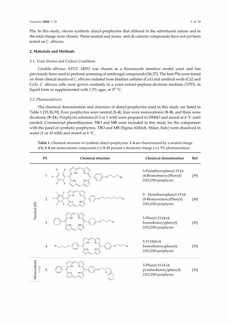
PSs. In this study, eleven synthetic diaryl-porphyrins that differed in the substituent nature and in the total charge were chosen. These neutral and mono- and di-cationic compounds have not yet been tested on C. albicans.
2. Materials and Methods
2.1. Yeast Strains and Culture Conditions
Candida albicans ATCC 14053 was chosen as a fluconazole sensitive model yeast and has previously been used to perform screening of antifungal compounds [36,37]. The best PSs were tested on three clinical strains of C. albicans isolated from bladder catheter (Ca1) and urethral swab (Ca2 and Ca3). C. albicans cells were grown routinely in a yeast extract-peptone-dextrose medium (YPD), in liquid form or supplemented with 1.5% agar, at 37 °C.
2.2. Photosensitizers
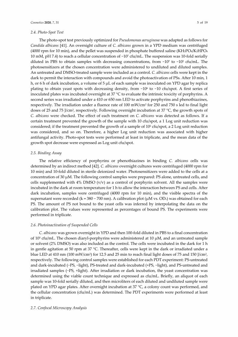
The chemical denomination and structure of diaryl-porphyrins used in this study are listed in Table 1 [35,38,39]. Four porphyrins were neutral (1–4), four were monocationic (5–8), and three were dicationic (9–11). Porphyrin solutions (0.5 or 1 mM) were prepared in DMSO and stored at 4 °C until needed. Commercial phenothiazines TBO and MB were included in this study for the comparison with the panel of synthetic porphyrins. TBO and MB (Sigma Aldrich, Milan, Italy) were dissolved in water (1 or 10 mM) and stored at 4 °C.
Table 1. Chemical structure of synthetic diaryl-porphyrins. 1–4 are characterized by a neutral charge (Ø); 5–8 are monocationic compounds (+); 9–11 present a dicationic charge (++). PS, photosensitizer.
PS Chemical structure Chemical denomination Ref
Neu
tral
(Ø)
1
5-Pentafluorophenyl-15-[4-(4-Bromobutoxy)Phenyl]-21H,23H-porphyrin
[39]
2
5- Pentafluorophenyl-15-[4-(8-Bromooctaoxy)Phenyl]-21H,23H-porphyrin
[39]
3
5-Phenyl-15-[4-(4-bromobutoxy)phenyl]-21H,23H-porphyrin
[35]
4
5,15-Di[4-(4-bromobutoxy)phenyl]-21H,23H-porphyrin
[35]
Mon
ocat
ioni
c(+
)
5
5-Phenyl-15-[4-(4-pyridinobutoxy)phenyl]-21H,23H-porphyrin
[35]
Cosmetics 2020, 7, 31 4 of 19
6
5-Phenyl-15-[4-(4-pyridinooctaoxy)phenyl]-21H,23H-porphyrin
[35]
7
5-Pentafluorophenyl-15-[4-(4-Pyridinobutoxy)Phenyl]-21H,23H-porphyrin
[39]
8
5-Pentafluorophenyl-15-[4-(4-Pyridinooctaoxy)Phenyl]-21H,23H-porphyrin
[39]
Dic
atio
nic
(++)
9
5,15-di(N-benzyl-4-pyridyl)porphyrin
[38]
10
5,15-Di[4-(4-pyridinobutoxy)phenyl]-21H,23H-porphyrin
[35]
11
5,15-Di[4-(4-pyridinooctaoxy)phenyl]-21H,23H-porphyrin
[35]
2.3. Light Source
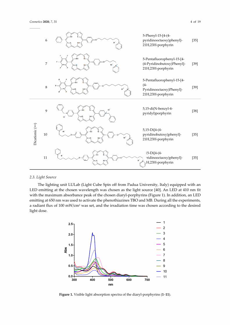
The lighting unit LULab (Light Cube Spin off from Padua University, Italy) equipped with an LED emitting at the chosen wavelength was chosen as the light source [40]. An LED at 410 nm fit with the maximum absorbance peak of the chosen diaryl-porphyrins (Figure 1). In addition, an LED emitting at 650 nm was used to activate the phenothiazines TBO and MB. During all the experiments, a radiant flux of 100 mW/cm2 was set, and the irradiation time was chosen according to the desired light dose.
Figure 1. Visible light absorption spectra of the diaryl-porphyrins (1–11).
Cosmetics 2020, 7, 31 5 of 19
2.4. Photo-Spot Test
The photo-spot test previously optimized for Pseudomonas aeruginosa was adapted as follows for Candida albicans [41]. An overnight culture of C. albicans grown in a YPD medium was centrifuged (4000 rpm for 10 min), and the pellet was suspended in phosphate buffered saline (KH2PO4/K2HPO4 10 mM, pH 7.4) to reach a cellular concentration of ~107 cfu/mL. The suspension was 10-fold serially diluted in PBS to obtain samples with decreasing concentrations, from ~107 to ~102 cfu/mL. The photosensitizers at the chosen concentration were administered to undiluted and diluted samples. An untreated and DMSO-treated sample were included as a control. C. albicans cells were kept in the dark to permit the interaction with compounds and avoid the photoactivation of PSs. After 10 min, 1 h, or 6 h of dark incubation, a volume of 5 µL of each sample was inoculated on YPD agar by replica plating to obtain yeast spots with decreasing density, from ~106 to ~10 cfu/spot. A first series of inoculated plates was incubated overnight at 37 °C to evaluate the intrinsic toxicity of porphyrins. A second series was irradiated under a 410 or 650 nm LED to activate porphyrins and phenothiazines, respectively. The irradiation under a fluence rate of 100 mW/cm2 for 250 and 750 s led to final light doses of 25 and 75 J/cm2, respectively. Following overnight incubation at 37 °C, the growth spots of C. albicans were checked. The effect of each treatment on C. albicans was detected as follows. If a certain treatment prevented the growth of the sample with 10 cfu/spot, a 1 Log unit reduction was considered; if the treatment prevented the growth of a sample of 102 cfu/spot, a 2 Log unit reduction was considered, and so on. Therefore, a higher Log unit reduction was associated with higher antifungal activity. Photo-spot tests were performed at least in triplicate, and the mean data of the growth spot decrease were expressed as Log unit cfu/spot.
2.5. Binding Assay
The relative efficiency of porphyrins or phenothiazines in binding C. albicans cells was determined by an indirect method [42]. C. albicans overnight cultures were centrifuged (4000 rpm for 10 min) and 10-fold diluted in sterile deionized water. Photosensitizers were added to the cells at a concentration of 30 µM. The following control samples were prepared: PS alone, untreated cells, and cells supplemented with 4% DMSO (v/v) as a control of porphyrin solvent. All the samples were incubated in the dark at room temperature for 1 h to allow the interaction between PS and cells. After dark incubation, samples were centrifuged (4000 rpm for 10 min), and the visible spectra of the supernatant were recorded (k = 380 − 700 nm). A calibration plot (µM vs. ODx) was obtained for each PS. The amount of PS not bound to the yeast cells was inferred by interpolating the data on the calibration plot. The values were represented as percentages of bound PS. The experiments were performed in triplicate.
2.6. Photoinactivation of Suspended Cells
C. albicans was grown overnight in YPD and then 100-fold diluted in PBS to a final concentration of 105 cfu/mL. The chosen diaryl-porphyrins were administered at 10 µM, and an untreated sample or solvent (2% DMSO) was also included as the control. The cells were incubated in the dark for 1 h in gentle agitation at 50 rpm at 37 °C. Thereafter, cells were kept in the dark or irradiated under a blue LED at 410 nm (100 mW/cm2) for 12.5 and 25 min to reach final light doses of 75 and 150 J/cm2, respectively. The following control samples were established for each PDT experiment: PS-untreated and dark-incubated (−PS, −light), PS-treated and dark-incubated (+PS, −light), and PS-untreated and irradiated samples (−PS, +light). After irradiation or dark incubation, the yeast concentration was determined using the viable count technique and expressed as cfu/mL. Briefly, an aliquot of each sample was 10-fold serially diluted, and then microliters of each diluted and undiluted sample were plated on YPD agar plates. After overnight incubation at 37 °C, a colony count was performed, and the cellular concentration (cfu/mL) was determined. The PDT experiments were performed at least in triplicate.
2.7. Confocal Microscopy Analysis
Cosmetics 2020, 7, 31 6 of 19
The effect of PDT on C. albicans cells by chosen diaryl-porphyrins was evaluated through confocal laser scanning microscopy (CLSM; Leica Microsystems, Wetzlar, Germany). C. albicans cells were photoinactivated following the treatment for suspended samples and irradiated at 150 J/cm2. After irradiation, a fluorescent boron-dipyrromethene dye (BODIPY 4,4-difluoro-1,3,5,7-tetramethyl-8-(2-methoxyphenyl)-4-bora-3a,4a-diaza-s-indacene) was added to the yeast suspension (5 µM) [43]. Cells were incubated with the fluorochrome for 30 min at 37 °C on a shaker at 50 rpm. Afterward, samples were centrifuged at 10,000 rpm for 10 min, and the pellet was suspended in 20 µL of PBS. A volume of 10 µL of suspension was placed between a microscope glass and a coverslip slide for CLSM acquisition. Pictures were obtained using a 63/5.0 objective lens (488 nm laser) and were edited by the free open source program ImageJ (National Institute of Health, Bethesda, USA). To assess the efficacy of the photodynamic treatment, the cell viability was checked through the viable count technique as previously described.
2.8. Biofilm Photoinactivation Assay
Candida albicans strains grown overnight in a YPD medium were centrifuged at 4000 rpm for 10 min. The pellets were 100-fold diluted in deionized water, and a volume of 250 µL was dispensed in 24 well microtiter plates. Cells were treated for 1 h in the dark at 37 °C with 20 µM porphyrins or 2% DMSO v/v, and untreated cells were also kept as a control. Afterwards, cells were kept in the dark or irradiated by 410 nm light (150 J/cm2). After dark incubation or irradiation, wells were filled with a further 250 µL of YPD medium and incubated at 37 °C for 24 h, to allow biofilm formation. Biofilm biomass was determined by crystal violet staining: upon removal of the supernatants, zero-point-five milliliters of a crystal violet (Sigma Aldrich, Milan, Italy) 0.1% solution were added, and after 20 min, a wash with deionized water was performed. After solubilization with 33% v/v acetic acid for 10 min, spectrophotometric measurement was performed at 590 nm. Adherent and planktonic populations of biofilm were also evaluated by a colony count method. Briefly, planktonic biomass was collected by the supernatant, and the adherent phase was collected by the wells upon the removal of the supernatant and the scraping of adherent cells in 0.5 mL of phosphate buffer. Collected samples were 10-fold serially diluted in a sterile phosphate buffer and plated on YPD agar plates. Upon overnight incubation, colonies were counted, and cfu/mL and cfu/well values were determined for planktonic and adherent populations, respectively. All experiments were independently repeated at least three times.
2.9. Statistical Analyses
Photoinactivation experiments on yeast suspended cells and biofilm formation were performed at least three times with different cultures, and statistical significance was assessed by one way ANOVA.
3. Results
3.1. Effect of Diaryl-Porphyrins on Candida albicans Viability
In the literature, most of the studies in the field of photoinactivation of Candida albicans have been performed with commercial phenothiazines, toluidine blue O (TBO) and methylene blue (MB). In the present study, we investigated a panel of synthetic diaryl-porphyrins as potential photosensitizers. Since in photodynamic therapy, the ideal PS should not display intrinsic toxicity, this feature was investigated. Furthermore, as porphyrins were dissolved in DMSO, the effect of this solvent on C. albicans viability was ruled out (Figure 2). C. albicans cells showed a different degree of sensitivity to porphyrins administered at 10 µM (Figure 2). The neutral porphyrins (1–4) did not display any toxicity to yeast cells, even at the longest dark incubation time. Among the monocationic diaryl-porphyrins, two PSs (5,6) showed a low degree of toxicity, causing the depletion of the samples with the lowest cellular densities (10 and 102 cfu/spot). The dicationic porphyrins were the most intrinsically toxic, and among these, Porphyrin 10 prevented the growth of the sample of 104 cfu/spot
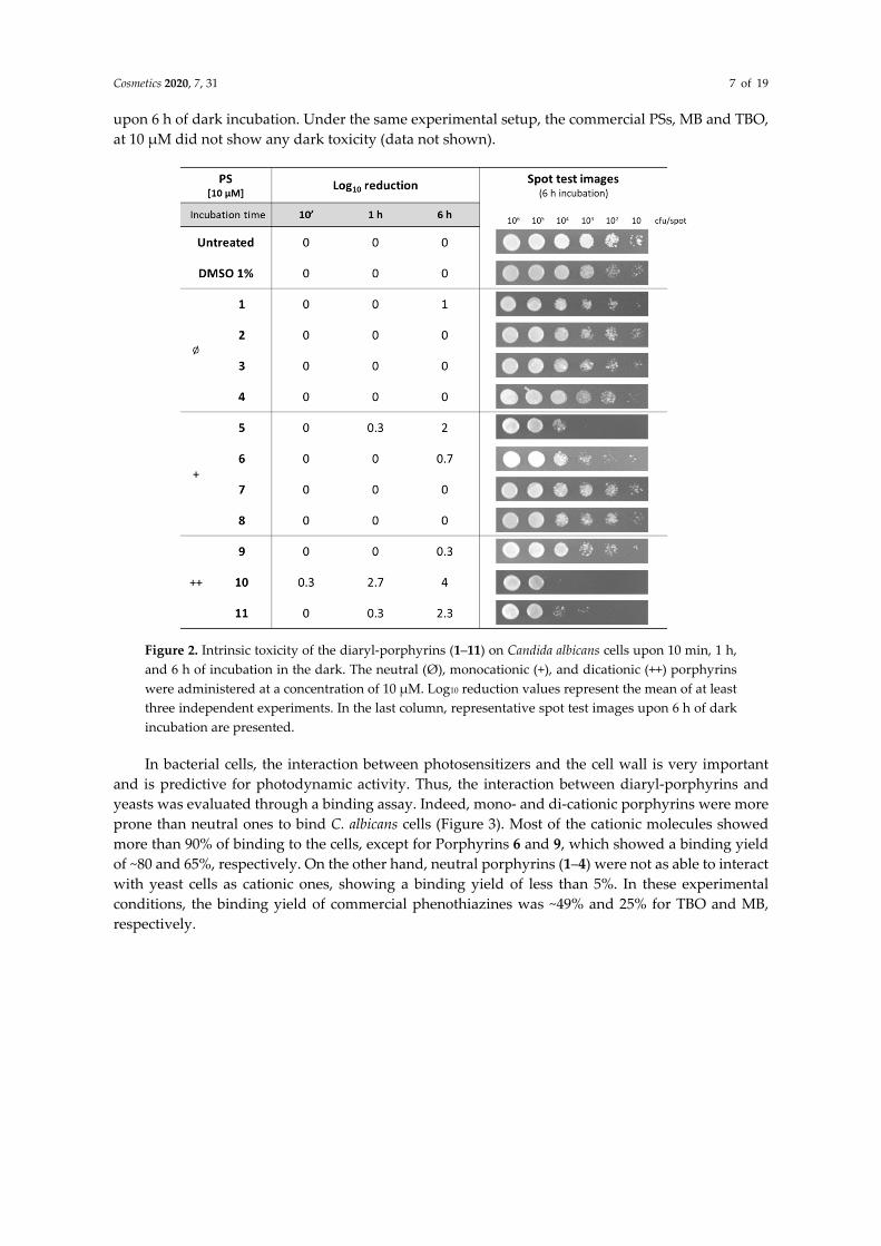
Cosmetics 2020, 7, 31 7 of 19
upon 6 h of dark incubation. Under the same experimental setup, the commercial PSs, MB and TBO, at 10 µM did not show any dark toxicity (data not shown).
Figure 2. Intrinsic toxicity of the diaryl-porphyrins (1–11) on Candida albicans cells upon 10 min, 1 h, and 6 h of incubation in the dark. The neutral (Ø), monocationic (+), and dicationic (++) porphyrins were administered at a concentration of 10 µM. Log10 reduction values represent the mean of at least three independent experiments. In the last column, representative spot test images upon 6 h of dark incubation are presented.
In bacterial cells, the interaction between photosensitizers and the cell wall is very important and is predictive for photodynamic activity. Thus, the interaction between diaryl-porphyrins and yeasts was evaluated through a binding assay. Indeed, mono- and di-cationic porphyrins were more prone than neutral ones to bind C. albicans cells (Figure 3). Most of the cationic molecules showed more than 90% of binding to the cells, except for Porphyrins 6 and 9, which showed a binding yield of ~80 and 65%, respectively. On the other hand, neutral porphyrins (1–4) were not as able to interact with yeast cells as cationic ones, showing a binding yield of less than 5%. In these experimental conditions, the binding yield of commercial phenothiazines was ~49% and 25% for TBO and MB, respectively.
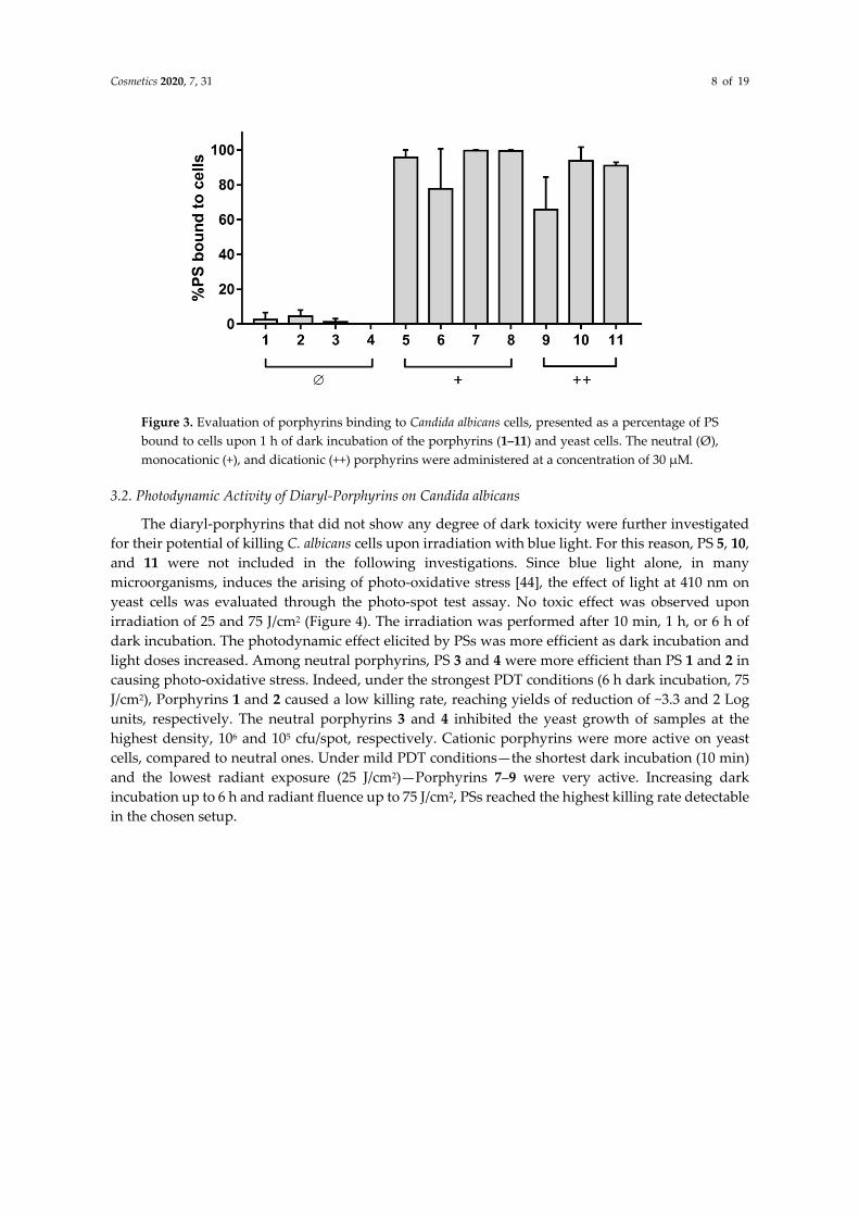
Cosmetics 2020, 7, 31 8 of 19
Figure 3. Evaluation of porphyrins binding to Candida albicans cells, presented as a percentage of PS bound to cells upon 1 h of dark incubation of the porphyrins (1–11) and yeast cells. The neutral (Ø), monocationic (+), and dicationic (++) porphyrins were administered at a concentration of 30 µM.
3.2. Photodynamic Activity of Diaryl-Porphyrins on Candida albicans
The diaryl-porphyrins that did not show any degree of dark toxicity were further investigated for their potential of killing C. albicans cells upon irradiation with blue light. For this reason, PS 5, 10, and 11 were not included in the following investigations. Since blue light alone, in many microorganisms, induces the arising of photo-oxidative stress [44], the effect of light at 410 nm on yeast cells was evaluated through the photo-spot test assay. No toxic effect was observed upon irradiation of 25 and 75 J/cm2 (Figure 4). The irradiation was performed after 10 min, 1 h, or 6 h of dark incubation. The photodynamic effect elicited by PSs was more efficient as dark incubation and light doses increased. Among neutral porphyrins, PS 3 and 4 were more efficient than PS 1 and 2 in causing photo-oxidative stress. Indeed, under the strongest PDT conditions (6 h dark incubation, 75 J/cm2), Porphyrins 1 and 2 caused a low killing rate, reaching yields of reduction of ~3.3 and 2 Log units, respectively. The neutral porphyrins 3 and 4 inhibited the yeast growth of samples at the highest density, 106 and 105 cfu/spot, respectively. Cationic porphyrins were more active on yeast cells, compared to neutral ones. Under mild PDT conditions—the shortest dark incubation (10 min) and the lowest radiant exposure (25 J/cm2)—Porphyrins 7–9 were very active. Increasing dark incubation up to 6 h and radiant fluence up to 75 J/cm2, PSs reached the highest killing rate detectable in the chosen setup.
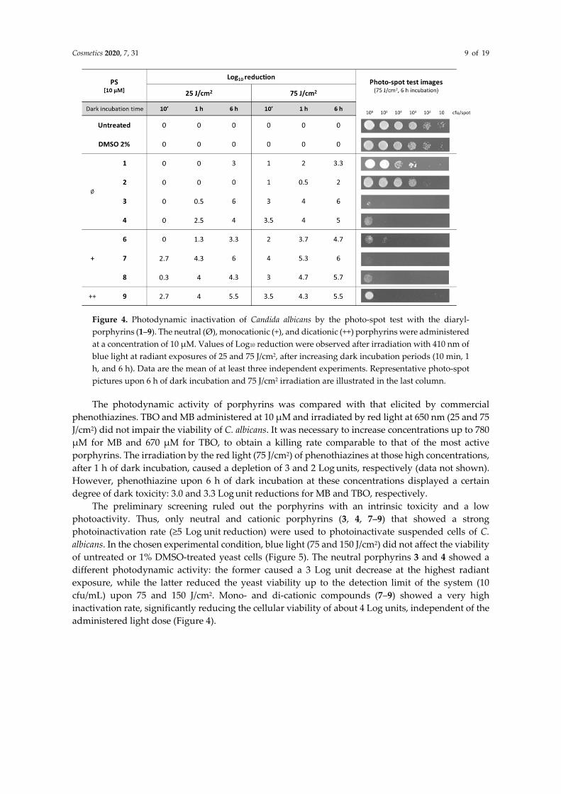
Cosmetics 2020, 7, 31 9 of 19
Figure 4. Photodynamic inactivation of Candida albicans by the photo-spot test with the diaryl-porphyrins (1–9). The neutral (Ø), monocationic (+), and dicationic (++) porphyrins were administered at a concentration of 10 µM. Values of Log10 reduction were observed after irradiation with 410 nm of blue light at radiant exposures of 25 and 75 J/cm2, after increasing dark incubation periods (10 min, 1 h, and 6 h). Data are the mean of at least three independent experiments. Representative photo-spot pictures upon 6 h of dark incubation and 75 J/cm2 irradiation are illustrated in the last column.
The photodynamic activity of porphyrins was compared with that elicited by commercial phenothiazines. TBO and MB administered at 10 µM and irradiated by red light at 650 nm (25 and 75 J/cm2) did not impair the viability of C. albicans. It was necessary to increase concentrations up to 780 µM for MB and 670 µM for TBO, to obtain a killing rate comparable to that of the most active porphyrins. The irradiation by the red light (75 J/cm2) of phenothiazines at those high concentrations, after 1 h of dark incubation, caused a depletion of 3 and 2 Log units, respectively (data not shown). However, phenothiazine upon 6 h of dark incubation at these concentrations displayed a certain degree of dark toxicity: 3.0 and 3.3 Log unit reductions for MB and TBO, respectively.
The preliminary screening ruled out the porphyrins with an intrinsic toxicity and a low photoactivity. Thus, only neutral and cationic porphyrins (3, 4, 7–9) that showed a strong photoinactivation rate (≥5 Log unit reduction) were used to photoinactivate suspended cells of C. albicans. In the chosen experimental condition, blue light (75 and 150 J/cm2) did not affect the viability of untreated or 1% DMSO-treated yeast cells (Figure 5). The neutral porphyrins 3 and 4 showed a different photodynamic activity: the former caused a 3 Log unit decrease at the highest radiant exposure, while the latter reduced the yeast viability up to the detection limit of the system (10 cfu/mL) upon 75 and 150 J/cm2. Mono- and di-cationic compounds (7–9) showed a very high inactivation rate, significantly reducing the cellular viability of about 4 Log units, independent of the administered light dose (Figure 4).
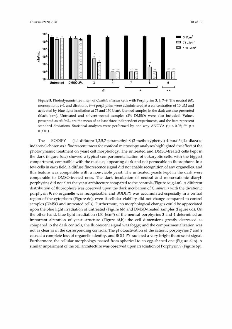
Cosmetics 2020, 7, 31 10 of 19
Figure 5. Photodynamic treatment of Candida albicans cells with Porphyrins 3, 4, 7–9. The neutral (Ø), monocationic (+), and dicationic (++) porphyrins were administered at a concentration of 10 µM and activated by blue light irradiation at 75 and 150 J/cm2. Control samples in the dark are also presented (black bars). Untreated and solvent-treated samples (2% DMSO) were also included. Values, presented as cfu/mL, are the mean of at least three independent experiments, and the bars represent standard deviations. Statistical analyses were performed by one way ANOVA (*p < 0.05; *** p < 0.0001).
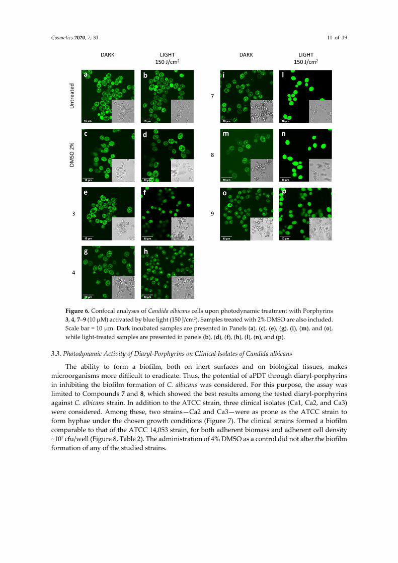
The BODIPY (4,4-difluoro-1,3,5,7-tetramethyl-8-(2-methoxyphenyl)-4-bora-3a,4a-diaza-s-indacene) chosen as a fluorescent tracer for confocal microscopy analyses highlighted the effect of the photodynamic treatment on yeast cell morphology. The untreated and DMSO-treated cells kept in the dark (Figure 6a,c) showed a typical compartmentalization of eukaryotic cells, with the biggest compartment, compatible with the nucleus, appearing dark and not permeable to fluorophore. In a few cells in each field, a diffuse fluorescence signal did not enable recognition of any organelles, and this feature was compatible with a non-viable yeast. The untreated yeasts kept in the dark were comparable to DMSO-treated ones. The dark incubation of neutral and mono-cationic diaryl-porphyrins did not alter the yeast architecture compared to the controls (Figure 6e,g,i,m). A different distribution of fluorophore was observed upon the dark incubation of C. albicans with the dicationic porphyrin 9: no organelle was recognizable, and BODIPY was accumulated especially in a central region of the cytoplasm (Figure 6o), even if cellular viability did not change compared to control samples (DMSO and untreated cells). Furthermore, no morphological changes could be appreciated upon the blue light irradiation of untreated (Figure 6b) and DMSO-treated samples (Figure 6d). On the other hand, blue light irradiation (150 J/cm2) of the neutral porphyrins 3 and 4 determined an important alteration of yeast structure (Figure 6f,h): the cell dimensions greatly decreased as compared to the dark controls; the fluorescent signal was foggy; and the compartmentalization was not as clear as in the corresponding controls. The photoactivation of the cationic porphyrins 7 and 8 caused a complete loss of organelle identity, and BODIPY radiated a very bright fluorescent signal. Furthermore, the cellular morphology passed from spherical to an egg-shaped one (Figure 6l,n). A similar impairment of the cell architecture was observed upon irradiation of Porphyrin 9 (Figure 6p).
Cosmetics 2020, 7, 31 11 of 19
Figure 6. Confocal analyses of Candida albicans cells upon photodynamic treatment with Porphyrins 3, 4, 7–9 (10 µM) activated by blue light (150 J/cm2). Samples treated with 2% DMSO are also included. Scale bar = 10 µm. Dark incubated samples are presented in Panels (a), (c), (e), (g), (i), (m), and (o), while light-treated samples are presented in panels (b), (d), (f), (h), (l), (n), and (p).
3.3. Photodynamic Activity of Diaryl-Porphyrins on Clinical Isolates of Candida albicans
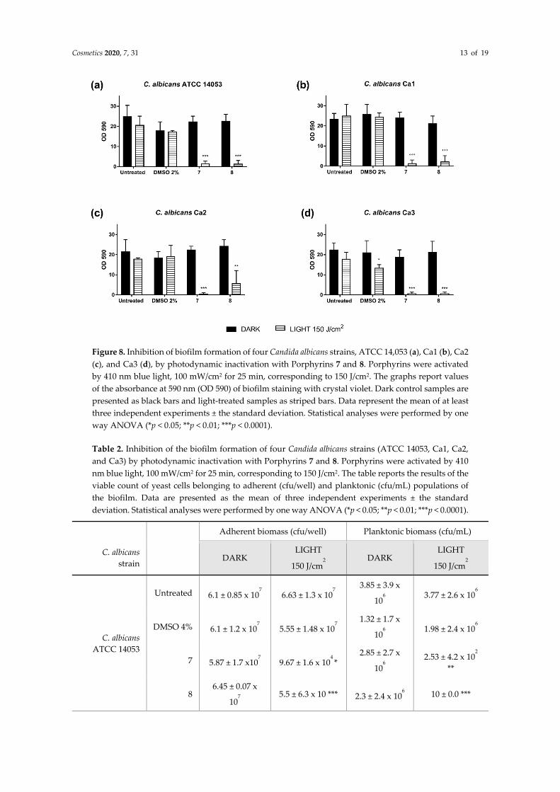
The ability to form a biofilm, both on inert surfaces and on biological tissues, makes microorganisms more difficult to eradicate. Thus, the potential of aPDT through diaryl-porphyrins in inhibiting the biofilm formation of C. albicans was considered. For this purpose, the assay was limited to Compounds 7 and 8, which showed the best results among the tested diaryl-porphyrins against C. albicans strain. In addition to the ATCC strain, three clinical isolates (Ca1, Ca2, and Ca3) were considered. Among these, two strains—Ca2 and Ca3—were as prone as the ATCC strain to form hyphae under the chosen growth conditions (Figure 7). The clinical strains formed a biofilm comparable to that of the ATCC 14,053 strain, for both adherent biomass and adherent cell density ~107 cfu/well (Figure 8, Table 2). The administration of 4% DMSO as a control did not alter the biofilm formation of any of the studied strains.
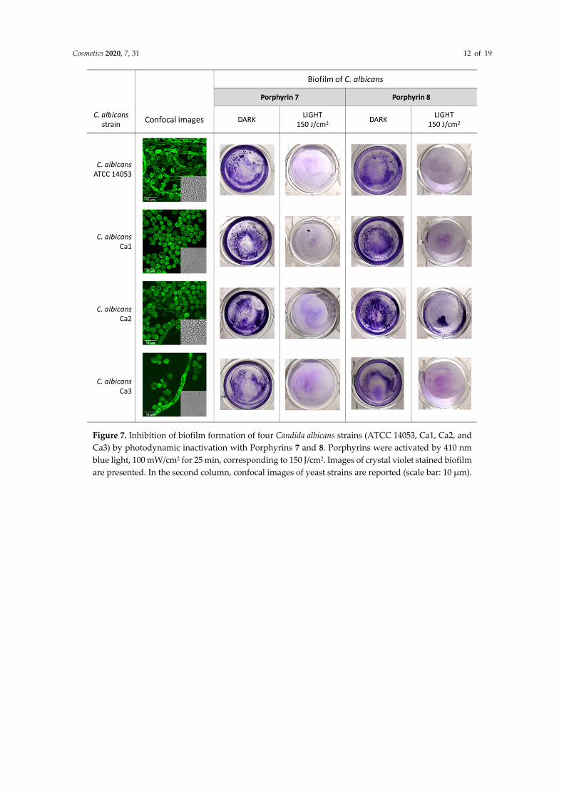
Cosmetics 2020, 7, 31 12 of 19
Figure 7. Inhibition of biofilm formation of four Candida albicans strains (ATCC 14053, Ca1, Ca2, and Ca3) by photodynamic inactivation with Porphyrins 7 and 8. Porphyrins were activated by 410 nm blue light, 100 mW/cm2 for 25 min, corresponding to 150 J/cm2. Images of crystal violet stained biofilm are presented. In the second column, confocal images of yeast strains are reported (scale bar: 10 µm).
Cosmetics 2020, 7, 31 13 of 19
Figure 8. Inhibition of biofilm formation of four Candida albicans strains, ATCC 14,053 (a), Ca1 (b), Ca2 (c), and Ca3 (d), by photodynamic inactivation with Porphyrins 7 and 8. Porphyrins were activated by 410 nm blue light, 100 mW/cm2 for 25 min, corresponding to 150 J/cm2. The graphs report values of the absorbance at 590 nm (OD 590) of biofilm staining with crystal violet. Dark control samples are presented as black bars and light-treated samples as striped bars. Data represent the mean of at least three independent experiments ± the standard deviation. Statistical analyses were performed by one way ANOVA (*p < 0.05; **p < 0.01; ***p < 0.0001).
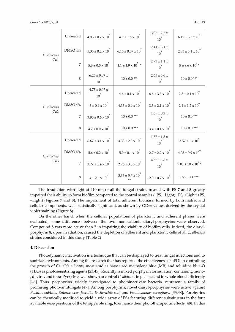
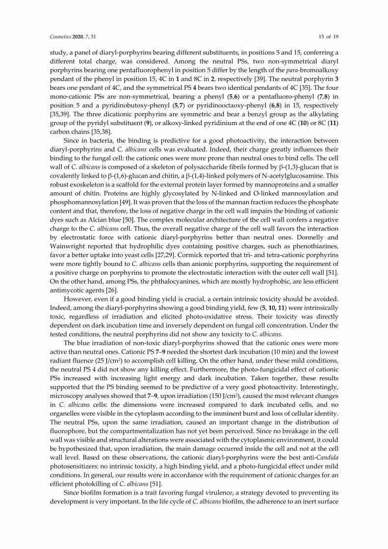
Table 2. Inhibition of the biofilm formation of four Candida albicans strains (ATCC 14053, Ca1, Ca2, and Ca3) by photodynamic inactivation with Porphyrins 7 and 8. Porphyrins were activated by 410 nm blue light, 100 mW/cm2 for 25 min, corresponding to 150 J/cm2. The table reports the results of the viable count of yeast cells belonging to adherent (cfu/well) and planktonic (cfu/mL) populations of the biofilm. Data are presented as the mean of three independent experiments ± the standard deviation. Statistical analyses were performed by one way ANOVA (*p < 0.05; **p < 0.01; ***p < 0.0001).
Adherent biomass (cfu/well) Planktonic biomass (cfu/mL)
C. albicans strain
DARK LIGHT
150 J/cm2
DARK LIGHT
150 J/cm2
C. albicans ATCC 14053
Untreated 6.1 ± 0.85 x 10⁷ 6.63 ± 1.3 x 10
7
3.85 ± 3.9 x
106
3.77 ± 2.6 x 10⁶
DMSO 4% 6.1 ± 1.2 x 10⁷ 5.55 ± 1.48 x 10
7
1.32 ± 1.7 x
106
1.98 ± 2.4 x 10⁶
7 5.87 ± 1.7 x10⁷ 9.67 ± 1.6 x 10
4 *
2.85 ± 2.7 x
106
2.53 ± 4.2 x 10²
**
8 6.45 ± 0.07 x
107
5.5 ± 6.3 x 10 *** 2.3 ± 2.4 x 10⁶ 10 ± 0.0 ***
Cosmetics 2020, 7, 31 14 of 19
C. albicans Ca1
Untreated 4.93 ± 0.7 x 10⁷ 4.9 ± 1.6 x 10
7
3.87 ± 2.7 x
106
6.17 ± 3.5 x 10⁶
DMSO 4% 5.35 ± 0.2 x 10⁷ 6.15 ± 0.07 x 10
7
2.41 ± 3.1 x
106
2.83 ± 3.1 x 10⁶
7 5.3 ± 0.5 x 10⁷ 1.1 ± 1.9 x 10
5 * 2.73 ± 1.1 x
106
5 ± 8.6 x 104 *
8 6.25 ± 0.07 x
107
10 ± 0.0 *** 2.65 ± 3.6 x
106
10 ± 0.0 ***
C. albicans Ca2
Untreated 4.75 ± 0.07 x
107
4.6 ± 0.1 x 10⁷ 6.6 ± 3.3 x 10
6 2.3 ± 0.1 x 10
6
DMSO 4% 5 ± 0.4 x 10⁷ 4.35 ± 0.9 x 10
7 3.5 ± 2.1 x 10
6 2.4 ± 1.2 x 10
6
7 3.95 ± 0.6 x 10⁷ 10 ± 0.0 ***
1.65 ± 0.2 x
106
10 ± 0.0 ***
8 4.7 ± 0.0 x 10⁷ 10 ± 0.0 *** 3.4 ± 0.1 x 10
6 10 ± 0.0 ***
C. albicans Ca3
Untreated 6.67 ± 3.1 x 10⁷ 3.33 ± 2.3 x 10
7
1.57 ± 1.5 x
107
3.57 ± 1 x 10⁶
DMSO 4% 5.6 ± 0.2 x 10⁷ 5.9 ± 0.4 x 10
7 2.7 ± 2.2 x 10
6 4.05 ± 0.9 x 10
6
7 3.27 ± 1.4 x 10⁷ 2.26 ± 3.8 x 10
5
4.57 ± 3.6 x
106
9.01 ± 10 x 103 *
8 4 ± 2.6 x 10⁷
3.36 ± 5.7 x 10³
** 2.9 ± 0.7 x 10
6 16.7 ± 11 ***
The irradiation with light at 410 nm of all the fungal strains treated with PS 7 and 8 greatly impaired their ability to form biofilm compared to the control samples (−PS, −Light; −PS, +Light; +PS, −Light) (Figures 7 and 8). The impairment of total adherent biomass, formed by both matrix and cellular components, was statistically significant, as shown by OD590 values derived by the crystal violet staining (Figure 8).
On the other hand, when the cellular populations of planktonic and adherent phases were evaluated, some differences between the two monocationic diaryl-porphyrins were observed. Compound 8 was more active than 7 in impairing the viability of biofilm cells. Indeed, the diaryl-porphyrin 8, upon irradiation, caused the depletion of adherent and planktonic cells of all C. albicans strains considered in this study (Table 2)
4. Discussion
Photodynamic inactivation is a technique that can be displayed to treat fungal infections and to sanitize environments. Among the research that has reported the effectiveness of aPDI in controlling the growth of Candida albicans, most studies have used methylene blue (MB) and toluidine blue-O (TBO) as photosensitizing agents [23,45]. Recently, a mixed porphyrin formulation, containing mono-, di-, tri-, and tetra-Py(+)-Me, was shown to control C. albicans in plasma and in whole blood efficiently [46]. Thus, porphyrins, widely investigated to photoinactivate bacteria, represent a family of promising photo-antifungals [47]. Among porphyrins, novel diaryl-porphyrins were active against Bacillus subtilis, Enterococcus faecalis, Escherichia coli, and Pseudomonas aeruginosa [35,38]. Porphyrins can be chemically modified to yield a wide array of PSs featuring different substituents in the four available meso positions of the tetrapyrrole ring, to enhance their phototherapeutic effects [48]. In this
Cosmetics 2020, 7, 31 15 of 19
study, a panel of diaryl-porphyrins bearing different substituents, in positions 5 and 15, conferring a different total charge, was considered. Among the neutral PSs, two non-symmetrical diaryl porphyrins bearing one pentafluorophenyl in position 5 differ by the length of the para-bromoalkoxy pendant of the phenyl in position 15, 4C in 1 and 8C in 2, respectively [39]. The neutral porphyrin 3 bears one pendant of 4C, and the symmetrical PS 4 bears two identical pendants of 4C [35]. The four mono-cationic PSs are non-symmetrical, bearing a phenyl (5,6) or a pentafluoro-phenyl (7,8) in position 5 and a pyridinobutoxy-phenyl (5,7) or pyridinooctaoxy-phenyl (6,8) in 15, respectively [35,39]. The three dicationic porphyrins are symmetric and bear a benzyl group as the alkylating group of the pyridyl substituent (9), or alkoxy-linked pyridinium at the end of one 4C (10) or 8C (11) carbon chains [35,38].
Since in bacteria, the binding is predictive for a good photoactivity, the interaction between diaryl-porphyrins and C. albicans cells was evaluated. Indeed, their charge greatly influences their binding to the fungal cell: the cationic ones were more prone than neutral ones to bind cells. The cell wall of C. albicans is composed of a skeleton of polysaccharide fibrils formed by β-(1,3)-glucan that is covalently linked to β-(1,6)-glucan and chitin, a β-(1,4)-linked polymers of N-acetylglucosamine. This robust exoskeleton is a scaffold for the external protein layer formed by mannoproteins and a smaller amount of chitin. Proteins are highly glycosylated by N-linked and O-linked mannosylation and phosphomannosylation [49]. It was proven that the loss of the mannan fraction reduces the phosphate content and that, therefore, the loss of negative charge in the cell wall impairs the binding of cationic dyes such as Alcian blue [50]. The complex molecular architecture of the cell wall confers a negative charge to the C. albicans cell. Thus, the overall negative charge of the cell wall favors the interaction by electrostatic force with cationic diaryl-porphyrins better than neutral ones. Donnelly and Wainwright reported that hydrophilic dyes containing positive charges, such as phenothiazines, favor a better uptake into yeast cells [27,29]. Cormick reported that tri- and tetra-cationic porphyrins were more tightly bound to C. albicans cells than anionic porphyrins, supporting the requirement of a positive charge on porphyrins to promote the electrostatic interaction with the outer cell wall [51]. On the other hand, among PSs, the phthalocyanines, which are mostly hydrophobic, are less efficient antimycotic agents [26].
However, even if a good binding yield is crucial, a certain intrinsic toxicity should be avoided. Indeed, among the diaryl-porphyrins showing a good binding yield, few (5, 10, 11) were intrinsically toxic, regardless of irradiation and elicited photo-oxidative stress. Their toxicity was directly dependent on dark incubation time and inversely dependent on fungal cell concentration. Under the tested conditions, the neutral porphyrins did not show any toxicity to C. albicans.
The blue irradiation of non-toxic diaryl-porphyrins showed that the cationic ones were more active than neutral ones. Cationic PS 7–9 needed the shortest dark incubation (10 min) and the lowest radiant fluence (25 J/cm2) to accomplish cell killing. On the other hand, under these mild conditions, the neutral PS 4 did not show any killing effect. Furthermore, the photo-fungicidal effect of cationic PSs increased with increasing light energy and dark incubation. Taken together, these results supported that the PS binding seemed to be predictive of a very good photoactivity. Interestingly, microscopy analyses showed that 7–9, upon irradiation (150 J/cm2), caused the most relevant changes in C. albicans cells: the dimensions were increased compared to dark incubated cells, and no organelles were visible in the cytoplasm according to the imminent burst and loss of cellular identity. The neutral PSs, upon the same irradiation, caused an important change in the distribution of fluorophore, but the compartmentalization has not yet been perceived. Since no breakage in the cell wall was visible and structural alterations were associated with the cytoplasmic environment, it could be hypothesized that, upon irradiation, the main damage occurred inside the cell and not at the cell wall level. Based on these observations, the cationic diaryl-porphyrins were the best anti-Candida photosensitizers: no intrinsic toxicity, a high binding yield, and a photo-fungicidal effect under mild conditions. In general, our results were in accordance with the requirement of cationic charges for an efficient photokilling of C. albicans [51].
Since biofilm formation is a trait favoring fungal virulence, a strategy devoted to preventing its development is very important. In the life cycle of C. albicans biofilm, the adherence to an inert surface
Cosmetics 2020, 7, 31 16 of 19
or tissue is essential to the following initiation step and maturation [52]. This study identified 7 and 8 as optimal diaryl-porphyrins to photoinactivate C. albicans cells. Both cationic PSs inhibited the formation of an adherent biomass. In particular, Compound 8, with the longest chain, was more efficient than 7, at inhibiting the planktonic and sessile cellular populations of the biofilm of both C. albicans ATCC 14053 and three C. albicans clinical isolates. It is worth noting that, in C. albicans, the response to PDT was strain-dependent, as previously observed in bacterial species. Indeed, it was observed that the bactericidal effect of the PDI in Staphylococcus aureus was strain-dependent [53]. Since there are few studies aimed at investigating the effectiveness of photodynamic therapy to treat C. albicans infections [54], an optimized PDT protocol based on diaryl-porphyrins should be applied to host cell lines to rule out any toxic effects and should be tested in vivo to evaluate their photoactivity in skin and oral districts. For this aim, photoactivable keratin sponges functionalized with porphyrins were proposed as a scaffold for the antimicrobial treatment of skin [55].
5. Conclusions
In conclusion, the resistance to antifungals developed by C. albicans strains is an issue that makes the management of infected patients difficult. This study supported the great potential of a light-based antimicrobial approach, known as photodynamic therapy, as an antifungal treatment. In particular, monocationic diaryl-porphyrins were shown to be optimal photosensitizers under studied settings. The presence of one positive charge on the porphyrin periphery favored the binding to the fungal cell without eliciting an intrinsic toxic effect. Upon irradiation with blue light, the candidate PSs killed the C. albicans cells and inhibited biofilm formation, compromising the viability of both adherent and planktonic phases. These porphyrins represent potential broad-spectrum photo-antimicrobial agents.
Author Contributions: Conceptualization, V.T.O. and E.C.; LED technology and software, N.T.; investigation, E.M., M.P., F.B., and A.B.; data curation, E.M. and V.T.O.; writing, original draft preparation, V.T.O. and E.M.; writing, review and editing, V.T.O., E.C., O.M, and A.B.; supervision, V.T.O.; funding acquisition, V.T.O. All authors read and agreed to the published version of the manuscript.
Funding: This research was supported by the University of Insubria (Fondo di Ateneo per la Ricerca).
Acknowledgments: E.M. is a PhD student of the Life Science and Biotechnology course at Università degli Studi dell’Insubria.
Conflicts of Interest: The authors declare that there is no conflict of interest.
References
1. Kim, S.; Woo, E.R.; Lee, D.G. Synergistic Antifungal Activity of Isoquercitrin: Apoptosis and Membrane Permeabilization Related to Reactive Oxygen Species in Candida albicans. IUBMB Life 2019, 71, 283–292.
2. Wang, C.; Yang, Z.; Peng, Y.; Guo, Y.; Yao, M.; Dong, J. Application of 460 nm visible light for the elimination of Candida albicans in vitro and in vivo. Mol. Med. Rep. 2018, 18, 2017–2026.
3. Feldman, M.; Shenderovich, J.; Al-Quntar, A.A.A.; Friedman, M.; Steinberg, D. Sustained release of a novel anti-quorum-sensing agent against oral fungal biofilms. Antimicrob. Agents Chemother. 2015, 59, 2265–2272.
4. Dai, T.; Bil De Arce, V.J.; Tegos, G.P.; Hamblin, M.R. Blue dye and red light, a dynamic combination for prophylaxis and treatment of cutaneous Candida albicans infections in mice. Antimicrob. Agents Chemother. 2011, 55, 5710–5717.
5. Cieplik, F.; Deng, D.; Crielaard, W.; Buchalla, W.; Hellwig, E.; Al-Ahmad, A.; Maisch, T. Antimicrobial photodynamic therapy–what we know and what we don’t. Crit. Rev. Microbiol. 2018, 44, 571–589.
6. Costa, A.C.B.P.; Campos Rasteiro, V.M.; Da Silva Hashimoto, E.S.H.; Araújo, C.F.; Pereira, C.A.; Junqueira, J.C.; Jorge, A.O.C. Effect of erythrosine- and LED-mediated photodynamic therapy on buccal candidiasis infection of immunosuppressed mice and Candida albicans adherence to buccal epithelial cells. Oral Surg. Oral Med. Oral Pathol. Oral Radiol. 2012, 114, 67–74.
7. Cavalheiro, M.; Teixeira, M.C. Candida Biofilms: Threats, challenges, and promising strategies. Front. Med. 2018, 5, 1–15.
8. Abe, M.; Nakamura, S.; Kinjo, Y.; Masuyama, Y.; Mitsuyama, J.; Kaku, M.; Miyazaki, Y. Efficacy of T-2307,
Cosmetics 2020, 7, 31 17 of 19
a novel arylamidine, against ocular complications of disseminated candidiasis in mice. J. Antimicrob. Chemother. 2019, 74, 1327–1332.
9. Machado-de-Sena, R.M.; Corrêa, L.; Kato, I.T.; Prates, R.A.; Senna, A.M.; Santos, C.C.; Picanço, D.A.; Ribeiro, M.S. Photodynamic therapy has antifungal effect and reduces inflammatory signals in Candida albicans-induced murine vaginitis. Photodiagnosis Photodyn. Ther. 2014, 11, 275–282.
10. Nobile, C.J.; Johnson, A.D. Candida albicans Biofilms and Human Disease. Annu. Rev. Microbiol. 2015, 69, 71–92.
11. Passarelli, P.C.; De Leonardis, M.; Piccirillo, G.B.; Desantis, V.; Papa, R.; Rella, E.; Bonaviri, G.N.M.; Papi, P.; Pompa, G.; Pasquantonio, G.; et al. The effectiveness of chlorhexidine and air polishing system in the treatment of candida albicans infected dental implants: An experimental in vitro study. Antibiotics 2020, 9, 1–12.
12. Benzaid, C.; Belmadani, A.; Djeribi, R.; Rouabhia, M. The effects of mentha × piperita essential oil on C. Albicans growth, transition, biofilm formation, and the expression of secreted aspartyl proteinases genes. Antibiotics 2019, 8, 10.
13. Rosa, L.P.; da Silva, F.C.; Viana, M.S.; Meira, G.A. In vitro effectiveness of 455-nm blue LED to reduce the load of Staphylococcus aureus and Candida albicans biofilms in compact bone tissue. Lasers Med. Sci. 2016, 31, 27–32.
14. Huang, M.C.; Shen, M.; Huang, Y.J.; Lin, H.C.; Chen, C.T. Photodynamic inactivation potentiates the susceptibility of antifungal agents against the planktonic and biofilm cells of Candida albicans. Int. J. Mol. Sci. 2018, 19, 434.
15. Ghannoum, M.A.; Rice, L.B. Antifungal agents: Mode of action, mechanisms of resistance, and correlation of these mechanisms with bacterial resistance. Clin. Microbiol. Rev. 1999, 12, 501–517.
16. Rodrigues, M.E.; Silva, S.; Azeredo, J.; Henriques, M. Novel strategies to fight Candida species infection. Crit. Rev. Microbiol. 2016, 42, 594–606.
17. Gupta, A.K.; Ahmad, I.; Summerbell, R.C. Fungicidal activities of commonly used disinfectants and antifungal pharmaceutical spray preparations against clinical strains of Aspergillus and Candida species. Med. Mycol. 2002, 40, 201–208.
18. Sibata, C.H.; Colussi, V.C.; Oleinick, N.L.; Kinsella, T.J. Photodynamic therapy: A new concept in medical treatment. Brazilian J. Med. Biol. Res. 2000, 33, 869–880.
19. Kato, I.T.; Prates, R.A.; Sabino, C.P.; Fuchs, B.B.; Tegos, G.P.; Mylonakis, E.; Hamblin, M.R.; Ribeiro, M.S. Antimicrobial photodynamic inactivation inhibits Candida albicans virulence factors and reduces in vivo pathogenicity. Antimicrob. Agents Chemother. 2013, 57, 445–451.
20. Lin, C.H.; Chien, H.F.; Lin, M.H.; Chen, C.P.; Shen, M.; Chen, C.T. Chitosan inhibits the rehabilitation of damaged microbes induced by photodynamic inactivation. Int. J. Mol. Sci. 2018, 19, 19.
21. Queiroga, A.S.; Trajano, V.N.; Lima, E.O.; Ferreira, A.F.M.; Queiroga, A.S.; Limeira, F.A. In vitro photodynamic inactivation of Candida spp. by different doses of low power laser light. Photodiagnosis Photodyn. Ther. 2011, 8, 332–336.
22. Costa, A.C.B.P.; Rasteiro, V.M.C.; Pereira, C.A.; Rossoni, R.D.; Junqueira, J.C.; Jorge, A.O.C. The effects of rose bengal- and erythrosine-mediated photodynamic therapy on Candida albicans. Mycoses 2012, 55, 56–63.
23. Pupo, Y.M.; Gomes, G.M.; Santos, E.B.; Chaves, L.; Michel, M.D.; Kozlowski, V.A.; Gomes, O.M.M.; Gomes, J.C. Susceptibility of Candida albicans to photodynamic therapy using methylene blue and toluidine blue as photosensitizing dyes. Acta Odontol. Latinoam. 2011, 24, 188–192.
24. Munin, E.; Giroldo, L.M.; Alves, L.P.; Costa, M.S. Study of germ tube formation by Candida albicans after photodynamic antimicrobial chemotherapy (PACT). J. Photochem. Photobiol. B Biol. 2007, 88, 16–20.
25. Teichert, M.C.; Jones, J.W.; Usacheva, M.N.; Biel, M.A. Treatment of oral candidiasis with methylene blue-mediated photodynamic therapy in an immunodeficient murine model. Oral Surg. Oral Med. Oral Pathol. Oral Radiol. Endod. 2002, 93, 155–160.
26. Calzavara-Pinton, P.G.; Venturini, M.; Sala, R. A comprehensive overview of photodynamic therapy in the treatment of superficial fungal infections of the skin. J. Photochem. Photobiol. B Biol. 2005, 78, 1–6.
27. Donnelly, R.F.; McCarron, P.A.; Tunney, M.M. Antifungal photodynamic therapy. Microbiol. Res. 2008, 163, 1–12.
28. Peloi, L.S.; Soares, R.R.S.; Biondo, C.E.G.; Souza, V.R.; Hioka, N.; Kimura, E. Photodynamic effect of light emitting diode light on cell growth. J. Biosci. 2008, 33, 231–237.
Cosmetics 2020, 7, 31 18 of 19
29. Wainwright, M.; Giddens, R.M. Phenothiazinium photosensitisers: Choices in synthesis and application. Dye. Pigment. 2003, 57, 245–257.
30. Ma, J.; Shi, H.; Sun, H.; Li, J.; Bai, Y. Antifungal effect of photodynamic therapy mediated by curcumin on Candida albicans biofilms in vitro. Photodiagnosis Photodyn. Ther. 2019, 27, 280–287.
31. Hsieh, Y.H.; Zhang, J.H.; Chuang, W.C.; Yu, K.H.; Huang, X.B.; Lee, Y.C.; Lee, C.I. An in vitro study on the effect of combined treatment with photodynamic and chemical therapies on Candida albicans. Int. J. Mol. Sci. 2018, 19, 337.
32. Janeth Rimachi Hidalgo, K.; Cabrini Carmello, J.; Carolina Jordão, C.; Aboud Barbugli, P.; de Sousa Costa, C.A.; Garcia de Oliveira Mima, E.; Pavarina, A.C. Antimicrobial photodynamic therapy in combination with nystatin in the treatment of experimental oral candidiasis induced by candida albicans resistant to fluconazole. Pharmaceuticals 2019, 12, 140.
33. Hsieh, Y.H.; Chuang, W.C.; Yu, K.H.; Jheng, C.P.; Lee, C.I. Sequential photodynamic therapy with phthalocyanine encapsulated chitosan-tripolyphosphate nanoparticles and flucytosine treatment against candida tropicalis. Pharmaceutics 2019, 11, 16.
34. Diogo, P.; Faustino, M.F.A.; Neves, G.M.P.M.S.; Palma, P.J.; Baptista, I.P.; Gonçalves, T.; Santos, J.M. An insight into advanced approaches for photosensitizer optimization in endodontics—a critical review. J. Funct. Biomater. 2019, 10, 44.
35. Caruso, E.; Malacarne, M.C.; Banfi, S.; Gariboldi, M.B.; Orlandi, V.T. Cationic diarylporphyrins: In vitro versatile anticancer and antibacterial photosensitizers. J. Photochem. Photobiol. B Biol. 2019, 197, 111548.
36. Gunasegar, S.; Himratul-Aznita, W.H. Nicotine enhances the thickness of biofilm and adherence of Candida albicans ATCC 14053 and Candida parapsilosis ATCC 22019. FEMS Yeast Res. 2019, 19, 1–11.
37. Campana, R.; Ciandrini, E.; Baffone, W. Experimental approach for a possible integrated protocol to determine sanitizer activity against both planktonic bacteria and related biofilms. Food Res. Int. 2018, 111, 472–479.
38. Orlandi, V.T.; Caruso, E.; Tettamanti, G.; Banfi, S.; Barbieri, P. Photoinduced antibacterial activity of two dicationic 5,15-diarylporphyrins. J. Photochem. Photobiol. B Biol. 2013, 127, 123–132.
39. Caruso, E.; Cerbara, M.; Malacarne, M.C.; Marras, E.; Monti, E.; Gariboldi, M.B. Synthesis and photodynamic activity of novel non-symmetrical diaryl porphyrins against cancer cell lines. J. Photochem. Photobiol. B Biol. 2019, 195, 39–50.
40. Martegani, E.; Bolognese, F.; Trivellin, N.; Orlandi, V.T. Effect of blue light at 410 and 455 nm on Pseudomonas aeruginosa biofilm. J. Photochem. Photobiol. B Biol. 2020, 204, 111790.
41. Orlandi, V.T.; Martegani, E.; Bolognese, F. Catalase A is involved in the response to photooxidative stress in Pseudomonas aeruginosa. Photodiagnosis Photodyn. Ther. 2018, 22, 233–240.
42. Orlandi, V.T.; Caruso, E.; Banfi, S.; Barbieri, P. Effect of organic matter on the in Vitro Photoeradication of Pseudomonas aeruginosa by means of a cationic tetraaryl-porphyrin. Photochem. Photobiol. 2012, 88, 557–564.
43. Sunahara, H.; Urano, Y.; Kojima, H.; Nagano, T. Design and synthesis of a library of BODIPY-based environmental polarity sensors utilizing photoinduced electron-transfer-controlled fluorescence ON/OFF switching. J. Am. Chem. Soc. 2007, 129, 5597–5604.
44. Wang, Y.; Wang, Y.; Wang, Y.; Murray, C.K.; Hamblin, M.R.; Hooper, D.C.; Dai, T. Antimicrobial blue light inactivation of pathogenic microbes: State of the art. Drug Resist. Updat. 2017, 33–35, 1–22.
45. Lyon, J.P.; Rezende, R.R.; Rabelo, M.P.; de Lima, C.J.; Moreira, L.M. Synergic Effect of Photodynamic Therapy with Methylene Blue and Surfactants in the Inhibition of Candida albicans. Mycopathologia 2013, 175, 159–164.
46. Sousa, V.; Gomes, A.T.P.C.; Freitas, A.; Faustino, M.A.F.; Neves, M.G.P.M.S.; Almeida, A. Photodynamic inactivation of candida albicans in blood plasma and whole blood. Antibiotics 2019, 8, 221.
47. Scanone, A.C.; Gsponer, N.S.; Alvarez, M.G.; Durantini, E.N. Porphyrins containing basic aliphatic amino groups as potential broad-spectrum antimicrobial agents. Photodiagnosis Photodyn. Ther. 2018, 24, 220–227.
48. Martinez De Pinillos Bayona, A.; Mroz, P.; Thunshelle, C.; Hamblin, M.R. Design features for optimization of tetrapyrrole macrocycles as antimicrobial and anticancer photosensitizers. Chem. Biol. Drug Des. 2017, 89, 192–206.
49. Kapteyn, J.C.; Montijn, R.C.; Dijkgraaf, G.J.P.; Van den Ende, H.; Klis, F.M. Covalent association of β-1,3-glucan with β-1,6-glucosylated mannoproteins in cell walls of Candida albicans. J. Bacteriol. 1995, 177, 3788–3792.
Cosmetics 2020, 7, 31 19 of 19
50. Hall, R.A.; Gow, N.A.R. Mannosylation in candida albicans: Role in cell wall function and immune recognition. Mol. Microbiol. 2013, 90, 1147–1161.
51. Cormick, M.P.; Alvarez, M.G.; Rovera, M.; Durantini, E.N. Photodynamic inactivation of Candida albicans sensitized by tri- and tetra-cationic porphyrin derivatives. Eur. J. Med. Chem. 2009, 44, 1592–1599.
52. Gulati, M.; Nobile, C.J. Candida albicans biofilms: Development, regulation, and molecular mechanisms. Microbes Infect. 2016, 18, 310–321.
53. Grinholc, M.; Szramka, B.; Kurlenda, J.; Graczyk, A.; Bielawski, K.P. Bactericidal effect of photodynamic inactivation against methicillin-resistant and methicillin-susceptible Staphylococcus aureus is strain-dependent. J. Photochem. Photobiol. B Biol. 2008, 90, 57–63.
54. Pereira Gonzales, F.; Maisch, T. Photodynamic inactivation for controlling Candida albicans infections. Fungal Biol. 2012, 116, 1–10.
55. Ferroni, C.; Sotgiu, G.; Sagnella, A.; Varchi, G.; Guerrini, A.; Giuri, D.; Polo, E.; Orlandi, V.T.; Marras, E.; Gariboldi, M.; et al. Wool Keratin 3D Scaffolds with Light-Triggered Antimicrobial Activity. Biomacromolecules 2016, 17, 2882–2890.
© 2020 by the authors. Licensee MDPI, Basel, Switzerland. This article is an open access article distributed under the terms and conditions of the Creative Commons Attribution (CC BY) license (http://creativecommons.org/licenses/by/4.0/).