Embed Size (px)
Citation preview
Crystal Engineering on Industrial Diaryl Pigments Using Lattice Energy Minimizations andX-ray Powder Diffraction
Martin U. Schmidt,* ,† Robert E. Dinnebier,‡ and Holger Kalkhof§
Institute for Inorganic and Analytical Chemistry, Johann Wolfgang Goethe-UniVersity, Max-Von-Laue-Strasse 7,D-60438 Frankfurt am Main, Germany, Max-Planck-Institute for Solid State Research, Heisenbergstrasse 1,D-70569 Stuttgart, Germany, and Computational Chemistry, Jerini AG, InValidenstrasse 130,D-10115 Berlin, Germany
ReceiVed: March 2, 2007
Diaryl azo pigments play an important role as yellow pigments for printing inks, with an annual pigmentproduction of more than 50,000 t. The crystal structures of Pigment Yellow 12 (PY12), Pigment Yellow 13(PY13), Pigment Yellow 14 (PY14), and Pigment Yellow 83 (PY83) were determined from X-ray powderdata using lattice energy minimizations and subsequent Rietveld refinements. Details of the lattice energyminimization procedure and of the development of a torsion potential for the biphenyl fragment are given.The Rietveld refinements were carried out using rigid bodies, or constraints. It was also possible to refine allatomic positions individually without any constraint or restraint, even for PY12 having 44 independent non-hydrogen atoms per asymmetric unit. For PY14 (23 independent non-hydrogen atoms), additionally all atomicisotropic temperature factors could be refined individually. PY12 crystallized in a herringbone arrangementwith twisted biaryl fragments. PY13 and PY14 formed a layer structure of planar molecules. PY83 showeda herringbone structure with planar molecules. According to quantum mechanical calculations, the twistingof the biaryl fragment results in a lower color strength of the pigments, whereas changes in the substitutionpattern have almost no influence on the color strength of a single molecule. Hence, the experimentally observedlower color strength of PY12 in comparison with that of PY13 and PY83 can be explained as a pure packingeffect. Further lattice energy calculations explained that the four investigated pigments crystallize in threedifferent structures because these structures are the energetically most favorable ones for each compound.For example, for PY13, PY14, or PY83, a PY12-analogous crystal structure would lead to considerably poorerlattice energies and lower densities. In contrast, lattice energy calculations revealed that PY12 could adopt aPY13-type structure with only slightly poorer energy. This structure was found experimentally as a metastablegamma phase of PY12. Calculations on mixed crystals (solid solutions) showed that mixed crystals of PY12and PY13 should adopt the PY13 structure with planar molecules, resulting in high color strengths; this wasproven experimentally (Pigment Yellow 188). Similarly, the high color strength of mixed crystals consistingof PY13 and PY14 (Pigment Yellow 174), and PY13/PY83 (Pigment Yellow 176) is explained by the crystalstructures.
1. IntroductionOrganic pigments are nowadays most commonly used for
coloring paints and plastics and for most printing applications.1
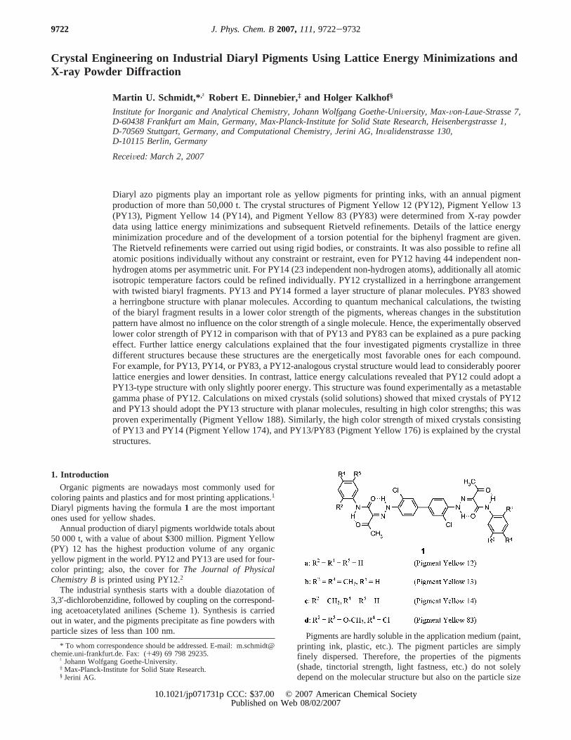
Diaryl pigments having the formula1 are the most importantones used for yellow shades.
Annual production of diaryl pigments worldwide totals about50 000 t, with a value of about $300 million. Pigment Yellow(PY) 12 has the highest production volume of any organicyellow pigment in the world. PY12 and PY13 are used for four-color printing; also, the cover forThe Journal of PhysicalChemistry Bis printed using PY12.2
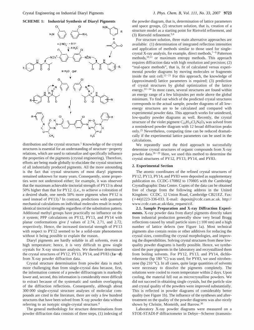
The industrial synthesis starts with a double diazotation of3,3′-dichlorobenzidine, followed by coupling on the correspond-ing acetoacetylated anilines (Scheme 1). Synthesis is carriedout in water, and the pigments precipitate as fine powders withparticle sizes of less than 100 nm.
Pigments are hardly soluble in the application medium (paint,printing ink, plastic, etc.). The pigment particles are simplyfinely dispersed. Therefore, the properties of the pigments(shade, tinctorial strength, light fastness, etc.) do not solelydepend on the molecular structure but also on the particle size
* To whom correspondence should be addressed. E-mail: [email protected]. Fax: (+49) 69 798 29235.
† Johann Wolfgang Goethe-University.‡ Max-Planck-Institute for Solid State Research.§ Jerini AG.
9722 J. Phys. Chem. B2007,111,9722-9732
10.1021/jp071731p CCC: $37.00 © 2007 American Chemical SocietyPublished on Web 08/02/2007
distribution and the crystal structure.1 Knowledge of the crystalstructures is essential for an understanding of structure-propertyrelations, which are used to rationalize and specifically influencethe properties of the pigments (crystal engineering). Therefore,efforts are being made globally to elucidate the crystal structuresof all industrially produced pigments. All the more astoundingis the fact that crystal structures of most diaryl pigmentsremained unknown for many years. Consequently, some proper-ties were not understood either; for example, it was observedthat the maximum achievable tinctorial strength of PY13 is about50% higher than that for PY12. (i.e., to achieve a coloration ofa desired shade, one needs 50% more pigment when PY12 isused instead of PY13).3 In contrast, predictions with quantummechanical calculations on individual molecules result in nearlyidentical tinctorial strengths regardless of the substitution pattern.Additional methyl groups have practically no influence on theπ system; PPP calculations on PY12, PY13, and PY14 withplanar conformations givef values of 2.74, 2.71, and 2.72,respectively. Hence, the increased tinctorial strength of PY13with respect to PY12 seemed to be a solid-state phenomenonwithout it being possible to explain the reason.
Diaryl pigments are hardly soluble in all solvents, even athigh temperature; hence, it is very difficult to grow singlecrystals for X-ray structure analysis. We therefore determinedthe crystal structures of PY12, PY13, PY14, and PY83 (1a-d)from X-ray powder diffraction data.
Crystal structure determination from powder data is muchmore challenging than from single-crystal data because, first,the information content of a powder diffractogram is markedlylower and, second, this information is considerably more difficultto extract because of the systematic and random overlappingof the diffraction reflections. Consequently, although about300 000 single-crystal structure analyses of molecular com-pounds are cited in the literature, there are only a few hundredstructures that have been solved from X-ray powder data withoutreferring to an isotypic single-crystal structure.4
The general methodology for structure determinations frompowder diffraction data consists of three steps, (1) indexing of
the powder diagram, that is, determination of lattice parametersand space groups, (2) structure solution, that is, creation of astructure model as a starting point for Rietveld refinement, and(3) Rietveld refinement.5,6
For structure solution, three main alternative approaches areavailable: (1) determination of integrated reflection intensitiesand application of methods similar to those used for single-crystal X-ray analysis, for example, direct methods,7-9 Pattersonmethods,10,11 or maximum entropy methods. This approachrequires diffraction data with high resolution and precision; (2)“real-space methods”, that is, fit of calculated versus experi-mental powder diagrams by moving molecules or fragmentsinside the unit cell.12-21 For this approach, the knowledge of(approximated) lattice parameters is required; (3) predictionof crystal structures by global optimization of the latticeenergy.22-24 In most cases, several structures are found withinan energy range of a few kilojoules per mole above the globalminimum. To find out which of the predicted crystal structurescorresponds to the actual sample, powder diagrams of all low-energy structures are to be calculated and compared withexperimental powder data. This approach works for unindexed,low-quality powder diagrams as well. Recently, the crystalstructure of the violet pigment C22H12Cl2N6O4 was solved froma nonindexed powder diagram with 12 broad diffraction peaksonly.25 Nevertheless, computing time can be reduced dramati-cally if the experimental lattice parameters can be used in thecalculations.
We repeatedly used the third approach to successfullydetermine crystal structures of organic compounds from X-raypowder data.26-29 Here, we used this method to determine thecrystal structures of PY12, PY13, PY14, and PY83.
2. Experimental Section
The atomic coordinates of the refined crystal structures ofPY12, PY13, PY14, and PY83 were deposited as supplementarypublication no. CCDC-170802 to 170805 with the CambridgeCrystallographic Data Centre. Copies of the data can be obtainedfree of charge from the following address in the UnitedKingdom: CCDC, 12 Union Road, Cambridge CB21EZ. Fax:(+44)1223-336-033. E-mail: [email protected]. http://www.ccdc.cam.ac.uk/data_request/cif.
2.1. Sample Preparation and X-ray Diffraction Experi-ments.X-ray powder data from diaryl pigments directly takenfrom industrial production generally show very broad Braggreflections caused by small particle sizes (e100 nm) and a highnumber of lattice defects (see Figure 1a). Most technicalpigments also contain resins or other additives for reducing thecrystal sizes, controlling the crystal morphologies, and improv-ing the dispersibilities. Solving crystal structures from these low-quality powder diagrams is hardly possible. Hence, we synthe-sized the pure pigments in the laboratory and recrystallized themfrom boiling solvents. For PY12, PY13, and PY14, dichlo-robenzene (bp 180°C) was used; for PY83, we used nitroben-zene (bp 210°C). In all cases, quite large quantities of solventswere necessary to dissolve the pigments completely. Thesolutions were cooled to room temperature within 2 days. Uponcooling, the material fell out as microcrystalline powders. Wedid not succeed in obtaining single crystals, but the particle sizeand crystal quality of the powders were improved substantially.This is reflected in powder diagrams of considerably betterquality (see Figure 1b). The influence of the syntheses and after-treatment on the quality of the powder diagrams was also nicelyshown by Christie, Monteith, and Barrow.30
Laboratory X-ray powder diagrams were measured on aSTOE-STADI-P diffractometer in Debye-Scherrer (transmis-
SCHEME 1: Industrial Synthesis of Diaryl Pigments
Crystal Engineering on Industrial Diaryl Pigments J. Phys. Chem. B, Vol. 111, No. 33, 20079723

sion) geometry. The diffractometer was equipped with a primaryGe(111) monochromator (Cu KR1 radiation,λ ) 1.54060 Å)and a linear position-sensitive detector. Powder diagramsrecorded in the range of 2θ ) 2-34°, with a total measuringtime of 2 h, turned out to be fully sufficient for indexing andcrystal structure solution. For the Rietveld refinements, high-resolution powder diffraction data were collected at the SUNY
X3B1 beamline at the National Synchrotron Light Source,Brookhaven National Laboratory, with the samples sealed inglass capillaries of 0.7 mm diameter for PY12, PY13, and PY83and of 1.0 mm for PY14. X-rays of wavelengths 1.14818(2) Åfor PY12, 1.14946(2) Å for PY13 and PY14, and 0.70002(2)Å for PY83 were selected by a double Si(111) monochromator.Wavelengths and the zero point have been determined from eight
Figure 1. X-ray powder diagrams of PY14. (a) Top (green), sample from industrial production (laboratory data,λ ) 1.541 Å); second (red),recrystallized product (laboratory data,λ ) 1.541 Å); third (blue), simulated powder diagram of the calculated crystal structure having the lowestenergy; bottom (black), recrystallized product (synchrotron data,λ ) 1.149 Å, displayed in the scale ofλ ) 1.541 Å). (b) Rietveld plot; unconstrainedRietveld refinement of all nonhydrogen atoms using synchrotron data (λ ) 1.149 Å): circles: experimental data; through line: calculated profile;bottom: difference curve.
9724 J. Phys. Chem. B, Vol. 111, No. 33, 2007 Schmidt et al.
well-defined reflections of the NBS1976 flat plate aluminastandard. The diffracted beam was analyzed with a Ge(111)crystal and detected with a Na(Tl)I scintillation counter with apulse height discriminator in the counting chain. The incomingbeam was monitored by an ion chamber for normalization forthe decay of the primary beam. In this parallel beam configu-ration, the resolution was determined by the analyzer crystalinstead of by slits. Data were taken with the followingparameters: PY12 (3.0-55.87° 2Θ; 0.005° 2Θ/step, 2.9 s/step);PY13 (5.0-42.19° 2Θ; 0.005° 2Θ/step, 3.3-5.3 s/step); PY14(4.0-64.995° 2Θ; 0.005° 2Θ/step, 3.3-6.3 s/step); and PY83(2.0-37.288° 2Θ; 0.004° 2Θ/step, 3.1-8.1 s/step). AlthoughΘ scans did not show serious crystallite size effects, the sampleswere spun aroundΘ during measurement for better particlestatistics. Low-angle diffraction peaks showed a strong asym-metry due to axial divergence and had a full-width at half-maximum (fwhm) of 0.01° 2Θ for PY83 and PY12, 0.022° 2Θfor PY13, and 0.017° 2Θ for PY14. Whereas the fwhm of thereflections of PY83 and PY12 was close to the resolution ofthe spectrometer, the reflections of PY13 and PY14 did showsome sample-dependent isotropic peak broadening.
2.2. Structure Solution by Lattice Energy Minimization.2.2.1. Molecular Geometry.For global lattice energy optimiza-tions, the knowledge of an (approximated) molecular geometryis required as input. Azo pigments can exist as azo or hydrazotautomers (Scheme 1). Whereas in solution both tautomers arepopulated, in the solid state, all yellow azo pigments exist asthe hydrazo tautomer because the acetoacetyl substituents formintramolecular hydrogen bonds between the acetyl and thehydrazo groups. This has been shown by single-crystal analysesof many mono-azo pigments of the Hansa Yellow type.31,32
(Monoazo pigments are much more soluble than diazo pigments;therefore, single crystals of sufficient size and quality can begrown.)
The molecular geometry was derived from crystal structuredata of similar molecules and fragments. A suitable model forPY12 would be the compound2a (reaction product of diazotized2-chloro-aniline andN-acetoacetyl-aniline), which is a quasi-half of a molecule of PY12. Hence, we synthesized andcrystallized the compound2a and determined the structure bysingle-crystal diffraction.33 However, the structure of2a iseffected by disorder. Therefore, we chose the crystal structureof 2b (reaction product of diazotized aniline andN-acetoacetyl-2-methyl-aniline)34 as a basis for the molecular geometry of1a-d, and only the geometry in the vicinity of the chlorineatom was taken from2a. For the central C-C bond, a lengthof 1.487 Å was assumed.35 Additional substituents (methyl,chlorine, and methoxy) to the terminal phenyl fragments wereadded according to standard values35 and data from theCambridge Structural Database.36 Furthermore, we assumed thatoverall molecular geometry is less influenced by the substituents.
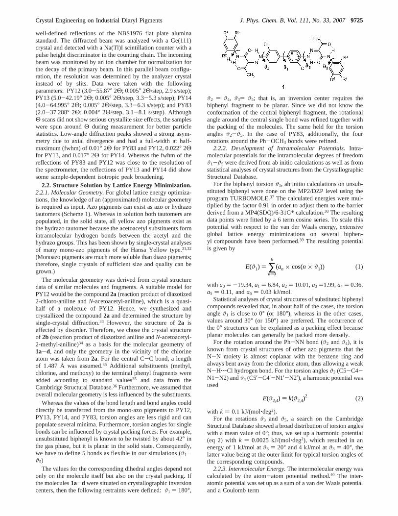
Whereas the values of the bond length and bond angles coulddirectly be transferred from the mono-azo pigments to PY12,PY13, PY14, and PY83, torsion angles are less rigid and canpopulate several minima. Furthermore, torsion angles for singlebonds can be influenced by crystal packing forces. For example,unsubstituted biphenyl is known to be twisted by about 42° inthe gas phase, but it is planar in the solid state. Consequently,we have to define 5 bonds as flexible in our simulations (ϑ1-ϑ5)
The values for the corresponding dihedral angles depend notonly on the molecule itself but also on the crystal packing. Ifthe molecules1a-d were situated on crystallographic inversioncenters, then the following restraints were defined:ϑ1 ) 180°,
ϑ2 ) ϑ4, ϑ3) ϑ5; that is, an inversion center requires thebiphenyl fragment to be planar. Since we did not know theconformation of the central biphenyl fragment, the rotationalangle around the central single bond was refined together withthe packing of the molecules. The same held for the torsionanglesϑ2-ϑ5. In the case of PY83, additionally, the fourrotations around the Ph-OCH3 bonds were refined.
2.2.2. DeVelopment of Intramolecular Potentials.Intra-molecular potentials for the intramolecular degrees of freedomϑ1-ϑ5 were derived from ab initio calculations as well as fromstatistical analyses of crystal structures from the CrystallographicStructural Database.
For the biphenyl torsionϑ1, ab initio calculations on unsub-stituted biphenyl were done on the MP2/DZP level using theprogram TURBOMOLE.37 The calculated energies were mul-tiplied by the factor 0.91 in order to adjust them to the barrierderived from a MP4(SDQ)/6-31G* calculation.38 The resultingdata points were fitted by a 6 term cosine series. To scale thispotential with respect to the van der Waals energy, extensiveglobal lattice energy minimizations on several biphen-yl compounds have been performed.39 The resulting potentialis given by
with a0 ) -19.34,a1 ) 6.84,a2 ) 10.01,a3 )1.99,a4 ) 0.36,a5 ) 0.11, anda6 ) 0.03 kJ/mol.
Statistical analyses of crystal structures of substituted biphenylcompounds revealed that, in about half of the cases, the torsionangleϑ1 is close to 0° (or 180°), whereas in the other cases,values around 30° (or 150°) are preferred. The occurrence ofthe 0° structures can be explained as a packing effect becauseplanar molecules can generally be packed more densely.
For the rotation around the Ph-NN bond (ϑ2 andϑ4), it isknown from crystal structures of other azo pigments that theN-N moiety is almost coplanar with the benzene ring andalways bent away from the chlorine atom, thus allowing a weakN-H‚‚‚Cl hydrogen bond. For the torsion anglesϑ2 (C5-C4-N1-N2) andϑ4 (C5′-C4′-N1′-N2′), a harmonic potential wasused
with k ) 0.1 kJ/(mol‚deg2).For the rotationsϑ3 and ϑ5, a search on the Cambridge
Structural Database showed a broad distribution of torsion angleswith a mean value of 0°; thus, we set up a harmonic potential(eq 2) with k ) 0.0025 kJ/(mol‚deg2), which resulted in anenergy of 1 kJ/mol atϑ3 ) 20° and 4 kJ/mol atϑ3 ) 40°, thelatter value being at the outer limit for typical torsion angles ofthe corresponding compounds.
2.2.3. Intermolecular Energy.The intermolecular energy wascalculated by the atom-atom potential method.40 The inter-atomic potential was set up as a sum of a van der Waals potentialand a Coulomb term
E(ϑ1) ) ∑n)0
6
(an × cos(n × ϑ1)) (1)
E(ϑ2,4) ) k(ϑ2,4)2 (2)
Crystal Engineering on Industrial Diaryl Pigments J. Phys. Chem. B, Vol. 111, No. 33, 20079725
whererij is the interatomic distance between atomsi andj, Aij,Bij, andCij are the empirical van der Waals parameters for theinteraction between atomsi and j,25,41 the q’s are the atomiccharges, andε is the relative dielectric constant,ε ) 1.
Atomic chargesqi were calculated by the charge iterationEHT method with the ICON program.42 The resulting charges,which are comparable to the Gasteiger43 charges, were multi-plied with an empirical factor of 1.1 to scale them with respectto the applied van der Waals potential. These charges have beenproved to work well in combination with our van der Waalsparameters.44
Hydrogen bond terms were not necessary in the descriptionof the intermolecular energy since the pigments1a-d did notform intermolecular hydrogen bonds.
2.2.4. Lattice Energy Minimizations.The energy minimiza-tions were performed with the CRYSCA45,46 program, whichcarries out global optimization of the lattice energy. Theminimizations started from a set of several thousand crystalstructures with given lattice parameters and given possible spacegroups but random position and spatial orientation of themolecules. For the intramolecular degrees of freedomϑ1-ϑ5,random start values within reasonable limits were similarlyassumed. The energy was optimized by a special steepestdescent method, varying the position and orientation of themolecules as well as the intramolecular torsion anglesϑ1-ϑ5.The lattice parameters were kept fixed. The obtained crystalstructures were ranked according to energy and checked forhigher symmetries.
2.3. Rietveld Refinement.The Rietveld refinements werecarried out with the program package GSAS.47 First of all, aLeBail fit48 without structural model was performed to refinethe lattice parameters and to optimize the peak shape parameters.The peak profile function was modeled using a multitermSimpson’s rule integration of the pseudo-Voigt function.49 Thestrong asymmetry in the low angle region was described by alater implemented function,50 which accounts for the asymmetrydue to axial divergence. The background was manually deter-mined with the program GUFI.51 The resulting polygon wascombined with a refinable 4 term cosine series. All profile andlattice parameters were fixed at the beginning of the Rietveldrefinements.
By comparing the number of atoms (44-54 non-hydrogenper molecule) and the number of distinguishable peaks (110-225), it seemed quite unfavorable to refine all atomic coordinatesindividually. The number of possible degrees of freedom couldtherefore be dramatically reduced by the introduction of rigidbodies.52 The effect was a strong increase of the ratio betweenlinear independent observations and parameters, leading to astabilized process of refinement. Furthermore, since the mo-
lecular fragments were forced to shift as a whole, meaninglesschanges could not occur.
In GSAS, a rigid body can be set up in various ways. Theposition of each atom within the rigid body is described as acombination of vectors given in Cartesian coordinates. Thelengths of the vectors can be refined through a multiplier. Forsmall molecules, the rigid body can be set up in a way thatthese vectors correspond to interatomic bonds, and thus, theposition of an atom is the sum of several refinable interatomicvectors. For complex molecules like the diaryl pigments, thisquickly becomes difficult to handle. A more comfortable wayis to use the Cartesian coordinates of all atoms directly. Insteadof refining individual bond lengths, an overall scale factor forthe size of the molecule can be refined. The width and the heightof the molecule can be refined individually by applying differentmultipliers to the three base vectors of the Cartesian coordinatesystem.
For the Rietveld refinements, the molecules of PY12-PY14were divided in 3-6 rigid groups, which were held together bysoft constraints, allowing constrained translations but individualrotations of the different groups. The number of refinedparameters was increased step by step.
Because of the high quality of the powder pattern of PY83,the rigid bodies were replaced by soft constraints (44 for bondlengths, 55 for bond angles, and 7 for planarity), leading to animproved refinement.
As a final test for the correctness of the molecular geometry,all atomic positions were refined independently for threestructures, after excluding the hydrogen atoms from the refine-ment. Although, the presumably flat molecular moieties likeC6 rings were slightly buckled, the positions of all non-hydrogenatoms remained close to their rigid-body positions, conformingthe correctness of the molecular and structural model. Subse-quent difference Fourier analysis revealed no considerableadditional or missing electron density.
Drawings of the crystal structures were made withSCHAKAL.53
3. Results
3.1. X-ray Diffraction and Energy Minimization. Thecomparison of the X-ray powder diagrams of1a-d showedthat only PY13 (1b) and PY14 (1c) are isotypic, whereas PY12(1a) and PY83 (1d) have different crystal structures.
After recrystallizing the pigments, the laboratory powderdiagrams of all pigments were indexed by TREOR54 withoutambiguity. The resulting lattice parameters are compiled in Table1. The number of molecules per unit cell (Z) was determinedby the rule “18 Å3 per non-hydrogen atom”.55 Since pigmentsare known to pack more densely than general organic com-pounds, we used a value of 16 Å3 instead of 18 Å3. The resultingvalues forZ are included in Table 1.
E )1
2∑
i∑
j(-Aijrij
-6 + Bije-Cijrij +
1
4πεε0
qiqj
rij) (3)
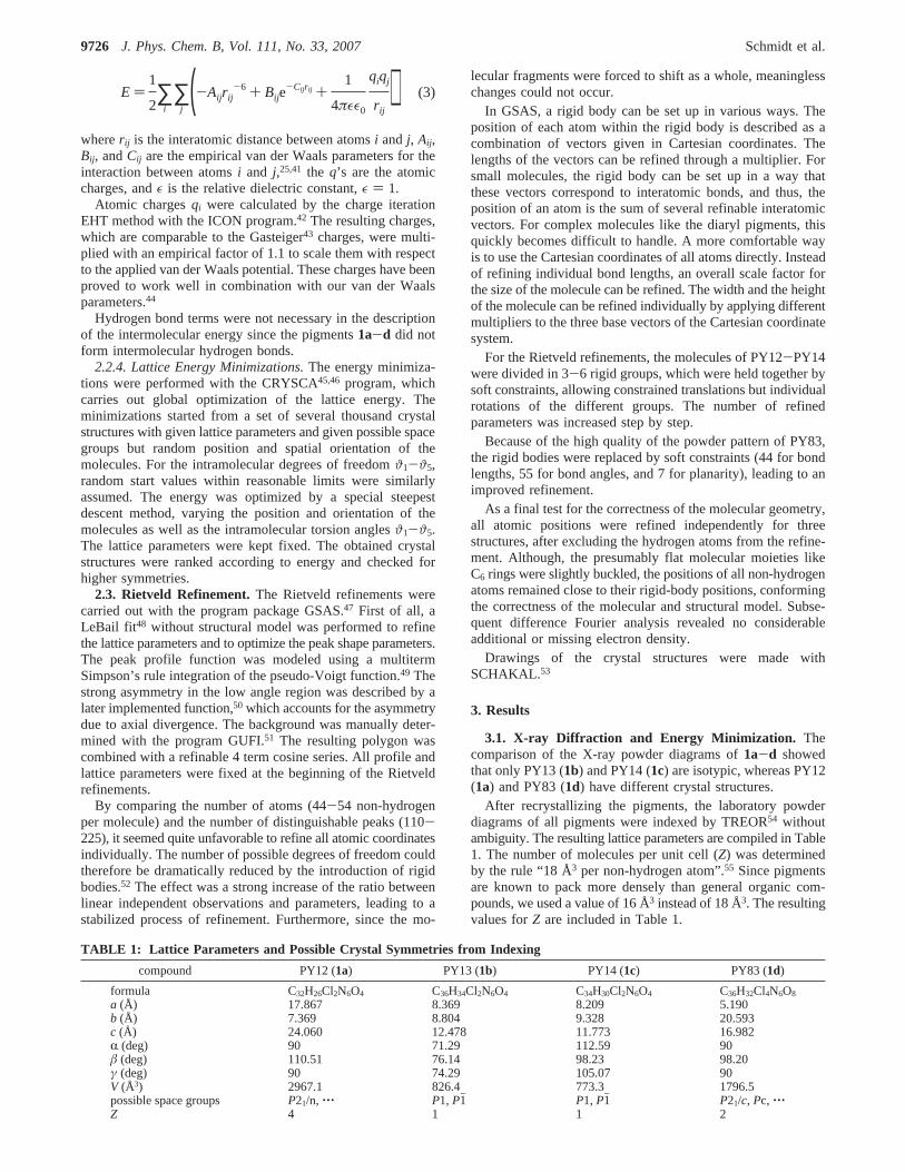
TABLE 1: Lattice Parameters and Possible Crystal Symmetries from Indexing
compound PY12 (1a) PY13 (1b) PY14 (1c) PY83 (1d)
formula C32H26Cl2N6O4 C36H34Cl2N6O4 C34H30Cl2N6O4 C36H32Cl4N6O8
a (Å) 17.867 8.369 8.209 5.190b (Å) 7.369 8.804 9.328 20.593c (Å) 24.060 12.478 11.773 16.982R (deg) 90 71.29 112.59 90â (deg) 110.51 76.14 98.23 98.20γ (deg) 90 74.29 105.07 90V (Å3) 2967.1 826.4 773.3 1796.5possible space groups P21/n, ‚‚‚ P1, P1h P1, P1h P21/c, Pc, ‚‚‚Z 4 1 1 2
9726 J. Phys. Chem. B, Vol. 111, No. 33, 2007 Schmidt et al.
For PY13 and PY14, the space group could either beP1 orP1h. If it was P1h, the molecule was situated on an inversioncenter. Since we did not know in advance if the molecule wasplanar and adopted inversion symmetry in the crystal, the energyminimizations had to be carried out without the inversionsymmetry, that is, in space groupP1. During the energyminimizations, in the lowest-energy packings and also manyother packings, the molecules adopted conformations withinversion centers, that is, the resulting crystal structures couldbe described in space groupP1h.
For PY12, the systematic extinctions led to the extinctionsymbol “P - 21/n -”, corresponding to the space groupP21/n.56 The determination of systematic extinctions frompowder diffraction data is, because of peak overlap, generallyless reliable than that from single data. Thus, other space groups,(P21, Pn, P21/m, P2/n, P2, Pm, or P2/m) could not be excluded.All of these space groups are statistically less frequent thanP21/n, especially forZ ) 4;57 therefore, the lattice energyminimization were started inP21/n, Z ) 4, with one moleculeper asymmetric unit, the molecule being located on a generalposition. The resulting low-energy packings did not showadditional symmetries.
The systematic extinctions of PY83 led to the probable spacegroupsP21/c or Pc (and some other, less frequently observedsymmetries likeP2/c). Lattice energy calculations were per-formed inP21/c, Z ) 2, with molecules on inversion centers,and in Pc, Z ) 2, with molecules on general positions. Thelow-energy structures of both calculations were identical, allexhibiting P21/c symmetry with the molecule situated on acrystallographic inversion center.
For all predicted low-energy structures, X-ray powderdiagrams were simulated. In all cases, packings with the best(lowest) energy gave simulated powder diagrams that werealmost identical to the experimental powder diffractograms (seeFigure 1c). Powder diagrams are very sensitive to smalldeviations in the crystal structures. The high similarity betweencalculated and experimental powder diagrams shows that thedifferences between the calculated and the actual crystalstructures are on the order of 0.1 Å only.
We also investigated whether the crystal structures could alsohave been solved if the powder diagrams were not indexable.Therefore, calculations were carried out in the statistically mostfrequent space groups, with some thousand starting structureshaving random lattice parameters; the unit cells were optimizedtogether with the position and orientation of the molecules andthe intramolecular degrees of freedom. The calculation timeswere considerably higher than those for calculations with givenunit cells. Nevertheless, for all four compounds, a minimumwith good energy can be found, showing a simulated powderdiagram similar to the experimental one. This minimumcorresponds, in all cases, to the actual crystal structure. Theaccuracy of the crystal structures calculated without specificationof the unit cell was, by far, sufficient for the Rietveld refinement.Hence, the crystal structures could also have been solved fromnonindexable X-ray powder diagrams.
3.2. Rietveld Refinements.The calculated crystal structureswere refined by the Rietveld method. Since we were interestedin structural details such as torsion angles, high-resolutionsynchrotron powder diffractograms of all four compounds wererecorded. The refined crystal structures were almost identicalto the crystal structures calculated by energy minimization.
In order to prove that the molecules of PY13, PY14, andPY83 (1b-d) are planar, additional Rietveld refinements werecarried out without the inversion centers, that is, in the
corresponding subgroups. The refinements resulted in structuresagain showing inversion symmetries. Thus, the crystal sym-metries found by lattice energy minimizations (P21/n, P1h, andP21/c) were confirmed by the Rietveld refinements.
The quality of the synchrotron powder data was so high thatfor PY12, PY13, and PY14 we tried for to refine all non-hydrogen atom positions individually without any constraintsor restraints. To our astonishment, the refinements converged,even in the case of PY12 having 44 independent non-hydrogenatoms per asymmetric unit. This is, to our knowledge, one ofthe largest number of atoms in an unconstrained refinementhitherto. The atomic positions changed only slightly duringrefinement. In the case of PY14, it was even possible to refineall atomic isotropic temperature factors individually to reason-able values (Figure 1b, Table 2)!
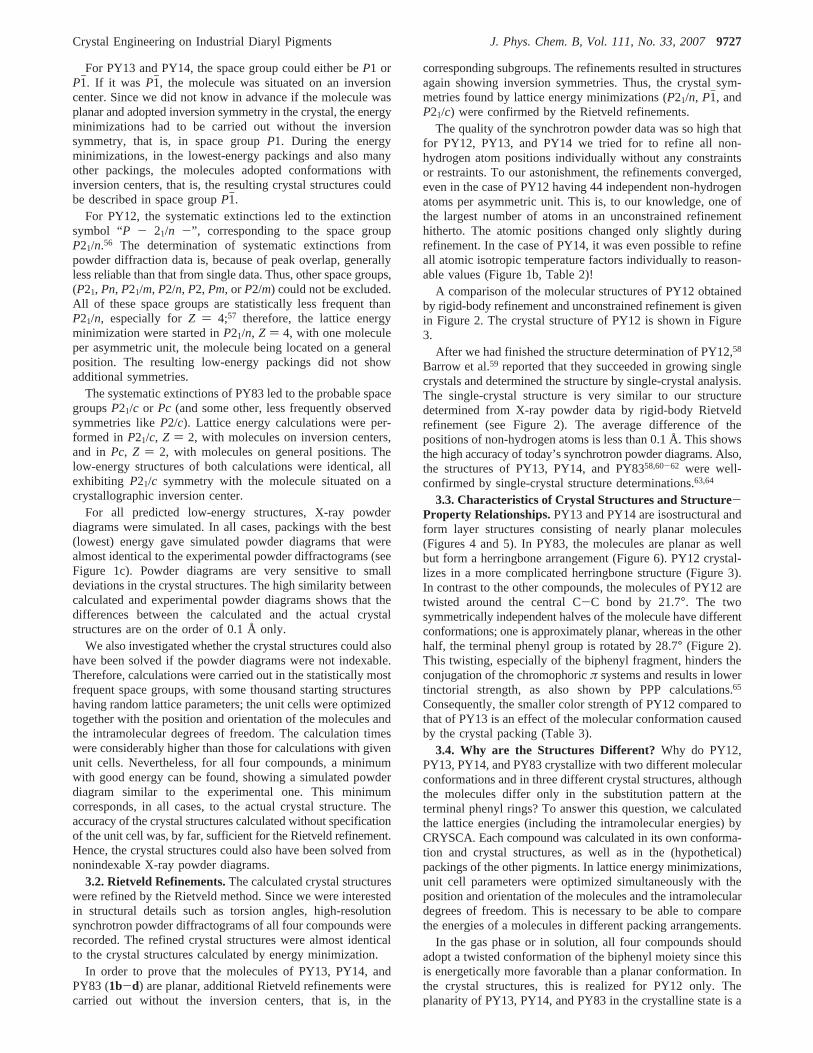
A comparison of the molecular structures of PY12 obtainedby rigid-body refinement and unconstrained refinement is givenin Figure 2. The crystal structure of PY12 is shown in Figure3.
After we had finished the structure determination of PY12,58
Barrow et al.59 reported that they succeeded in growing singlecrystals and determined the structure by single-crystal analysis.The single-crystal structure is very similar to our structuredetermined from X-ray powder data by rigid-body Rietveldrefinement (see Figure 2). The average difference of thepositions of non-hydrogen atoms is less than 0.1 Å. This showsthe high accuracy of today’s synchrotron powder diagrams. Also,the structures of PY13, PY14, and PY8358,60-62 were well-confirmed by single-crystal structure determinations.63,64
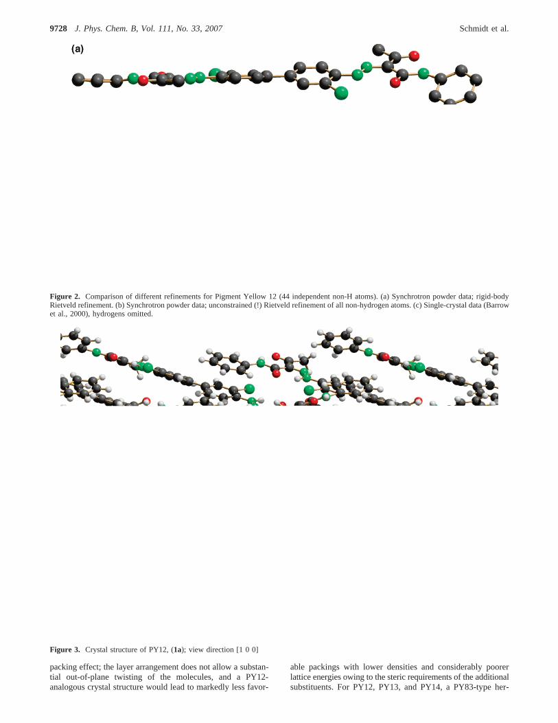
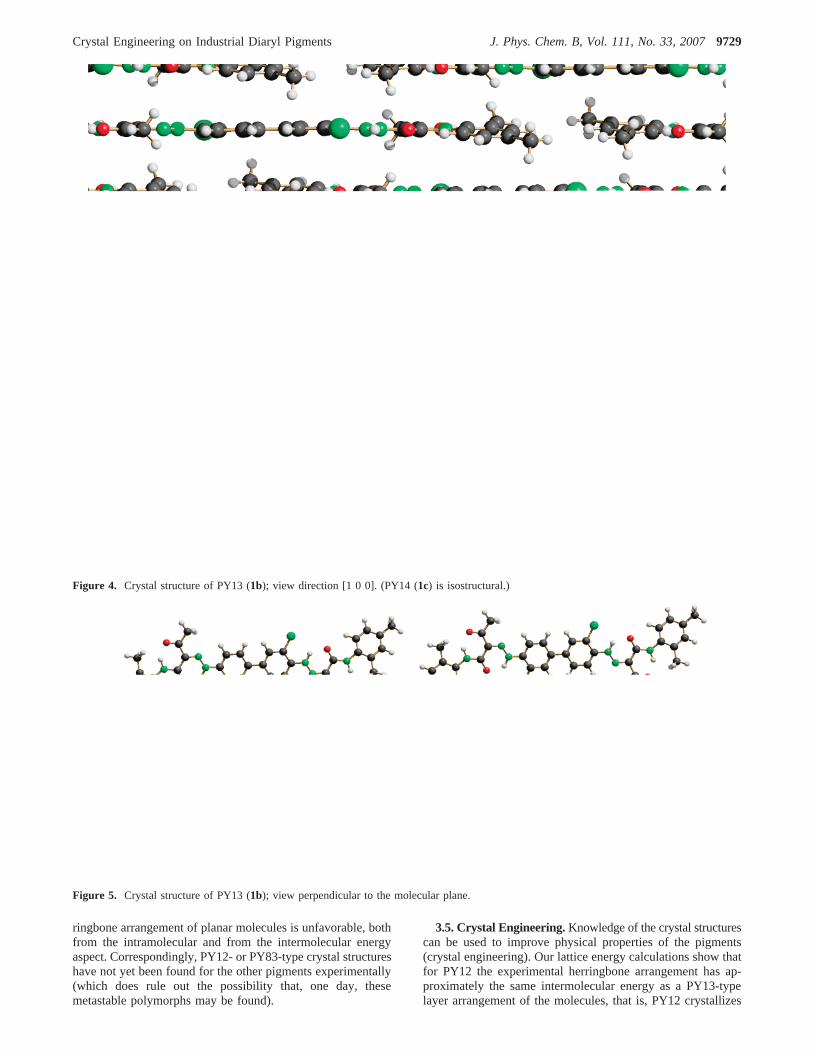
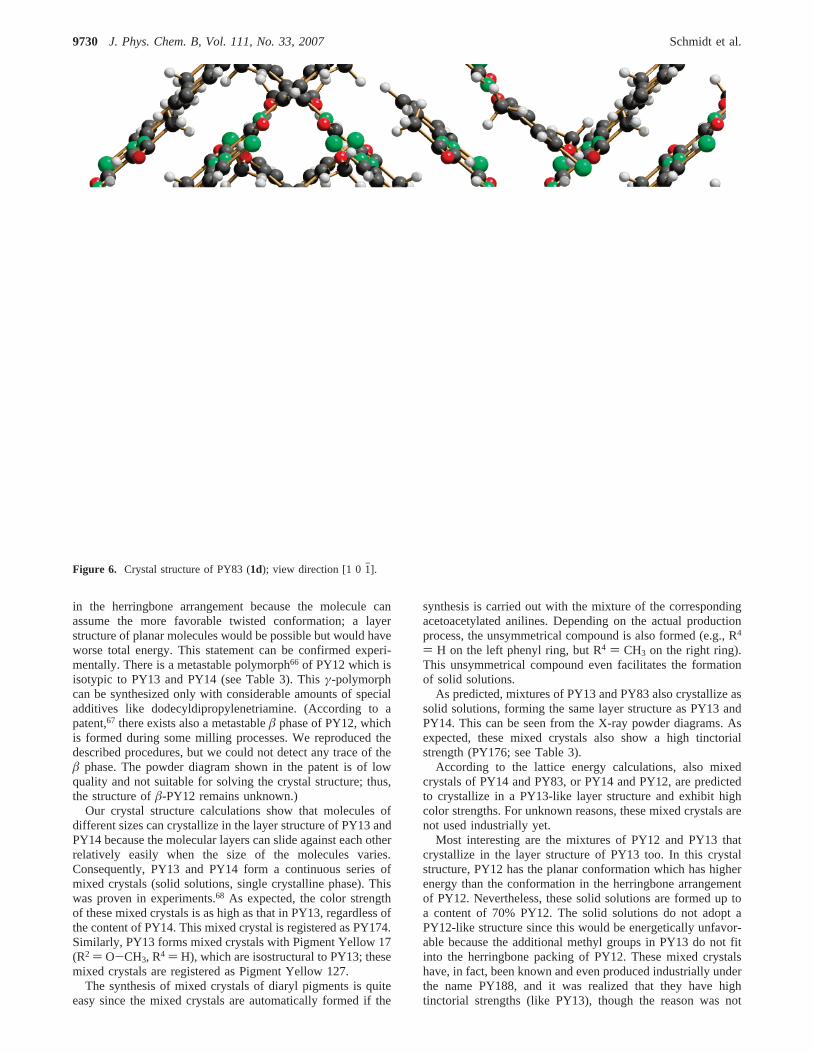
3.3. Characteristics of Crystal Structures and Structure-Property Relationships.PY13 and PY14 are isostructural andform layer structures consisting of nearly planar molecules(Figures 4 and 5). In PY83, the molecules are planar as wellbut form a herringbone arrangement (Figure 6). PY12 crystal-lizes in a more complicated herringbone structure (Figure 3).In contrast to the other compounds, the molecules of PY12 aretwisted around the central C-C bond by 21.7°. The twosymmetrically independent halves of the molecule have differentconformations; one is approximately planar, whereas in the otherhalf, the terminal phenyl group is rotated by 28.7° (Figure 2).This twisting, especially of the biphenyl fragment, hinders theconjugation of the chromophoricπ systems and results in lowertinctorial strength, as also shown by PPP calculations.65
Consequently, the smaller color strength of PY12 compared tothat of PY13 is an effect of the molecular conformation causedby the crystal packing (Table 3).
3.4. Why are the Structures Different? Why do PY12,PY13, PY14, and PY83 crystallize with two different molecularconformations and in three different crystal structures, althoughthe molecules differ only in the substitution pattern at theterminal phenyl rings? To answer this question, we calculatedthe lattice energies (including the intramolecular energies) byCRYSCA. Each compound was calculated in its own conforma-tion and crystal structures, as well as in the (hypothetical)packings of the other pigments. In lattice energy minimizations,unit cell parameters were optimized simultaneously with theposition and orientation of the molecules and the intramoleculardegrees of freedom. This is necessary to be able to comparethe energies of a molecules in different packing arrangements.
In the gas phase or in solution, all four compounds shouldadopt a twisted conformation of the biphenyl moiety since thisis energetically more favorable than a planar conformation. Inthe crystal structures, this is realized for PY12 only. Theplanarity of PY13, PY14, and PY83 in the crystalline state is a
Crystal Engineering on Industrial Diaryl Pigments J. Phys. Chem. B, Vol. 111, No. 33, 20079727
packing effect; the layer arrangement does not allow a substan-tial out-of-plane twisting of the molecules, and a PY12-analogous crystal structure would lead to markedly less favor-
able packings with lower densities and considerably poorerlattice energies owing to the steric requirements of the additionalsubstituents. For PY12, PY13, and PY14, a PY83-type her-
Figure 2. Comparison of different refinements for Pigment Yellow 12 (44 independent non-H atoms). (a) Synchrotron powder data; rigid-bodyRietveld refinement. (b) Synchrotron powder data; unconstrained (!) Rietveld refinement of all non-hydrogen atoms. (c) Single-crystal data (Barrowet al., 2000), hydrogens omitted.
Figure 3. Crystal structure of PY12, (1a); view direction [1 0 0]
9728 J. Phys. Chem. B, Vol. 111, No. 33, 2007 Schmidt et al.
ringbone arrangement of planar molecules is unfavorable, bothfrom the intramolecular and from the intermolecular energyaspect. Correspondingly, PY12- or PY83-type crystal structureshave not yet been found for the other pigments experimentally(which does rule out the possibility that, one day, thesemetastable polymorphs may be found).
3.5. Crystal Engineering.Knowledge of the crystal structurescan be used to improve physical properties of the pigments(crystal engineering). Our lattice energy calculations show thatfor PY12 the experimental herringbone arrangement has ap-proximately the same intermolecular energy as a PY13-typelayer arrangement of the molecules, that is, PY12 crystallizes
Figure 4. Crystal structure of PY13 (1b); view direction [1 0 0]. (PY14 (1c) is isostructural.)
Figure 5. Crystal structure of PY13 (1b); view perpendicular to the molecular plane.
Crystal Engineering on Industrial Diaryl Pigments J. Phys. Chem. B, Vol. 111, No. 33, 20079729
in the herringbone arrangement because the molecule canassume the more favorable twisted conformation; a layerstructure of planar molecules would be possible but would haveworse total energy. This statement can be confirmed experi-mentally. There is a metastable polymorph66 of PY12 which isisotypic to PY13 and PY14 (see Table 3). Thisγ-polymorphcan be synthesized only with considerable amounts of specialadditives like dodecyldipropylenetriamine. (According to apatent,67 there exists also a metastableâ phase of PY12, whichis formed during some milling processes. We reproduced thedescribed procedures, but we could not detect any trace of theâ phase. The powder diagram shown in the patent is of lowquality and not suitable for solving the crystal structure; thus,the structure ofâ-PY12 remains unknown.)
Our crystal structure calculations show that molecules ofdifferent sizes can crystallize in the layer structure of PY13 andPY14 because the molecular layers can slide against each otherrelatively easily when the size of the molecules varies.Consequently, PY13 and PY14 form a continuous series ofmixed crystals (solid solutions, single crystalline phase). Thiswas proven in experiments.68 As expected, the color strengthof these mixed crystals is as high as that in PY13, regardless ofthe content of PY14. This mixed crystal is registered as PY174.Similarly, PY13 forms mixed crystals with Pigment Yellow 17(R2 ) O-CH3, R4 ) H), which are isostructural to PY13; thesemixed crystals are registered as Pigment Yellow 127.
The synthesis of mixed crystals of diaryl pigments is quiteeasy since the mixed crystals are automatically formed if the
synthesis is carried out with the mixture of the correspondingacetoacetylated anilines. Depending on the actual productionprocess, the unsymmetrical compound is also formed (e.g., R4
) H on the left phenyl ring, but R4 ) CH3 on the right ring).This unsymmetrical compound even facilitates the formationof solid solutions.
As predicted, mixtures of PY13 and PY83 also crystallize assolid solutions, forming the same layer structure as PY13 andPY14. This can be seen from the X-ray powder diagrams. Asexpected, these mixed crystals also show a high tinctorialstrength (PY176; see Table 3).
According to the lattice energy calculations, also mixedcrystals of PY14 and PY83, or PY14 and PY12, are predictedto crystallize in a PY13-like layer structure and exhibit highcolor strengths. For unknown reasons, these mixed crystals arenot used industrially yet.
Most interesting are the mixtures of PY12 and PY13 thatcrystallize in the layer structure of PY13 too. In this crystalstructure, PY12 has the planar conformation which has higherenergy than the conformation in the herringbone arrangementof PY12. Nevertheless, these solid solutions are formed up toa content of 70% PY12. The solid solutions do not adopt aPY12-like structure since this would be energetically unfavor-able because the additional methyl groups in PY13 do not fitinto the herringbone packing of PY12. These mixed crystalshave, in fact, been known and even produced industrially underthe name PY188, and it was realized that they have hightinctorial strengths (like PY13), though the reason was not
Figure 6. Crystal structure of PY83 (1d); view direction [1 0 1h].
9730 J. Phys. Chem. B, Vol. 111, No. 33, 2007 Schmidt et al.
known. Now, we can understand the high color strengths onthe basis of the crystal structure of these solid solutions.
From the industrial point of view, mixed crystals of PY12and PY13 have the advantage that they have similar physicalproperties as PY13 (i.e., high color strengths), but the productioncosts are lower because the starting materials of PY12 arecheaper than those for PY13 (because aniline is cheaper than2,4-dimethylaniline. The production process itself is similar asthat for pure PY13). This is a nice example of where crystalengineering helps to save millions of dollars per year.
4. Conclusions
Crystal structures of the diaryl pigments PY12, PY13, PY14,and PY83 were determined from X-ray powder diagrams bymeans of lattice energy minimizations and subsequent Rietveldrefinements. The energy calculations also showed the reasonsfor the existence of three different crystal structures differingnot only in their packing but also in molecular conformation.By combination of quantum mechanical calculations (for theindividual molecules) and force-field calculations (for the latticeenergy), we could explain the packings and their influence onthe different coloristic properties of the pigments. Subsequently,crystal engineering was applied for the formation of mixedcrystals having high color strengths.
The diaryl pigments are, at present, among the largest crystalstructures solved by means of X-ray powder diffraction.However, neither energy minimization nor Rietveld refinementis restricted to molecules with 70-90 atoms. There are manycompounds with 50-200 atoms (including active pharmaceuti-cal ingredients), whose crystal structures arouse great interestbut which cannot be solved by single-crystal X-ray structureanalysis because single crystals cannot been grown. We areconvinced that most of these crystal structures can be solved inthe next few years from X-ray powder diagrams. As shown,the knowledge of the crystal structures can subsequently be usedfor crystal engineering, that is, for the targeted synthesis ofcompounds having improved solid-state properties.
Acknowledgment. The authors thank Professor Dr. ErichF. Paulus and Dipl-Ing. Ursula Conrad (both formerly at HoechstAG, Frankfurt am Main) for the X-ray powder measurements,Professor Peter W. Stephens (SUNY at Stony Brook) for thesynchrotron measurements, Dr. Heinz Schiffer (Hoechst AG,now at Clariant, Basel) for ab initio calculations, Professor Dr.Hans Jo¨rg Lindner (Technical University of Darmstadt) for PPPcalculations, and Dr. Hans Joachim Metz for ensuring excellent
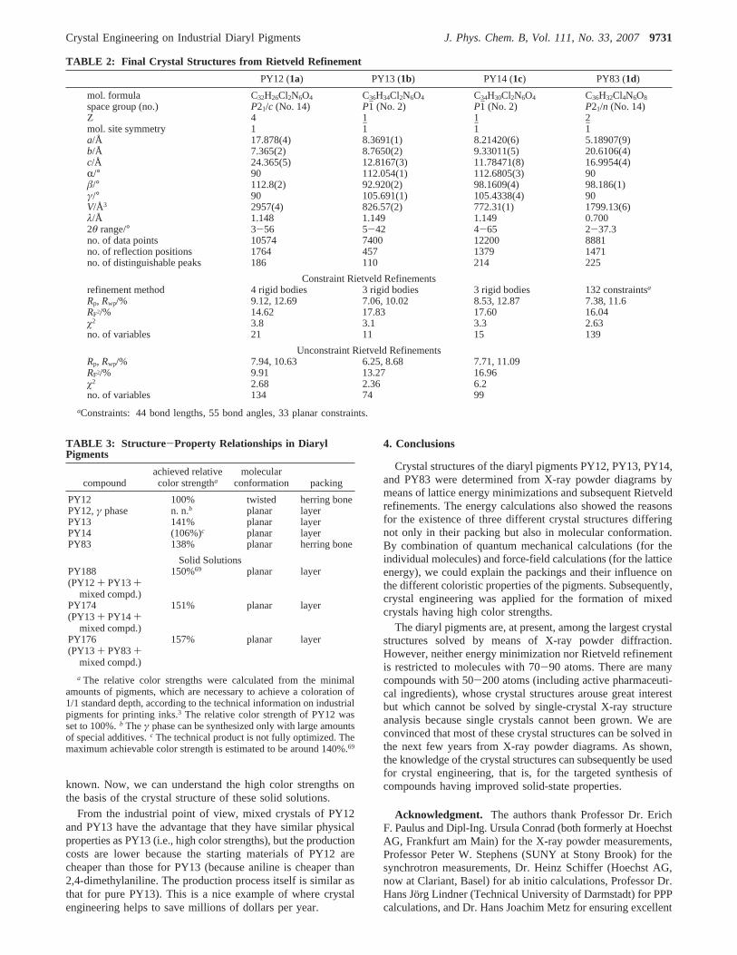
TABLE 2: Final Crystal Structures from Rietveld Refinement
PY12 (1a) PY13 (1b) PY14 (1c) PY83 (1d)
mol. formula C32H26Cl2N6O4 C36H34Cl2N6O4 C34H30Cl2N6O4 C36H32Cl4N6O8
space group (no.) P21/c (No. 14) P1h (No. 2) P1h (No. 2) P21/n (No. 14)Z 4 1 1 2mol. site symmetry 1 1h 1h 1ha/Å 17.878(4) 8.3691(1) 8.21420(6) 5.18907(9)b/Å 7.365(2) 8.7650(2) 9.33011(5) 20.6106(4)c/Å 24.365(5) 12.8167(3) 11.78471(8) 16.9954(4)R/° 90 112.054(1) 112.6805(3) 90â/° 112.8(2) 92.920(2) 98.1609(4) 98.186(1)γ/° 90 105.691(1) 105.4338(4) 90V/Å3 2957(4) 826.57(2) 772.31(1) 1799.13(6)λ/Å 1.148 1.149 1.149 0.7002θ range/° 3-56 5-42 4-65 2-37.3no. of data points 10574 7400 12200 8881no. of reflection positions 1764 457 1379 1471no. of distinguishable peaks 186 110 214 225
Constraint Rietveld Refinementsrefinement method 4 rigid bodies 3 rigid bodies 3 rigid bodies 132 constraintsa
Rp, Rwp/% 9.12, 12.69 7.06, 10.02 8.53, 12.87 7.38, 11.6RF2/% 14.62 17.83 17.60 16.04ø2 3.8 3.1 3.3 2.63no. of variables 21 11 15 139
Unconstraint Rietveld RefinementsRp, Rwp/% 7.94, 10.63 6.25, 8.68 7.71, 11.09RF2/% 9.91 13.27 16.96ø2 2.68 2.36 6.2no. of variables 134 74 99
aConstraints: 44 bond lengths, 55 bond angles, 33 planar constraints.
TABLE 3: Structure -Property Relationships in DiarylPigments
compoundachieved relativecolor strengtha
molecularconformation packing
PY12 100% twisted herring bonePY12,γ phase n. n.b planar layerPY13 141% planar layerPY14 (106%)c planar layerPY83 138% planar herring bone
Solid SolutionsPY188(PY12+ PY13+
mixed compd.)
150%69 planar layer
PY174(PY13+ PY14+
mixed compd.)
151% planar layer
PY176(PY13+ PY83+
mixed compd.)
157% planar layer
a The relative color strengths were calculated from the minimalamounts of pigments, which are necessary to achieve a coloration of1/1 standard depth, according to the technical information on industrialpigments for printing inks.3 The relative color strength of PY12 wasset to 100%.b Theγ phase can be synthesized only with large amountsof special additives.c The technical product is not fully optimized. Themaximum achievable color strength is estimated to be around 140%.69
Crystal Engineering on Industrial Diaryl Pigments J. Phys. Chem. B, Vol. 111, No. 33, 20079731
working conditions at Clariant. The analyses of the yellowpigments used in the printed edition for the cover ofThe Journalof Physical Chemistry Bwere carried out by Fatima Cheurfa inthe Clariant Analytical Services group (Frankfurt am Main).This project was financially supported by Clariant (DivisionPigments & Additives, Pigments Research, Frankfurt), theDeutsche Forschungsgemeinschaft (German Research Society),and the U.S. Department of Energy, Division of Basic EnergySciences (synchrotron measurements). This paper is dedicatedto Professor Dr. Erich F. Paulus on the occasion of his 70thbirthday.
References and Notes
(1) Herbst, W.; Hunger, K.Industrial Organic Pigments, 3rd ed.;Wiley-VCH: Weinheim, Germany, 2004.
(2) Mass spectrometrical analysis of a small piece of the cover of theprinted edition ofThe Journal of Physical Chemistry B.
(3) Colorants for Printing Inks; Technical information brochure,Clariant GmbH: Frankfurt am Main, 1999; update, 2000.
(4) Le Bail, A. Structure Determination from Powder Diffraction -Database. http://sdpd.univ-lemans.fr/iniref.html (2007).
(5) Rietveld, H. M.Acta Crystallogr.1967, 22, 151.(6) Rietveld, H. M.J. Appl. Crystallogr.1969, 2, 65.(7) Cascarano, G.; Favia, L.; Giacovazzo, C.J. Appl. Crystallogr.1992,
25, 310.(8) Altomare, A.; Burla, M. C.; Camalli, M.; Carrozzini, B.; Cascarano,
G. L.; Giacovazzo, C.; Guagliardi, A.; Moliterni, A. C. G.; Polidori, G.;Rizzi, R. J. Appl. Crystallogr.1999, 32, 339.
(9) Chan, F. C.; Anwar, J.; Cernik, R.; Barnes, P.; Wilson, R. M.J.Appl. Crystallogr.1999, 32, 436.
(10) Wagner, K.; Hirschler, J.; Egert, E.Z. Kristallogr. 2001, 216, 565.(11) Lasocha, W.; Czapkiewicz, J.; Milart, P.; Schenk, H.Z. Kristallogr.
2001, 216, 291.(12) Harris, K. D. M.; Tremayne, M.Chem. Mater.1996, 8, 2554.(13) Harris, K. D. M.; Tremayne, M.; Kariuki, B. M.Angew. Chem.
2001, 113, 1674;Angew. Chem., Int. Ed.2001, 40, 1626.(14) Hammond, R. B.; Roberts, K. J.; Docherty, R.; Edmondson, M.;
Gairns, R.J. Chem. Soc., Perkin Trans. 21996, 1527.(15) Tremayne, M.; Kariuki, B. M.; Harris, K. D. M.J. Appl. Crystallogr.
1996, 29, 211.(16) Andreev, Yu. G.; Lightfoot, P.; Bruce, P. G.J. Appl. Crystallogr.
1997, 30, 294.(17) Chernyshev, V. V.; Schenk, H.Z. Kristallogr. 1998, 213, 1.(18) David, W. I. F.; Shankland, K.; Shankland, N.Chem. Commun.
1998, 931.(19) Engel, G. E.; Wilke, S.; Ko¨nig, O.; Harris, K. D. M.; Leusen, F. J.
J. J. Appl. Crystallgr.1999, 32, 1169.(20) Kariuki, B. M.; Calcagno, P.; Harris, K. D. M.; Philp, D.; Johnston,
R. L. Angew. Chem.1999, 111, 860; Angew. Chem., Int. Ed.1999, 38,831.
(21) Dinnebier, R. E.; Sieger, P.; Nar, H.; Shankland, K.; David, W. I.F. J. Pharm. Sci.2000, 89, 1465.
(22) Lommerse, J. P. M.; Motherwell, W. D. S.; Ammon, H. L.; Dunitz,J. D.; Gavezzotti, A.; Hofmann, D. W. M.; Leusen, F. J. J.; Mooij, W. T.M.; Price, S. L.; Schweizer, B.; Schmidt, M. U.; van Eijck, B. P.; Verwer,P.; Williams, D. E.Acta Crystallogr., Sect. B2000, 56, 697.
(23) Motherwell, W. D. S.; Ammon, H. L.; Dunitz, J. D.; Dzyabchenko,A.; Erk, P.; Gavezzotti, A.; Hofmann, D. W. M.; Leusen, F. J. J.; Lommerse,J. P. M.; Mooij, W. T. M.; Price, S. L.; Scheraga, H.; Schweizer, B.;Schmidt, M. U.; van Eijck, B. P.; Verwer, P.; Williams, D. E.ActaCrystallogr., Sect. B2002, 58, 647.
(24) Day, G. M.; Motherwell, W. D. S.; Ammon, H. L.; Boerrigter, S.X. M.; Della Valle, R. G.; Venuti, E.; Dzyabchenko, A.; Dunitz, J. D.;Schweizer, B.; van Eijck, B. P.; Erk, P.; Facelli, J. C.; Bazterra, V. E.;Ferraro, M. B.; Hofmann, D. W. M.; Leusen, F. J. J.; Liang, C.; Pantelides,C. C.; Karamertzanis, P. G.; Price, S. L.; Lewis, T. C.; Nowell, H.; Torrisi,A.; Scheraga, H. A.; Arnautova, Y. A.; Schmidt, M. U.; Verwer, P.ActaCrystallogr., Sect. B2005, 61, 511.
(25) Schmidt, M. U.; Ermrich, M.; Dinnebier, R. E.Acta Crystallogr.,Sect. B2005, 61, 37.
(26) Schmidt, M. U.; Dinnebier, R. E.J. Appl. Crystallogr.1999, 32,178.
(27) Schmidt, M. U. InCrystal Engineering: From Molecules andCrystals to Materials; Braga, D., Grepioni, F., Orpen, A. G., Eds. KluwerAcademic Publishers: Dordrecht, The Netherlands, 1999; pp 331-348.
(28) Schmidt, M. U.; Ermrich, M.; Dinnebier, R. E.Acta Crystallogr.Sect. B2005, 61, 37.
(29) Schmidt, M. U.; Hofmann, D. W. M.; Buchsbaum, C.; Metz, H. J.Angew. Chem.2006, 118, 1335; Schmidt, M. U.; Hofmann, D. W. M.;Buchsbaum, C.; Metz, H. J.Angew. Chem., Int. Ed.2006, 45, 1313.
(30) Christie, R. M.; Monteith, J. E.; Barrow, M. J.Surf. Coat. Int.,Part B 2003, 86, 247.
(31) Paulus, E. F.Z. Kristallogr. 1983, 165, 137.(32) Whitaker, A.Acta Crystallogr., Sect. C1987, 43, 2141.(33) Paulus, E. F. Unpublished results.(34) Paulus, E. F.; Rieper, W.Acta Crystallogr., Sect. A1984, 40, C277.(35) Allen, F. H.; Kennard, O.; Watson, D. G.; Brammer, L.; Orpen, A.
G.; Taylor, R.J. Chem. Soc., Perkin Trans.1987, 2, S1.(36) Cambridge Structural Database; Cambridge Crystallographic Data
Centre: 12 Union Road, Cambridge, England.(37) Ahlrichs, R.; von Arnim, M.Methods and Techniques in Compu-
tational Chemistry METECC-95; STEF: Cagliari, 1995, pp 509-554.(38) Tsusuki, S.; Tanabe, K.J. Phys. Chem.1991, 95, 139.(39) Kalkhof, H. Dissertation, Technical University, Darmstadt, Ger-
many, 2002.(40) Pertsin, A. J.; Kitajgorodsky, A. I.The Atom-Atom Potential
Method; Springer-Verlag: New York, 1987.(41) Schmidt, M. U. InCrystal Engineering: From Molecules and
Crystals to Materials; Braga, D., Orpen, G.; Kluwer Academic Publishers:Dordrecht, The Netherlands, 1999, pp 331-348.
(42) Howell, J.; Rossi, A.; Wallace, D.; Haraki, K.; Hoffmann, R.Quantum Chemical Programs Exchange1977, 11, 344; QCPE No. 517.
(43) Gasteiger, J.; Marsili, M.Tetrahedron1980, 36, 3219.(44) Schmidt, M. U.Kristallstrukturberechnungen Metallorganischer
MolekulVerbindungen; Verlag Shaker: Aachen, Germany, 1995.(45) Schmidt, M. U.; Kalkhof, H.CRYSCA, Program for crystal
structure calculations of flexible molecules; Frankfurt am Main, 1997.(46) Schmidt, M. U.; Englert, U.J. Chem. Soc., Dalton Trans.1996,
2077.(47) Larson, C.; von Dreele R. B.Los Alamos National Laboratory
Report; LAUR 86-748, GSAS 1994, version 2002 V, 2002.(48) Le Bail, A.; Duroy, H.; Fourquet, J. L.Mater. Res. Bull.1988, 23,
447.(49) Thompson, P.; Cox, D. E.; Hastings, J. B.J. Appl. Crystallogr.
1987, 20, 79.(50) Finger, L. W.; Cox, D. E.; Jephcoat, A. P.J. Appl. Crystallogr.
1994, 27, 892.(51) Dinnebier, R. E.; Finger, L.Z. Kristallogr. Suppl.1998, 15, 148.(52) Dinnebier, R. E.Powder Diffr.1999, 14, 84.(53) Keller, E.SCHAKAL99. Kristallographisches Institut der Univer-
sitat: Freiburg, 1999.(54) Werner, P.-E.Z. Kristallogr. 1964, 120, 375.(55) Kempster, C. J. E.; Lipson, H.Acta Crystallogr., Sect. B1972, 28,
3764.(56) International Tables for Crystallography, Volume A: Space group
symmetry, 5th ed.; Hahn, T., Ed.; Kluwer Academic Publishers: Dordrecht,The Netherlands, 2000.
(57) Belsky, V. K.; Zorkaya, O. N.; Zorky, P. M.Acta Crystallogr.,Sect. A1995, 51, 473.
(58) Schmidt, M. U. InColour Science ’98, Vol. 1: Dye and PigmentChemistry; Griffiths, J. Ed.; University of Leeds: Leeds, U.K. 1999, pp72-81; ISBN 0-85316-196-8.
(59) Barrow, M. J.; Christie, R. M.; Lough, A. J.; Monteith, J. E.;Standring, P. N.Dyes Pigm.2000, 45, 153.
(60) Schmidt, M. U.; Dinnebier, R. E.; Kalkhof, H.Z. Kristallogr. Suppl.1998, 15, 72.
(61) Schmidt, M. U.; Dinnebier, R. E. Presentation at the IUCr-XVIIIConference, Glasgow, U.K., August 4-13, 1999.
(62) Schmidt, M. U.; Dinnebier, R. E.; Kalkhof, H. Presentation at the19th European Crystallographic Meeting, Nancy, France, August 26-31,2000; p 129.
(63) Barrow, M. J.; Christie, R. M.; Monteith, J. E.Dyes Pigm.2002,55, 79.
(64) Barrow, M. J.; Christie, R. M.; Badcock, T. D.Dyes Pigm.2003,57, 99.
(65) Christie, R. M.; Standring, P. N.Dyes Pigm.1989, 11, 109.(66) Tuck, B.; Stirling, J. A.; Farnocchi, C. J.; McKay, R. B. (Ciba SC),
European Patent EP 0790282 A2, 1997.(67) Kawamura, T. Japanese Patent JP 62153353, 1987.(68) Tanaka, K.; Takagi, K.; Haginoya, M.; Nakazima, E.Shikizai
Kyokaishi1963, 36, 392.(69) Ott, U. (Clariant), personal communication.
9732 J. Phys. Chem. B, Vol. 111, No. 33, 2007 Schmidt et al.