Embed Size (px)
Citation preview
HAL Id: hal-00532236https://hal.archives-ouvertes.fr/hal-00532236
Submitted on 4 Nov 2010
HAL is a multi-disciplinary open accessarchive for the deposit and dissemination of sci-entific research documents, whether they are pub-lished or not. The documents may come fromteaching and research institutions in France orabroad, or from public or private research centers.
L’archive ouverte pluridisciplinaire HAL, estdestinée au dépôt et à la diffusion de documentsscientifiques de niveau recherche, publiés ou non,émanant des établissements d’enseignement et derecherche français ou étrangers, des laboratoirespublics ou privés.
Detection of central nervous system tissues in meatproducts: Validation and standardization of a real-time
PCR-based detection systemHolger Schönenbrücher, Amir Abdulmawjood, Katrin-Annette Göbel, Michael
Bülte
To cite this version:Holger Schönenbrücher, Amir Abdulmawjood, Katrin-Annette Göbel, Michael Bülte. Detectionof central nervous system tissues in meat products: Validation and standardization of a real-time PCR-based detection system. Veterinary Microbiology, Elsevier, 2007, 123 (4), pp.336.�10.1016/j.vetmic.2007.04.003�. �hal-00532236�
Accepted Manuscript
Title: Detection of central nervous system tissues in meatproducts: Validation and standardization of a real-timePCR-based detection system
Authors: Holger Schonenbrucher, Amir Abdulmawjood,Katrin-Annette Gobel, Michael Bulte
PII: S0378-1135(07)00170-8DOI: doi:10.1016/j.vetmic.2007.04.003Reference: VETMIC 3647
To appear in: VETMIC
Please cite this article as: Schonenbrucher, H., Abdulmawjood, A., Gobel, K.-A., Bulte,M., Detection of central nervous system tissues in meat products: Validation andstandardization of a real-time PCR-based detection system, Veterinary Microbiology(2007), doi:10.1016/j.vetmic.2007.04.003
This is a PDF file of an unedited manuscript that has been accepted for publication.As a service to our customers we are providing this early version of the manuscript.The manuscript will undergo copyediting, typesetting, and review of the resulting proofbefore it is published in its final form. Please note that during the production processerrors may be discovered which could affect the content, and all legal disclaimers thatapply to the journal pertain.
Acce
pted
Man
uscr
ipt
1
Detection of central nervous system tissues in meat products: validation and 1
standardization of a real-time PCR-based detection system 2
3
Holger Schönenbrücher*, Amir Abdulmawjood, Katrin-Annette Göbel, Michael Bülte 4
5
Institute of Veterinary Food Science, Faculty of Veterinary Medicine, Justus-Liebig-6
University of Giessen, Frankfurter Str. 92, D-35392 Giessen, Germany 7
8
* Corresponding author at: 9
Institute of Veterinary Food Science, Faculty of Veterinary Medicine, Justus-Liebig-10
University of Giessen, Frankfurter Str. 92, D-35392 Giessen, Germany 11
Phone: +49-641-9938252 12
Fax: +49-641-9938259 13
E-mail: [email protected] (Holger Schönenbrücher) 14
15
Abstract 16
Several phenotypic as well as three genotypic methods have been published describing 17
the detection of central nervous system (CNS) tissues that are part of the bovine spongiform 18
encephalopathy (BSE) risk material in food products. However, none of these methods is able 19
to differentiate between CNS tissue of the banned ruminant species and tissues of other 20
animal species. 21
A quantitative and species-specific real-time RT-PCR method has been developed that 22
enables the reliable identification of CNS tissues in meat and meat products. This method is 23
based on a messenger (m)RNA assay that uses bovine, ovine and caprine glial fibrillary acidic 24
protein (GFAP) encoding gene sequences as markers. The in-house validation studies 25
evaluated the tissue specificity of up to 15 bovine tissues and the standardization of absolute 26
Page 1 of 26
Acce
pted
Man
uscr
ipt
2
as well as relative quantitative measurement. The specific amplification of spinal cord and 1
brain tissue GFAP cDNA has been shown previously. In addition, two commercially available 2
ELISA kits were used for the comparative analysis of artificially contaminated minced meat. 3
Small quantities of bovine brain that had been stored over the recommended period of 14 days 4
were examined. The real-time PCR method proved to be suitable for the detection of 0.1% 5
CNS tissue. No false negative results were observed. 6
The quantitative detection of GFAP mRNA using real-time RT-PCR seems a suitable 7
tool in routine diagnostic testing that assesses the illegal use of CNS tissue in meat and meat 8
products. The stability of the selected target region of the GFAP-mRNA also allows the 9
detection of CNS tissues after the meat has been processed. 10
11
Keywords: Real-time PCR, central nervous system tissues, BSE-risk material 12
13
14
Page 2 of 26
Acce
pted
Man
uscr
ipt
3
1. Introduction 1
Belonging to the group of diseases known as transmissible spongiform 2
encephalopathies (TSEs), bovine spongiform encephalopathy (BSE) has been causally 3
associated with a new variant of Creutzfeldt-Jakob disease (vCJD) in humans. To effectively 4
protect consumers from contracting vCJD, European legislators have set up numerous 5
guidelines to reduce risk, in which national eradication programmes including the area-wide 6
BSE testing of cattle and the prohibition of the use of specified risk material (SRM, e.g. brain 7
and spinal cord) of cattle, sheep and goat in the food chain have become of particular 8
importance. SRMs are age-dependent and their use is defined in the regulation (EC) No. 9
999/2001 (Annex V) of the European Commission. It should be mentioned that porcine CNS 10
tissues are not affected by this legislation. Ruminant brain and spinal cord have been shown to 11
contain the highest infectivity titre of the causative agent PrPSc (EFSA, 2005). There is also 12
strong evidence that BSE is most likely transferred to humans by way of the oral route of 13
infection (Bruce et al., 1997; Cousens et al., 2001; Comer and Huntly, 2003). 14
Methods enabling the detection of BSE risk material in food products include enzyme-15
linked immunosorbent assays (Schmidt et al., 1999; Schmidt et al., 2001), GC-MS 16
(Biedermann et al., 2004), Western blot and immunohistochemical methods (Lücker et al., 17
1999; Wenisch et al., 1999; Lücker et al., 2000). However, none of these methods allows the 18
differentiation of CNS of banned ruminant species from tissues of other animal species. 19
This study focussed on the validation and the standardisation of a real-time reverse 20
transcriptase polymerase chain reaction (RT-PCR) assay for the species-specific detection of 21
bovine, ovine and caprine CNS tissues in raw and heat-treated meat products. The evaluation 22
of the tissue specificity was based on up to 15 bovine tissues. In addition, real-time RT PCR 23
and two commercially available ELISA kits were compared for their efficiency in detecting 24
BSE pathogens in artificially contaminated minced meat. 25
Page 3 of 26
Acce
pted
Man
uscr
ipt
4
2. Materials and Methods 1
2.1. Parameters of the real-time PCR assay 2
The real-time PCR-based method used was standardised and validated according to 3
the requirements of the ISO documents TAG 3 N 145 rev, ISO TC 34/SC 9 N containing 4
general requirements for real-time PCR (anonymous, 2006a) and ISO/CD TAG 3 N 0144 rev, 5
ISO TC 34/SC 9 N ISO/CD containing general performance characteristics of molecular 6
detection methods (anonymous, 2006b). The parameters of the real-time PCR assay are 7
summarised in Table 1. Two-step reverse transcriptive PCR was carried out as described 8
previously (Schönenbrücher et al., 2004a; Abdulmawjood et al., 2005). For diagnostic quality 9
assurance, an internal amplification control (IAC) was included to avoid false negative 10
results. Briefly, the RNeasy® Lipid Tissue miniKit (Qiagen, Hilden, Germany) was used to 11
isolate total cellular RNA. To achieve an efficient cell lysis, the mixture of the sample or the 12
swab with the Qiazol lysis reagent (Qiagen, Hilden, Germany) was transferred to a glass 13
matrix tube (FastRNA Green, Q BIOgene, Heidelberg, Germany) processed in a spin/rotation 14
instrument (FastPrep® -120; Q BIOgene, Heidelberg). The total cellular RNA was 15
subsequently reverse transcribed into cDNA using the TaqMan® Reverse Transcriptase 16
Reagents kit with UNG (Applied Biosystems, Darmstadt, Germany) according to the 17
manufacturer’s protocol. The transcribed cDNA was stored at -20°C until further use. 18
19
2.2. Collection of tissue samples and quantification of GFAP content 20
To estimate tissue specificity, 15 bovine tissue samples from 3 different animals were 21
collected at an abattoir in Giessen, Germany, and immediately processed according to the 22
aforementioned protocol. The material included brain, spinal cord, peripheral nerves (in 23
particular sciatic and axillary nerves), fat, heart, kidney, liver, lung, lymph nodes, muscle, 24
spleen, adrenal gland, pancreas, parotid gland and thymus. Total cellular RNA of each tissue 25
Page 4 of 26
Acce
pted
Man
uscr
ipt
5
sample was prepared 3 times; all real-time PCR runs were performed in triplicate, resulting in 1
a set of 27 quantitative data per sample. The absolute quantification was standardised by 2
using a standard curve out of a serial dilution of a known bovine brain GFAP cDNA standard 3
in triplicate (Figure 1). The relative quantification was based on an 18S rRNA RT-PCR 4
(Applied Biosystems, Darmstadt, Germany) as a housekeeping gene (Abdulmawjood et al., 5
2005). For this purpose a selection of 10 tissue samples encompassing brain, spinal cord, 6
peripheral nerves (in particular sciatic and axillary nerves), heart, kidney, liver, lung, lymph 7
nodes, muscle and spleen was evaluated. 8
9
2.3. Production of internal reference material (IRM) 10
Minced meat and cooked sausages were prepared according to recipes provided in the 11
guidelines of the German Food Code (Bundesanzeiger, 1994) and artificially contaminated 12
with low concentrations of bovine brain homogenate. Minced meat (50% pork, 50% beef) 13
with 0.1% (duplicate), 0.2%, 0.5% and 1% of CNS tissue was prepared. Two batches of raw 14
IRM were prepared and stored at <+2°C for 14 days. Samples without CNS tissue and bovine 15
brain homogenate were processed using the same protocols for quality-control reasons. 16
The sausages were prepared as described by Lücker et al. (2000). The sausages were 17
boiled at 120°C for 15 min, representing strong heat treatment, or medium heated at 80°C for 18
90 min. The liver sausages were prepared by homogenising 80% porcine meat and 18% 19
porcine liver. Curing salt (2.5g/kg), emulsifier (0.5g/kg) and ready mixed herbs (5g/kg, 20
“Kalbsleberwurst Morenoperle”, Gewürzmüller, Korntal-Münchingen, Germany) were added. 21
The sausage meat was put in cans and heated for 60 minutes at 80°C. The liver sausages were 22
then stored at <+10°C. All RNA preparations were done in duplicate. 23
24
Page 5 of 26

Acce
pted
Man
uscr
ipt
6
1
2.4. Comparison of real-time PCR with commercially available ELISA kits 2
The minced meat was analysed using the ScheBo® Brainostic GFAP ELISA Kit 3
(ScheBo Biotech AG, Giessen, Germany) and the RIDASCREEN® Risk Material 10/5 ELISA 4
(R-Biopharm AG, Darmstadt, Germany) according to the manufacturers’ instructions. For the 5
RIDASCREEN® Risk Material 10/5 the protocol was modified by using an initial weight of 6
50 mg instead of swab samples. The optical density (OD) values of the samples were 7
examined by applying the Tecan SunriseTM plate reader (Tecan Austria GmbH, Grödig, 8
Austria). 9
10
3. Results 11
The oligonucleotide primer sequences were combined with a TaqMan®mgb fluorogenic 12
probe (Abdulmawjood et al., 2005) and the sequences were amplified according to the 13
parameters given in Table 1. The 100% limit of detection, which was calculated with a serial 14
dilution of a known quantity of a bovine brain GFAP cDNA standard, gave a value of 1 15
picogram bovine GFAP cDNA per PCR reaction (Abdulmawjood et al., 2005). The standard 16
curve showed a slope of -3.124 and a correlation coefficient (R) > 0.99 (data not shown). 17
18
3.1 Examination of the tissue specificity including absolute and relative quantitative 19
measurements 20
For the examination of the tissue specificity, absolute as well as relative quantitative 21
measurements were standardised for the real-time PCR technique. Both methods showed that 22
the highest amounts of GFAP were expressed in bovine neuronal organs, spinal cord and 23
brain. The same was found for sheep and goat neuronal tissues (data not shown). 24
Considerably lower amounts of GFAP were detected in sciatic and axillary nerves. With 25
gxPage 6 of 26
Acce
pted
Man
uscr
ipt
7
regard to the absolute quantification and according to the logarithmic scale presented in 1
Figure 1, the arithmetic mean of the GFAP cDNA content ranged from 531.5 in the spinal 2
cord, 78.89 in the brain and 0.94 in the peripheral nerves. The GFAP cDNA values ( ) 3
obtained for the non-neuronal soft tissues varied between 0.0013 in fat and 0.0406 in adrenal 4
glands. The corresponding threshold (Ct)-values, including the standard deviation for each 5
tested tissue sample quantified against the known GFAP standard, are given in Figure 2. The 6
Ct-values of all non-neuronal tissues investigated were greater than 34, except that of the 7
adrenal gland (29.5). The peripheral nerves gave a value of 27.9. The relative quantitative 8
consideration of ten selected tissues ranged from 1.88E+06 in the spinal cord, 1.46E+05 in the 9
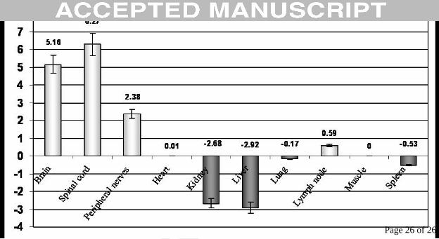
brain, 2.41E+02 in the peripheral nerves down to 2.00E-03 in the kidney (Figure 3). 10
Independent from the quantification technique used, no fluorescent signal was detected in any 11
of the DNA samples prepared from the different tissues used as quality and negative controls 12
or blank values. 13
14
3.2 Comparison of commercially available ELISA kits and the real time-PCR 15
The limit of detection (LOD) of the real-time PCR assay was assessed by comparing 16
the results with those obtained with the ScheBo® Brainostic GFAP ELISA Kit and the 17
RIDASCREEN® Risk Material 10/5 ELISA. These two enzyme immunoassays are based on 18
the detection of the GFAP antigen and are the only ELISA kits that were commercially 19
available in Germany at the time of the investigation. The LOD of the real-time PCR assay in 20
artificially contaminated minced meat was 0.1% bovine brain as described earlier 21
(Schönenbrücher et al., 2004a; Abdulmawjood et al., 2005). The 0.1% IRM was prepared in 22
duplicate to minimise the effect of a potentially inhomogeneous distribution of the 0.1% 23
content of bovine brain which would have resulted in false negative results. All three test 24
systems allowed the detection of 0.5% of bovine brain over the 14-day testing period (Table 25
gx
Page 7 of 26
Acce
pted
Man
uscr
ipt
8
2). The real-time PCR protocol still enabled the correct detection of as little as 0.1% CNS 1
tissue after 14 days. With regard to the criteria described in Table 1 for the classification of 2
positive or negative results, the Brainostic GFAP ELISA Kit enabled the identification of 3
0.2% and 0.1% CNS tissues. In contrast, the RIDASCREEN® Risk Material 10/5 ELISA 4
detected 0.2%, but failed to detect of 0.1% CNS tissues on four and three days out of the 14-5
day trial period, respectively. 6
The real-time PCR did not show any false positive nor false negative results for the 7
samples without CNS tissue (0%), the blank values, bovine DNA and porcine (negative 8
controls) when using the RIDASCREEN® Risk Material 10/5 ELISA kit (data not shown). 9
The ScheBo® Brainostic GFAP ELISA Kit gave two false positive results out of 10 samples 10
that did not contain CNS tissue. The diagnostic sensitivity of the real-time PCR assay was 11
100%. The sensitivity of the ScheBo® Brainostic GFAP ELISA Kit was 92.5%. With the 12
swab sampling technique and a cut off value of 0.1% (as recommended by the supplier), the 13
RIDASCREEN® Risk Material 10/5 ELISA gave a sensitivity of 52.5%. A cut off value of 14
0.2% raised the sensitivity to 95%. Subsequently, the sample preparation of the 15
RIDASCREEN® Risk Material 10/5 ELISA was modified by using 50 mg instead of a swab 16
sample. This resulted in a significant increase in sensitivity up to 90 % (cut off 0.1%, data not 17
shown). 18
19
4. Discussion 20
A real-time PCR based method was standardised and evaluated in terms of the 21
species-specific detection of CNS tissues in raw meat and heat-treated samples. The 22
experimental studies were conducted according to the ISO documents ISO/CD TAG 3 N 145 23
rev, ISO TC 34/SC 9 N containing general requirements for real-time PCR (anonymous, 24
2006a) and ISO/CD TAG 3 N 0144 rev ISO TC 34/SC 9 N, outlining general performance 25
Page 8 of 26
Acce
pted
Man
uscr
ipt
9
characteristics of molecular detection methods (anonymous, 2006b). Hence, two 1
commercially available ELISA kits were included in the evaluation as well as an external 2
validation that was conducted in a multicentre trial (Abdulmawjood et al., 2006). 3
Since food matrices vary considerably, sample preparation is a crucial step in mRNA 4
assays. Different sampling techniques were evaluated in which swab sampling was seen as an 5
easy-to-use technique for the examination of surfaces. An initial weight of 100mg is 6
recommended for processed meat and heat-treated meat products (Table 1). 7
8
4.1. Examination of the tissue specificity including absolute and relative quantitative 9
measurements 10
The absolute and relative expression levels of GFAP mRNA were determined. The 11
applicability of the standard curve used for absolute quantification (Figure 1) was proven by a 12
slope of -3.124 and a correlation coefficient (R) of >0.99. The relative expression of GFAP 13
mRNA was achieved by using the 2∆∆Ct methods with muscle as a calibrator tissue as 14
described earlier (Abdulmawjood et al., 2005). The endogenous control served as 15
normalisation for the mRNA added to the reverse transcription PCR and as control reaction to 16
avoid false negative results. Both methods revealed that the highest amounts of GFAP were 17
expressed in bovine brain and spinal cord (Figures 2 and 3). Because of their cross-sectional 18
dimension and large anatomic expansion, sciatic and axillary nerves represent the most 19
important peripheral nerves in meat processing. The ratio of the absolute values generated for 20
the peripheral nerves corresponded to about an 80 times higher GFAP mRNA content for the 21
brain and a 530 times higher GFAP mRNA content for the spinal cord. Relative quantification 22
gave a ratio of 1000- (brain) and 10, 000-fold (spinal cord, Abdulmawjood et al., 2005). Low 23
levels of GFAP in non-neuronal tissues could be detected (Figures 2 and 4). A GFAP signal 24
obtained from 100% adrenal gland tissue corresponded to 0.1% bovine brain, examined by 25
Page 9 of 26

Acce
pted
Man
uscr
ipt
10
absolute quantification. A low signal was also measured in fat, which could only be explained 1
by a minor contamination of this tissue by peripheral nerves during sampling. As can be seen 2
in the Ct-values generated (Figure 3), the diagnostic sensitivity was not affected by the low 3
GFAP mRNA content described above. 4
5
4.2. Comparison of commercially available ELISA kits and real-time PCR 6
Since 2004, enzyme-linked immunosorbent assay tests based on the GFAP antigen 7
have been the official German reference method for the detection of CNS tissues according to 8
§ 64 of the Foodstuff, Commodities and Animal Feed Act (anonymous, 2005). The GFAP 9
antigen is almost exclusively found in the CNS (Schmidt et al., 1999; Schmidt et al., 2001; 10
Herde et al., 2005; Rencova, 2005; Hossner et al., 2006). The multicentre trial presented by 11
Agazzi et al. (2002) proved the suitability of ELISA methods for the analysis of heat-treated 12
samples. The ScheBo® Brainostic GFAP ELISA Kit offered the highest sensitivity of 92.5%. 13
Application of the recommended swab sampling technique gave a sensitivity of 52.5% for the 14
RIDASCREEN® Risk Material 10/5 ELISA. Hughson et al. (2003) evaluated the ELISA kit 15
by using minced meat provided by the UK Food Standards Agency but could not detect CNS 16
concentrations as low as 0.1%. Hossner et al. (2006) could not detect 0.2% of CNS tissue and 17
reported highly variable results for the RIDASCREEN® Risk Material 10/5 ELISA. The 18
results gained in the present study suggest that the sensitivity of the assay can be increased by 19
modifying the preparation method of the samples. As an alternative, a cut-off value of 0.2% 20
could be used to improve the sensitivity up to 90%. No differences were observed between 21
the two 0.1% batches. This showed the comparability and the homogenous distribution of the 22
two batches. The real-time PCR assay enabled the reproducible detection of 0.1% bovine 23
brain after a storage time of 14 days. 24
25
Page 10 of 26

Acce
pted
Man
uscr
ipt
11
Furthermore, the performance of the real-time PCR assay was evaluated by a 1
multicentre trial including government laboratories as well as private bioanalytical 2
companies. The investigation showed that the detection limit of the methods is at least as low 3
as 0.1 when analysing strongly heated or medium-heated material. A detection limit of 0.2% 4
was obtained for liver sausages. A detailed statistical analysis of the data is given by 5
Abdulmawjood et al. (2006). The second part of the ring trial will investigate the effect of 6
sample preparation as well as the results obtained with different thermocycler models. 7
Previously Seybold et al. (2003) and Lange et al. (2003) showed the possible 8
applicability of conventional PCR methods for the detection of CNS tissues. Seybold et al. 9
(2003) used a restriction fragment length polymorphism (RFLP) system based on the GFAP 10
mRNA to differentiate between several animal species. This approach showed cross-reactions 11
with raw heart as well as muscle tissues. Lange et al. (2003) used GFAP mRNA sequences as 12
well as myelin basic protein (MBP) mRNA sequences. The oligonucleotide primers selected 13
for the marker GFAP enabled a non species-specific detection of brain tissue. The MBP 14
oligonucleotide primers differentiated bovine, ovine and caprine from porcine brain tissues. 15
The negative PCR results for brain tissues of goose, ostrich and chicken were considered to be 16
preliminary, indicating the need for further investigation of the species-specific detection of 17
CNS tissues. However, both groups did not present reliable data on the critical influence of 18
peripheral nervous system (PNS) tissues. Seybold et al. (2003) lacked the investigation of 19
peripheral nerves. Lange et al. (2003) did not explain the consequences of their detection of 20
GFAP in sciatic nerve (e. g. on the detection limit of the method). The occurrence of the 21
mRNA target region of the MBP in sciatic nerve was not conducted. 22
With regard to the immunohistochemical methods, Lücker et al. (1998) combined the 23
detection of cholesterol and neuron-specific enolase (NSE). Furthermore, NSE and the CNS-24
specific GFAP were combined for Western blot analysis (Lücker et al., 2000), which proved 25
Page 11 of 26

Acce
pted
Man
uscr
ipt
12
unsuitable for strongly heat-treated meat products (>80°C). Immunohistological staining of 1
NSE was also unsuitable for testing heat-treated meat products (Aupperle et al., 2002; 2
Tersteeg et al., 2002). Herde et al. (2005) showed that MBP was still detectable by Western 3
Blot analysis after meat processing, which also included the addition of spices as well as heat 4
treatment and took storage stability into account. 5
By using gas chromotography-mass spectrometry (GC-MS), Lücker et al. (2004) 6
identified cerebronic acid as a reliable target for the tissue-specific detection of CNS tissues in 7
meat products. The technique offers the potential for the species- and also the age-dependent 8
quantification of the CNS content. Therefore, several analytical steps are required. At first, the 9
identification of a CNS positive sample can be achieved by using cerebronic acid. The 10
relationship of isomers of the tetracosenic acid is used to investigate species and age of the 11
CNS (Biedermann et al., 2004). As a prerequisite, standards of CNS containing CNS of the 12
adequate species and age must be used. Further and more extensive studies are needed to 13
elucidate the limits of the age- and species-specific detection. Poerschmann et al. (2006) 14
proved the sequential pressurized liquid extraction to be superior to the commonly used 15
exhaustive lipid extraction method. As a consequence, the GC-MS technique achieves further 16
methodological improvement. 17
However, all of the methods published so far have some drawbacks and do not allow 18
the reliable detection of specified risk material taking into account the age of the animals or 19
the differentiation of CNS tissues obtained from countries with or without a geographical 20
BSE risk (e. g. Argentina). Knowledge about the spread of the causative BSE agent is steadily 21
increasing (Thomzig et al., 2004; Angers et al., 2006) which underlines the importance of 22
setting up reference methods. 23
As an important part of public health concerns, the EU recommends the development 24
of reliable methods for the detection of CNS in food. The specific detection of CNS tissues of 25
Page 12 of 26
Acce
pted
Man
uscr
ipt
13
banned animal species using the real-time PCR-based method presented here can be 1
conducted in a single run. Porcine CNS tissues can be specifically detected in a second real-2
time PCR assay (Schönenbrücher et al., 2004b). As far as the authors know this is the first 3
report that evaluates a real-time PCR-based method in conjunction with a multicentre trial 4
according to ISO requirements and it is also the first report that compares it with two 5
commercially available ELISA kits. This study showed that, in minced meat, the GFAP 6
mRNA target region remains detectable with RT-PCR after several days of storage. With 7
regard to potential economic consequences for the meat-producing industry, should a CNS 8
positive sample be detected, there is a very low risk of obtaining false positive and false 9
negative responses. 10
The validation data presented here offer a highly suitable method for routine use as 11
well as a large sample throughput test. However, a further multicentre trial considering the 12
sample preparation will have to be conducted before the method can be put into routine 13
practice. 14
15
Acknowledgements 16
This work was undertaken as part of the “AZ.: 01HS022/1” project funded by the 17
Federal Ministry of Consumer Protection, Food and Agriculture, Germany. The authors 18
would like to thank K. Simon and C. Walter for excellent technical assistance. 19
20
21
Page 13 of 26
Acce
pted
Man
uscr
ipt
14
References 1
Abdulmawjood, A., Schönenbrücher, H., Bülte, M., 2005. Novel molecular method for 2
detection of bovine-specific central nervous system tissues as bovine spongiform 3
encephalopathy risk material in meat and meat products. J. Mol. Diagn. 7, 368-374. 4
Abdulmawjood, A., Schönenbrücher, H., Bülte, M., 2006. Collaborative trial for validation of 5
a real time RT-PCR assay for detection of central nervous system tissues as BSE risk 6
material: Part 1. J. AOAC Int. 89, 1335-1340. 7
Agazzi, M.E., Barrero-Moreno, J., Lücker, E., v. Holst, C., Anklam, E., 2002. Performance 8
comparison of two analytical methods for the detection of tissues of the central 9
nervous system in sausages: results of an interlaboratory study. Eur. Food Res. 10
Technol. 215, 334-339. 11
Angers, R.C., Browning, S.R., Seward, T.S., Sigurdson, C.J., Miller, M.W., Hoover, E.A., 12
Telling, G.C., 2006. Prions in skeletal muscles of deer with chronic wasting disease. 13
Science 311, 1117. 14
Anonymous, 2006a. Microbiology of food and animal feeding stuffs - Polymerase chain 15
reaction (PCR) for the detection of food-borne pathogens – Performance 16
characteristics of molecular detection methods. ISO/CD TAG 3 N 0144 rev, ISO TC 17
34/SC 9 N, 01.02.2006. 18
Anonymous, 2006b. Microbiology of food and animal feeding stuffs - Real time polymerase 19
chain reaction (PCR) for the detection of food-borne pathogens - General requirements 20
and definitions ISO/CD TAG 3 N 145 rev, ISO TC 34/SC 9 N, 01.02.2006. 21
Aupperle, H., Lücker, E., Overhoff M., Schoon, H.A., 2002. Verfahren zum Nachweis von im 22
Hinblick auf die BSE unerwünschten Zutaten in Fleischerzeugnissen. 23
Immunhistologischer Nachweis von zentralem und peripherem Nervengewebe in 24
Fleischerzeugnissen. Fleischw. 3, 100-104. 25
Page 14 of 26

Acce
pted
Man
uscr
ipt
15
Biedermann, W., Lücker, E., Porschmann, J., Lachhab, S., Truyen U., Hensel, A. 2004. 1
Structural characterisation of some fatty acids from the brain as biomarkers of BSE 2
risk material. Anal. Bioanal. Chem. 379, 1031-1038. 3
Bruce, M.E., Will, R.G., Ironside, J.W., McConnell, I., Drummond, D., Suttie, A., McCardle, 4
L., Chree, A., Hope, J., Birkett, J., Cousens, S., Fraser H., Bostock, C.J., 1997. 5
Transmissions to mice indicate that 'new variant' CJD is caused by the BSE agent. 6
Nature 2, 498-501. 7
Bundesanzeiger (Federal Herald), 1994. Guiding principles for meat and meat products. 8
German food code. Bundesanzeiger, Köln, pp. 49-150. 9
Cousens, S., Smith, P.G., Ward, H., Everington, D., Knight, R.S., Zeidler, M., Stewart, G., 10
Smith-Bathgate, E.A., Macleod, M.A., Mackenzie J., Will, R.G., 2001. Geographical 11
distribution of variant Creutzfeldt-Jakob disease in Great Britain, 1994-2000. Lancet 12
357, 1002-1007. 13
EFSA, 2005. Quantitative assessment of the residual BSE risk in bovine-derived products, 14
EFSA QRA report 2004 – working document. EFSA J. 307, 1-135. 15
European Commission, 2001. Commission Regulation EC No 999/2001 OJ L 147 16
31/05/2001. EC, Luxemburg, p1, last amendment by Commission Regulation (EC) No 17
1041/2006 of 7 July 2006 of the European Parliament OJ L 187, 8.7.2006. 18
Comer, P.J. Huntly. P.J., 2003. TSE risk assessments: a decision support tool. Stat. Methods 19
Med. Res. 12, 279-291. 20
Herde, K., Bergmann, M., Lang, C., Leiser R., Wenisch S., 2005. Glial fibrillary acidic 21
protein and myelin basic protein as markers for the immunochemical detection of 22
bovine central nervous tissue in heat-treated meat products. J. Food Prot. 68, 823-827. 23
Hossner, K. L., Yemm, R.S., Sonnenshein, S.E., Mason, G.L., Cummings, B.A., Reddy, 24
M.C., Sofos, J.N., Scanga, J.A., Tatum, J.D., Smith, G.C., Belk K.E., 2006. 25
Page 15 of 26
Acce
pted
Man
uscr
ipt
16
Comparison of immunochemical (enzyme-linked immunosorbent assay) and 1
immunohistochemical methods for the detection of central nervous system tissue in 2
meat products. J. Food Prot. 69, 644-650. 3
Hughson, E., Reece, P., Dennis, M. J., S. Oehlschlager, S., 2003. Comparative evaluation of 4
the performance of two commercial kits for the detection of central nervous system 5
tissue in meat. Food Addit. Contam. 20, 1034-1043. 6
Lange, B., Alter, T., Froeb, A., Lücker, E., 2003. Molecular biological detection of tissues of 7
central nervous system in meat products. Berl. Münch. Tierärztl. Wochenschr. 116, 8
467-473. 9
Lücker, E.H., Eigenbrodt, E., Wenisch, S., Failing, K. Leiser R., Bülte, M., 1999. 10
Development of an integrated procedure for the detection of central nervous tissue in 11
meat products using cholesterol and neuron-specific enolase as markers. J. Food. Prot. 12
62, 268-276. 13
Lücker, E. H., Eigenbrodt, E., Wenisch, S., Leiser, R., Bülte, M., 2000. Identification of 14
central nervous system tissue in retail meat products. J. Food Prot. 63, 258-263. 15
Lücker, E., Horlacher S., Eigenbrodt, E., 2001. Brain in human nutrition and variant 16
Creutzfeldt-Jakob disease risk (vCJD): detection of brain in retail liver sausages using 17
cholesterol and neuron specific enolase (NSE) as markers. Br. J. Nutr. 86 Suppl. 1, 18
115-119. 19
Lücker, E., Biedermann, W., Lachhab, S., Truyen U., Hensel, A., 2004. GC-MS detection of 20
central nervous tissues as TSE risk material in meat products: analytical quality and 21
strategy. Anal. Bioanal. Chem. 380, 866-870. 22
Poerschmann J., Trommler, U., Biedermann, W., Truyen U., Lücker, E., 2006. Sequential 23
pressurized liquid extraction to determine brain-originating fatty acids in meat 24
Page 16 of 26

Acce
pted
Man
uscr
ipt
17
products as markers in bovine spongiform encephalopathy risk assessment studies. J. 1
Chromatogr. A. 16, 26-33 2
Rencova, E., 2005. Comparison of commercially available antibodies for the detection of 3
central nervous system tissue in meat products by enzyme-linked immunosorbent 4
assay. J. Food Prot. 68, 630-632. 5
Reddy, M.C., Hossner, K.L., Belk, K.E., Scanga, J.A., Yemm, R.S., Sofos J.N., Smith, G.C., 6
2006. Detection of central nervous system tissue on meat and carcass-splitting band 7
saw blade surfaces using modified fluorescent glial fibrillary acidic protein enzyme-8
linked immunosorbent assay sampling and extraction procedures. J. Food Prot. 69, 9
1966-1970. 10
Schmidt, G. R., Hossner, K.L., Yemm, R.S., Gould, D.H., O`Callaghan, J.P., 1999. An 11
enzyme-linked immunosorbent assay for glial fibrillary acidic protein as an indicator 12
of the presence of brain or spinal cord in meat. J. Food. Protect. 62, 394-397. 13
Schmidt, G.R., Yemm, R.S., Childs, K.D., O`Callaghan, J.P., Hossner, K.L., 2001. The 14
detection of central nervous system tissue on beef carcasses and in comminuted beef. 15
J. Food Prot. 64, 2047-2052. 16
Schönenbrücher H., Abdulmawjood, A., Bülte, M., 2004a. Neuartiger tierartspezifischer 17
Nachweis von GFAP in prozessierten Lebensmitteln mit einem Real Time-PCR-18
Verfahren. Fleischw. 6, 114-117. 19
Schönenbrücher H., Abdulmawjood A., Bülte, M., 2004b. Nachweis von GFAP - Spezifischer 20
Nachweis in prozessierten Lebensmitteln mit einem SybrGreen® Real Time-PCR-21
Verfahren. Fleischw. 8, 90-92. 22
Seyboldt C., John A., v. Mueffling T., Nowak, B. Wenzel, S., 2003. Reverse transcription-23
polymerase chain reaction assay for species-specific detection of bovine central 24
nervous system tissue in meat and meat products. J. Food Prot. 66, 644-651. 25
Page 17 of 26

Acce
pted
Man
uscr
ipt
18
Tersteeg, M.H.G., Koolmees, P.A., v. Knapen, F., 2002. Immunhistochemical detection of 1
brain tissue in heated meat products. Meat Science 61, 67-72. 2
Thomzig, A., Schulz-Schaeffer, W., Kratzel, C., Mai, J., Beekes, M., 2004. Preclinical 3
deposition of pathological prion protein PrPSc in muscles of hamsters orally exposed 4
to scrapie. J. Clin. Invest. 113, 1465-1472. 5
Wenisch, S., Lücker, E., Eigenbrodt, E., Leiser, R., Bülte, M., 1999. Detection of central 6
nervous tissue in meat products – an immunhistological approach. Nutr. Res. 19, 7
1165-1172. 8
9
10
11
Page 18 of 26
Acce
pted
Man
uscr
ipt
19
Table 1: Parameters of the real-time PCR assay 1
2
Target gene Glial fibrillary acidic protein (GFAP)
messenger (m) RNA
Detected animal species cattle, sheep and goat
Fluorescent probes TaqMan®mgb-probes
Quantitative measurement absolute or relative
Internal amplification control* puc19-plasmid
Samples raw meat, swab samples** and heat-treated
meat products (F-values 5.4)
Initial weight 100 mg
Time requirement 5 hours
* if using absolute quantification 3
** swab samples taken from carcasses, heads of cattle used for meat cutting, pieces of 4
meat 5
6
7
8
9
Page 19 of 26

Acce
pted
Man
uscr
ipt
20
Table 2: Comparative study of real-time PCR and two ELISA kit results of artificially contaminated minced meat 1
2
Page 20 of 26
Acce
pted
Man
uscr
ipt
21
Day 0 Day 1 Day 3 Day 7 Day 14 Storage time Brain concentrations
Sample A
Sample B
Sample A
Sample B
Sample A
Sample B
Sample A
Sample B
Sample A
Sample B
0% RISK MATERIAL 10/51)
- - - - - - - - - -
Brainostic2) - - - - (+) (+) - - - -
Real-time PCR3) - - - - - - - - - -
0.1% RISK MATERIAL 10/5 - - - - - - - - - - Brainostic - (+) - + (+) (+) + + + +
Real-time PCR + + + + + + + + + +
0.1% RISK MATERIAL 10/5 - (+) - - - (+) - - - -
Brainostic (+) (+) (+) + (+) (+) + + + +
Real-time PCR + + + + + + + + + +
0.2% RISK MATERIAL 10/5 (+) (+) + (+) (+) (+) (+) (+) (+) - Brainostic - + + (+) + (+) + + + +
Real-time PCR + + + + + + + + + +
0.5% RISK MATERIAL 10/5 + + + + + + (+) (+) (+) (+)
Brainostic + + + + + + + + + +
Real-time PCR + + + + + + + + + + 1 1) RISASCREEN® Risk Material 10/5-ELISA: + Optical Density (OD)Sample > OD of Standard (S)2 (0.1%); (+) ODSample > 2xODS1 (0%); 2
- ODSample < 2xODS1 (0%) 3
Page 21 of 26

Acce
pted
Man
uscr
ipt
22
2) Brainostic GFAP-ELISA: + Optical Density (OD)Sample > OD of Standard (S)1 (0.1%); (+) ODSample > 70% of ODS2; - ODSample < 70% of S2 1
3) + Threshold cycle (Ct) < 32; - Ct > 32 2
3
Page 22 of 26

Acce
pted
Man
uscr
ipt
23
Figure legends Figure 1: Absolute quantification: GFAP cDNA-contents ( ) per PCR-reaction [ng] 1
presented with a logarithmic scale 2
3
Figure 2: Absolute quantification: Corresponding Ct-values 4
5
Figure 3: Relative quantification: Logarithmic values of the GFAP cDNA-contents ( ) per PCR reaction [ng] 6
7
8
gx
gx
Page 23 of 26

Acc
epte
dM
anus
crip
tPage 24 of 26
Acc
epte
dM
anus
crip
tPage 25 of 26
Acc
epte
dM
anus
crip
t
Page 26 of 26