Embed Size (px)
Citation preview
DETERMINATION OF CARDIAC SIZE FROM CHEST ROENTGENOGRAMS FOLLOWING SKYLAB MISSIONS
A. Nicogossian, Y.D. *, G . W. Hoffler, V. D. R. L. Johnson, V. D. * and R. J. Gowen, Ph.D. 1
ABSTRACT
Decreased cardiothoracic transverse diameter ra t ios following Mercury, Gemini and Apollo space f l i gh t s have been reported previously. To evaluate further changes in cardiac s ize , standard posteroanterior chest films in systole and diastole were obtained before f l i g h t and within a few hours a f t e r recovery on each of the Skylab astronauts. P o s t f l i g h t chest X-rays were visually compared to the preflight roentgenograms for possible changes i n pulmonary vasculature, l u n g parenchyma, bony or soft t issue structures. the following measurements were obtained: verse diameters, cardiothoracic transverse diameter r a t i o , cardiac area from the product of both diagonal diameters, cardiac s i lhouet te area by planimetry, thoracic cage area and cardiothoracic a r a ra t io . The postfl ight frontal cardiac s i lhouet te s izes were signif cantly decreased when compared w i t h the respective preflight values (PO 05 or 0.01). The observed changes are t h o u g h t t o be related t o postf i g h t decrease i n the intracardiac chamber volume.
From these roentgenograms cardiac and thoracic trans-
INTRODUCTION
Determination of s i ze i s a major factor i n the c l inical evaluation of the healthy or fa i l ing heart. interpretation of both electrocardiographic and hemodynamic information. The evaluation o f changes i n cardiac s ize has been important i n the overall cardiovascular assessment of or thostat ic intolerance observed among the majority of astronauts following space missions.
Knowledge of heart s ize a s s i s t s the
Decreased cardiothoracic transverse diameter ra t ios following Mercury, Gemini and Apollo f l i g h t s have been reported ea r l i e r from our labora- tory ( 1 , 2 ) . More recently similar data following space missions of
*National Aeronautics and Space Administration - Lyndon B. Johnson
'U .S.A. F. Academy, Colorado Spr ings , Colorado.
Space Center, Houston, TX.
785
longer duration have been presented ( 3 ) . The majority of crewmembers who exhibited postf l ight decreases i n the cardiac s i lhouet te s i ze also showed a decreased or thostat ic tolerance to lower body negative pressure. Similar findings were also reported by the Soviet investi- gators following 30-day bed r e s t studies (4) and i n cosmonauts upon return from space missions. T h i s paper presents further radiological data from a l l three Skylab manned missions and discusses the physio- logical factors possibly involved i n the cardiac s i lhouet te changes.
METHODS AND MATERIALS
Standard posteroanterior chest films were obtained before and as soon a s possible a f t e r f l i g h t on each of the Skylab astronauts following extended space f l i gh t s of d i f fe ren t durations: 28, 59 and 84 days. X-ray exposures were 150 m i 11 iseconds i n duration. d ias to l ic exposures were triggered electronically from the electro- cardiographic R-wave peak by a special device interposed w i t h the X-ray equipment control. The electronic t r igger device delayed the roentgenographic exposures from the R-wave peak according t o the instantaneous heart ra te (the preceding RR in te rva l ) . delays were 175 to 325 milliseconds, corresponding t o heart ra tes ranging from 140 to 40 beats per minute, and fo r diastole 385 to 1165 milliseconds for heart ra tes ranging from 140 to 44 beats per minute.
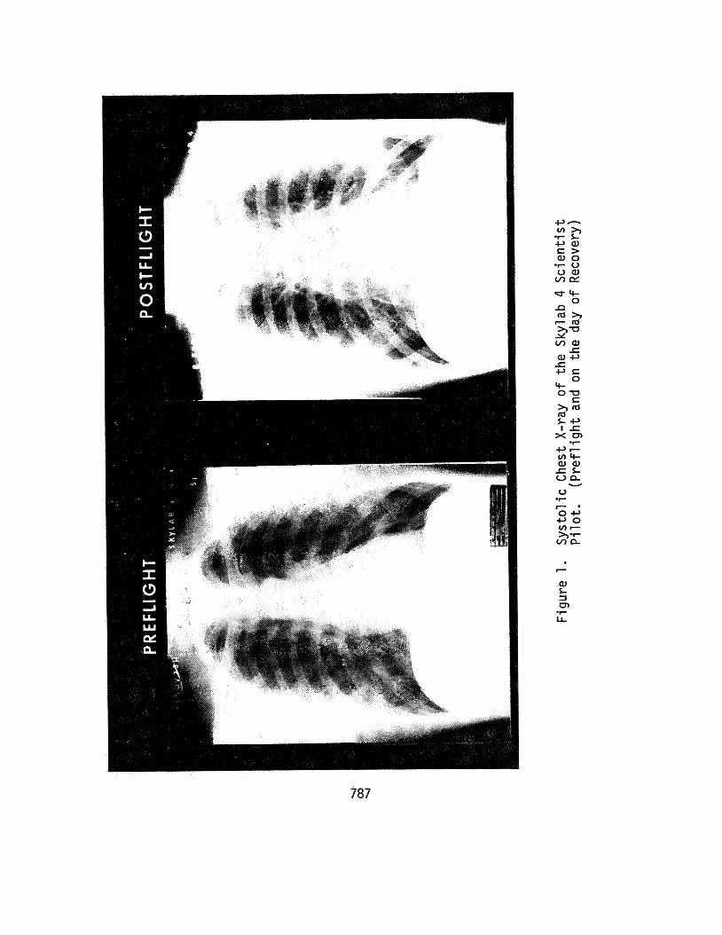
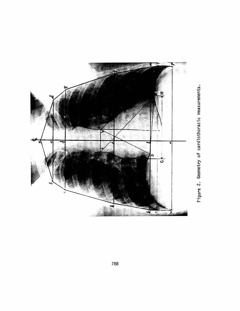
Postfl ight chest X-rays were visually compared w i t h the pref l ight films fo r possible changes which m i g h t have occurred in pulmonary vasculature, lung parenchyma, bony or s o f t t issue structures. One or two additional postf l ight films were taken several days following splashdown to assess trends. While many of the film pairs showed readily apparent postfl ight decreases i n heart s i ze ( f i g . 1 ) , several measures have been adopted to determine this change quantitatively. u t i l i zed i n determining thoracic and cardiac areas. The thoracic cage area was obtained by a modified method as described by Barnhard (5) and by Loyd (6 ) . After the inner border of the ribs was outlined, the thoracic center was determined by drawing the l i ne (CL) along the vertebral column. Next, perpendicular l ines to ( C L ) were drawn a t 2.5, 5.0 and 15.0 centimeters from the f i r s t thoracic inter- vertebral space - point of origin (a) creating three upper polygonal segments. A horizontal l ine (k-m) drawn halfway between the level of the apices of the r i g h t and l e f t hemidiaphragms delineated the lower border of the fourth segment. horizontal 1 ine (n-p) drawn a t mid-distance between the two costophrenic angles; actual area of this segment was modified by c i rcu lar deductions fo r infra diaphragm space. the computed area of each of the above f ive segments.
All Systol i c and
For systole the
Figure 2 shows the geometry
The last segment was delimited by a
The to ta l thoracic area was summed from
786
787
rc 0
788
To obtain uniform evaluation of the cardiac area, the heart s i lhouet te was outlined i n the following manner.
O The lower border of the heart was defined as the l ine ( B - D ) between the intersections of the right and l e f t cardiac borders and the respective hemidiaphragms ,
O the r ight cardiac border (A-D) fo superior vena cava,
O the l e f t cardiac border (B-C) was regression l i ne of the l e f t heart the l e f t margin of the descending
lowed the r igh t atrium and
completed by drawing a border intersecting with aorta ,
O the upper cardiac border was outlined by a perpendicular ( A - C ) t o the center l ine a t the level of the upper l e f t heart border.
The following parameters were measured and/or computed:
O Cardiothoracic transverse diameter r a t io (C/TD).
O Cardiac s i lhouet te area, by planimetry.
O Cardiothoracic area r a t i o (C/TA) .
O Cardiac area from the product of long and short diagonal diameters .
I t i s hoped t h a t the additional information inherent in this technique will r e f l ec t more accurate s i ze values and help compensate for s l igh t variations i n body position and inspiratory level. All postfl ight d a t a were compared t o the respective preflight values using the Student's t - t e s t and regression analysis.
RESULTS
No roentgenological abnormalities were observed on e i ther preflight or postfl ight films. of recovery were of poor quali ty and n o t amenable t o analysis; a l l other X-rays were of acceptable quali ty. Differences between pref l ight and postfl ight sys to l ic and d ias to l ic cardiothoracic diameter and area ra t ios are presented i n tables I and 11. Both C/T and C/T showed a decrease i n the individual values postfl ight.
The chest X-rays of the Skylab 3 Pi lo t on the day
In general t b r e was
789
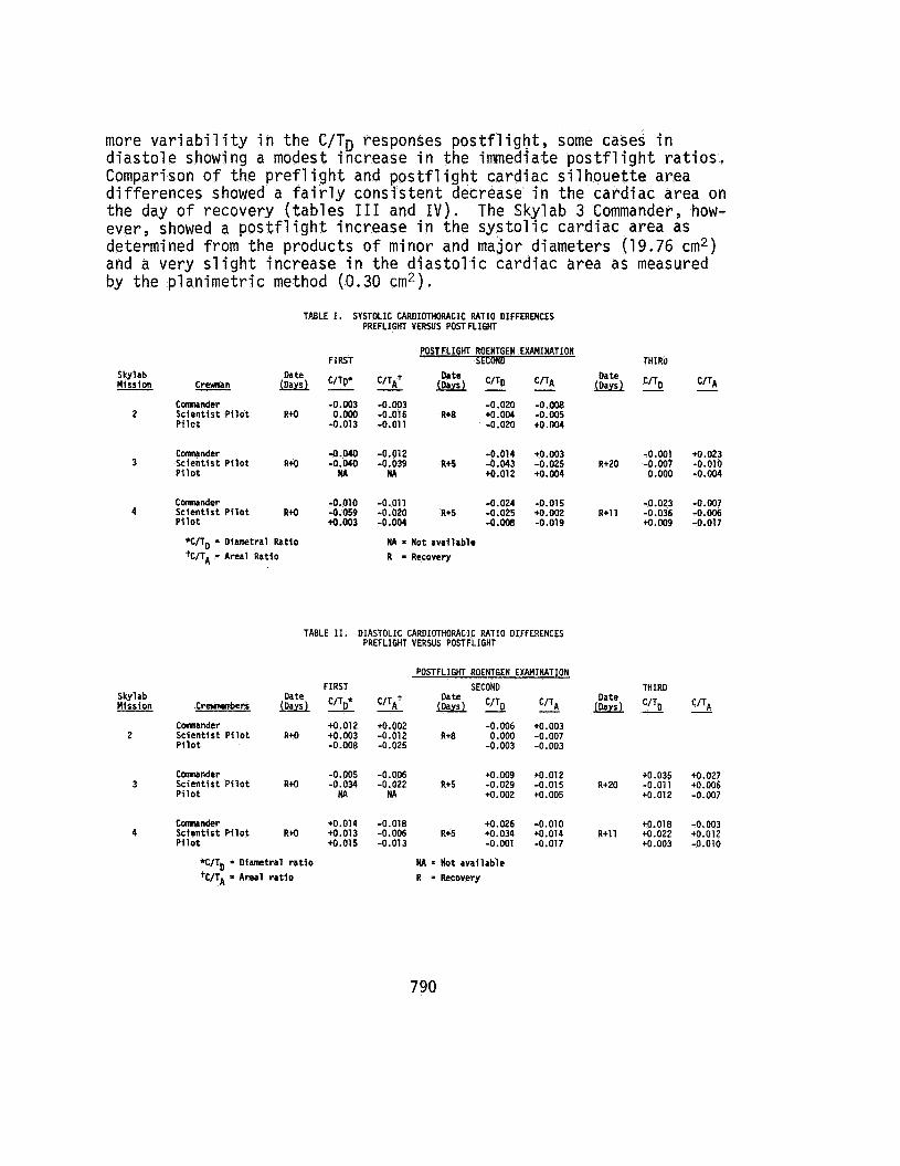
more v a r i a b i l i t y i n the C/TD responses p o s t f l i g h t , some cases i n d i a s t o l e showing a modest increase i n the immediate p o s t f l i g h t r a t i o s . Comparison of t he p r e f l i g h t and p o s t f l i g h t card iac s i l h o u e t t e area d i f f e rences showed a f a i r l y cons i s ten t decrease i n the card iac area on t he day o f recovery ( tab les I11 and I V ) . The Skylab 3 Commander, how- ever, showed a p o s t f l i g h t increase i n t h e s y s t o l i c card iac area as determined from the products o f minor and major diameters (19.76 cm2) and a very s l i g h t increase i n the d i a s t o l i c card iac area as measured by the p lan ime t r i c method (0.30 cm2).
TABLE I. SYSTOLIC CARDIOTHORACIC RATIO DIFFERENCES PREFLIGHT VERSUS POSTFLIGHT
Skylab Uission
2
-
3
4
Oa te CreMMn -
Connander Scientist Pilot R+O Pilot
Comnander Scientist Pilot R+O Pilot
Cannander Scientist Pilot R+O Pilot
*C/TD = Diametral Ratio k/TA = Areal Ratlo
FIRST
C/TD* - -0.003 0.000 -0.013
-0.040 - 0 . W NA
-0.010 -0.059 *0.003
POSTFLIGHT ROENTGEN EXAMINATION THIRO
- C/TAt & C/TD - C/TA & C/To - C/TA
-0.003 -0.020 -0.008 -0.015 Rt8 +O.O04 -0.005 -0.011 -0.020 +O.004
-0.012 -0.014 +0.003 -0.001 +O.023 -0.039 R+5 -0.043 -0.025 R+20 -0.007 -0.010 HA +O.O12 +0.004 0.000 -0.004
-0.011 -0.024 -0.015 -0.023 -0.007 -0.020 R+5 -0.025 +0.002 R+11 -0.036 -0.006 -0.004 -0.008 -0.019 +O.M -0.017
NA = Mot avallablc R = Recovery
TABLE XI. DIASTOLIC CAROIOTHORACIC RATIO DIFFERENCES PREFLIGHT VERSUS POSTFLIGHT
POSTFLIGHT ROENTGEN EXAMINATION FIRST SECOND THIRO
Oate C/TD C/TA Skylab Misston C r e e r s & C/TO. CITA+ - - 'ITA
Connander +O.012 +0.002 -0.006 +0.003 2 Scientist Pilot R+O +0.003 -0.012 R+8 O.Oo0 -0.007
Pilot -0.008 -0.025 -0.003 -0.003
Comnander -0.005 -0.006 +0.009 +0.012 +0.035 +0.027 3 Scientist Pilot R+O -0.034 -0.022 R+5 -0.029 -0.015 R+20 -0.011 +0.006
Pilot NA NA +0.002 +0.005 +0.012 -0.007
Cwnander +O.014 -0.018 +0.026 -0.010 +0.018 -0.003 4 Scientist Pilot R+O +0.013 -0.006 R+5 +0.034 +0.014 R+11 +0.022 +0.012
Pilot +0.015 -0.013 -0.001 -0.017 +0.003 -0.010
%/TD = Diametral ratio +C/T,, = Areal ratio
NA = Not available R = Recovery
790
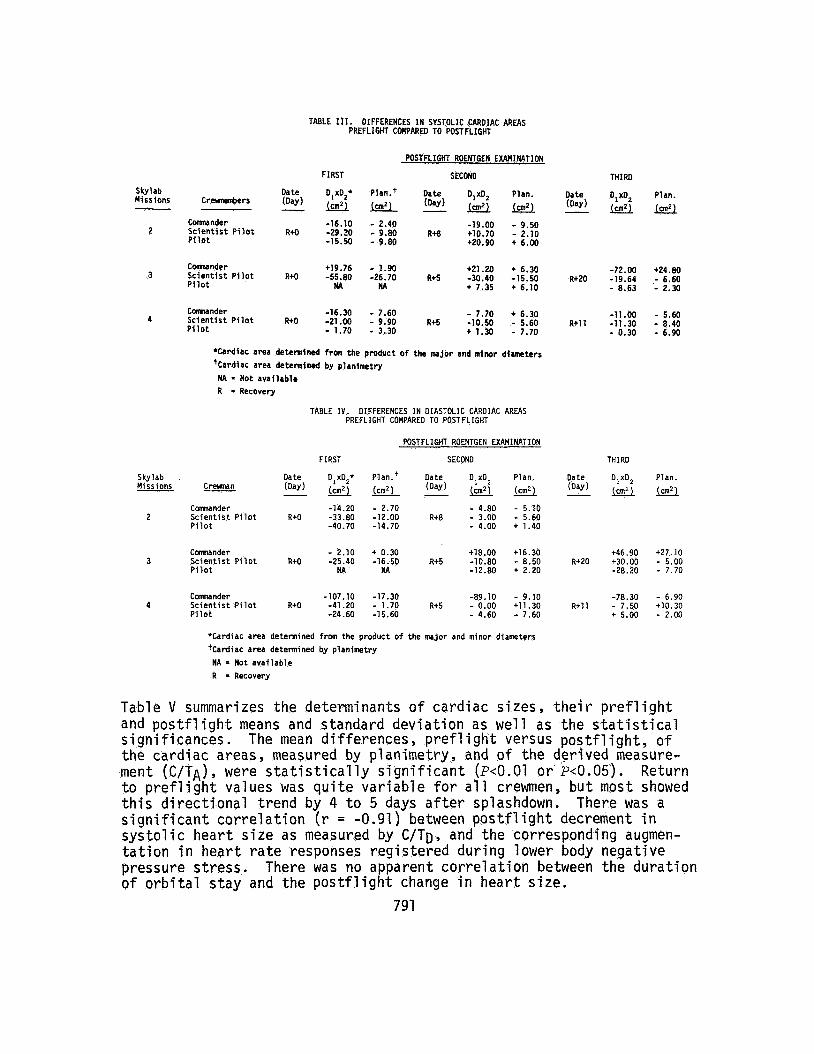
TABLE 111. DIFFERENCES I N SYSTOLIC CARDIAC AREAS PREFLIGHT COMPARE0 TO POSTFLIGHT
Skylab Missions
2
3
4
Skylab Missions
2
3
4
Table V
POSTFLIGHT ROENTGEN EXAMINATION
FIRST SECONO
Date olx0,* Plan.+ Date D,XO, Plan. Date Cremnembers (Day) (cm2) (cm2) (Day) (cm2) (an2) (Day) - -
-16.10 - 2.40 -19.00 - 9.50 Comnander S c i e n t i s t P i l o t R+O -29.20 - 9.80 ~ + 8 i10.70 - 2.10 P i l o t -15.50 - 9.80 i20.90 + 6.00
Comnander +19.76 - 1.90 +21.20 t 6.30 Sc ien t is t P i l o t R i O -55.80 -26.70 R i 5 -30.40 -15.50
i 7.35 + 6.10 R+20
P i l o t M HA
Connander -16.30 - 7.60 - 7.70 i 6.30
P i l o t - 1.70 - 3.30 i 1.30 - 7.70 R t l l Sc ien t is t P i l o t R i O -21.00 - 9.90 R+S -10.50 - 5.60
"Cardiac area detcnnincd f ran the product of the major and minor diameters 'Cardiac area detewined by planimetry NA = Not avallablm R = Recovery
TABLE I V . DIFFERENCES I N DIASTOLIC CARDIAC AREAS PREFLIGHT COMPARED TO POSTFLIGHT
POSTFLIGHT ROENTGEN EXAMINATION
FIRST SECONO
Comnander -14.20 - 2.70 - 4.80 - 5.10 Sc ien t is t P i l o t R+O -33.80 -12.00 Rt8 - 3.00 - 5.60 P i l o t -40.70 -14.70 - 4.00 + 1.40
Comnander - 2.10 + 0.30 t18.00 t16.30 Sc ien t is t P i l o t R+O -25.40 -16.50 R+5 -10.80 - 8.50 Rt20 Pi l o t HA NA -12.80 t 2.20
Connander -107.10 -17.30 -89.10 - 9.10 Sc ien t is t P i l o t R+O -41.20 - 1.70 R+5 - 0.00 t11.30 Rt11 P i l o t -24.60 -15.64 - 4.60 - 7.60
'Cardiac area determined from the product of the major and minor diameters +Cardiac area detennined by planimetry
NA = Not avai lable R = Recovery
summarizes the determinants of cardiac sizes, their
THIRD
DlxO, Plan.
-72.00 i24.80 -19.64 - 6.60 - 8.63 * 2.30
-11.00 - 5.60 -11.30 - 8.40 - 0.30 - 6.90
THIRD
O,xO, Plan.
+46.90 t27.10 t30.00 - 5.00 -28.20 - 7.70
-78.30 - 6.90 - 7.50 t10.30 t 5.00 - 2.00
prefl isht and postflight means and standard deviation as well as the statistical significances. The mean differences, preflight versus postflight, of the cardiac areas, measured by planimetry, and o f the derived measure- ment (C/TA) , were statistically significant (P<O.o1 or ~ 0 . 0 5 ) . Return to preflight values was quite variable for all crewmen, but most showed this directional trend by 4 to 5 days after splashdown. significant correlation (r = -0.91) between postflight decrement in systolic heart size as measured by C/TD, and the corresponding augmen- tation in heart rate responses registered during lower body negative pressure stress. of orbital stay and the postflight change in heart size.
There was a
There was no apparent correlation between the duration
791
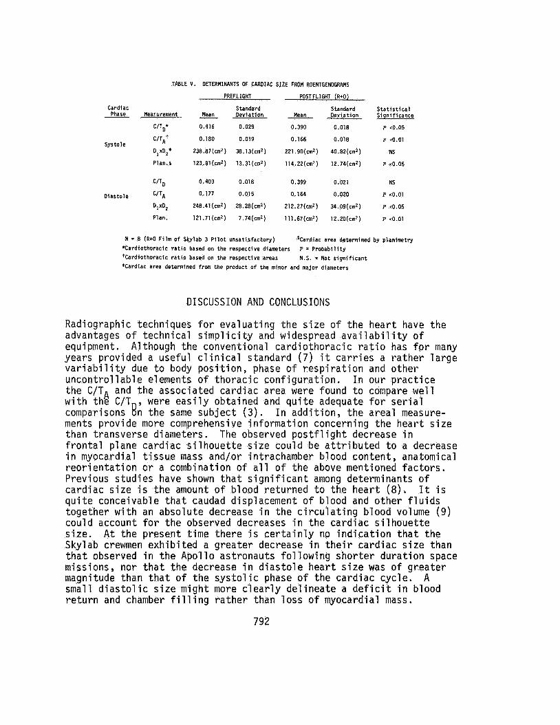
TABLE V . DETERMINANTS OF CARDIAC SIZE FROM ROENTGENOGRAMS
PREFLIGHT POSTFLIGHT (R+O)
Cardiac Standard Standard Statistical Phase Meacurement - Mean Deviation Mean Deviation Sisnificance
UTD* 0.416 0.029 0.390 0.018 P ~0.05
C/TAI 0.180 0.019 0.166 0.018 P co.01 Systole
DlxOzt 238.87(cm2 38.1 3(cm2 ) 221 .90(cm2 ) 40.82(cmZ ) NS
Plan.§ 123.81(crn2) 13.31(cm2) 114.22(cm2) 12.74(cm2) P <0.05
C/TD 0.403 0.018 0.399 0.021 NS
Diastole C/TA 0.177 0.015 0.164 0.020 P .0.01
DlXD, 248.41(cm2) 28.28(cm2) 212.27(cmZ) 34.09(cmZ) P <0.05
Plan. 121.71(cm2) 7.74(un2) 111.67(cm2) 12.20(cm2) P <0.01
N = 8 (R+O Film of Skylab 3 Pilot unsatisfactory) 'Cardiothoracic ratio based on the respective diameters +Cardiothoracic ratio based on the respective areas *Cardiac area determined from the product of the minor and major diameters
'Cardiac area determined by planimetry P = Probability N.S. = Not significant
DISCUSSION AND CONCLUSIONS
Radiographic techniques for evaluating the s i ze of the heart have the advantages of technical simp1 i c i ty and widespread avai 1 ab i 1 i ty of equipment. Although the conventional cardiothoracic r a t io has for many years provided a useful c l inical standard ( 7 ) i t car r ies a rather large var iab i l i ty due to body position, phase of respiration and other uncontrollable elements of thoracic configuration. In our practice the C/TA and the associated cardiac area were found t o compare well with the C/T , were easi ly obtained and qui te adequate fo r se r ia l comparisons 8 n the same subject ( 3 ) . ments provide more comprehensive information concerning the heart s i ze t h a n transverse diameters. frontal plane cardiac s i lhouet te s ize could be at t r ibuted to a decrease i n myocardial t i s sue mass and/or intrachamber blood content, anatomical reorientation c r a combination of a l l of the above mentioned factors. Previous studies have shown tha t s ignif icant among determinants of cardiac s i ze i s the amount of blood returned to the heart (8) . I t i s quite conceivable tha t caudad displacement of blood and other f lu ids together w i t h an absolute decrease i n the circulating blood volume (9 ) could account fo r the observed decreases i n the cardiac s i lhouet te s ize . A t the present time there i s cer ta inly no indication tha t the Skylab crewmen exhibited a greater decrease i n t he i r cardiac s i ze than tha t observed i n the Apollo astronauts following shorter duration space missions, nor t ha t the decrease i n d ias tole heart s i ze was of greater magnitude than tha t of the sys to l ic phase of the cardiac cycle. A small d ias to l ic s i ze m i g h t more clear ly delineate a d e f i c i t i n blood return and chamber f i l l i n g rather than loss of myocardial mass.
I n addition, the areal measure-
The observed p o s t f l i g h t decrease i n
792

Further studies d u r i n g the Shut t le e ra should be directed toward a better understanding of the intracardiac chamber and myocardial tissue components possibly involved i n the reported X-ray findings.
ACKNOWLEDGEMENT
The authors gra te fu l ly acknowledge the assis tance of M. M . Ward i n the preparation of this paper.
REFERENCES
1.
2.
3.
4.
5.
6.
7.
8.
9.
Berry, C . A. 1973. Chapter 8 In Bioastronautics Data Book, 2nd Ed., NASA SP-3006.
Hoffler, G . W., R . A. Wolthuis and R . L . Johnson. 1974. Apollo Space Crew Cardiovascular Evaluations. Aero. V e d . , 45:807.
Nicogossian, A . , G . W . Hoffler, R . L . Johnson and R . J . Gowen. Determination of Cardiac Size Following Space Missions: The Second Manned Skylab Mission. Presented a t the Annual Aero- space Medical Meeting, Washington, D . C . , May 1974.
Krasnykh, I . C . 1974. Roentgenological study of cardiac function and mineral saturat ion of bone tissue a f t e r t h i r t y days of hypo- kinesia. Kosm. BioZ. Av. Med. 8: 98. (Russian).
Barnhard, H . J . , J . A. Pierce, J . W. Joyce and J . H . Bates. 1960. Roentgenographic Determination of Total Lung Capacity. Amer. J . Med., 28:51.
Loyd, H . M . , T . S t r i n g and A . B. DuBois. 1972. Radiographic and Plethysmographic Determination of Total Lung Capacity. RadioZogy 56: 881.
Sutton, D. 1969. Textbook of Radiology. Ed. Livingstone, Edin- burgh & London, England.
Larsson, H . and R . S. Kjellberg. 1948. Roentgenological hear t volume determination w i t h special regard t o pulse r a t e and the posi t ion of the body'. Acta Rad., 29: 159.
Johnson, P . C . Blood Volume Changes. Presented a t the Skylab Life Sciences Symposium, August 1974, NASA - JSC, Houston, Texas.
793