Embed Size (px)
Citation preview

Determination of Hematocrit (Hct)(Packed Cell Volume; PCV)
Physiology Lab-4
March, 2019
Lect. Asst. Zakariya A. Mahdi
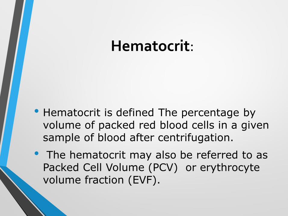
Hematocrit:
• Hematocrit is defined The percentage by volume of packed red blood cells in a given sample of blood after centrifugation.
• The hematocrit may also be referred to as Packed Cell Volume (PCV) or erythrocyte volume fraction (EVF).
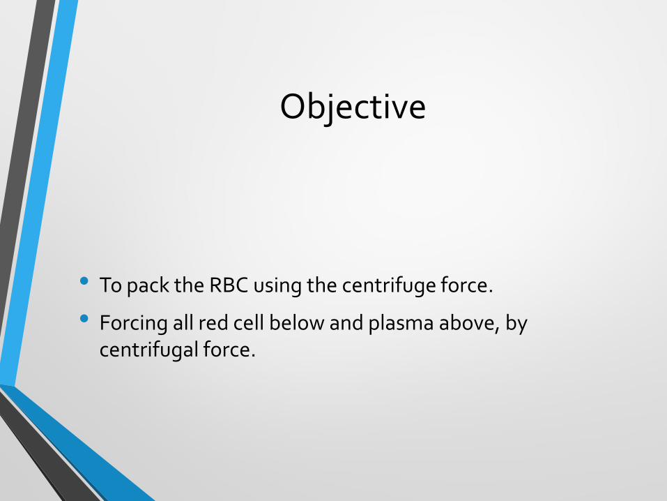
Objective
• To pack the RBC using the centrifuge force.
• Forcing all red cell below and plasma above, by centrifugal force.
Relevance
• Measurement of hematocrit (Hct) or packed cell volume (PCV) is the most accurate and simplest of all tests in clinical hematology for detecting the presence and degree of anemia or polycythemia. In comparison, hemoglobin estimation is less accurate, and RBC count far less accurate.
Methods
• Microhaematocrit
• Electronic cell counting
Material and instruments
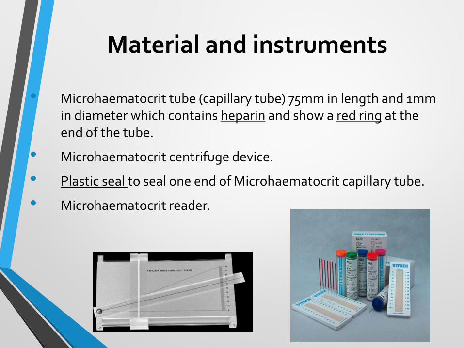
• Microhaematocrit tube (capillary tube) 75mm in length and 1mm in diameter which contains heparin and show a red ring at the end of the tube.
• Microhaematocrit centrifuge device.
• Plastic seal to seal one end of Microhaematocrit capillary tube.
• Microhaematocrit reader.
Procedure:
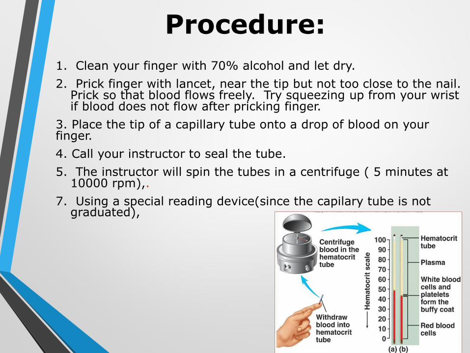
1. Clean your finger with 70% alcohol and let dry.
2. Prick finger with lancet, near the tip but not too close to the nail. Prick so that blood flows freely. Try squeezing up from your wrist if blood does not flow after pricking finger.
3. Place the tip of a capillary tube onto a drop of blood on your finger.
4. Call your instructor to seal the tube.
5. The instructor will spin the tubes in a centrifuge ( 5 minutes at 10000 rpm),.
7. Using a special reading device(since the capilary tube is not graduated),
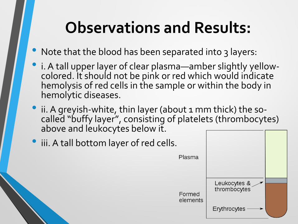
Observations and Results:
• Note that the blood has been separated into 3 layers:
• i. A tall upper layer of clear plasma—amber slightly yellow-colored. It should not be pink or red which would indicate hemolysis of red cells in the sample or within the body in hemolytic diseases.
• ii. A greyish-white, thin layer (about 1 mm thick) the so-called “buffy layer”, consisting of platelets (thrombocytes) above and leukocytes below it.
• iii. A tall bottom layer of red cells.
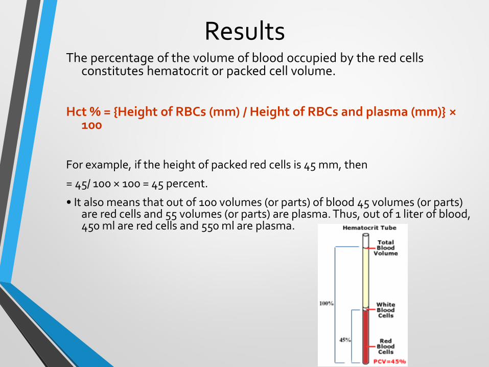
ResultsThe percentage of the volume of blood occupied by the red cells
constitutes hematocrit or packed cell volume.
Hct % = {Height of RBCs (mm) / Height of RBCs and plasma (mm)} ×100
For example, if the height of packed red cells is 45 mm, then
= 45/ 100 × 100 = 45 percent.
• It also means that out of 100 volumes (or parts) of blood 45 volumes (or parts) are red cells and 55 volumes (or parts) are plasma. Thus, out of 1 liter of blood, 450 ml are red cells and 550 ml are plasma.
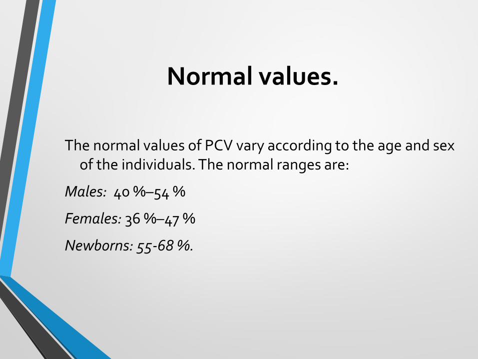
Normal values.
The normal values of PCV vary according to the age and sex of the individuals. The normal ranges are:
Males: 40 %–54 %
Females: 36 %–47 %
Newborns: 55-68 %.
Clinical implications:
• PCV is affected by the shape, & the number of the RBCs & the plasma volume.
• High PCV either indicates either increase in number of circulating RBCs or decrease in plasma volume seen in cholera due to loss of water in the stool
• A low PCV indicates either decrease in RBC or increase in plasma volume
Clinical implications:
A lower than normal hematocrit may indicate:
• An insufficient supply of healthy red blood cells (anemia)
• A large number of white blood cells — usually a very small portion of your blood — due to long-term illness, infection, leukemia, lymphoma or other disorders of white blood cells.
• Acute kidneydisease (lower Erythropoietin production lead to less RBCs production by the bone marrow).
• Pregnancy may lead to women having additional fluid in blood. This could potentially lead to a small drop in hematocrit levels
Clinical implications:
• A higher than normal hematocrit may indicate:
• Abnormal increase in red blood cells (erythrocytosis)
• A disorder, such as polycythemia vera that causes your body to produce too many red blood cells (in polycythemia it may rise to as high as 70 %).
• At higher altitudes, there is a lower oxygen supply in the air and thus hematocrit levels may increase over time.
• Low blood oxygen levels (hypoxia)
• Lung or heart disease — if the body senses low oxygen levels, it will make more red blood cells in an effort to increase the amount of oxygen in the blood
• Dehydration.
• Burn( due to loss of plasma)
Sources of error and comments
1. An increased amount of anti-coagulant decreases the Hct reading as a result of erythrocyte shrinking.
2. Improper sealing of the capillary tube causes a decreased Hct reading as a result of loss of blood during centrifugation. a higher number of erythrocytes are lost in relation to the plasma.
3. The microhematocrit centrifuge should never be forced to stop by applying pressure to the metal cover plate. This will cause the RBC layer to “sling” forward and results in a falsely elevated value.
Sources of error and comments
7. The buffy coat of the specimen should not be included in the Hct reading, because its inclusion falsely elevates the result.
8. A decrease or increase in the readings may be seen if the microhematocrit reader is not used properly.
9. If too much time elapses between when the centrifuge stops and the capillary tube is removed, the red cells can begin to settle out and cause a false reading of the hematocrit.

Thank you