Embed Size (px)
Citation preview
Vol:.(1234567890)
Annals of Nuclear Medicine (2018) 32:60–68https://doi.org/10.1007/s12149-017-1220-1
1 3
ORIGINAL ARTICLE
Diagnostic value of 99mTc-ethambutol scintigraphy in tuberculosis: compared to microbiological and histopathological tests
A. H. S. Kartamihardja1 · Y. Kurniawati2 · R. Gunawan3
Received: 16 October 2017 / Accepted: 20 November 2017 / Published online: 5 December 2017 © The Author(s) 2017. This article is an open access publication
AbstractObjective Tuberculosis (TB) still remains the world’s endemic infection. TB affects the lungs and any part of the body other than the lung. The diagnosis of TB has not changed much over the decades. Ethambutol is one of the first line treatments for TB. It can be labeled using 99mTc. 99mTc-ethambutol will be accumulated in the site of TB lesion and can be imaged using gamma camera. The aim of this study was to evaluate the diagnostic value of 99mTc-ethambutol scintigraphy in detecting and localizing of TB.Methods Retrospective cross-sectional study was done. Subjects were patients suspected of having TB infection. Whole body and SPECT-CT imaging at the suspected area was done 1 and 4 h after injection of 370–555 MBq 99mTc-ethambutol. 99mTc-ethambutol scintigraphy was analyzed visually. The results were compared with that of histopathological or micro-biological tests. Statistical analysis was done to determine the sensitivity, specificity, PPV, NPV and accuracy.Results One hundred and sixty-eight subjects were involved in this study. There were 110 men and 58 women with mean age of 34.52 ± 11.94 years. There were concordance results in 156 (92.86%) and discordant in 12 (7.14%) subjects between 99mTc-ethambutol scintigraphy and histopathological or microbiological result. The sensitivity, specificity, PPV, NPV and accuracy of 99mTc-ethambutol scintigraphy in the diagnosis of pulmonary TB were 93.9, 85.7, 93.9, 85.7 and 91.4%, respec-tively, for extra-pulmonary TB 95.5, 77.8, 97.9, 63.6, and 85.1%, respectively, and for total tuberculosis 94.9, 83.3, 96.3, 78.1 and 92.8%, respectively. There was no side effect observed in this study.Conclusion 99mTc-ethambutol scintigraphy is a useful diagnostic imaging technique to detect and localize intra- and extra-pulmonary TB. It is safe to be performed even in pediatric patient. Consuming ethambutol less than 2 weeks did not influ-ence the result.
Keywords Pulmonary · Extra-pulmonary tuberculosis · SPECT/CT imaging · 99mTc-ethambutol
Introduction
Tuberculosis (TB) is an infectious disease caused by the bacillus Mycobacterium tuberculosis. TB is not only typi-cally affecting the lungs known as pulmonary TB, but also affects any part of the body as single or multiple sites known
as extra-pulmonary TB [1–3]. Tuberculous bacillus causes a focal infection in the site where it is deposited after inha-lation [4]. TB remained one of the top 10 causes of death worldwide. It still remains the world’s endemic infection although between 2000 and 2015 the number of deaths fell by 22%. The TB epidemic is larger than previously esti-mated. There were an estimated 10.4 million incident TB cases worldwide in 2015, while the rate of decline incidence from 2014 to 2015 remained at only 1.5% although the treat-ment averted a million deaths. This condition could be due to persisting gaps between treatment and diagnostic modali-ties. According to WHO report, one-third of pulmonary TB was undiagnosed or delayed diagnosed caused continued transmission in communities [1].
The diagnosis of TB remained unchanged for many dec-ades and it probably would have no progress. The ultimate
* A. H. S. Kartamihardja [email protected]
1 Department of Nuclear Medicine and Molecular Medicine, Dr. Hasan Sadikin General Hospital, Faculty of Medicine Universitas Padjadjaran, Bandung, Indonesia
2 Faculty of Medicine, Universitas Andalas Padang, Padang, Indonesia
3 National Nuclear Energy Agency of Indonesian, Jakarta, Indonesia
61Annals of Nuclear Medicine (2018) 32:60–68
1 3
diagnosis of TB depends on the recognition of Mycobacte-rium tuberculosis on histological examination and/or bacte-riological culture. Several diagnostic tests for TB have been developed including sputum smear microscopy more than 100 years ago [1]. Unfortunately, the most of undiagnosed pulmonary TB was cases with negative smear sputum micro-scopic. A systemic review showed that the sensitivity of this test is only 31–69% [5, 6]. Molecular diagnostic tools have been developed, but it cannot be routinely used for diagnosis of pulmonary TB [5, 7–9]. Currently, the Xpert® MTB/RIF assay is recommended by WHO for the diagnosis of TB. The test has much better accuracy than microscopy and culture methods. The limitation of this test is requiring more devel-oped laboratory capacity and sometimes difficult to collect adequate specimen. Other limitation is it takes 2 weeks to provide results [1, 10]. The diagnosis of extra-pulmonary TB is more difficult compared with that of pulmonary TB. Extra-pulmonary TB involves relatively inaccessible sites for bacteriological confirmation as well [11, 12]. In these situations, rapid non-invasive imaging modality is necessary to detect and localize the site of TB.
Nuclear medicine technique is a non-invasive diagnos-tic modality which is highly sensitive and specific to detect and localize the lesion at early stage and less time consum-ing. A wide variety of radiopharmaceuticals have been used for infection/inflammation imaging. 67Ga-citrate is a high-sensitive agent for infection/inflammation imaging, but non-specific, since malignancy diseases will provide similar result [13, 14]. 99mTc-albumin, 99mTc-nanocolloid, 99mTc-tetrofosmin, FDG-PET, and 99mTc-MIBI are other sensitive and non-specific radiopharmaceutical agents used for diag-nosis of infections including TB [15–18]. 111In-oxine-WBC, 99mTc-HMPAO-WBC, 99mTc-HIg and peptide labeled are specific process agents for infection/inflammation imag-ing. However, these radiopharmaceuticals are basically not specific to separate bacterial infection from sterile inflam-mation. 99mTc-ciprofloxacin can be a more specific radiop-harmaceutical for bacterial infection imaging, because it is taken up by living bacteria and it inactivates DNA gyrase [5, 19–21]. Unfortunately 99mTc-ciprofloxacin cannot differen-tiate TB from other bacterial infection, since ciprofloxacin acts as a broad-spectrum antibiotic that can be taken up by any living bacteria [19–24]. 99mTc-INH has been developed to image TB, but clinically is not widely use [25].
Ethambutol is an active specific antibiotic against myco-bacterium. It inhibits mycolic acid in bacterial cell mem-brane [11, 26–29]. Verma et al. showed that 99mTc-labeled ethambutol is specifically taken up by Mycobacterium tuberculosis, and can be image using gamma camera [30]. In vivo studies showed that the whole body bio-distribution of 99mTc-ethambutol was consistent with the pharmacoki-netic characteristics of ethambutol as anti-tuberculosis drug [30–35]. 99mTc-ethambutol remains in tubercular lesion as
it is bound to mycolic acid in the cell wall of bacteria, but will be cleared out from non-tubercular lesions [34, 36]. 99mTc-ethambutol was not seen in dormant TB cases. The advantages of 99mTc-ethambutol scintigraphy are it is a non-invasive procedure and provides result faster than the cyto-logical test with minimal or no side effects [35–37].
The aim of this study was to evaluate the diagnostic value of 99mTc-ethambutol scintigraphy in detecting and localizing of TB infection.
Materials and methods
Retrospective cross-sectional study was done in the Depart-ment of Nuclear Medicine and Molecular Imaging, Dr. Hasan Sadikin General Hospital/Faculty of Medicine, Uni-versitas Padjadjaran Bandung, Indonesia. Secondary data were collected from medical record. Subjects were patients who were referred for 99mTc-ethambutol scintigraphy to confirm or exclude TB infection from 2009 to 2015. The duration between the start of ethambutol drug and the day of 99mTc-ethambutol scintigraphy test was recorded. Side effect from injection of radiopharmaceuticals was observed. Sub-jects without histopathological or microbiological data and under TB treatment for more than 2 weeks were excluded from this study. This study was approved by Health Research Ethic Committee Faculty of Medicine Universitas Padjadja-ran no. 592/UN6.C1.3.2/KEPK/PN/2015.
Ethambutol cold kit was developed by Center for Radio-isotope and Radiopharmaceuticals Technology, National Nuclear Energy Agency of Indonesian. Whole body and SPECT/CT imaging in the suspected areas were done at 1 and 4 h following 370–740 MBq intravenous injection of 99mTc-ethambutol. SPECT/CT was done in the suspected area of TB based on whole body imaging. Twenty-four hours imaging was done if necessary. The results of 99mTc-etham-butol scintigraphy were compared with that of histopatho-logical or microbiological test.
99mTc‑ethambutol labeling of procedure
Ethambutol cold kit comes in 2 vials. One vial (A) contains 3.5 mg ethambutol and 5 mg mannitol. The other vial (B) contains 400 µg SnCl2.2H2O and 2 mg sodium pyrophos-phate. One mL aquades was added to vial A and mixed well. All the solution was taken from vial A and put into vial B using sterilized fine syringe, and mixed well. 370–740 MBq freshly eluted 99mTc was added to vial B. The mixture in vial B was swirled and allowed to react for 5–10 min at room temperature. Instant thin-layer chromatography (ITLC) was performed for quality control using acetone as a solvent to determine radiochemical purity of 99mTc-ethambutol. The radiochemical purity should be more than 90% to be used in
62 Annals of Nuclear Medicine (2018) 32:60–68
1 3
this study. This based on the formulation for the pre-deter-mined radiochemical purity that 88–94% radiochemical purity of 99mTc-ethambutol can be used [38].
Imaging protocol
There is no specific preparation for ethambutol scan. Ante-rior and posterior whole body and SPECT/CT images were done using a standard dual-head Gamma Camera SPECT/CT (GE-Infinia®) with Low-Energy High-Resolution (LEHR) Collimator at 1, 4 and if necessary 24 h follow-ing intravenous injection of 99mTc-ethambutol. Injection dose of 99mTc-ethambutol was 370–740 MBq. SPECT/CT images were done covering lungs and other suspected area to improve the sensitivity in detection of lesions. The subjects were observed for any signs and symptoms from side effect of radiopharmaceutical injection. Common side effects include problems with vision, headaches, nausea, joint pain, tiredness and allergic reaction.
Interpretation
The interpretation of image was based on the quality of tracer uptake. Images were analyzed visually for qualitative analysis. Normal distribution of 99mTc-ethambutol is seen as high uptake in kidney, urinary bladder, liver and spleen. There is no tracer uptake shown on bone, bone marrow,, epiphysis, stomach and thyroid, soft tissue and lung as well. Significant urinary activity was seen in 1 h, since the kidney is the main excretory organ [34]. Any increased pathological uptake of 99mTc-ethambutol seen in the area out of normal distribution compared with that of the opposite area was considered as positive results. The pathological uptake is increasing gradually at 4- or 24-h images compared with that of normal uptake. Negative result was considered if normal radioactivity distribution was seen without any pathological tracer uptake, or increased tracer uptake at 1 h, but decreas-ing at 4 h image [30, 32].
Statistic analysis
Statistic analysis was done to determine the sensitivity, specificity, negative and positive predictive value as well as accuracy by comparing the result of 99mTc-ethambutol scintigraphy with that of histopathological or microbiologi-cal using a 2 × 2 table.
Results
One hundred and sixty-eight subjects out of 221 fulfilled the inclusion and exclusion criteria. They were included in this study. There were 110 (65.5%) males and 58 (34.5%) females
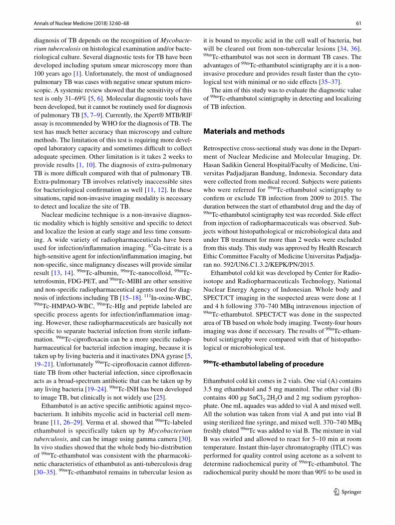
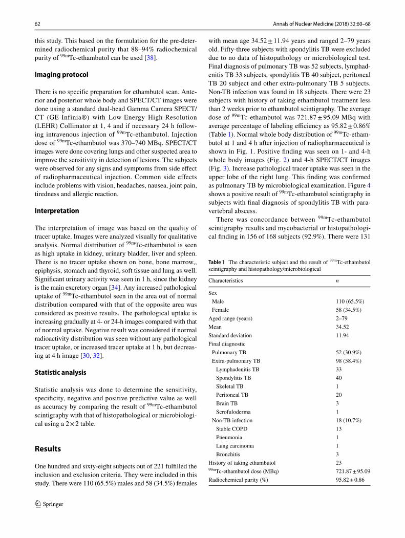
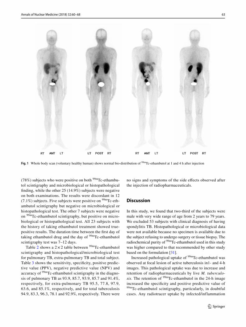
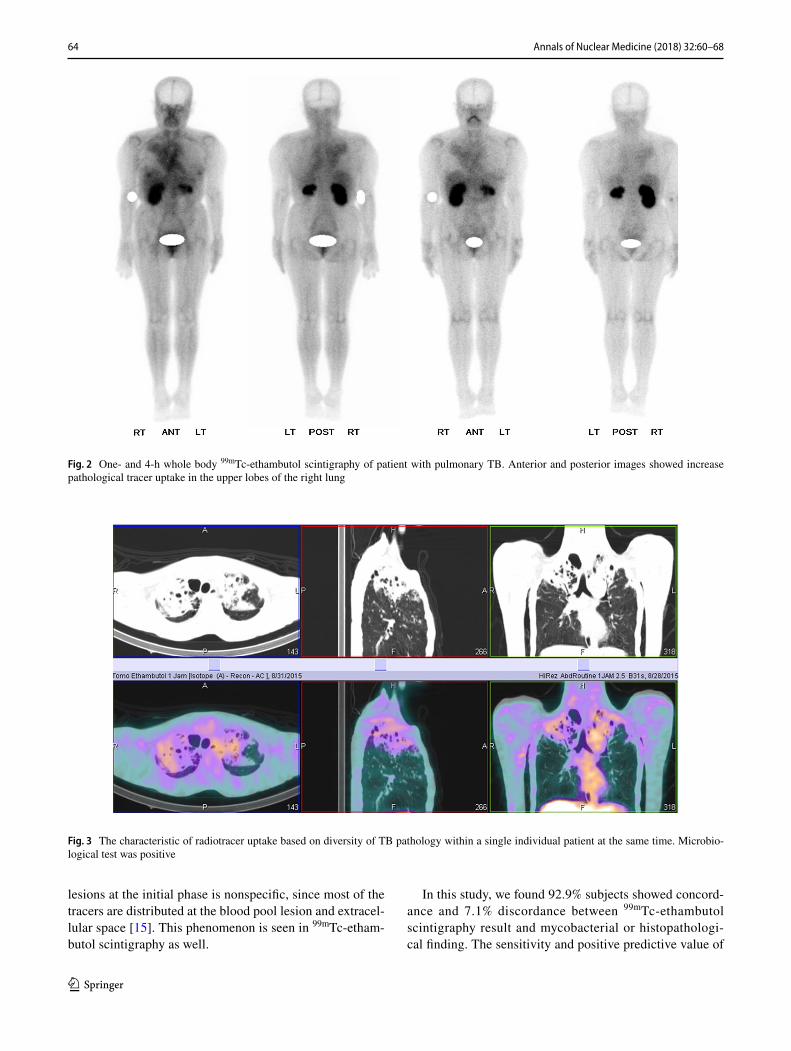
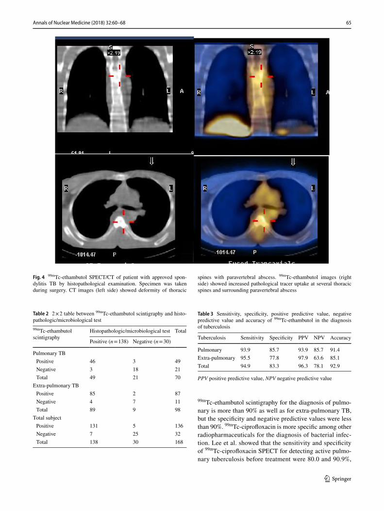
with mean age 34.52 ± 11.94 years and ranged 2–79 years old. Fifty-three subjects with spondylitis TB were excluded due to no data of histopathology or microbiological test. Final diagnosis of pulmonary TB was 52 subjects, lymphad-enitis TB 33 subjects, spondylitis TB 40 subject, peritoneal TB 20 subject and other extra-pulmonary TB 5 subjects. Non-TB infection was found in 18 subjects. There were 23 subjects with history of taking ethambutol treatment less than 2 weeks prior to ethambutol scintigraphy. The average dose of 99mTc-ethambutol was 721.87 ± 95.09 MBq with average percentage of labeling efficiency as 95.82 ± 0.86% (Table 1). Normal whole body distribution of 99mTc-etham-butol at 1 and 4 h after injection of radiopharmaceutical is shown in Fig. 1. Positive finding was seen on 1- and 4-h whole body images (Fig. 2) and 4-h SPECT/CT images (Fig. 3). Increase pathological tracer uptake was seen in the upper lobe of the right lung. This finding was confirmed as pulmonary TB by microbiological examination. Figure 4 shows a positive result of 99mTc-ethambutol scintigraphy in subjects with final diagnosis of spondylitis TB with para-vertebral abscess.
There was concordance between 99mTc-ethambutol scintigraphy results and mycobacterial or histopathologi-cal finding in 156 of 168 subjects (92.9%). There were 131
Table 1 The characteristic subject and the result of 99mTc-ethambutol scintigraphy and histopathology/microbiological
Characteristics n
Sex Male 110 (65.5%) Female 58 (34.5%)
Aged range (years) 2–79Mean 34.52Standard deviation 11.94Final diagnostic Pulmonary TB 52 (30.9%) Extra-pulmonary TB 98 (58.4%) Lymphadenitis TB 33 Spondylitis TB 40 Skeletal TB 1 Peritoneal TB 20 Brain TB 3 Scrofuloderma 1
Non-TB infection 18 (10.7%) Stable COPD 13 Pneumonia 1 Lung carcinoma 1 Bronchitis 3
History of taking ethambutol 2399mTc-ethambutol dose (MBq) 721.87 ± 95.09Radiochemical purity (%) 95.82 ± 0.86
63Annals of Nuclear Medicine (2018) 32:60–68
1 3
(78%) subjects who were positive on both 99mTc-ethambu-tol scintigraphy and microbiological or histopathological finding, while the other 25 (14.9%) subjects were negative on both examinations. The results were discordant in 12 (7.1%) subjects. Five subjects were positive on 99mTc-eth-ambutol scintigraphy but negative on microbiological or histopathological test. The other 7 subjects were negative on 99mTc-ethambutol scintigraphy, but positive on micro-biological or histopathological test. All 23 subjects with the history of taking ethambutol treatment showed true-positive results. The duration time between the first day of taking ethambutol drug and the day of 99mTc-ethambutol scintigraphy test was 7–12 days.
Table 2 shows a 2 × 2 table between 99mTc-ethambutol scintigraphy and histopathological/microbiological test for pulmonary TB, extra-pulmonary TB and total subject. Table 3 shows the sensitivity, specificity, positive predic-tive value (PPV), negative predictive value (NPV) and accuracy of 99mTc-ethambutol scintigraphy in the diagno-sis of pulmonary TB as 93.9, 85.7, 93.9, 85.7 and 91.4%, respectively, for extra-pulmonary TB 95.5, 77.8, 97.9, 63.6, and 85.1%, respectively, and for total tuberculosis 94.9, 83.3, 96.3, 78.1 and 92.9%, respectively. There were
no signs and symptoms of the side effects observed after the injection of radiopharmaceuticals.
Discussion
In this study, we found that two-third of the subjects were male with very wide range of age from 2 years to 79 years. We excluded 53 subjects with clinical diagnosis of having spondylitis TB. Histopathological or microbiological data were not available because no specimen is available due to the subject refusing to undergo surgery or tissue biopsy. The radiochemical purity of 99mTc-ethambutol used in this study was higher compared to that recommended by other study based on the formulation [31].
Increased pathological uptake of 99mTc-ethambutol was observed at focal lesion of active tuberculosis in1- and 4-h images. This pathological uptake was due to increase and retention of radiopharmaceuticals by live M. tuberculo-sis. The retention of 99mTc-ethambutol in the 24-h image increased the specificity and positive predictive value of 99mTc-ethambutol scintigraphy, particularly, in doubtful cases. Any radiotracer uptake by infected/inflammation
Fig. 1 Whole body scan (voluntary healthy human) shows normal bio-distribution of 99mTc-ethambutol at 1 and 4 h after injection
64 Annals of Nuclear Medicine (2018) 32:60–68
1 3
lesions at the initial phase is nonspecific, since most of the tracers are distributed at the blood pool lesion and extracel-lular space [15]. This phenomenon is seen in 99mTc-etham-butol scintigraphy as well.
In this study, we found 92.9% subjects showed concord-ance and 7.1% discordance between 99mTc-ethambutol scintigraphy result and mycobacterial or histopathologi-cal finding. The sensitivity and positive predictive value of
Fig. 2 One- and 4-h whole body 99mTc-ethambutol scintigraphy of patient with pulmonary TB. Anterior and posterior images showed increase pathological tracer uptake in the upper lobes of the right lung
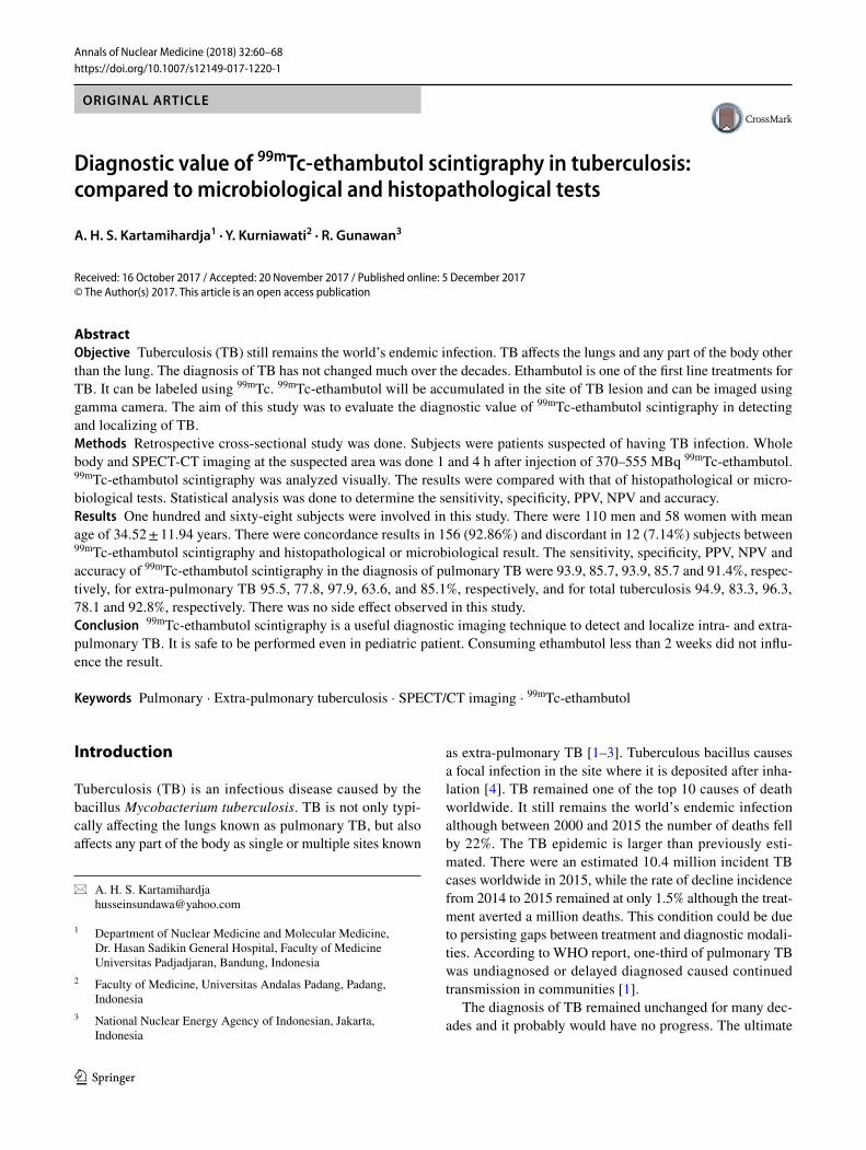
Fig. 3 The characteristic of radiotracer uptake based on diversity of TB pathology within a single individual patient at the same time. Microbio-logical test was positive
65Annals of Nuclear Medicine (2018) 32:60–68
1 3
99mTc-ethambutol scintigraphy for the diagnosis of pulmo-nary is more than 90% as well as for extra-pulmonary TB, but the specificity and negative predictive values were less than 90%. 99mTc-ciprofloxacin is more specific among other radiopharmaceuticals for the diagnosis of bacterial infec-tion. Lee et al. showed that the sensitivity and specificity of 99mTc-ciprofloxacin SPECT for detecting active pulmo-nary tuberculosis before treatment were 80.0 and 90.9%,
Fig. 4 99mTc-ethambutol SPECT/CT of patient with approved spon-dylitis TB by histopathological examination. Specimen was taken during surgery. CT images (left side) showed deformity of thoracic
spines with paravertebral abscess. 99mTc-ethambutol images (right side) showed increased pathological tracer uptake at several thoracic spines and surrounding paravertebral abscess
Table 2 2 × 2 table between 99mTc-ethambutol scintigraphy and histo-pathologic/microbiological test99mTc-ethambutol scintigraphy
Histopathologic/microbiological test Total
Positive (n = 138) Negative (n = 30)
Pulmonary TB Positive 46 3 49 Negative 3 18 21 Total 49 21 70
Extra-pulmonary TB Positive 85 2 87 Negative 4 7 11 Total 89 9 98
Total subject Positive 131 5 136 Negative 7 25 32 Total 138 30 168
Table 3 Sensitivity, specificity, positive predictive value, negative predictive value and accuracy of 99mTc-ethambutol in the diagnosis of tuberculosis
PPV positive predictive value, NPV negative predictive value
Tuberculosis Sensitivity Specificity PPV NPV Accuracy
Pulmonary 93.9 85.7 93.9 85.7 91.4Extra-pulmonary 95.5 77.8 97.9 63.6 85.1Total 94.9 83.3 96.3 78.1 92.9
66 Annals of Nuclear Medicine (2018) 32:60–68
1 3
respectively. The positive predictive value was 88.9% and the negative predictive value was 83.3% [5]. We found that the sensitivity, negative and positive predictive values of 99mTc-ethambutol scintigraphy were higher compared to that in the study by Lee et al., but in the contrary, the specificity was slightly lower. Theoretically, the specificity of 99mTc-ethambutol should be higher compared with that of 99mTc-ciprofloxacin, since 99mTc-ethambutol is more specific for Mycobacterium tuberculosis. Low negative predictive value in the diagnosis of TB could be due to a limited number of subjects with non-TB. Negative TB was found in only 30 subjects, while positive TB was found in 138 subjects. Less number of subjects with negative TB were found in extra-pulmonary TB compared with that for pulmonary TB. It is recommended to do another study with comparable number of subjects between positive and negative TB.
Tuberculosis lesions in human are very complex with a wide range of pathological features. This variety of lesions could be observed within a single individual patient simul-taneously [39–41]. Due to their characteristic; these lesions will provide either false-positive or false-negative results on 99mTc-ethambutol scintigraphy. False-positive result could be caused by: (1) hypervascularization of non-specific infec-tion or inflammation [30], (2) difficulties to get the adequate specimen [41], (3) and at least 104 number of acid-fast bacilli/mL of specimen culture are required to provide posi-tive result [42, 43]. In this study, we found 5 false-positive subjects: 3 subjects from the group of pulmonary TB and the other 2 subjects from the group of extra-pulmonary TB showed false-positive result. Hypervascularization could be found in TB granuloma. High uptake of radioactivity seen at solid lesions or nodules in lung parenchyma suggested granuloma cellular uptakes just around the lesions of largely filled cavities. Granuloma contains predominantly intracellu-lar M. tuberculosis with good vascularization which showed high uptake of 99mTc-ethambutol. Negative smear sputum microscopic test in those subjects could be because granu-loma did not contact with airway lung structure [39, 40, 44]. Subject with false positive on 99mTc-ethambutol scin-tigraphy, but abnormal clinical feature and chest X-ray were considered as suggestive for active pulmonary tuberculosis. Those subjects were treated with anti-tuberculosis drugs. On the follow-up, they showed better improve clinical features. This false-positive case can be considered as true positive based on good response following anti-tuberculosis treat-ment, although smear sputum microscopic test was negative. Performing serial late images could minimize false-positive result of 99mTc-ethambutol scintigraphy due to hypervascu-larization. Twenty-hour images following injection of 99mTc-ethambutol could be done if necessary.
In this study, we found false-negative results in 7 subjects, 3 subjects belong to pulmonary TB group and 4 subjects belong to extra-pulmonary TB group. They showed negative
result on 99mTc-ethambutol scintigraphy, but positive on microbiological or histopathological test. False-negative result could be found in necrotic caseous lesions that spread and destroy vasculature which lead to lack of radiopharma-ceutical supply from blood [16]. These necrotic lesions can remain solid with very few bacilli. Necrotic caseous lesions can be seen on X-ray without radiotracer uptake that caused false negative results on 99mTc-ethambutol scintigraphy with positive smear sputum microscopic test [39, 40].
During the period of 5 years performing 99mTc-etham-butol scintigraphy, we found 13 negative 99mTc-ethambutol scintigraphy results in patients suspected of having TB. All of them were under intensive phase treatment using etham-butol. Since the characteristic of 99mTc-ethambutol is similar to ethambutol used for treatment, the question arises whether negative result is true negative or false negative due to com-petition between 99mTc-ethambutol as radiopharmaceutical and ethambutol as anti-tuberculosis drug. In our study, 23 subjects with positive pulmonary TB based on smear spu-tum microscopic test were positive on 99mTc-ethambutol scintigraphy. All of these subjects showed positive result on the second 99mTc-ethambutol scintigraphy performed after taking ethambutol drug for 7–12 days. This finding showed that taking drug during intensive treatment less than 2 weeks would not affect the result of 99mTc-ethambutol scintigraphy.
In this study we did not find any sign and symptom related to injection of 99mTc-ethambutol. Problem in vision due to optic neuropathy or cardio-hepatotoxicity occurs very rarely after several months of therapeutics. Ethambutol is freely given to pediatric patients. Adverse effects of ethambutol are rare and dose dependent. The diagnostic dose administered is only less than 3.5 mg. In clinical context, it was consid-ered as a safe radiopharmaceutical, even in children [31].
Conclusion
This study showed that 99mTc-ethambutol scintigraphy is a useful diagnostic imaging technique to detect and localize both intra- and extra-pulmonary tuberculosis. 99mTc-etham-butol scintigraphy is safe to be performed even in pediatric patient. Consuming ethambutol less than 2 weeks does not influence the result of 99mTc-ethambutol scintigraphy.
Open Access This article is distributed under the terms of the Creative Commons Attribution 4.0 International License (http://creativecom-mons.org/licenses/by/4.0/), which permits unrestricted use, distribu-tion, and reproduction in any medium, provided you give appropriate credit to the original author(s) and the source, provide a link to the Creative Commons license, and indicate if changes were made.
67Annals of Nuclear Medicine (2018) 32:60–68
1 3
References
1. The World Health Organization (WHO). The Global Tuberculosis (TB) Report 2016. Available on (https://communitymedicine4as-ses.wordpress.com/2016/10/13/who-releases-global-tuberculosis-report-2016-13-october-2016/). 26 Aug 2017.
2. Fanning A. Tuberculosis extra-pulmonary disease. CMAJ 1999; 160: 1597–603.
3. Iseman MD. Tuberculosis in relation to human immunodeficiency virus and acquired immunodeficiency syndrome. In: Iseman MD, editor. A clinician’s guide to tuberculosis. Philadelphia: Lippincott Williams and Wilkins; 2000. pp. 199–252.
4. Dutt AK, Stead WW. Epidemiology. In: Schlossberg D ed. Tuber-culosis and non-tuberculous mycobacterial infection. Philadel-phia: W.B. Saunders Company; 1999. pp. 3–16.
5. Lee M, Yoon M, Hwang KH, Choe W. Tc-99m ciprofloxacin SPECT of pulmonary tuberculosis. Nucl Med Mol Imaging. 2010;44(2):116–22.
6. World Health Organization. Expert Group Meeting Report. Using the Xpert MTB/RIF assay to detect pulmonary and extra-pulmo-nary tuberculosis and rifampicin resistance in adults and children. World Health Organization, Geneva 2013.
7. World Health Organization. Systemic screening for active tubercu-losis, principles and recommendation. World Health Organization, Geneva 2013.
8. World Health Organization. Improving the diagnosis and treat-ment of smear negative pulmonary and extra-pulmonary tubercu-losis among adults and adolescent. Stop TB Department of HIV/AIDS. World Health Organization, Geneva 2007.
9. World Health Organization. Global tuberculosis control. World Health Organization Report, Geneva 2009.
10. Daniel TM, De Banne SM. The serodiagnosis of tuberculosis and other mycobacterial diseases by enzyme-linked immunosorbent assay. Am Rev Respir Dis. 1987;135:1137–51.
11. Bhan S, Nag V. Skeletal tuberculosis. In: Sharma SK, Mohan A, edit Tuberculosis. New Delhi: Jaypee Brothers Medical Publisers; 2001:pp. 237 – 60.
12. Sharrard WInfections of bones and joints. In Paediatric Ortho-paedics and Fractures, edited by WJW Sharrard. 3rd Ed,. Oxford: Blackwell Scientific, 1993: pp. 1247–84.
13. Seabold JE, Palestreo CJ, Brown ML, et al. Procedure guide-line for gallium scintigraphy in inflammation. J Nucl Med. 1997;38(6):994–7.
14. Ergun P, Turay UY, Ortapamuk H, Biber C, et al. The role of gal-lium-67 scintigraphy and high resolution computed tomography as predictors of disease activity in sputum smear negative pulmonary tuberculosis. Turk Respir J. 2003;4(3):123–6.
15. Degirmenci B, Kilinc O, Cirak KA, et al. 99mTc-tetrofos-min scintigraphy in pulmonary tuberculosis. J Nucl Med. 1998;39(12):2116–20.
16. Raziel G, Masjedi MR, Fotouhi F, et al. The role of Tc-99m MIBI scintigraphy in the management of patients with pulmonary tuber-culosis. Eur Rev Med Pharmacol Sci 2012; 16: 622–62.
17. Sathekge M, Maes A, Kgomo M, Stoltz A, Van de Wiele C. Use of 18F-FDG PET to predict response to first-line tuberculostatics in HIV-associated tuberculosis. J Nucl Med. 2011;52:880–5.
18. Bakheet SMB, Powe J, Kandil A, Ezzat A, Rostom A, Amartey J. F-18 FDG uptake in breast infection and inflammation. Clin Nucl Med. 2000;25(2):100–3.
19. Mora Ríos FG, Isunza Ramírez A, López Marmolejo A, Palma Rosillo RM, Guízar Cuevas S, et al. Sensitivity and specificity of the Tc-99m ciprofloxacin scan in pediatric osteomyelitis. Acta Ortop Mex. 2010;24(4):248 – 51.
20. Britton KE, Wareham DW, Das SS, Solanki KK, Amaral H, Bhatnagar A, Kartamihardja AHS, et al. Imaging bacterial
infection with Tc-99m–ciprofloxacin (infecton). J Clin Pathol. 2002;55:817–23.
21. Kartamihardja AHS. Pencitraan 99mTc-cyprofloxacine dibanding-kan dengan 99mTc-HMPAO-WBC pada Penderita Infeksi Tulang dan Sendi. MKB 2006; 38 (2): 59–65.
22. Hidayat B, Anwar IB, Kartamihardja AHS, Masjhur JS. Peranan pencitraan 99mTc-infecton dalam mendeteksi spondilitis tuberku-losa. MKB 2000; 32 (2): 123–9.
23. Hall AV, Solanki KK, Vijanmuri S, et al. Evaluation of efficacy of 99mTc-infecton, a novel agent for detecting site of infection. J Clin Pathol. 1998;51:215–9.
24. Kartini N, Nurlaila Z. Microbiological characterization of 99mTc-cyprofloxacin and 99mTc-ethambutol labeled compounds as the infection imaging radiopharmaceuticals. Regional seminar on pharmaceuticals and biomedical analysis, School of Pharmacy. Institut Teknologi Bandung, 2005.
25. Namrata S, Bathnagar A. Clinical evaluation of Tc-99m 2IT-INH in normal subjects and patients with tubercular lesions. Afr J Pharm Pharmacol. 2009;3(4):110–9.
26. Hugh G, Watts, Lifeso RM. Current concepts review-tuberculosis of bones and joints. J Bone Jt Surg Am. 1996; 78: 288–99.
27. World Health Organization. Treatment of tuberculosis. Guideline for National Programmers. 3rd ed. World Health Organization Report, Geneva: 2003.
28. Forbes M, Peets EA, Kuck NA. Effect of ethambutol on Mycobac-terium. Ann NY Acad Sci. 1966;135:726–31.
29. Takayama K, Amstrong EL, Kunugi KA, Kilburn JQ. Inhibi-tion by ethambutol of mycolic acid transfer into the cell wall of Mycobacterium smegmatis. Antimicrob Agents Chemother. 1979;16:240–2.
30. Verma J, Bathnagar A, Sen S, Singh AK, Bose M. Radio-labeling ethambutol with technetium-99m and its evaluation for detection tuberculosis. World J Nucl Med. 2005;4(1):35–46.
31. Singh N, Bhatnagar A. Clinical evaluation of efficacy of TC-ethambutol in tubercular lesion imaging. Tuberc Res Treat 2010. doi:https://doi.org/10.1155/2010/618051.
32. Oekar NK, Kartamihardja AHS. Pencitraan dengan Radionuklida 99mTc-ethambutol untuk diagnosis tuberkulosis extra pulmonal (penelitian pada hewan percobaan). MKB 2006.; 38 (3): 116–21.
33. Forbes M, Kuck NA, Peets A. Mode of action of ethambutol. J Bacteriol. 1962;84(5):1099–103.
34. Oekar NK. Kit Diagnostik Berbasis Teknik Nuklir dalam Penata-laksanaan Tuberkulosis. Maj Kedokt Indon. 2008;58(10):388–94.
35. Wangsaatmadja H, dan Kartini N. Profil Farmakokinetika Radi-ofarmaka Etambutol Bertanda Teknesium-99m sebagai sediaan sidik tuberkulosis. J Farm Indones. 2011;5(4):188–94.
36. Kartamihardja AHS. 99m Tc-ethambutol imaging in detecting and localizing of tuberculosis. 9th World Congress of Nuclear Medi-cine and Biology. Seoul, 2006.
37. Namrata S, Bhatnagar A. Clinical evaluation of 99mTc-ethambutol in tubercular lesion imaging. Tuberc Res Treat 2010. Download from http://www.researchgate.net/publication.
38. Juwita R, Sumpena Y, Eka M, Sriyani, Kartini K. Biological evaluation of 99mTc-ethambutol for early detection of tuberculosis infection in animal model. Maj Farm Indones. 2009;20(2):55–61.
39. Dartois V. The Path of anti-tuberculosis drugs: from blood to lesions to mycobacterial cells. Nat Rev Microbiol. 2014;12:159–67.
40. Lenaerts A, Barry CE, Dartos V. Heterogenity in tuberculosis pathology, microenvironments and therapeutic responses. Immu-nol Rev. 2015;264(1):200–308.
41. de Souza NMV, de Lima Ferreira M, Pinheiro AC, Saraiva MF, de Almeida MV and Valle MS. Synthesis and biological aspects of mycolic acid: an important target againts Mycobacterium tuber-culosis. Sci World J. 2008;8:720–51.
68 Annals of Nuclear Medicine (2018) 32:60–68
1 3
42. Glassroth J. Diagnosis of tuberculosis. Tuberculosis. A compre-hensive international approach, Reichman LB, Hershfield ES, editors. New York, Marcel Dekker, 1993: pp. 149–65.
43. Rooney JJ, Crocco JA, Kramer S, Lyons HA. Further observa-tions on tuberculin reactions in active tuberculosis. Am J Med. 1976;60:517–22.
44. de Anthony H. Management of an HIV-positive patients with smear negative pulmonary tuberculosis. Tuberc Compr Clin Ref 2009;98.


![Thyroid pathophysiology scintigraphy[1]](https://img.pdfslide.net/doc/110x75/588a7dc81a28abad628b4ebd/thyroid-pathophysiology-scintigraphy1.jpg)