Embed Size (px)
Citation preview

1
Digestive System
Function of the Digestive Tract The digestive system (1) processes food, (2) extracts nutrients from food, and (3) eliminates
undigested material. Food spends about 1-2 hrs in the stomach, 2-4 hours in the small intestine. Organs of Digestive Tract (tract – series of interconnected tube-like organs) 1. Digestive Tract – tube that extends from the mouth to the anus (about 30’ in length). a. It is also known as the alimentary canal or GI tract (stomach and intestine). b. The digestive tract consists of the oral cavity (mouth), pharynx (throat), esophagus, stomach,
small intestine, and the large intestine. 2. Accessory organs (aid digestion): teeth, tongue, salivary glands, liver, gallbladder, and pancreas. The
accessory organs provide chewing, buffers, and enzymes that aid in the mechanical and chemical breakdown of food.

2
Four Stages of Food Processing 1. Ingestion – eating food 2. Digestion a. enzymatic breakdown of food in the lumen of the GI tract from large organic molecules (complex
carbohydrates, protein, fats) to small molecules that can be absorbed across plasma membranes and used by cells of the body.
b. Some nutrients in the diet are already in usable form and are absorbed without being digested (e.g., vitamins, water, minerals, glucose, amino acids)
c. digestion in the lumen of the stomach and intestine is extracellular since it doesn’t occur in cells 3. Absorption – uptake of nutrients into the epithelial cells that line the digestive tract, then through
them and out the other side where they are taken up by the blood or lymph 4. Elimination – disposal of undigested material as the feces Importance of Food to Body Cells 1. ATP synthesis by cellular respiration a. food molecules like glucose, fatty acids, and amino acids are used by cells to make ATP b. 60-70% of organic food molecules are used to generate ATP to support the basal metabolism of
cells to include 1. maintenance of muscle tone 2. generate body heat 3. maintain ATP-dependent processes such as heart function, urine production, and neuronal
activity within the brain 2. organic nutrients are used to build proteins, carbohydrates, lipids, and nucleic acids that cells need
for growth, maintenance, and repair 3. body cells need water, vitamins, and minerals Diet 1. organic component consists mostly of large polymers (starch, glycogen, protein) that cannot be
absorbed so they are first broken down to smaller molecules called monomers by water-splitting hydrolases
Organic Compounds Digestion Monomers (absorbable)
carbohydrate Starch and glycogen Sucrose (table sugar) Maltose
Glucose Fructose (corn syrup sweetener)
protein Protein Amino acids
lipid Triglycerides or fat molecules 2 Fatty acids and monoglyceride
Nucleic Acids DNA, RNA Nucleotides
Human diet in U.S. roughly 50-60% carbohydrate, 10-15% protein, and 20-30% fat 2. Water, Minerals, and Vitamins: Diet also includes water, minerals (inorganic elements such as
calcium, sodium, phosphorus, potassium, iron, sulfur, and magnesium), and vitamins (small organic compounds that are necessary for cell metabolism; most vitamins cannot be synthesized by the cells of the body and must be provided in the diet or made by intestinal bacteria).

3
Components of the Digestive Tract Mouth (oral cavity) – the mouth is also known as the oral cavity; the opening into the mouth is called
the oral orifice
Salivary Glands (exocrine gland) 1. secrete about 1 to 1.5 liters of saliva per day in humans into ducts that open into the mouth a. cattle produce upwards of 140 liters of saliva per day (about 37 gallons) b. sheep produce 10 L per day 2. Functions of saliva a. moisten and lubricate food 1. food sticks together into a ball called a bolus that can be swallowed 2. mucus in saliva makes bolus slippery (lubricant) b. digestion 1. salivary amylase is a hydrolytic enzyme in saliva that breaks down starch to mostly maltose
(disaccharide made up of 2 glucose molecules) a. salivary amylase is active in the mouth and within the upper portion of the stomach for
another 15 or so minutes before it is denatured by stomach acidity 1. amylase stays active in the internal areas of the bolus that is free of acid for a while 2. the bolus is eventually broken apart as it mixes with the gastric juice to form chyme b. maltose is a disaccharide consisting of 2 glucose molecules bonded together c. this helps to reduce the carbohydrates that would otherwise coat teeth 2. lingual lipase a. breaks down fats by hydrolysis; it is optimally active at a pH of around 5

4
b. it is somewhat active in the mouth for a short time c. the enzyme travels with food into the stomach where it is active for a longer period of
time d. about 10% of lipid digestion occurs in the stomach as a result of the activity of this
enzyme.
TG → 2 FA’s + monoglyceride c. defense 1. lysozyme is an antibacterial enzyme that kills bacteria by breaking down their cell walls
causing the cell to swell with water and burst or lyse as their plasma membrane stretches and breaks. Saliva is hypotonic to cytoplasm of bacteria.
2. lysozyme is also in lacrimal fluid that flows across the surface of the eye from tear glands 3. rinse away foods into the pharynx that bacteria might eat d. taste 1. taste-stimulating chemicals must dissolve in saliva in order to interact with the gustatory cells
of taste buds 2. 5 tastes: sweet, sour, bitter, salty, umami (meaty) e. Poison 1. venomous reptiles have special salivary glands that produce highly toxic chemicals that flow
into hollow fangs and are then injected into prey 2. most venoms are enzymes that attack neurons and muscle cells
2. Salivation Center is in medulla oblongata of brainstem. a. Cells of salivary gland stimulated by parasympathetic neurons that send out impulses in the
facial nerve (VII) off the pons. 1. Acetylcholine mediates the parasympathetic effect at cholinergic synapses. 2. Digestive centers associated with the autonomic nervous center are in the medulla.

5
b. chemoceptors and pressoceptors on the sides of the mouth discharge impulses to the brainstem in response to food in the mouth to stimulate salivation
c. sight, sound, and taste of food elicit a cephalic effect (thoughts about food in the cerebral cortex) that sends impulses that stimulate parasympathetic motor neurons in medulla to stimulate salivary glands
Pharynx (throat) 1. auditory tube off the pharynx leads to the middle ear cavity 2. glottis is an opening on the side of the pharynx that opens into the larynx (voicebox); the glottis is
guarded by a flap called the epiglottis 3. the pharynx becomes the esophagus 4. mouth, pharynx and esophagus are lined with stratified squamous (goblet cells secrete mucin) Esophagus 1. long tube about 10-12 inches in length in humans that penetrates the diaphragm and opens below
the diaphragm into the stomach 2. 4-layered wall construction of esophagus, stomach, and intestine a. mucosa (lining layer next to lumen b. submucosa – mostly dense irregular and areolar connective tissue that is rich in blood and lymph
vessels c. muscularis 1. smooth muscle layer with elastic connective tissue 2. peristalsis begins here and continues to the anus; alternating cycles of contraction and
relaxation that apply pressure to the food in the lumen of the alimentary tract in order to move food towards the anus
d. serosa (also called the visceral peritoneum in the abdominal cavity) 1. serous membrane that covers the GI tract (epithelial layer on loose connective tissue) 2. keeps the surface of the esophagus, stomach, and intestine moist and slippery so that the
tube doesn’t stick to itself or anything else in the abdominal cavity 3. Peristalsis: A bolus of food entering the esophagus causes its wall to stretch. This triggers the onset
of peristaltic contractions – waves of contractions that push the food into the stomach. a. One can swallow and move food forward as a result of peristalsis regardless of body position b. Liquid reaches the stomach within 1 to 2 seconds, whereas a food bolus takes about 4 to 8
seconds 4. gastroesophageal sphincter (cardiac sphincter, lower esophageal sphincter) a. sphincter muscle at the base of the esophagus that closes off when food is being processed in
the stomach b. prevents chyme in the stomach from spurting back up into the esophagus c. Heartburn (GERD = Gastroesophageal Reflux Disease). Prevacid and Prilosec are proton pump
inhibitors that are taken to control acidity
Mouth to Stomach Stomach Small Intestine Large Intestine
Food: 7 sec Liquid: 1-2 sec
2-4 hrs 5-6 hrs 12-18 hrs
16-24 hrs: Time from swallowing to defecation

6
Stomach 1. Pouch-like organ directly under the diaphragm in humans 2. Primary Function: mix food with gastric juice to produce chyme a. powerful peristaltic muscular contractions create mixing waves at the rate of about one every
15-20 seconds that mix food with gastric juice b. chyme - thin greenish pasty fluid (similar to vomit) that moves from the stomach into the
duodenal segment of the small intestine; chyme is semi-digested food c. the stomach empties all its contents into the duodenum within 2 to 6 hours after a meal
(average is about 4 hours) d. muscle churning of an empty stomach causes hunger pains e. the epithelial cells that line the stomach are joined by tight junctions to prevent seepage
between them 3. Pyloric sphincter a. powerful sphincter muscle at the base of the stomach as it joins the duodenum. Normally
contracted b. waves of stomach peristalsis eventually move chyme through pyloric sphincter in 10-15 ml
spurts (2-3 teaspoons)(1 tbsp = 15 ml) 4. Gastric Pits and Gastric glands a. The gastric mucosa contains depressions called gastric pits. b. Gastric glands are long tubular arrangements of cells that extend from the base of the gastric pit. c. Gastric glands open into the gastric pits. Stem cells in the gastric pits divide continuously to
replace the cells of the gastric mucosa (they migrate upwards as they form) and replace the cells of the gastric glands (they migrate downwards). The stomach mucosa is replaced every 3 days in humans

7
d. Mucous cells (goblet cells) a. line the gastric pits and secrete mucopolysaccharides that produce a thick mucous coat (1-3
mm thick) on the stomach wall that serves as a barrier between it and the gastric juice b. mucus lubricates the stomach lining c. mucus protects the stomach lining from the acidic gastric juice and from the protease pepsin

8
HCl secretion from parietal cells
5. Gastric Glands 2 types of secretory cells secrete products into stomach lumen to form gastric juice (about 2 to 3 L
per day): parietal, Chief (zymogenic). The gastric juice consists primarily of water, hydrochloric acid, and pepsin.
a. Parietal (oxyntic) cells – secrete hydrochloric acid (HCl) and intrinsic factor (IF) 1. HCl secretion - cells contain transporter pumps for H+ and Cl- a. pH of 1 to 2 (highly acidic) - very caustic environment resulting in the death of about ½
million cells per minute (all cells are replaced every 3-7 days; very high turnover rate). Proton pumps inhibited by acid reflux medicine like Zantac and Pevacid and Prilosec
b. HCl along with lysozyme in saliva kill most bacteria and other microbes that come in with food (sterilize food)
c. Denatures proteins – lose their 3D shape as large proteins uncoil to expose more peptide bonds for enzyme breakdown. This acts to soften food so that it is more easily digested.

9
d. HCl converts pepsinogen to pepsin (activation of pepsin by HCl) 1. pepsinogen is an inactive enzyme when it is secreted into gastric juice by chief
(zymogenic) cells 2. HCl cleaves off a small fragment of pepsinogen converting it to pepsin 3. pepsin then acts on other pepsinogen molecules to convert them to pepsin a. pepsin is autocatalytic in that it makes more of itself from pepsinogen b. autocatalytic (self-activating) is a process where the active form of an enzyme
(pepsin) creates more of the active form by acting on the precursor (pepsinogen) 4. pepsin is a powerful protease a. pepsin is an enzyme that breaks down large proteins into protein fragments
(peptides or small amino acid chains) b. protein digestion begins in the stomach (15%) e. Softens food by breaking down the intercellular material that binds meat and plant cells
together f. a concentrated solution of HCl off a chemical stock room shelf with a pH of 2 can cause
serious chemical burns to the skin and can damage the eyes. It must be handled with care.
2. Mechanisms of HCl secretion by Parietal Cells a. H+ secreted into gastric lumen by a primary H+/K+ ATPase active transport pump on the
gastric luminal border. Parietal cells have a lot of ATP-producing mitochondria to operate the H+ pumps
b. K+ transported into cell then leave through K+ diffusion channels in the luminal membrane of the parietal cell back out to gastric lumen (recycling of K+)
c. the secreted H+ is derived from the breakdown of H2O into H+ and OH- d. Carbonic anhydrase in parietal cells combines CO2 + OH- gives HCO3- (bicarbonate ion) 1. This reaction causes H2O to split into H+ and OH- serving as the course of H+ for
secretion. 2. CO2 diffuses into cell from blood 3. There is a high concentration of CA in parietal cells e. CA makes sure that there is a high concentration of HCO3- in the cell. 1. HCO3- is then transported out of the cell by a Cl-/HCO3- antiporter in the basolateral
membrane of the parietal cells 2. HCO3- are exchanged for Cl- which come into the cell against their concentration
gradient f. Cl- build up inside the cell. Cl- then move out of the cell into the gastric lumen by
diffusion along its electrochemical gradient through a Cl- leakage channel in the luminal membrane
3. Heartburn - regurgitation of acidic chyme from the stomach into esophagus causes a burning sensation behind the heart.
a. gastroesophageal sphincter weakens with age b. Can treat with antacids that reduce the acidity of the stomach (Rolaids, Tums, Maalox) c. antacids are acids themselves with a pH around 4.5 d. Heartburn has nothing to do with the heart. It is solely produced by a burning sensation
that results from cell damage in response to acid reflux of stomach contents into the esophagus.

10
4. Intrinsic Factor (glycoprotein, GIF – gastric intrinsic factor) – secreted by parietal cells a. necessary for absorption of vitamin B12 by the small intestine. B12 is not absorbed across
the stomach wall. The production of IF is the only essential role of the stomach b. Intrinsic factor binds to B12 in the stomach, then the complex moves into the small
intestine where the complex is actively absorbed across the small intestinal wall by receptor-mediated endocytosis
c. Vitamin B12 (water soluble vitamin also known as cobalamin)) is essential for the synthesis of hemoglobin, hence the formation of RBC’s. In the absence of vitamin B12, one develops pernicious anemia (decr RBC and decr O2-carrying capacity of blood).
d. Intrinsic factor synthesis is the only critical function of the stomach. If the stomach is removed, then a person must take vitamin B12 by injection or vitamin B12 and intrinsic factor orally
e. sources of B12 – fish, meat, eggs, poultry, milk, fortified breakfast cereals. Generally not available from plant foods so vegetarians may need to supplement diet with Vit B12

11
Pepsinogen activation in stomach
d. Chief (zymogenic) cells – most numerous cells of the gastric glands 1. secrete pepsinogen (327 aa) which is activated to pepsin (283 aa) by HCl (and to a lesser
extent gastric lipase); chief cells are sometimes called zymogenic cells. A zymogen is the general name for an inactive protein or proenzyme.
2. pepsin – initially secreted as pepsinogen, an inactive enzyme that becomes active once it moves through the mucus coat of the stomach and mixes in with the acidic chyme. HCl removes several amino acids from pepsinogen to convert it to pepsin.
3. Pepsin - active form of enzyme (hydrolase) a. powerful protease that is optimally active at acid pH of around 2 (unusual for an enzyme).
Pepsin becomes denatured and inactive at pH of 6.5 b. accounts for 10-15% of all protein digestion c. once formed, pepsin is autocatalytic

12
c. pepsin breaks peptide bonds between certain aromatic amino acids (e.g., phe, trp, tyr) that break large proteins into shorter peptide chains
d. pepsin was one of the first enzymes known; discovered in 1836 e. Enteroendocrine (G cells) – secrete Gastrin 1. secrete the hormone gastrin into the bloodstream to help regulate digestion (these cells
produce at least 6 other hormones as well); Gastrin circulates within the blood back to the stomach where it exerts its effects
2. Function of Gastrin (acts locally on the stomach) a. stimulates parietal (HCl) and chief cells (pepsinogen) to secrete gastric juice b. increase stomach peristalsis (mixing waves) by stimulating smooth muscle contractions in
wall of stomach f. Regenerative Stem cells 1. stem cells are found within the gastric pits 2. these stem cells continually divide by mitosis to produce new cells for the gastric mucosa and
replace glandular cells as well 3. the epithelial cells that line the stomach only live for 3-6 days. They are continually
regenerated throughout life.

13
6. Regulation of Gastric Secretion and Motility: 2 major reflexes a. Cephalic Reflex - sight, smell, taste, and the thought of food excite neurons in the cerebrum to
send impulses to a digestive center in the medulla to transmit parasympathetic output via the vagus (X) nerves to the stomach. The acetylcholine effects are as follows:
1. stimulate secretion of gastric juice by parietal cells of gastric glands - accounts for 1/3rd of gastric juice
2. stimulate enteroendocrine cells to release gastrin 3. promote peristaltic mixing waves (increase contractility of smooth muscle in muscularis of
stomach) to create chyme 4. cephalic reflex also sends parasympathetic impulses to the liver, pancreas, small intestine,
and salivary glands 5. Emotions such as fear and anxiety trigger sympathetic outflow (norepinephrine and
epinephrine) which inhibits gastric activity (slow down digestion) b. Gastric Reflex 1. Initiated by food entering the stomach a. Stretch Receptors - Distention (stretching) of the stomach stimulates stretch receptors in
the wall of the stomach b. Chemoceptors - stimulated when food enters the stomach and causes its pH to increase
(become less acidic) 2. Sensory output travels to digestive center in the medulla to trigger parasympathetic
impulses down vagi (cranial nerve X) to release ACh and stimulate (1) secretion of gastric

14
juice by parietal cells, (2) stimulate smooth muscle contractions in stomach wall to increase peristaltic mixing waves, and (3) release gastrin from enteroendocrine cells
3. accounts for about 2/3rd of gastric juice
Small Intestine (SI) – coiled tube in abdominal cavity with a diameter of around 1”, food spends 3-6
hours in the small intestine 1. Most digestion (80-90%) and about 90% of the absorption occurs in the small intestine (other 10%
of absorption occurs in stomach and large intestine). Some digestion occurs in the mouth and stomach, but there is no significant absorption of nutrients across either wall.
2. receives secretions from the gall bladder and pancreas 3. 3 sections: duodenum (8-10”), jejunum (8’), and ileum (12’). 4. Duodenum is a very important section of the small intestine a. receives chyme from the stomach, bile from the liver and pancreatic juice from the pancreas b. Stomach acid is neutralized in the duodenum to a pH of around 7 c. Fats are emulsified by bile salts into tiny fat droplets d. Pancreatic enzymes begin digestion 5. Folds of the small intestine (not true of large intestine) - increase absorptive surface area a. villus (villi) - microscopic finger-like folds of the mucosa b. microvilli 1. microscopic hair-like projections of the plasma membranes of the simple columnar epithelial
cells that line the lumen (3000-6000 per epithelial cell) 2. microvilli do not wave back and forth like cilia 3. can only be seen with an electron microscope 4. like the stiff bristles of a brush 5. brush border enzymes embedded in plasma membranes of microvilli

15
c. Brush Border - description of the brush-like microvilli that line the lumen (only in small intestine) d. Small intestine surface area about 900 m2 (1800 sq ft of floor space of a typical house) 6. the epithelial cells that line the small intestine are joined by tight junctions to prevent seepage
between them

16
Digestion within the Small Intestine (digestion is the enzymatic breakdown of large organic compounds (e.g., glycogen, proteins) to small absorbable units (e.g., glucose, amino acids)
1. Digestive Enzymes of Small Intestine - Most nutrients are ingested in large, complex form and must be broken down to simpler compounds before they can be absorbed across the gut wall
a. Sources of Intestinal Enzymes 1. Pancreatic juice 2. Brush Border Enzymes a. enzymes that are embedded in the microvilli of the plasma membranes of epithelial cells
that line the lumen of the small intestine b. These enzymes of the small intestine are not released into the lumen. Chyme must come
into contact with them for digestion to occur.

17
2. Carbohydrate Digestion - most carbohydrates are taken in with the diet as disaccharides or
polysaccharides and digested in the small intestine, although carbohydrate digestion begins in the mouth and stomach (salivary amylase). Only monosaccharides like glucose, galactose and fructose can be absorbed.
a. Pancreatic amylase (from pancreas, intestinal cells do not make amylase) - breaks down starch to maltose (disaccharide consisting of two glucose units). Amylase cannot digest cellulose, hence it ends up in the feces as dietary fiber. Amylase does not digest glycogen.
b. Brush Border Enzymes: maltase, sucrase and lactase c. maltase - splits maltose into 2 glucose units; glucose is then absorbed into the blood

18
d. sucrase - splits sucrose (table sugar) into glucose and fructose; glucose and fructose are both
absorbed into blood; fructose is taken to the liver where it is converted to glucose; fruits and vegetables contain sucrose
e. lactase 1. splits lactose, milk sugar present in dairy products (e.g., milk, cheese, yogurt), into glucose
and galactose; galactose is absorbed and converted by liver cells to glucose 2. Lactose Intolerance a. lactase generally produced in all children until at least the age of 4 b. in some individuals, however, the mucosal cells fail to continue to produce lactase which
results in a condition known as lactose intolerance c. Lactose remains in intestine and causes diarrhea and cramping 1. increase in osmotic pressure of intestinal juice causes diarrhea 2. bacterial digestion of lactose produces CO2 which can cause abdominal cramping d. Treatment 1. add lactase drops to milk 2. take a lactase tablet before a meal 3. buy lactose-free foods e. glucose and galactose are transported into mucosal cells by secondary active transport (SGLT),
whereas fructose moves by facilitated diffusion (GLUT5); sugars then move out of epithelial cells to capillaries by facilitated diffusion

19

20
3. Protein digestion a. protein digestion begins in the stomach with pepsin (10-15%) to produce peptides (protein
fragments) b. enterokinase 1. brush-border enzyme made by small intestinal cells 2. enterokinase converts trypsinogen (zymogen or proenzyme) from the pancreas to trypsin, a
powerful protease 3. trypsin then converts chymotrypsinogen to chymotrypsin and procarboxypeptidase to
carboxypeptidase 4. trypsinogen, chymotrypsinogen and procarboxypeptidase are zymogens (inactive enzymes)
when they are secreted by exocytosis from pancreatic cells c. Pancreatic proteases that are active in small intestine (trypsin, chymotrypsin, carboxypeptidase) 1. source: pancreatic acinar cells 2. break proteins into peptides (fragments of proteins that are under 50 aa in length) d. Intestinal peptidases (also called aminopeptidases) 1. brush border peptidases hydrolyze peptide fragments to amino acids that can be absorbed 2. source: small intestinal epithelial cells e. amino acids are actively transported into epithelial cells and moved out the other side towards
the capillaries by facilitated diffusion

21
4. Lipid Digestion - small intestine is the major site for lipid digestion (10% TG digestion by lingual
lipase in stomach) a. most lipid ingested as triglycerides (glycerol and 3 fatty acids) 1. fats are not water soluble; they are hydrophobic 2. fats clump up into fat globules that float when placed in water unless attached to water
soluble molecules b. Pancreatic lipase 1. source: pancreatic acinar cells 2. intestinal cells do not make lipases

22
3. There is enough pancreatic lipase in the pancreatic juice that flows into the duodenum to digest all of the fat molecules in chyme within a few minutes.
4. lingual lipase from the salivary glands is active in the stomach and accounts for about 10% of lipid digestion
c. bile salts from the liver aid in lipid digestion 1. bile salts emulsify or break up fat globules to small droplets of fat (emulsification) called
emulsion droplets 2. this increases the total surface area for pancreatic lipase to act upon d. lipids are broken down by lipases to 2 fatty acids and a monoglyceride. The fatty acids and
monogycerides are absorbed into the epithelial cells that line the intestine where they are converted back to triglycerides.
e. Micelles 1. Triglycerides are broken down by lipases in the intestinal lumen to 2 fatty acids and a
monoglyceride. 2. Bile salts arrive in the intestine with the bile from the liver. Bile salts in the intestine surround
these digested lipids along with phospholipids, fat-soluble vitamins (A, D, E, and K) and cholesterol to form micelles.
3. Micelles then migrate by diffusion through the juices within the intestinal lumen and make contact with the plasma membranes of epithelial cells that line the small intestine where the micelle breaks up.
4. Fatty acids and monoglycerides (lipids) leave the micelles and diffuse through the plasma membranes and into the absorptive epithelial cells.
a. The bile salts of the micelles can then be reused to pick up more lipids within chyme for transport to the epithelial cells.

23
b. once all of the fat is absorbed, the bile salts themselves are absorbed through epithelial cells into the blood where they return to the liver for reuse in the bile
5. Micelles are essential to lipid absorption. Without micelles, the small intestine only absorbs about 40 to 50% of dietary fat and almost no cholesterol.
f. Chylomicrons 1. The smooth endoplasmic reticulum of the epithelial cells that line the intestine contain
enzymes that recombine the absorbed fatty acids and monoglycerides into triglycerides (fat molecules).
2. The triglycerides combine with cholesterol, phospholipids, fat-soluble vitamins (A, D, E, K) and lipoproteins to form chylomicrons (a complex of protein and lipid).
3. The chylomicrons are then packaged by the Golgi into secretory vesicles and exocytosed to the outside of the cell.
4. Chylomicrons are too large to cross capillary walls. Instead they enter lymph capillaries and eventually make their way into the bloodstream. Lacteals are lymphatic capillaries that absorb dietary fat in the villi of the SI.
5. Chylomicrons generally deliver the lipid they carry to adipocytes for fat storage. Endothelial cells of the blood capillaries have a surface enzyme called lipoprotein lipase that hydrolyzes triglycerides into monoglycerides and free fatty acids. These products then pass through the capillary wall into adipocytes where they are resynthesized into storage triglycerides.
6. Chylomicrons are around 90% triglycerides, 6% phospholipids, 2% cholesterol, 2% lipoproteins
5. Nucleic Acid Digestion a. involve nucleases from small intestine that digest DNA and RNA b. nucleic acids are broken down to pentose sugars, phosphate ions, and nitrogen bases. All of
these are then absorbed. Enteroendocrine cells of small intestine 1. enteroendocrine cells of the small intestine secrete two peptide hormones into bloodstream a. Secretin (SCT) – first hormone discovered in 1902 b. Cholecystokinin (CCK) 2. Major stimulus for the release of both hormones is the movement of chyme into the small intestine 3. Action of SCT and CCK Hormones a. Pancreas: Both hormones stimulate the pancreas (pancreatic juice) 1. Secretin: stimulates pancreatic duct cells to secrete a pancreatic juice rich in NaHCO3 that
neutralizes the acidity of chyme in the duodenum 2. CCK: stimulates acinar cells of the pancreas to secrete a pancreatic juice rich in enzymes
(lipase and amylase) and proenzymes (e.g., trypsinogen) for digestion in the duodenum b. Gall Bladder: CCK stimulates smooth muscle within the wall of the gall bladder to contract which
moves bile into ducts that open into the duodenum. It also relaxes the hepatopancreatic sphincter at the end of the bile duct so that bile can flow into the duodenum.
4. The enteroendocrine cells of the gastric glands secrete gastrin.

24
Water and Mineral Absorption in small intestine 1. Almost 90% of water absorption occurs in the small intestine. The rest occurs in the large intestine 2. Minerals (electrolytes like Na+, K+, Cl-) are usually absorbed along the entire length of the small
intestine regardless of metabolic needs leaving it to the kidneys to excrete any excess in the urine. Iron and calcium are unusual in that they are absorbed in proportion to the body needs.
3. The kidneys secrete any minerals in excess.
Large Intestine - approximately 5 feet in length and 2.5 inches in diameter 1. Components: cecum, colon, rectum, anal canal, anus 2. Vermiform appendix a. blind-ended tube off the cecum b. appendicitis - ruptured appendix allowing fecal material and bacteria to enter abdominal cavity
and set up a massive infection of the peritoneum (serous membrane that lines cavity and covers organs). Extremely painful and life-threatening if not aggressively treated with hospitalization and antibiotic therapy
3. Anus - opening to outside of the body that is guarded by two muscles a. internal anal sphincter - smooth muscle in wall of anal canal that is under reflexive autonomic
control b. external anal sphincter - skeletal muscle around the wall of the anal canal that is under voluntary
control

25
4. Defecation Reflex - emptying feces out of the rectum (bowel movement) a. Takes about 3 to 10 hours to compact feces in the rectum b. Stretch receptors in wall of rectum send impulses into lateral horns of sacral segments of spinal
cord to trigger a defecation center to send parasympathetic impulses outward that 1. contract smooth muscle within the wall of the rectum that puts pressure on the feces 2. relaxes internal anal sphincter c. Control timing of defecation by consciously regulating activity of external anal sphincter
(difficult for toddler to control this muscle until the age of 2½ or 3) d. Contraction of abdominal muscles increases the pressure within the abdominal cavity that helps
in defecation 5. Functions of Large Intestine (two main functions: absorb water from undigested food; make feces) a. Water Absorption - one of the functions of the large intestine is to absorb water from food;
most water absorption occurs by osmosis in the small intestine, although some occurs in large intestine
b. compact feces (water absorption helps with this) Bacteria in Large Intestine 1. The large intestine is populated by hundreds of species of bacteria. There are 100’s of trillions of
bacteria living in the large intestine. Human body contains 2-6 lbs of bacteria. a. Bacteria acting on food produce intestinal gases that come out the anus called flatulents. The
gases include hydrogen sulfide (H2S) mixed with two small amines (indole and skatole) that produce a bad odor. This is the odor associated with flatus (farts) and feces.
b. The relationship between bacteria and humans is a symbiotic mutualism 1. symbiosis – intimate relationship between two different organisms 2. mutualism – symbiotic relationship in which both organisms benefit a. bacteria get a warm, moist living environment with ample supply of food and humans
receive vitamins (especially B vitamins and vitamin K) that are synthesized by the bacteria and released into the lumen when they die. B vitamins include B1 (thiamine), B2 (riboflavin), B3 (niacin), B5 (pantothenic acid), B6 (folate), B7 (biotin), B12 (cobalamin)
b. These vitamins are then absorbed across the gut wall into the bloodstream. 3. parasitism is another type of symbiotic relationship. Parasitism occurs when one organism
benefits from the relationship to the detriment of the other 4. commensalism is when 1 organism benefits and the other is neither helped or harmed 2. Vitamin Synthesis: Bacteria within the large intestine produce significant amounts of B vitamins that
are used as the coenzymes, NAD (niacin or Vit B3) and FAD (Vit B2 or riboflavin), that are involved with ATP production. Vitamin K is necessary for synthesis of prothrombin (blood clotting)
3. Prolonged use of antibiotics can reduce bacterial populations in the intestine and result in vitamin deficiencies
4. Coliform count a. Escherichia coli is a common bacterium found in the large intestines of animals. Its presence in
water (e.g. Lake Lanier beaches) indicates that the water has been contaminated with feces from septic system overflow, geese droppings in an embayment along the shores of Lake Lanier, chicken plant waste, or overflow from sewage treatment plants
b. Coliform count - water sample taken from body of water and cultured to determine bacteria content from sewage contamination

26
5. Composition of Feces a. Feces is a mix of water and solids 1. 75% of feces is water 2. 25% of fecal material is solids b. Solid material in feces 1. 30% intestinal bacteria 2. 30% undigested fiber (indigestible plant parts like cellulose and lignin) found in vegetables,
grains (wheat and corn), and fruits 3. 10-20% fat. Interestingly, this is not fat from the diet which is almost always completely
digested and absorbed in the small intestine. Instead, this fat comes from dead bacteria and the membranes of dead epithelial cells
4. 20-25% dead epithelial cells that slough off stomach and intestinal lining

27
Pancreas 1. Dual Function a. Endocrine - islet cells (2% of pancreas) secrete insulin and glucagon which act to regulate blood
glucose levels and process other nutrients as well b. Digestive (exocrine function) – approx. 98% of the pancreas consists of acinar cells and duct cells
that produce and secrete a pancreatic juice into two pancreatic ducts that leave the pancreas and open up into the duodenum (one of the pancreatic ducts merges with the bile duct, then opens into the duodenum via ampulla)

28
2. Pancreatic Juice - alkaline solution of digestive enzymes (1200 to 1500 ml per day) and HCO3- a. Major Enzymes – secreted from acinar cells 1. Pancreatic amylase - important enzyme in polysaccharide digestion that breaks down starch,
but not cellulose or glycogen; active form of enzyme secreted by pancreas 2. Pancreatic lipase a. principle triglyceride-digesting enzyme in adult body; active form of enzyme secreted b. intestinal cells do not make a lipase or amylase c. if the pancreatic acinar cells are damaged, then up to 60-70% of ingested fat may be
excreted in the feces 3. Proteolytic enzymes (secreted from the pancreas as proenzymes) a. 3 major proteolytic enzymes from the pancreatic acinar cells are are secreted as
proenzymes. They include 1. trypsinogen 2. chymotrypsinogen 3. procarboxypeptidase b. each of these 3 protein-splitting enzymes attacks a different type of peptide linkage c. Trypsinogen - once this inactive enzyme reaches the duodenum, it is converted to the
active protease trypsin by an intestinal activating enzyme called enterokinase (secreted by mucosal cells).
1. This mechanism prevents the pancreas from digesting itself. Trypsin is only active in the small intestine
2. other protein-digesting enzymes include chymotrypsinogen and carboxypeptidase d. Trypsin converts chymotrypsinogen (inactive) to chymotrypsin (active enzyme) and
procarboxypeptidase (inactive zymogen) to carboxypeptidase (active enzyme) b. Sodium bicarbonate (NaHCO3) – secreted by duct cells 1. Function: Decreases the acidity of chyme as it enters the duodenum. Bicarbonate ions bind to
H+ removing them from solution. 2. this makes the pancreatic juice alkaline with a pH of about 7.5 (7.1 to 8.2) 3. NaHCO3 dissociates in water to liberate Na+ + HCO3
- 4. HCO3- in the duodenum combines with H+ -> H2CO3 -> H2O + CO2
5. net effect is to decrease the [H+] of chyme as it mixes with the pancreatic juice in the duodenum
6. reducing the acidity helps protect the intestinal mucosa 7. Intestinal enzymes operate best at neutral pH (around 7) 3. Regulation of Pancreatic Secretions a. Cephalic and Gastric Reflexes - parasympathetic effect via vagus (X) nerve to stimulate pancreas 1. Cephalic - smell, think, taste food stimulates medulla to send parasympathetic impulses to
pancreas (as well as to stomach to increase gastric juice secretion and motility) 2. Gastric - stretching of the stomach results in parasympathetic output from medulla b. Chyme entering duodenum of small intestine stimulates the release of SCT (secretin) and CCK
(cholecystokinin). 1. these peptide hormones are released into the blood 2. both of them stimulate pancreatic cells to make and secrete a pancreatic juice

29
b. Intestinal Hormones: Secretin and CCK, released by duodenal enteroendocrine mucosal cells in
response to the acid and fat content of chyme entering the duodenum. 1. Secretin (SCT, secretin was the first hormone to be discovered; it occurred in 1902) a. stimulates secretion of pancreatic duct cells to secrete NaHCO3 into pancreatic juice b. hormone released from duodenal enteroendocrine cells in response to an acidic chyme
entering the duodenum c. the amount of SCT release is proportional to the amount of acid entering duodenum with
chyme 2. CCK (cholecystokinin) a. hormone released from duodenal enteroendocrine cells in response to fat and protein in
chyme as it enters the duodenum b. contracts smooth muscle in the wall of the gallbladder to force bile into the bile duct c. stimulates pancreatic acinar cells to make digestive enzymes and add them to the
pancreatic juice that then flows into pancreatic duct d. stimulates the relaxation of the hepatopancreatic sphincter (sphincter of Oddi) to relax
allowing bile and the pancreatic juice to enter the duodenum e. CCK acts on the brain to signal one to stop eating. CCK helps to suppress the appetite by
giving one a sense of fullness (satiety). CCK inhibits hypothalamic neurons in the diencephalon from releasing NPY.
30
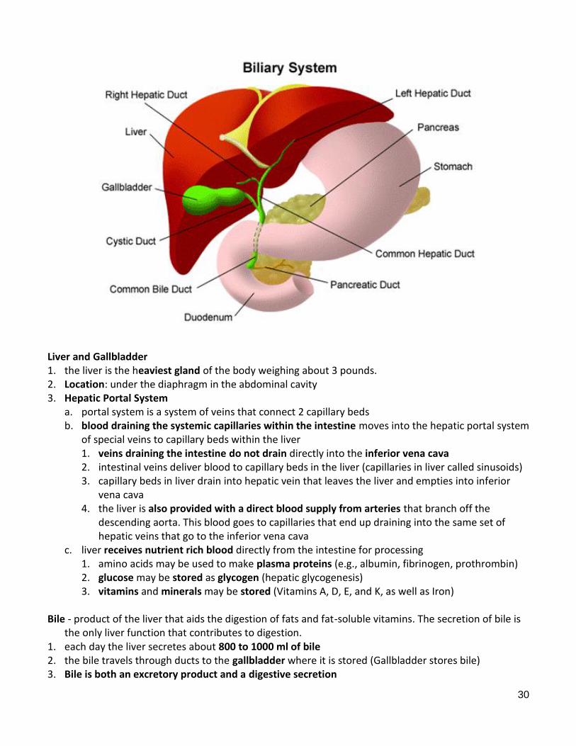
Liver and Gallbladder 1. the liver is the heaviest gland of the body weighing about 3 pounds. 2. Location: under the diaphragm in the abdominal cavity 3. Hepatic Portal System a. portal system is a system of veins that connect 2 capillary beds b. blood draining the systemic capillaries within the intestine moves into the hepatic portal system
of special veins to capillary beds within the liver 1. veins draining the intestine do not drain directly into the inferior vena cava 2. intestinal veins deliver blood to capillary beds in the liver (capillaries in liver called sinusoids) 3. capillary beds in liver drain into hepatic vein that leaves the liver and empties into inferior
vena cava 4. the liver is also provided with a direct blood supply from arteries that branch off the
descending aorta. This blood goes to capillaries that end up draining into the same set of hepatic veins that go to the inferior vena cava
c. liver receives nutrient rich blood directly from the intestine for processing 1. amino acids may be used to make plasma proteins (e.g., albumin, fibrinogen, prothrombin) 2. glucose may be stored as glycogen (hepatic glycogenesis) 3. vitamins and minerals may be stored (Vitamins A, D, E, and K, as well as Iron) Bile - product of the liver that aids the digestion of fats and fat-soluble vitamins. The secretion of bile is
the only liver function that contributes to digestion. 1. each day the liver secretes about 800 to 1000 ml of bile 2. the bile travels through ducts to the gallbladder where it is stored (Gallbladder stores bile) 3. Bile is both an excretory product and a digestive secretion

31
a. Excretory 1. hepatocytes take excess cholesterol and bilirubin (from breakdown of hemoglobin from
phagocytosed RBCs) out of the blood and secrete them into bile. RBC’s are phagocytized by macrophages that live mostly in the spleen
2. Bilirubin is called a bile pigment since it is yellowish-brown. It is degraded by intestinal bacteria to urobilinogen which colors feces brown.
3. bile formation is the body’s only way of getting rid of excess cholesterol (HDLs are lipoproteins in plasma that carry excess cholesterol in the body to the liver for elimination in the bile)
b. Digestion - bile salts 4. Bile Salts (2 Functions: fat emulsification and micelle formation) a. biles salts are fat emulsifiers that are synthesized by hepatocytes (liver cells) from cholesterol b. during digestion of chyme in the small intestine, bile moves out of the gallbladder and down the
cystic duct to the common bile duct and finally into the duodenum c. bile salts mix with chyme and emulsify fats d. Emulsification - break fat globules into tiny emulsion droplets (increase surface area for enzyme
action) to 1. increase the effectiveness of lipases 2. aids in the absorption of lipid-soluble vitamins A, D, E, and K e. without emulsification, insoluble fats tend to clump together and resist digestion f. within the small intestine, bile salts mix with fatty acids and monoglycerides that result from
lipase activity to form micelles. Micelles carry the fatty acids and monoglycerides to epithelial cells where they uncouple from the micelle and are absorbed into the columnar epithelial cells. They are resynthesized inside the mucosal cells to form triglycerides and then complexed with protein to form chylomicrons.
g. bile salts are eventually reabsorbed from the last part of the small intestine (ileum) and recycled by the blood back to the liver for reuse. Only about 5% of the bile salts secreted with bile into the small intestine end up in the feces
5. Regulation of Bile Secretion a. Vagal stimulation of the liver results in the production of bile b. CCK 1. CCK is released from enteroendocrine cells of duodenum in response to acidic chime 2. CCK stimulates the smooth muscle in the wall of the gallbladder to contract expelling bile
towards the duodenum 3. bile does not usually enter the duodenum until the gallbladder is stimulated by CCK

32
Some of the Important Roles of the Liver in Nutrient Processing 1. After a meal, the liver processes digested nutrients that flow to it with the hepatic portal system
circulation 2. the liver removes glucose, amino acids, vitamins, and other nutrients from the blood and shunts
them into biochemical pathways within hepatocytes (liver cells) a. The liver stores glucose as glycogen (hepatic glycogenesis) and converts fatty acids and glycerol
to triglycerides (hepatic lipogenesis) b. the liver converts amino acids into proteins (protein synthesis). The liver makes most of the
plasma proteins (e.g., prothrombin, fibrinogen, albumin) c. hepatocytes secrete a variety of proteins to include albumin, lipoproteins, clotting factors, and
angiotensinogen d. between meals, the liver converts amino acids to glucose by gluconeogenesis to maintain blood
sugar levels Carbohydrate Metabolism in liver 1. liver helps to store glucose as glycogen and maintain blood glucose levels between meals (glucose
homeostasis). Glucose in the blood is often called blood sugar. Glucose is the only significant simple sugar that circulates within the blood.
2. Carbohydrate in the Body: A normal, well-nourished individual has about 440 gms of carbohydrate in the body that can be found primarily in 3 places within the body.
a. Muscle glycogen (400 g, 74% of total, 0.9 lbs) – over 600 skeletal muscles that make up around 40% of total body mass
b. Liver glycogen (100 g, 23%, 0.2 lbs) – can be used to regulate blood sugar (glucose) levels c. Blood glucose (15 g, 3%) 3. Glycogen a. Glucose is not stored within the cells of the body as a simple sugar. Instead, glucose molecules
are polymerized into glycogen and stored in liver or muscle cells. Glycogen is the glucose-

33
storage molecule within the body. Each glycogen consists of hundreds if not thousands of glucose molecules bonded together.
b. about 25% of glycogen is stored in the liver, whereas about 75% is in skeletal muscle (plus very small amounts in cardiac muscle and other tissue cells).
c. The glucose that comes from the breakdown of glycogen in muscles cells can only be used in muscle cells.
d. The glucose that comes from the breakdown of glycogen in liver cells can be paid out into the blood to maintain blood sugar levels.
4. Hepatic Glycogenesis (anabolic) – Glucose homeostasis a. synthesis of glucose into glycogen for storage b. promoted by insulin during a meal (insulin stimulates glycogen synthase) c. Absorptive state – processing a meal in the intestine 5. Hepatic Glycogenolysis (catabolic) - Glucose homeostasis a. hydrolysis of glycogen to provide glucose to the blood b. promoted by glucagon, cortisol, epinephrine. c. Glycogenolysis releases glucose to the blood between meals (post-absorptive state) 6. Hepatic Gluconeogenesis - Glucose homeostasis a. formation of glucose from non-carbohydrate compounds like amino acids and glycerol. Fatty
acids cannot be used for gluconeogenesis. b. gluconeogenesis occurs primarily in the liver, but mostly in a fasting state (i.e., 8-12 hrs after last
meal or overnight when one wakes up in the morning). Gluconeogenesis can occur in kidney cells as well, but only under conditions in which the body is starving for food (i.e., run out of liver glycogen)
c. promoted by cortisol and glucagon d. the amino acids that are used by liver cells to make glucose generally come from the breakdown
or proteolysis of muscle proteins like actin and myosin. Muscle protein is preferred over other kinds of proteins like enzymes and collagen. Collagen is also broken down.
Carbohydrate in Liver Muscle Lipid in Adipocytes
Meal Insulin Hepatic glycogenesis Protein synthesis Lipogenesis (also within the liver)
Between Meals Glucagon Epinephrine
Hepatic glycogenolysis Lipolysis
Fasting State* Cortisol Epinephrine
Hepatic gluconeogenesis Proteolysis Lipolysis
*Fasting is a post-absorptive state during which one does not eat. Lipid Metabolism in Liver 1. The liver carries out most of the body’s lipogenesis. Excess organic nutrients such as glucose and
amino acids that come in with the food can be shunted into biochemical pathways that modify and extend their carbon skeletons into fat molecules
2. liver cells produce VLDLs (special protein-lipid complexes) that carry the triglycerides (fat molecules) to fat cells (adipocytes) for storage. Liver cells store some of the fats that they make, but not near to the same extent as that stored within fat cells. Fats are stored primarily in fat cells.
3. Produces the lipoproteins that transport cholesterol and triglycerides to and from body cells

34
4. the liver synthesizes 85% of the body’s cholesterol (cholesterol is used to make steroid hormones, Vitamin D, and bile salts and is used to strengthen plasma membranes)
Urea Formation in Liver 1. Cells deaminate amino acids (remove the amine group) and use the resultant carbon skeleton for
ATP production or for the production of carbohydrates and fats. Deamination produces ammonia which is released from cells into the bloodstream.
2. The liver takes in ammonia and combines it with CO2 to form urea. 3. Urea is moved out of the liver into the bloodstream, where it travels to the kidney for elimination in
urine. Liver damage can lead to increased ammonia in the blood. 4. Death occurs within days from ammonia toxicity which kills off brain neurons. Synthesis of Plasma Proteins (some examples) in liver 1. Albumin - plasma protein that helps to maintain the osmotic pressure of blood plasma which
opposes the excess filtration of water out of the arteriolar end of the capillary and promote reabsorption at the venule end
2. Fibrinogen and Prothrombin - necessary to form blood clots Storage of Vitamins and Minerals 1. Vitamins A, D, E, K, and B12 are stored in the liver if dietary intake exceeds the body’s needs 2. Iron and Copper Phagocytosis of worn-out RBCs and WBCs in liver and spleen (most phagocytosis occurs as blood travels
through venous sinuses of the spleen) Vitamins and Minerals 1. Vitamins: A (rhodopsin for function of rod so see in dim vision), D (calcium absorption from the
intestine so can maintain a strong bony matrix), K (prothrombin for blood clotting), B12 (RBC formation in red marrow)
a. vitamins are small organic compounds that most often come in with the diet b. vitamins are necessary for the metabolism of cells c. vitamins are named with letters in the order of their discovery and also as in the case of B
vitamins as a result of structural similarities d. with a few exceptions (e.g., vitamin D), vitamins can’t be synthesized by cells, thus they must be
provided in the diet or made by intestinal bacteria 2. Minerals: Calcium (bones and teeth, muscle contraction), phosphorus (bones and teeth, ATP), iron
(Hemoglobin), iodine (thyroxin), sodium and potassium (resting potential) Classification of Animals by Primary Feeding Methods 1. Filter or Suspension Feeders a. Trap organic material suspended in water (e.g., dead particles, bacteria, algae, small animals) b. Baleen whales, sponges 2. Detritivores a. Feed on dead and living material in sediments b. Earthworm 3. Fluid feeders

35
a. Suck or lick fluids of larger animals without eating the whole animal b. mosquitoes, vampire bats 4. Carnivores a. primarily eat other animals; specialize in capturing live prey and usually require high-protein diet b. lion 5. Herbivores a. eat algal or plant material like kelp blades or leaves, fruits and seeds b. deer, goat 6. Omnivores a. eat both animals and other organisms like plants b. humans, monkey, pigs 7. Symbiont autotroph-bearing animals in the ocean a. autotrophs are primary producers (e.g., photosynthesis) b. reef-building Cnidarians (coral) with dinoflagellate symbionts