Embed Size (px)
Citation preview

저 시-비 리- 경 지 2.0 한민
는 아래 조건 르는 경 에 한하여 게
l 저 물 복제, 포, 전송, 전시, 공연 송할 수 습니다.
다 과 같 조건 라야 합니다:
l 하는, 저 물 나 포 경 , 저 물에 적 된 허락조건 명확하게 나타내어야 합니다.
l 저 터 허가를 면 러한 조건들 적 되지 않습니다.
저 에 른 리는 내 에 하여 향 지 않습니다.
것 허락규약(Legal Code) 해하 쉽게 약한 것 니다.
Disclaimer
저 시. 하는 원저 를 시하여야 합니다.
비 리. 하는 저 물 리 목적 할 수 없습니다.
경 지. 하는 저 물 개 , 형 또는 가공할 수 없습니다.
Sinus Augmentation using BMP-2
in a Bovine Hydroxyapatite/Collagen
Carrier in Dogs
Jae-Kook Cha
Department of Dentistry
The Graduate School, Yonsei University
Sinus Augmentation using BMP-2
in a Bovine Hydroxyapatite/Collagen
Carrier in Dogs
Directed by Professor Seong-Ho Choi
A Doctoral Dissertation
submitted to the Department of Dentistry
the Graduate School of Yonsei University
in partial fulfillment of the requirements for the degree of
Ph.D. in Dental Science
Jae-Kook Cha
December 2015
This certifies that the Doctoral Dissertation
of Jae-Kook Cha is approved.
Thesis Supervisor: Seong-Ho Choi
Ui-Won Jung
Jung-Seok Lee
Sungtae Kim
Young-Taek Kim
The Graduate School
Yonsei University
December 2015
감사의 글
본 논문이 완성되기까지 부족한 저에게 지도와 격려를 아끼지 않
으신 아버지와 같은 최성호 교수님, 정의원 교수님, 이중석 교수님,
김성태 교수님, 김영택 교수님께 깊은 감사를 드립니다. 그리고 부
족한 논문임에도 진심 어린 조언으로 격려해주시고 따뜻한 관심으
로 지켜봐 주신 김종관 교수님, 채중규 교수님, 조규성 교수님, 김
창성 교수님께 감사드립니다.
연구 내내 많은 도움을 준 치주과 수련 동기들과, 선후배님들께
모두 진심으로 감사드립니다.
마지막으로 어려움이 있을 때마다 항상 저의 버팀목이 되어주시
고, 물심양면으로 도움을 주신 아버지, 어머니와 장인, 장모님께
깊은 사랑과 감사를 드립니다. 무엇보다도 아이를 돌보며 내조를
아끼지 않은 제 인생의 가장 좋은 친구이자 동반자인 아내 이영주
에게 저의 온 마음을 담아 감사와 사랑을 전합니다.
아울러 학업을 핑계로 많은 시간 같이하지 못했음에도 항상 아빠
에게 큰 힘을 주었던 아들 차건호에게도 고마움을 전합니다.
2015년 12월
저자 씀
i
Table of Contents
List of Figures ii
List of Tables iii
Abstract (English) iv
I. Introduction 1
II. Materials and Methods 3
1. Experimental Animals 3
2. Experimental Design 3
3. Surgical Procedures 4
4. Radiographic Analysis 5
5. Histologic Analysis 6
6. Histomorphometric Analysis 6
7. Statistics 8
III. Results 9
1. Clinical Findings 9
2. Radiographic Analysis 9
3. Histologic Findings 10
4. Histometric Analysis 11
IV. Discussion 14
References 18
Figure Legends 22
Figures 24
Abstract (Korean) 29
ii
List of Figures
Figure 1. (a-d) Surgical procedures, (e) The coronally sectioned radiographic
image, (f) Schematic drawing of the histometric analysis.
Figure 2. Representative 3D reconstructed and coronally sectioned micro-
computed tomography images of nasal sinuses.
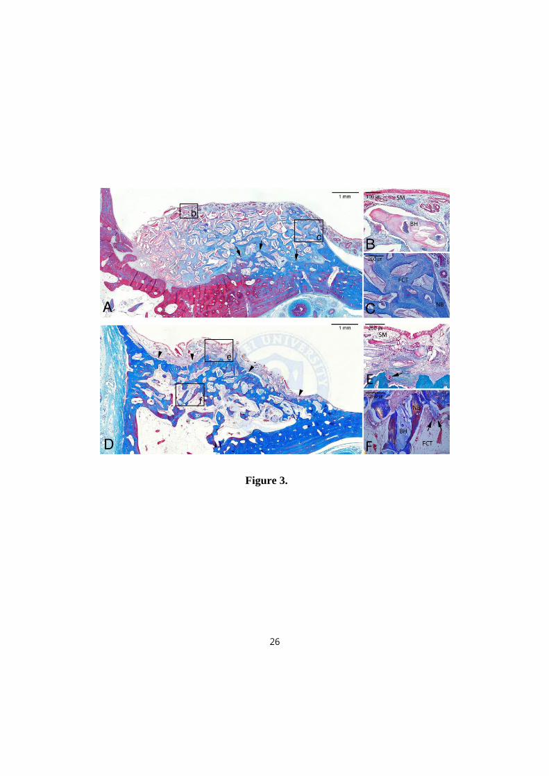
Figure 3. Histologic photomicrographs from the control group (a, b and c),
and the 0.1 mg/ml BMP-2-treated group (d, e and f).
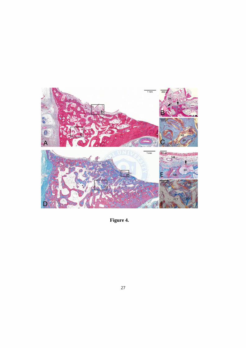
Figure 4. Histologic photomicrographs from the 0.5 mg/ml BMP-2-treated
group (a, b and c), and the 1.5 mg/ml BMP-2-treated group (d, e and f).
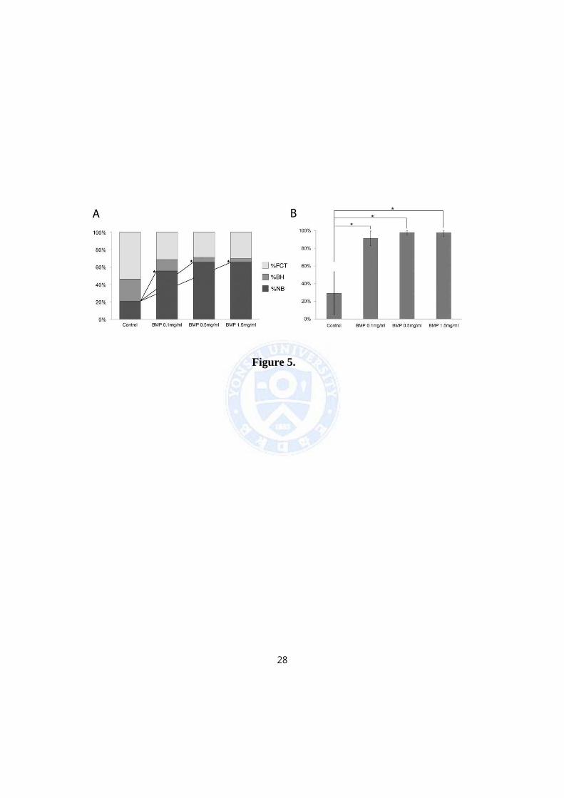
Figure 5. (a) Composition of the total augmented area. (b) Proportion of the
number of BH particles surrounded by NB.

iii
List of Tables
Table 1. Composition of the total augmented area.
Table 2. Percentage of NB between the peripheral and central area of the sinus
cavity.
iv
Abstract
Sinus Augmentation Using BMP-2 in a Bovine
Hydroxyapatite/Collagen Carrier in Dogs
Jae Kook Cha, D.D.S., M.S.D.
Department of Dentistry
The Graduate School, Yonsei University
(Directed by Professor Seong-Ho Choi, D.D.S., M.S.D., PhD.)
Objective: The objective of this study was to determine the efficacy of bone
morphogenetic protein 2 (BMP-2) in a bovine hydroxyapatite/collagen (BHC) carrier
to augment bone formation in a canine nasal sinus model.
Material and Methods: Eight mongrel dogs, approximately 12 months old and 30 kg
in weight were used. Following preparation of bilateral sinus access windows, BHC
alone (control) or loaded with E.coli-derived BMP-2 at 0.1 mg/mL was implanted in
4 animals, and BHC loaded with E.coli-derived BMP-2 at 0.5 and 1.5 mg/mL was
implanted in 4 animals. The animals were euthanized at 20 weeks when block
sections were obtained for micro-computed tomography and histometric analyses.
Results: Total augmented volumes did not differ significantly between groups.

v
Histometric analysis showed significantly enhanced bone formation for the BMP-2
groups compared with control.
Conclusion: BMP-2 in a BHC carrier, even at the low 0.1-mg/mL concentration,
induces osteogenic activity, enhancing local bone formation in a canine sinus model.
KEYWORDS: bone substitutes, bone tissue engineering, bone regeneration, sinus
augmentation
1
Sinus Augmentation Using BMP-2 in a Bovine
Hydroxyapatite/Collagen Carrier in Dogs
Jae Kook Cha, D.D.S., M.S.D.
Department of Dentistry
The Graduate School, Yonsei University
(Directed by Professor Seong-Ho Choi, D.D.S., M.S.D., PhD.)
I. Introduction
A number of studies have demonstrated extensive bone formation using bone
morphogenetic protein 2 (BMP-2) with different carriers in various animal sinus
augmentation models (Park, 2009). Among them, BMP-2 in absorbable collagen
sponge (ACS) carrier was shown to be more effective than autogenous bone grafts
and have been considered the new gold standard for this indication (Lee et al., 2013).
Along with the superior effects of BMP-2/ACS in preclinical models, clinical studies
have demonstrated successful results following the use of BMP-2/ACS for maxillary
sinus augmentation (Boyne et al., 2005; Triplett et al., 2009).
2
The concentration of BMP-2 to induce the most effective bone formation is still
unclear and it is influenced by various factors; e.g. delivery system, release kinetics
and recipient site (Seeherman et al., 2002). In vitro and in vivo studies using strict
dose analysis have shown an inverse correlation between bone maturation and BMP-2
dose (Park et al., 2012; Song et al., 2011; Wikesjo et al., 2008; Zara et al., 2011).
However, in regards to sinus augmentation, the study of BMP-2 concentration matters
relative to bone formation/maturation, and occurrence/severity of adverse events has
not yet been reported. A wide range of BMP-2 concentrations have been used in
preclinical sinus augmentation models (Choi et al., 2012; Gutwald et al., 2010;
Hanisch et al., 1997; Lee et al., 2013). We should consider that these studies were
performed in different experimental animals with various carrier systems and also that
the biologic response varies between recipient sites, thus, the results of these studies
could not be directly compared. Therefore, a study to determine the effects of BMP-2
depending on the concentration in sinus model is needed.
The objective of this study was to determine the efficacy of BMP-2 in a bovine
hydroxyapatite/collagen (BHC) carrier to augment bone formation in the canine nasal
sinus model.
3
II. Materials and Methods
1. Experimental Animals
Eight mongrel dogs aged about 12 months and weighing approximately 30 kg
were used. All had healthy dentitions and periodontal tissues without any systemic
disease. The selection and management of experimental animals and surgical
procedures followed a protocol approved for this study by the Institutional Animal
Care and Use Committee, College of Medicine, Yonsei University (09-021).
2. Experimental Design
E. coli derived BMP-2 was provided from the Research and Development
Institute of Cowellmedi (Busan, Korea). The BMP-2 expressed by E. coli has been
described in detail previously (Lee et al., 2010). BMP-2 was reconstituted and diluted
in a buffer to obtain concentrations of 0.1, 0.5, and 1.5 mg/ml, and then 250 mg of
BHC (Bio-Oss Collagen, Geistlich Pharma AG, Wolhusen, Switzerland) was loaded
with 200 l of one of the three different concentrations of BMP-2 or saline (control).
The BHC blocks were a uniform volume of 9 x 9 x 8 mm in width, length and height.
BMP-2 was loaded using an auto pipette in a sterilized culture dish, and after
allowing 10 min for the BMP-2 to adsorb onto the surface of BHC, the blocks were
4
placed into the nasal sinuses. The experimental sites were divided into four groups
according to the dose of BMP-2 applied (n = 4 per group):
i) Control group: BHC loaded with normal saline.
ii) BHC loaded with BMP-2 at 0.1 mg/ml (total dose = 0.02 mg).
iii) BHC loaded with BMP-2 at 0.5 mg/ml (total dose = 0.10 mg).
iv) BHC loaded with BMP-2 at 1.5 mg/ml (total dose = 0.30 mg).
Control and 0.1 mg/ml BMP-2 group were assigned in each sinus of four dogs,
0.5 and 1.5mg/ml BMP-2 groups were assigned in the other four dogs.
3. Surgical Procedure
The maxillary premolars were extracted bilaterally prior to the experimental
surgery and the experimental sites were allowed to heal for 2 months. The
experimental surgical procedure was performed under general anesthesia. The
anesthetic procedure was described in our previous report (Oh et al., 2011).
After healing following extraction, the edentulous region was accessed using
buccal incisions. A horizontal incision was made at the gingival crest from the first
premolar to the third premolar, from which a vertical incision was extended apically.
The full-thickness flap was elevated and the lateral bone wall was removed using an
8-mm-diameter trephine bur under sufficient saline irrigation. The position of the
lateral window was determined using intraoral radiographs of the edentulous area.
The membrane was carefully elevated from the floor (Fig. 1a) and lateral walls, and
5
then the BMP-2-loaded scaffold was applied to the created space (Fig. 1b and c). The
flap was repositioned using a suture material (4-0 Monosyn; glyconate absorbable
monofilament, B-Braun, Aesculap, Center Valley, PA, USA). The animals were
euthanized by anesthetic drug overdose at 20 weeks.
4. Radiographic Analysis
Block sections that included BHC were dissected and fixed in 10% neutral
buffered formalin for 10 days (Fig. 1d). The fixed specimens were scanned in a micro
computed tomography (micro-CT; SkyScan 1072, SkyScan, Aartselaar, Belgium) at a
resolution of 35 μm (100 kV, 100 μA). The scanned data were saved in DICOM
format, and the experimental area was reconstructed with OnDemand 3D software
(CyberMed, Seoul, Korea). In all sectioned planes, the total augmented areas were
identified by color coding and traced manually by one experienced examiner using
the software program. The composition of the augmented area were automatically
indicated according to the gray values of the threshold, and then modified manually
for fine distinction between bovine hydroxyapatite (BH) and new bone (NB) in all
sectioned planes (Fig. 1e). The gray values of threshold were standardized and they
ranged from 145 to 225 for BH and from 95 to 145 for NB. The overall dimensional
topography of experimentally grafted sinus cavities were visualized with the aid of
three-dimensionally (3D) reconstructed images. The total augmented volume and
6
volume of BH (mm3) were calculated by integration of the data from all tomographic
images.
5. Histologic Analysis
After rinsing the specimens in sterile water, the sections were decalcified in 5%
formic acid, dehydrated in a graded ethanol series, and embedded in paraffin. Serial
sections were cut at a thickness of 5 µm in an apicocoronal vertical plane. The two
most central sections of each grafted site were selected and stained with hematoxylin-
eosin and Masson’s trichrome. Histologic analysis was performed using a
stereomicroscope (MZFLIII, Leica, Wetzlar, Germany) and light microscope (BX-50,
Olympus Optical, Tokyo, Japan).
6. Histomorphometric Analysis
Micrographs were taken at a magnification of ×12.5 and assembled to enable
visualization of the entire sinus. After examination with a conventional light
microscope, histomorphometric measurements were made using a PC-based image-
analysis system (Image-Pro Plus, Media Cybernetics, Silver Spring, MD, USA). The
histomorphometric analysis was performed by one experienced, masked examiner
(J.S.L.). To minimize intraexaminer errors, several randomly selected samples were
measured twice with a 1-week interval. The intraexaminer reproducibility was
evaluated using the concordance correlation coefficient, which was 0.93-0.98
7
(Barnhart et al., 2007; Lin, 1989). Each photomicrograph was horizontally aligned
and imaginary horizontal line was established according to the base of nasal sinus
floor for linear measurement. The following measurements were made as primary
outcome variables (Fig. 1f):
i) Total augmented area (mm2) and height (mm): the experimentally augmented
area surrounded by the floor of nasal sinus and Schneiderian membrane,
including NB, residual BH particles, and FCT. Augmented height was
defined as the distance from the floor of the nasal sinus to the highest point of
the total augmented area.
ii) NB height proportion (%NBH; %): the proportion of the distance from the
floor of the nasal sinus to the highest point of NB within the sinus (NBH) in
relation to the total augmented height.
iii) Composition of the augmented area (%): the proportions of NB (%NB),
residual particles (%BH), and FCT (%FCT) relative to the total augmented
area.
The following additional measurements were made as secondary outcome
variables, to evaluate the healing around the residual particles and the homogeneity of
bone regeneration:
i) Proportions of BH particles surrounded by NB (%BHB): the proportion of
the number of BH particles surrounded by NB relative to the total number of
BH particles in the augmented area.
ii) Separated fractions of NB in the central and peripheral portions of the
8
augmented area (%; Fig. 1f): %NB in selected central and peripheral areas,
respectively. The central-most vertical and horizontal area, and the
horizontally central/most-coronal area were selected for this measurement.
7. Statistics
Group means (±SD) were calculated. Statistical analyses were performed
separately at the intra- and inter-subject levels. Wilcoxon signed-rank test was used
for the comparison between control and 0.1 mg/ml BMP-2 group, and 0.5 and 1.5
mg/ml groups. Mann-Whitney U test used to compare groups which were allocated in
different animals; control versus 0.5 and 1.5 mg/ml groups, and 0.1 mg/ml group
versus 0.5 and 1.5 mg/ml groups. The level of statistical significance was set at p <
0.05.
9
III. RESULTS
1. Clinical Findings
Surgical wound healing was uneventful during the experimental period. Nasal
bleeding occurred immediately after the surgical procedure in several dogs, but it
stopped within an hour. No complications – including membrane perforation, wound
dehiscence, severe swelling, and bleeding – were observed.
2. Radiographic Analysis
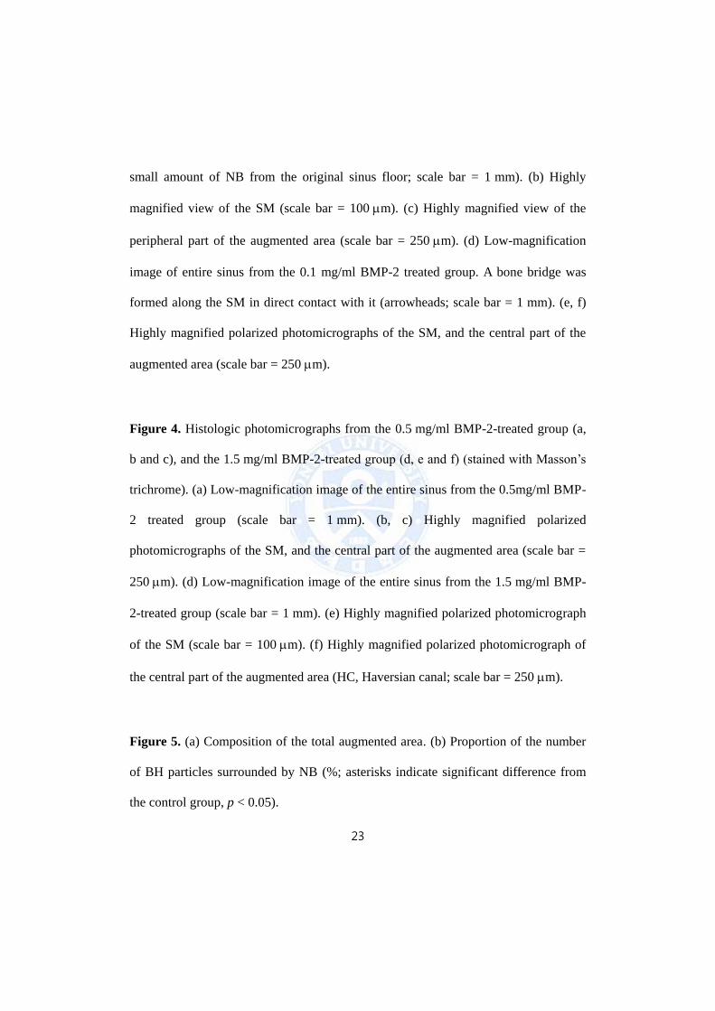
The 3D reconstructed images revealed the entire shape of the augmented area
(Fig. 2). In the control group, the BHC had almost maintained its original rectangular
block shape over the sinus floor. However, the BH had become spread out and
flattened on the sinus floor in the BMP-2-treated groups.
NB and BH could be observed in the cross-sectional images (Fig. 2). In the
control group, the BH particles were still evident at the floor of the sinus and only a
small amount of NB was observed inside the BHC. In the BMP-2-treated groups, the
BH particles appeared to have disassembled and were scattered over the sinus floor.
With a BMP-2 dose of 0.1 mg/ml, a bone bridge had formed within the BHC block so
that the BH and the NB had become commingled. In the groups with BMP-2 at 0.5
and 1.5 mg/ml, the amount of BH was significantly reduced and abundant NB was
10
observed, homogeneous with the original sinus floor. The volume of BH in the BMP-
2 treated groups tended to be lower than that in the control group, however the
differences between groups were not statistically significant on intra/inter subject
level tests (control group, 26.25 ± 15.01 mm3; 0.1 mg/ml BMP-2 group, 14.50 ±
10.32 mm3; 0.5 mg/ml BMP-2 group, 12.52 ± 16.42 mm3; 1.5 mg/ml BMP-2 group,
11.21 ± 5.25 mm3). Also, the total augmented volumes did not differ statistically
between the groups (control group, 253.99 ± 41.00 mm3; 0.1 mg/ml BMP-2 group,
346.61 ± 102.75 mm3; 0.5 mg/ml BMP-2 group, 246.93 ± 95.97 mm3; 1.5 mg/ml
BMP-2 group, 317.50 ± 95.78 mm3).
3. Histologic Findings
The nasal sinus cavity was surrounded by respiratory mucosa and a thin layer of
cortical bone. The Schneiderian membrane was intact, with no sign of inflammation
in all groups.
In the control group, most of the BH particles were encapsulated by dense
fibroblastic cells with smooth borders in the whole augmented area, and only a small
amount of NB could be detected in the original sinus floor. In the central portion of
the augmented area in particular, the NB was barely visible and multinucleated
osteoclast-like cells were absent (Fig. 3a, b and c).
In contrast, in the BMP-2 treated groups, a trabecular pattern of NB was observed
in direct contact along the Schneiderian membrane. NB was also seen at the tented
11
area lateral to the BHC. In the NB induced by BMP-2, concentric layers of bone
tissue were observed around Haversian canals and well-defined lamellar bone,
exhibiting characteristics of mature bone. The osteoclast-like cells were absent and
the osteoclastic activity was not observed around the BH particles with smooth and
intact borders (Fig. 3d, e and f, Fig. 4). Interestingly, with 0.5 mg/ml BMP-2 the BH
particles tightly attached to the NB had penetrated the alveolar bone beneath the
original sinus floor such that the original sinus floor was barely distinguishable from
the NB (Fig. 4a, b and c); moreover, with 1.5 mg/ml BMP-2, a large amount of
completely remodeled NB was observed throughout the entire sinus cavity, and the
BH particles were barely visible (Fig. 4d, e and f).
4. Histometric Analysis
The composition of the augmented area is summarized in Fig. 5a and Table 1. Both
the area of NB (mm2) and %NB were significantly larger in all of the BMP-2-treated
groups than in the untreated control. Furthermore, the bone formation observed with
BMP-2 was significantly larger than the control group not only in the central portion
of augmented area close to the basal bone of the sinus, but also in the peripheral
portion of augmented area near the Schneiderian membrane (Table 2).
%BHB and %NBH differed significantly between the control and 0.1 mg/ml BMP-
2 group or 0.5 and 1.5 mg/ml BMP-2 groups. The values in the BMP-2-treated groups
tended to increase with concentration, but the differences between the groups were
12
not statistically significant at all intra/inter subject level tests (Fig. 5b; %NBH: control
group, 52.3 ± 27.1%; 0.1 mg/ml BMP-2 group , 97.8 ± 4.3%; 0.5 mg/ml BMP-2
group, 98.8 ± 2.3%; 1.5 mg/ml BMP-2 group, 100%; %BHB: control group, 29.2 ±
24.3%; 0.1 mg/ml BMP-2 group , 91.4 ± 8.2%; 0.5 mg/ml BMP-2 group, 97.9 ±
2.6%; 1.5 mg/ml BMP-2 group, 97.7 ± 4.6%).
Table 1. Composition of the Total Augmented Area. (mean ± standard deviation)
Group NB (mm2) BH (mm2) FCT (mm2) AA (mm2)
Control 7.04 ± 2 .43 8.29 ± 1.29 17.75 ± 1.49 33.09 ± 3.04
BMP-2 (0.1 mg/ml) 18.50 ± 1.1* 4.36 ± 0.25* 10.51 ± 2.09* 33.36 ± 3.04
BMP-2 (0.5 mg/ml) 22.05 ± 1.91* 2.01 ± 1.02*,† 9.88 ± 2.49* 33.94 ± 5.01
BMP-2 (1.5 mg/ml) 24.26 ± 5.90* 1.52 ± 0.22*,† 10.88 ± 3.09* 36.65 ± 4.18
NB, newly formed bone; BH, bovine hydroxyapatite; FCT, fibrovascular connective tissue;
AA, total augmented area; BMP-2, bone morphogenetic protein 2.
* Significantly different from control group (p < 0.05).
†Significantly different from BMP-2 at 0.1 mg/ml (p < 0.05).
13
Table 2. Percentage of NB between the peripheral and central area of the sinus cavity.
(mean ± standard deviation)
Group Ce (%) Pe (%)
Control 5.18 ± 0.95 10.03 ± 1.00
BMP-2 (0.1 mg/ml) 44.30 ± 5.58* 45.11 ± 7.02*
BMP-2 (0.5 mg/ml) 38.67 ± 13.80* 42.70 ± 5.79*
BMP-2 (1.5 mg/ml) 42.29 ± 19.63* 44.58 ± 7.99*
Ce, central-most vertical and horizontal areas of the augmented graft; Pe,
horizontally central/most-coronal area of the augmented graft.
* Significantly different from control group (P < 0.05).
14
IV. DISCUSSION
The present study evaluated osteoinductive activities of BMP-2 in BHC using the
dog nasal sinus experimental model. Osteogenic activity of BMP-2 was observed
even at the lowest dose (0.1 mg/ml, corresponding to 0.02 mg of BMP-2), and this
value was 15 times lower than the approved concentration for human use (1.5mg/ml
with ACS), nevertheless induced twice the amount of bone regeneration compared to
the control.
Previous studies have demonstrated that low quality of bone is attributable to the
formation of adipose tissue when higher concentrations of BMP-2 are applied (Park et
al., 2012; Song et al., 2011). However, our findings refute this since we obtained
good-quality bone with limited adipogenic differentiation at all experimental sites
including the ones with low and high concentrations of BMP-2. Improved bone
qualities were shown even at the peripheral portion of augmented areas distant from
osteogenic sources in all experimental sites, while new bone formation was barely
observed in this area of the control group. This is concurrent with Choi et al. reporting
that osteoinductive potential of the Schneiderian membrane is activated at the early
stage of healing with BMP-2 (Choi et al., 2013). Despite the controversy of whether
the Schneiderian membrane contains the osteogenic source, the authors suggested that
newly formed bone from the Schneiderian membrane at the peripheral area could
protect the augmented space from the volume shrinkage by remodeling process.
15
All experimental sites showed comparable formation of newly formed bone,
regardless of the concentration of BMP-2. This indicates that increased dose of BMP-
2 beyond a certain threshold would not improve bone regeneration in sinus
augmentation, and the threshold concentration would be smaller than the present
experimental range of BMP-2 concentration (0.1-1.5 mg/ml in 200 l) in dog nasal
sinus model. The sinus augmentation model is a contained defect surrounded by the
sinus floor and the Schneiderian membrane; thus its healing process could be
accelerated even with the low BMP-2 concentration. And also characteristics of the
carrying system could have influenced the present result. BMP-2 may not have been
fully adsorbed onto the surface of BHC during preparation, and the BMP-2 might not
be biologically available along the concentration gradient. The adsorption and release
profiles of BMP-2/BHC have not been verified yet, thus this feature should be fully
investigated in future studies.
Interestingly, it was observed that local high dose of BMP-2 (0.5mg/ml) induced
remodeling of the sinus floor and beyond (1.5mg/ml). In the process of new bone
formation, a previous study has reported that the BMP-2 dose-dependently stimulated
osteoclastic bone resorption as well as acted as a mediator of the osteoblast-osteoclast
interaction (Kanatani et al., 1995). The scattered residual biomaterials surrounded by
newly formed bone in the present results may be a product of this vigorous
remodeling process enhanced by BMP-2. While the cortical portion of the original
sinus floor had been resorbed away in the early healing process, BHC would have
scattered and sequential new bone formation would have occurred around these
16
particles. Even though the collagen matrix in BHC could enhance the clinical
manageability, it could be suggested that the structural integrity of BHC might be
insufficient to support the maintenance of space during the healing process by BMP-2.
In the same vein, the present results demonstrated a tendency of decrease in the
proportion of remaining BH at sites that received BMP-2; histological analysis
revealed significant differences, but volumetric analysis on the reconstructed micro
CT images did not. This could be explained by excessive swelling in the early healing
phase of experimental sites and increased bone formation by BMP-2, which could
have migrated the grafted biomaterial to the lateral aspect of the augmented area. BH
has been considered as a non-resorbable biomaterial in craniofacial fields (Hallman et
al., 2001; Hallman and Thor, 2008; Schlegel and Donath, 1998). The previous results
showed that the BH particles remained similar size to the original BH particles even
in 7- and 10-year biopsy samples (Mordenfeld et al., 2010; Orsini et al., 2007).
This study incorporated the nasal sinus model of the dog, which is anatomically
adjacent to the maxillary premolars and has a similar bone structure to the human
maxillary sinus which contains alveolar bone and the lateral wall (Aerssens et al.,
1998). Histologically, it has similar compositions to the human Schneiderian
membrane as the dog sinus membrane is comprised of pseudostratified ciliated
columnar epithelium which is a respiratory epithelium (Lee et al., 2007). Additionally,
the intraoral surgical approach in the dog nasal sinus model has a high relevance to
the human clinical procedure (Wetzel et al., 1995). On the other hand, as the dog
nasal sinus is connected to the nose, it is subjected to more direct transmission of
17
positive respiratory pressure than the human (Haas et al., 1998). Hence the present
results may conservatively be interpreted for clinical application in human.
The present study used two separate statistical analyses; dependent test for
comparing groups within one animal, and independent test for groups between
animals. It was caused by the limitation in the number of sinuses of one subject
animal; therefore, these results should be interpreted conservatively. However, the
present study focused on whether the significantly reduced concentration (0.1mg/ml)
of BMP-2 could affect bone healing in sinus augmentation, and the results clearly
demonstrated extensive new bone formation in comparison to the control. These
results were comparable to that of the conventional concentrations (0.5 and 1.5mg/ml)
of BMP-2. Therefore, in continuation of the current focus of minimizing the dosage
of BMP for bone regeneration, the findings of our study indicate the necessity to
perform further studies with concentrations of BMP-2 lower than 0.1 mg/ml.
Within the limitations of this study, it can be concluded that BMP-2 in a BHC
carrier, even at the low 0.1-mg/mL concentration, induces osteogenic activity,
enhancing local bone formation in the canine sinus model.
18
REFERENCES
Aerssens J, Boonen S, Lowet G, Dequeker J: Interspecies differences in bone composition,
density, and quality: potential implications for in vivo bone research. Endocrinology
139(2): 663-670, 1998.
Barnhart HX, Lokhnygina Y, Kosinski AS, Haber M: Comparison of concordance correlation
coefficient and coefficient of individual agreement in assessing agreement. J Biopharm
Stat 17(4): 721-738, 2007.
Boyne PJ, Lilly LC, Marx RE, Moy PK, Nevins M, Spagnoli DB, et al.: De novo bone
induction by recombinant human bone morphogenetic protein-2 (rhBMP-2) in maxillary
sinus floor augmentation. J Oral Maxillofac Surg 63(12): 1693-1707, 2005.
Choi Y, Lee JS, Kim YJ, Kim MS, Choi SH, Cho KS, et al.: Recombinant human bone
morphogenetic protein-2 stimulates the osteogenic potential of the schneiderian
membrane: a histometric analysis in rabbits. Tissue Eng Part A 19(17-18): 1994-2004,
2013.
Choi Y, Yun JH, Kim CS, Choi SH, Chai JK, Jung UW: Sinus augmentation using absorbable
collagen sponge loaded with Escherichia coli-expressed recombinant human bone
morphogenetic protein 2 in a standardized rabbit sinus model: a radiographic and
histologic analysis. Clin Oral Implants Res 23(6): 682-689, 2012.
Gutwald R, Haberstroh J, Stricker A, Ruther E, Otto F, Xavier SP, et al.: Influence of rhBMP-2
on bone formation and osseointegration in different implant systems after sinus-floor
elevation. An in vivo study on sheep. J Craniomaxillofac Surg 38(8): 571-579, 2010.
Haas R, Mailath G, Dortbudak O, Watzek G: Bovine hydroxyapatite for maxillary sinus
19
augmentation: analysis of interfacial bond strength of dental implants using pull-out tests.
Clin Oral Implants Res 9(2): 117-122, 1998.
Hallman M, Lundgren S, Sennerby L: Histologic analysis of clinical biopsies taken 6 months
and 3 years after maxillary sinus floor augmentation with 80% bovine hydroxyapatite
and 20% autogenous bone mixed with fibrin glue. Clin Implant Dent Relat Res 3(2): 87-
96, 2001.
Hallman M, Thor A: Bone substitutes and growth factors as an alternative/complement to
autogenous bone for grafting in implant dentistry. Periodontol 2000 47: 172-192, 2008.
Hanisch O, Tatakis DN, Rohrer MD, Wohrle PS, Wozney JM, Wikesjo UM: Bone formation
and osseointegration stimulated by rhBMP-2 following subantral augmentation
procedures in nonhuman primates. Int J Oral Maxillofac Implants 12(6): 785-792, 1997.
Kanatani M, Sugimoto T, Kaji H, Kobayashi T, Nishiyama K, Fukase M, et al.: Stimulatory
effect of bone morphogenetic protein-2 on osteoclast-like cell formation and bone-
resorbing activity. J Bone Miner Res 10(11): 1681-1690, 1995.
Lee HJ, Choi BH, Jung JH, Zhu SJ, Lee SH, Huh JY, et al.: Maxillary sinus floor
augmentation using autogenous bone grafts and platelet-enriched fibrin glue with
simultaneous implant placement. Oral Surg Oral Med Oral Pathol Oral Radiol Endod
103(3): 329-333, 2007.
Lee J, Susin C, Rodriguez NA, de Stefano J, Prasad HS, Buxton AN, et al.: Sinus
augmentation using rhBMP-2/ACS in a mini-pig model: relative efficacy of autogenous
fresh particulate iliac bone grafts. Clin Oral Implants Res 24(5): 497-504, 2013.
Lee JH, Kim CS, Choi KH, Jung UW, Yun JH, Choi SH, et al.: The induction of bone
formation in rat calvarial defects and subcutaneous tissues by recombinant human BMP-
2, produced in Escherichia coli. Biomaterials 31(13): 3512-3519, 2010.
20
Lin LI: A concordance correlation coefficient to evaluate reproducibility. Biometrics 45(1):
255-268, 1989.
Mordenfeld A, Hallman M, Johansson CB, Albrektsson T: Histological and
histomorphometrical analyses of biopsies harvested 11 years after maxillary sinus floor
augmentation with deproteinized bovine and autogenous bone. Clin Oral Implants Res
21(9): 961-970, 2010.
Oh KC, Cha JK, Kim CS, Choi SH, Chai JK, Jung UW: The influence of perforating the
autogenous block bone and the recipient bed in dogs. Part I: a radiographic analysis. Clin
Oral Implants Res 22(11): 1298-1302, 2011.
Orsini G, Scarano A, Degidi M, Caputi S, Iezzi G, Piattelli A: Histological and ultrastructural
evaluation of bone around Bio-Oss particles in sinus augmentation. Oral Dis 13(6): 586-
593, 2007.
Park JB: Use of bone morphogenetic proteins in sinus augmentation procedure. J Craniofac
Surg 20(5): 1501-1503, 2009.
Park JC, Kim JC, Kim BK, Cho KS, Im GI, Kim BS, et al.: Dose- and time-dependent effects
of recombinant human bone morphogenetic protein-2 on the osteogenic and adipogenic
potentials of alveolar bone-derived stromal cells. J Periodontal Res, 2012.
Schlegel AK, Donath K: BIO-OSS--a resorbable bone substitute? J Long Term Eff Med
Implants 8(3-4): 201-209, 1998.
Seeherman H, Wozney J, Li R: Bone morphogenetic protein delivery systems. Spine (Phila Pa
1976) 27(16 Suppl 1): S16-23, 2002.
Song DS, Park JC, Jung IH, Choi SH, Cho KS, Kim CK, et al.: Enhanced adipogenic
differentiation and reduced collagen synthesis induced by human periodontal ligament
stem cells might underlie the negative effect of recombinant human bone morphogenetic
21
protein-2 on periodontal regeneration. J Periodontal Res 46(2): 193-203, 2011.
Triplett RG, Nevins M, Marx RE, Spagnoli DB, Oates TW, Moy PK, et al.: Pivotal,
randomized, parallel evaluation of recombinant human bone morphogenetic protein-
2/absorbable collagen sponge and autogenous bone graft for maxillary sinus floor
augmentation. J Oral Maxillofac Surg 67(9): 1947-1960, 2009.
Wetzel AC, Stich H, Caffesse RG: Bone apposition onto oral implants in the sinus area filled
with different grafting materials. A histological study in beagle dogs. Clin Oral Implants
Res 6(3): 155-163, 1995.
Wikesjo UM, Qahash M, Polimeni G, Susin C, Shanaman RH, Rohrer MD, et al.: Alveolar
ridge augmentation using implants coated with recombinant human bone morphogenetic
protein-2: histologic observations. J Clin Periodontol 35(11): 1001-1010, 2008.
Zara JN, Siu RK, Zhang X, Shen J, Ngo R, Lee M, et al.: High doses of bone morphogenetic
protein 2 induce structurally abnormal bone and inflammation in vivo. Tissue Eng Part A
17(9-10): 1389-1399, 2011.
22
Figure Legends
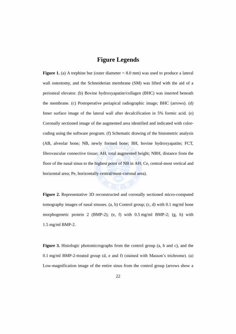
Figure 1. (a) A trephine bur (outer diameter = 8.0 mm) was used to produce a lateral
wall osteotomy, and the Schneiderian membrane (SM) was lifted with the aid of a
periosteal elevator. (b) Bovine hydroxyapatite/collagen (BHC) was inserted beneath
the membrane. (c) Postoperative periapical radiographic image; BHC (arrows). (d)
Inner surface image of the lateral wall after decalcification in 5% formic acid. (e)
Coronally sectioned image of the augmented area identified and indicated with color-
coding using the software program. (f) Schematic drawing of the histometric analysis
(AB, alveolar bone; NB, newly formed bone; BH, bovine hydroxyapatite; FCT,
fibrovascular connective tissue; AH, total augmented height; NBH, distance from the
floor of the nasal sinus to the highest point of NB in AH; Ce, central-most vertical and
horizontal area; Pe, horizontally central/most-coronal area).
Figure 2. Representative 3D reconstructed and coronally sectioned micro-computed
tomography images of nasal sinuses. (a, b) Control group; (c, d) with 0.1 mg/ml bone
morphogenetic protein 2 (BMP-2); (e, f) with 0.5 mg/ml BMP-2; (g, h) with
1.5 mg/ml BMP-2.
Figure 3. Histologic photomicrographs from the control group (a, b and c), and the
0.1 mg/ml BMP-2-treated group (d, e and f) (stained with Masson’s trichrome). (a)
Low-magnification image of the entire sinus from the control group (arrows show a
23
small amount of NB from the original sinus floor; scale bar = 1 mm). (b) Highly
magnified view of the SM (scale bar = 100 m). (c) Highly magnified view of the
peripheral part of the augmented area (scale bar = 250 m). (d) Low-magnification
image of entire sinus from the 0.1 mg/ml BMP-2 treated group. A bone bridge was
formed along the SM in direct contact with it (arrowheads; scale bar = 1 mm). (e, f)
Highly magnified polarized photomicrographs of the SM, and the central part of the
augmented area (scale bar = 250 m).
Figure 4. Histologic photomicrographs from the 0.5 mg/ml BMP-2-treated group (a,
b and c), and the 1.5 mg/ml BMP-2-treated group (d, e and f) (stained with Masson’s
trichrome). (a) Low-magnification image of the entire sinus from the 0.5mg/ml BMP-
2 treated group (scale bar = 1 mm). (b, c) Highly magnified polarized
photomicrographs of the SM, and the central part of the augmented area (scale bar =
250 m). (d) Low-magnification image of the entire sinus from the 1.5 mg/ml BMP-
2-treated group (scale bar = 1 mm). (e) Highly magnified polarized photomicrograph
of the SM (scale bar = 100 m). (f) Highly magnified polarized photomicrograph of
the central part of the augmented area (HC, Haversian canal; scale bar = 250 m).
Figure 5. (a) Composition of the total augmented area. (b) Proportion of the number
of BH particles surrounded by NB (%; asterisks indicate significant difference from
the control group, p < 0.05).
24
Figures
Figure 1.
25
Figure 2.
26
Figure 3.
27
Figure 4.
28
Figure 5.
29
국문요약
상악동 거상술 시 탈단백우골/콜라겐 전달체를 사용한 제 2
형 골형성 유도 단백질의 농도에 따른 골 형성
< 지도교수 최성호 >
연세대학교 대학원 치의학과
차 재 국
목적: 상악동에서 제 2형 골형성 유도 단백질(BMP-2)의 골 형성 효과
는 여러 선행 연구를 통해 증명되었지만, 아직까지 최적의 농도는 정립되
지 않았다. 상악동 거상술은 치과 술식 중 다량의 골이식재가 사용되는 술
식으로, 과량의 BMP-2의 사용에 따른 부작용이 쉽게 발생할 수 있으므로
골 형성 효과를 유지하며 부작용을 최소화할 수 있는 이상적인 농도를 정
립하는 과정이 필요하다. 따라서 이 연구의 목적은 개 상악동에서 탈단백
우골/콜라겐 (BHC) 전달체를 사용한 BMP-2의 농도에 따른 골 형성 효과
를 평가하여 최적의 농도를 결정하는 것이다.
재료 및 방법: 8마리의 잡견 양측에서 상악동 거상술을 시행하였다. 4마
리 개의 한쪽 상악동에는 생리식염수를 첨가한 BHC (대조군)를, 그리고
다른 한쪽 상악동에는 0.1 mg/ml 농도의 BMP-2를 첨가한 BHC를 이식하
30
였다. 나머지 4마리의 개의 상악동에는 각각 0.5, 1.5 mg/ml 농도의
BMP-2를 첨가한 BHC를 이식하였다. 20주의 치유기간 후, 방사선학적 분
석 및 조직 계측학적 분석을 시행하였다.
결과: 방사선학적 분석 결과 총 증대된 부피는 실험군과 대조군 간 유의
한 차이를 보이지 않았다. 조직계측학적 분석에 따르면 BMP-2를 첨가한
군들에서 대조군에 비해 유의하게 증가된 신생골의 면적과 높이를 보였다.
BMP-2의 농도가 증가할수록 신생골의 양은 증가하는 경향을 보였으나,
통계적으로 유의한 차이는 없었다. 상악동 내 부위에 따른 골 형성 효과를
분석한 결과 BMP 처치군은 상악동 중앙부와 주변부에서 모두 균일한 골
형성이 있었지만, 대조군의 상악동 중앙부에서는 신생골이 거의 관찰되지
않았다.
결론: 개 상악동에서 모든 BMP-2 처치군은 BHC를 전달체로 사용하여
대조군에 비해 현저하게 향상된 골 형성 효과를 보였다. 최소 농도인 0.1
mg/ml의 BMP-2도 고농도 BMP-2에 비해 유의한 차이 없는 골 형성 효
과를 보였다. 따라서 개 상악동에서 BMP-2의 최적의 농도는 0.1 mg/ml
이하일 것으로 사료되며 이에 대한 추가적인 연구가 필요하다.
핵심되는 말: 골재생, 골대체제, 골조직공학, 상악동 증대술