Embed Size (px)
Citation preview
LUND UNIVERSITY
PO Box 117221 00 Lund+46 46-222 00 00
Dissecting nasopharyngeal cancer. Studies on prerequisites for antigen-specific activeimmunotherapy.
Nilsson, Johan
2020
Document Version:Publisher's PDF, also known as Version of record
Link to publication
Citation for published version (APA):Nilsson, J. (2020). Dissecting nasopharyngeal cancer. Studies on prerequisites for antigen-specific activeimmunotherapy. Lund University, Faculty of Medicine.
Total number of authors:1
General rightsUnless other specific re-use rights are stated the following general rights apply:Copyright and moral rights for the publications made accessible in the public portal are retained by the authorsand/or other copyright owners and it is a condition of accessing publications that users recognise and abide by thelegal requirements associated with these rights. • Users may download and print one copy of any publication from the public portal for the purpose of private studyor research. • You may not further distribute the material or use it for any profit-making activity or commercial gain • You may freely distribute the URL identifying the publication in the public portal
Read more about Creative commons licenses: https://creativecommons.org/licenses/Take down policyIf you believe that this document breaches copyright please contact us providing details, and we will removeaccess to the work immediately and investigate your claim.
Dissecting nasopharyngeal cancerStudies on prerequisites for antigen-specific active immunotherapy
JOHAN NILSSON
OTORHINOLARYNGOLOGY AND HEAD & NECK SURGERY | LUND UNIVERSITY

Otorhinolaryngology and Head & Neck Surgery
Lund University, Faculty of Medicine Doctoral Dissertation Series 2020:55
ISBN 978-91-7619-916-9ISSN 1652-8220
From nanoparticles to terabytes
Nasopharyngeal cancer is a distinct malignancy frequently associated with the Epstein-Barr virus and often with an extensive intralesional infiltra-tion of immune cells. Utilising several tools rang-ing from nanoparticles to big data digital image techniques, this thesis explores certain aspects of interest in potential future antigen-specific active immunotherapy for this type of cancer.
Johan Nilsson, M.D., is a senior consultant in Otorhinolaryngology and Head & Neck Surgery at Skåne University Hospital, Lund, Sweden. Pho-to: Ulf Jönson, the Swedish Head & Neck Cancer Patient Association.
9789176
199169
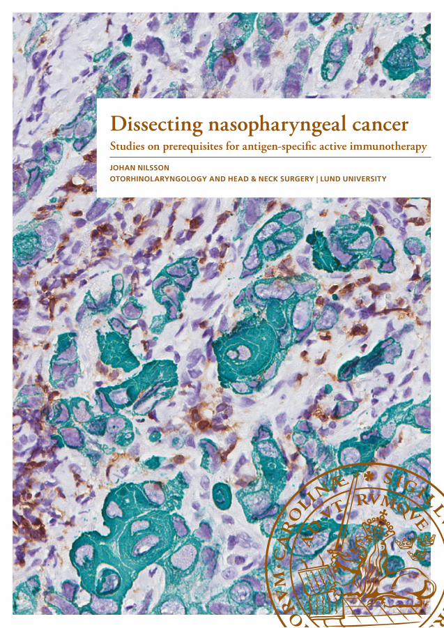
Front cover. Snapshot from the battle line: the human immune system vs. nasopharyngeal cancer. CD8 on T-lymphocytes is stained brown and cytokeratin in cancer cells is stained green. Photo: The author.

Dissecting nasopharyngeal cancer

Dissecting nasopharyngeal cancer Studies on prerequisites for
antigen-specific active immunotherapy
Johan Nilsson
DOCTORAL DISSERTATION
by due permission of the Faculty of Medicine, Lund University, Sweden. To be defended at the Torsten Landberg Lecture Hall, Department of Oncology
and Radiation Physics on May 15th 2020 at 13.15
Faculty opponent Professor Göran Laurell, Otorhinolaryngology and Head & Neck Surgery,
Department of Surgical Sciences, Uppsala University, Sweden
Organization LUND UNIVERSITY, Department of Clinial Sciences, Otorhinolaryngology and Head & Neck Surgery
Document name DOCTORAL DISSERTATION
Date of issue May 15th, 2020
Author: Johan Nilsson Sponsoring organization
Title and subtitle Dissecting nasopharyngeal cancer – Studies on prerequisites for antigen-specific active immunotherapy Abstract Background: Nasopharyngeal cancer (NPC) is frequently associated with the Epstein-Barr virus (EBV). Accordingly, attempts have been made to utilise the presence of non-human EBV-antigen on NPC cells therapeutically by immunological interventions. In this context, data are lacking with regard to levels of EBV antigen within the tumour as well as to intralesional presence and distribution of CD8+ T-cells and antigen-presenting dendritic cells (DCs) and their expression of targets that may favour cross-presentation of antigen (e.g., the pattern recognition receptor CD207). Arguably, such data may reflect specific “cancer immune phenotypes”, which in turn may be linked to clinical features of disease including survival. Furthermore, pattern recognition receptors may be targeted by specific adjuvants; γ-PGA-Phe nanoparticles may represent one such possibility. Methods: Fresh NPC biopsies were processed by multi-colour flow-cytometry focusing on DC subsets and their expression of the C-lectin receptor CD207. Formalin-fixated and paraffin-embedded NPC samples were retrieved and analysed for EBV-DNA quantitates and by immunohistochemistry focusing on CD207+ DCs and CD8+ T-cells. Specific cancer immune phenotypes were explored, based on presence and distribution of lymphocytes, and quantitates of intralesional CD207+ DCs and CD8+ T-cells were assessed with digital imaging. The association between the biological parameters and the clinical features of disease was examined. In a rat model, effects of topical exposure of γ-PGA-Phe nanoparticles on middle ear mucosa were examined. Results: EBV-DNA was present in NPC lesions (range: 0.0005-94617 copies/cell). The presence of CD8+ T-cells was particularly high in EBV-DNA-rich tumours. Subsets of DCs, i.e. CD123+ pDCs, CD1c+ mDCs, CD141+ mDCs, and CD1c-CD141-mDCs, were observed and a high frequency of CD207 expression was seen among CD1c+ mDCs compared to the other subsets. In NPC, CD207+ cells, likely representing CD1c+ mDCs, were particularly frequent in cancer cell areas. Specific “cancer immune phenotypes” were detected: “inflamed” (61.7%), “excluded” (29.8%), and “deserted” (8.5%) and CD8 ratios aggregated as “inflamed” > “excluded” > “deserted”. An EBV-DNA load of more than 70 copies/cell was associated with prolonged disease-free survival for EBV-DNA positive patients (p=0.046). Between immune phenotypes, a statistically significant difference in disease-free survival was observed between the “inflamed” and “excluded” subtypes (p=0.0090). γ-PGA-Phe nanoparticles produced a type-1 response characterized by generation of pro-inflammatory cytokines (IL-1α, IL-1β, IL-6, MIP-1α, and TNF-α) and inflammatory histopathological changes. Conclusions: Specific intralesional DC subpopulations are present in NPC and the C-lectin receptor CD207, involved in antigen cross-presentation, is frequently expressed. CD207+ DCs are constantly encountered among cancer cells in NPC. Lymphocyte-based cancer immune phenotypes can be demonstrated in NPC and this observation is confirmed by CD8+ T-cell quantification. The cancer immune phenotypes and intralesional EBV-DNA load predicts survival in NPC. γ-PGA-Phe NPs, antigen carriers with adjuvant properties, induces a local mucosal response compatible with a type-1 response. All these findings are relevant for future antigen-specific active immunotherapy for NPC.
Key words: nasopharyngeal cancer, Epstein-Barr virus, dendritic cells, CD207, CD8, cancer immune phenotypes, nanoparticles Classification system and/or index terms (if any)
Supplementary bibliographical information Language English
ISSN 1652-8220 ISBN 978-91-7619-916-9
Recipient’s notes Number of pages 92 Price
Security classification
I, the undersigned, being the copyright owner of the abstract of the above-mentioned dissertation, hereby grant to all reference sources permission to publish and disseminate the abstract of the above-mentioned dissertation.
Signature Date 2020-04-01
Dissecting nasopharyngeal cancer Studies on prerequisites for
antigen-specific active immunotherapy
Johan Nilsson
Copyright pp 1-92 Johan Nilsson
Article 1 © Oncology Letters, Spandidos Publications
Article 2 © Scientific Reports, Springer Nature
Article 3 © by the Authors (Unpublished manuscript)
Article 4 © Acta Oto-Laryngologica, Taylor & Francis
Department of Otorhinolaryngology and Head & Neck Surgery Faculty of Medicine Lund University
ISBN 978-91-7619-916-9 ISSN 1652-8220
Printed in Sweden by Media-Tryck, Lund University Lund 2020

Audere est Facere
Table of Contents
Abbreviations ........................................................................................................ 10 Original articles .................................................................................................... 13 Introduction .......................................................................................................... 15 Background ........................................................................................................... 17
The immune system ..................................................................................... 17 The immune system and cancer ................................................................... 21 Immunotherapy in cancer ............................................................................. 23 Nasopharyngeal cancer ................................................................................ 24 Epstein-Barr virus and cancer ...................................................................... 26 Epstein-Barr virus in nasopharyngeal cancer ............................................... 28 Human papilloma virus in nasopharyngeal cancer ....................................... 30 Current treatment of nasopharyngeal cancer ................................................ 31 The introduction of immunotherapy in nasopharyngeal cancer ................... 32 Antigen-specific immunotherapy in nasopharyngeal cancer ....................... 32 Intralesional CD8+ T-cells in nasopharyngeal cancer ................................. 35 Nanoparticles ................................................................................................ 35 Nanoparticles as carriers and adjuvants in antigen-specific active immunotherapy........................................................ 36 Certain prerequisites for antigen-specific active immunotherapy in nasopharyngeal cancer ........................................ 38
Materials and methods ......................................................................................... 39 I. ................................................................................................................... 39II. .................................................................................................................. 39III. ................................................................................................................. 40IV. ................................................................................................................ 40
Results .................................................................................................................... 41 I. ................................................................................................................... 41II. .................................................................................................................. 42III. ................................................................................................................. 44IV. ................................................................................................................ 48
Discussion .............................................................................................................. 49 Dendritic cells in nasopharyngeal cancer ..................................................... 49 Epstein-Barr virus in nasopharyngeal cancer ............................................... 50 Cancer immune phenotypes and CD8+ T-cells in nasopharyngeal cancer ................................................................................. 51 Prognostic factors in nasopharyngeal cancer ............................................... 52 The potential role for γ-PGA-Phe nanoparticles in antigen-specific active immunotherapy........................................................ 54 Methodological considerations .................................................................... 55
Conclusions ........................................................................................................... 59 Future perspectives and closing remarks ........................................................... 61 Sammanfattning på svenska ................................................................................ 63 Acknowledgements ............................................................................................... 67 Funding .................................................................................................................. 69 References ............................................................................................................. 71
10
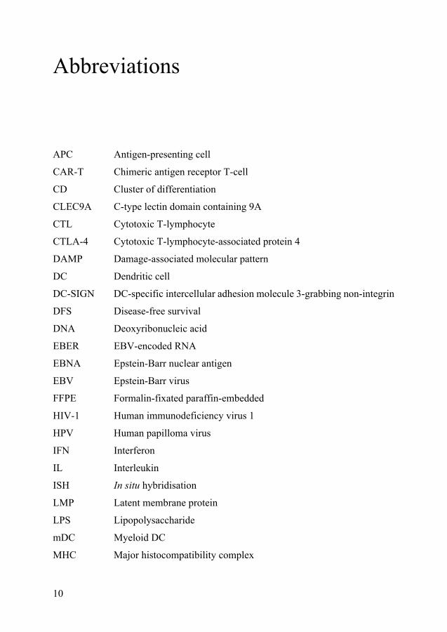
Abbreviations
APC Antigen-presenting cell
CAR-T Chimeric antigen receptor T-cell
CD Cluster of differentiation
CLEC9A C-type lectin domain containing 9A
CTL Cytotoxic T-lymphocyte
CTLA-4 Cytotoxic T-lymphocyte-associated protein 4
DAMP Damage-associated molecular pattern
DC Dendritic cell
DC-SIGN DC-specific intercellular adhesion molecule 3-grabbing non-integrin
DFS Disease-free survival
DNA Deoxyribonucleic acid
EBER EBV-encoded RNA
EBNA Epstein-Barr nuclear antigen
EBV Epstein-Barr virus
FFPE Formalin-fixated paraffin-embedded
HIV-1 Human immunodeficiency virus 1
HPV Human papilloma virus
IFN Interferon
IL Interleukin
ISH In situ hybridisation
LMP Latent membrane protein
LPS Lipopolysaccharide
mDC Myeloid DC
MHC Major histocompatibility complex
11
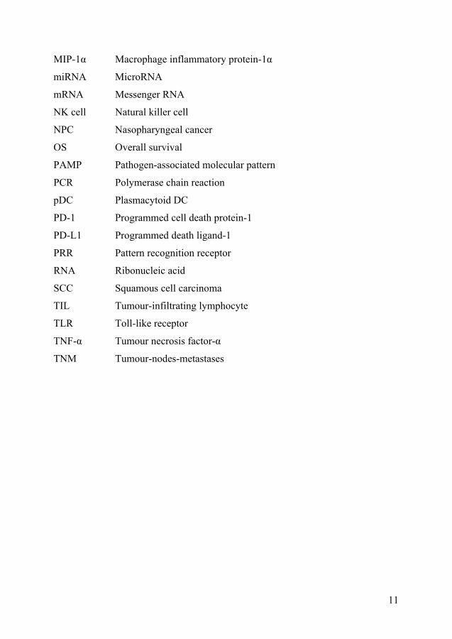
MIP-1α Macrophage inflammatory protein-1α
miRNA MicroRNA
mRNA Messenger RNA
NK cell Natural killer cell
NPC Nasopharyngeal cancer
OS Overall survival
PAMP Pathogen-associated molecular pattern
PCR Polymerase chain reaction
pDC Plasmacytoid DC
PD-1 Programmed cell death protein-1
PD-L1 Programmed death ligand-1
PRR Pattern recognition receptor
RNA Ribonucleic acid
SCC Squamous cell carcinoma
TIL Tumour-infiltrating lymphocyte
TLR Toll-like receptor
TNF-α Tumour necrosis factor-α
TNM Tumour-nodes-metastases

12
13
Original articles
I. Nilsson JS, Abolhalaj M, Lundberg K, Lindstedt M, Greiff L. Dendritic cell subpopulations in nasopharyngeal cancer. Oncol Lett. 2019; 17: 2557-61.
II. Nilsson JS, Forslund O, Andersson FC, Lindstedt M, Greiff L. Intralesional EBV-DNA load as marker of prognosis for nasopharyngeal cancer. Sci Rep. 2019; 9: 15432.
III. Nilsson JS, Sobti A, Erjefält JS, Forslund O, Lindstedt M, Greiff L. Immune phenotypes of nasopharyngeal cancer. Manuscript.
IV. Nilsson JS, Broos S, Akagi T, Akashi M, Hermansson A, Cayé-Thomasen P, Lindstedt M, Greiff L. Amphiphilic γ-PGA nanoparticles administered on rat middle ear mucosa produce adjuvant-like immunostimulation in vivo. Acta Otolaryngol. 2014; 134: 1034-41.

14
15
Introduction
Nasopharyngeal cancer (NPC) is a distinct entity among epithelial cancers of the upper aerodigestive tract, frequently associated with the Epstein-Barr virus (EBV) and with an often extensive infiltration of immune cells. In EBV-related cases, the cancer cells consistently express virus-specific markers that may be regarded as targets in potential antigen-directed treatment such as active immunotherapy. Furthermore, individual patterns of intralesional immune cell infiltration, and the particular presence of distinct immune cell subsets, may carry prognostic information and may be therapy-directing in terms of suitability for e.g. immunotherapy. In conclusion, nasopharyngeal cancer, in particular the EBV-driven form, is an interesting candidate to explore for antigen-specific active immunotherapy.
The overall objective of this thesis was to investigate certain aspects of NPC as necessary background information for future antigen-specific active immuno-therapy and for patient selection. In addition, the purpose was to investigate the local inflammatory effect on a target mucosa of an antigen carrier/adjuvant system that could be used in such an immunotherapeutic setting.
Specific aims were:
I. To study whether or not intralesional dendritic cells (DCs) are present in untreated NPC and, if so, which DC subtypes they represent and whether or not they express the C-lectin receptor CD207.
II. To quantitate intralesional EBV-DNA in untreated NPC and investigate its relationship with EBV-encoded RNA (EBER) and human papilloma virus (HPV) as well as with clinical presentation and prognosis.
III. To explore immune phenotypes in NPC and quantitate intralesional CD8+ T-cells and CD207+ DCs. Furthermore, to explore associations between these features and intralesional EBV-DNA/EBER and clinical/ prognostic information, respectively.
IV. To investigate whether or not specific nanoparticles (γ-PGA-Phe NPs), which are able to function as antigen carriers and adjuvants, may produce a desirable immune response in a target mucosa.

16
17
Background
The immune system The human immune system consists of two integrated arms. The innate arm (i.e. the native or non-specific immunity) reacts to non-specific molecular patterns, expressed by damaged cells and pathogens, and executes a rapid early consistent response. The adaptive arm (i.e. the acquired or specific immunity) reacts to highly specific molecular components (antigens) and regulate a subsequent tailored response that also induces a memory. Cells of the innate arm include natural killer (NK) cells and the myeloid immune cells: mast cells, eosinophils, basophils, neutrophils, macrophages and DCs. The innate defence also includes the complement system and the chemical, physiological and structural barriers of the human body. An important feature of the innate response is the activation of the adaptive response [1].
The adaptive response, mediated by B- and T-lymphocytes (B- and T-cells) comprises two main types of effector responses: the cell-mediated immunity and the humoral immunity. Activated CD8+ T-cells, i.e. cytotoxic T-lymphocytes (CTLs), and NK-cells are the main effector cells of the cell-mediated response, while the humoral response is primarily effectuated by B-cell produced antibodies. The response patterns are orchestrated by CD4+ T-cells, termed helper T-cells (Th-cells), which can be sub-classified as e.g. Th1-, Th2- and Th17-cells; activators and stimulators of different types of adaptive effector responses. The Th1-cells mediate the type-1 response, equalling the cell-mediated response, directed against intracellular pathogens, but also against defective cells such as cancer cells. The type-2 response, i.e. the humoral response, is mediated by Th2-cells and is typically directed against extracellular pathogens. Th17-cells are also mediators of the defence against extracellular pathogens. In addition, the different CD4+ cell subsets are also activators of myeloid immune cells of the innate arm: macrophages within the type-1 response, mast cells, eosinophils and basophils within the type-2 response, and neutrophils by CD4+ Th17-cells [1]. Notably, an additional lineage of CD4+ Th-cells exists, the rare Treg-cells, which are responsible for down-regulating the immune responses when appropriate [2-4].
The innate response partly relies on expression of different pattern recognition receptors (PRRs); membrane-bound or cytosolic receptors that react on danger signals. The most important PRRs are the Toll-like receptors (TLRs) and the C-
18
lectin receptors. PRRs are highly conserved receptor structures that bind to pathogen-associated molecular patterns (PAMPs) and damage-associated molecular patterns (DAMPs). PAMPs are structures unique for microbes that are also highly conserved; examples include lipopolysaccharides (LPS) of gram-negative bacteria and double-stranded RNA of replicating viruses. DAMPs are structures related to cell damage and cell death. PRRs are expressed on most cells of the innate immune system [5, 6]. PRR ligands that are not considered as PAMPs or DAMPs, which elicit an immune response, may be considered adjuvants.
Apart from detection of pathogens, an important element of the immune response is the identification and annihilation of faulty own cells, for example infected cells and tumour cells. For this purpose, the major histocompatibility complex (MHC) molecules constantly present peptides to the adaptive arm of the immune system. MHC class I molecules are found on most cells of the human body. They present intracellular antigen to effector cells, reflecting what resides inside the particular cell, and thus potentially evoking a response towards it. In contrast, MHC class II molecules are found solely on antigen-presenting cells (APCs), comprising macrophages, B-cells and DCs. These are cells that detect and internalise extracellular antigen and present it to CD4+ T-cells, thereby setting the stage for a possible adaptive effector response and linking the innate and the adaptive arm of the immune system. The interaction with T-cells is facilitated through binding of CD80 and CD86 on APCs to the co-stimulatory T-cell membrane protein CD28 [1, 7].
DCs, characterised in the early 1970s [8], are the most potent of the APCs and key regulators of the T-cell polarization [9] (Figure 1). The DC population is small and heterogeneous. DCs are often sub-classified as CD1c+, CD141+ and CD16+ myeloid DCs (mDCs), and CD123+ plasmacytoid DCs (pDCs) [10], but subpopulations exist that fall outside this way of categorisation, such as CD1c- CD141- mDCs [11, 12]. DCs express PRRs that differ between subsets, which can also be used for classification [10]. These differences also imply that the obtained effects at activation differs, skewing the response in various directions, for example into a type-1 or a type-2 response [13, 14].
19
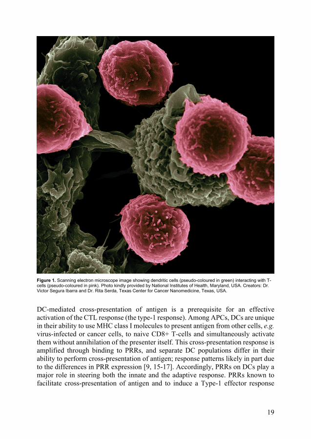
Figure 1. Scanning electron microscope image showing dendritic cells (pseudo-coloured in green) interacting with T-cells (pseudo-coloured in pink). Photo kindly provided by National Institutes of Health, Maryland, USA. Creators: Dr. Victor Segura Ibarra and Dr. Rita Serda, Texas Center for Cancer Nanomedicine, Texas, USA.
DC-mediated cross-presentation of antigen is a prerequisite for an effective activation of the CTL response (the type-1 response). Among APCs, DCs are unique in their ability to use MHC class I molecules to present antigen from other cells, e.g. virus-infected or cancer cells, to naive CD8+ T-cells and simultaneously activate them without annihilation of the presenter itself. This cross-presentation response is amplified through binding to PRRs, and separate DC populations differ in their ability to perform cross-presentation of antigen; response patterns likely in part due to the differences in PRR expression [9, 15-17]. Accordingly, PRRs on DCs play a major role in steering both the innate and the adaptive response. PRRs known to facilitate cross-presentation of antigen and to induce a Type-1 effector response
20
include TLR2, TLR4, Dectin-1, Dectin-2, DEC205, CLEC9A, DC-SIGN (CD209) and CD207 [18-21].
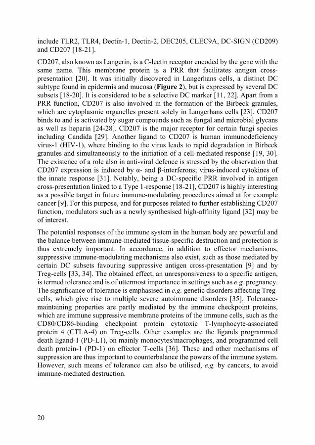
CD207, also known as Langerin, is a C-lectin receptor encoded by the gene with the same name. This membrane protein is a PRR that facilitates antigen cross-presentation [20]. It was initially discovered in Langerhans cells, a distinct DC subtype found in epidermis and mucosa (Figure 2), but is expressed by several DC subsets [18-20]. It is considered to be a selective DC marker [11, 22]. Apart from a PRR function, CD207 is also involved in the formation of the Birbeck granules, which are cytoplasmic organelles present solely in Langerhans cells [23]. CD207 binds to and is activated by sugar compounds such as fungal and microbial glycans as well as heparin [24-28]. CD207 is the major receptor for certain fungi species including Candida [29]. Another ligand to CD207 is human immunodeficiency virus-1 (HIV-1), where binding to the virus leads to rapid degradation in Birbeck granules and simultaneously to the initiation of a cell-mediated response [19, 30]. The existence of a role also in anti-viral defence is stressed by the observation that CD207 expression is induced by α- and β-interferons; virus-induced cytokines of the innate response [31]. Notably, being a DC-specific PRR involved in antigen cross-presentation linked to a Type 1-response [18-21], CD207 is highly interesting as a possible target in future immune-modulating procedures aimed at for example cancer [9]. For this purpose, and for purposes related to further establishing CD207 function, modulators such as a newly synthesised high-affinity ligand [32] may be of interest.
The potential responses of the immune system in the human body are powerful and the balance between immune-mediated tissue-specific destruction and protection is thus extremely important. In accordance, in addition to effector mechanisms, suppressive immune-modulating mechanisms also exist, such as those mediated by certain DC subsets favouring suppressive antigen cross-presentation [9] and by Treg-cells [33, 34]. The obtained effect, an unresponsiveness to a specific antigen, is termed tolerance and is of uttermost importance in settings such as e.g. pregnancy. The significance of tolerance is emphasised in e.g. genetic disorders affecting Treg-cells, which give rise to multiple severe autoimmune disorders [35]. Tolerance-maintaining properties are partly mediated by the immune checkpoint proteins, which are immune suppressive membrane proteins of the immune cells, such as the CD80/CD86-binding checkpoint protein cytotoxic T-lymphocyte-associated protein 4 (CTLA-4) on Treg-cells. Other examples are the ligands programmed death ligand-1 (PD-L1), on mainly monocytes/macrophages, and programmed cell death protein-1 (PD-1) on effector T-cells [36]. These and other mechanisms of suppression are thus important to counterbalance the powers of the immune system. However, such means of tolerance can also be utilised, e.g. by cancers, to avoid immune-mediated destruction.
21
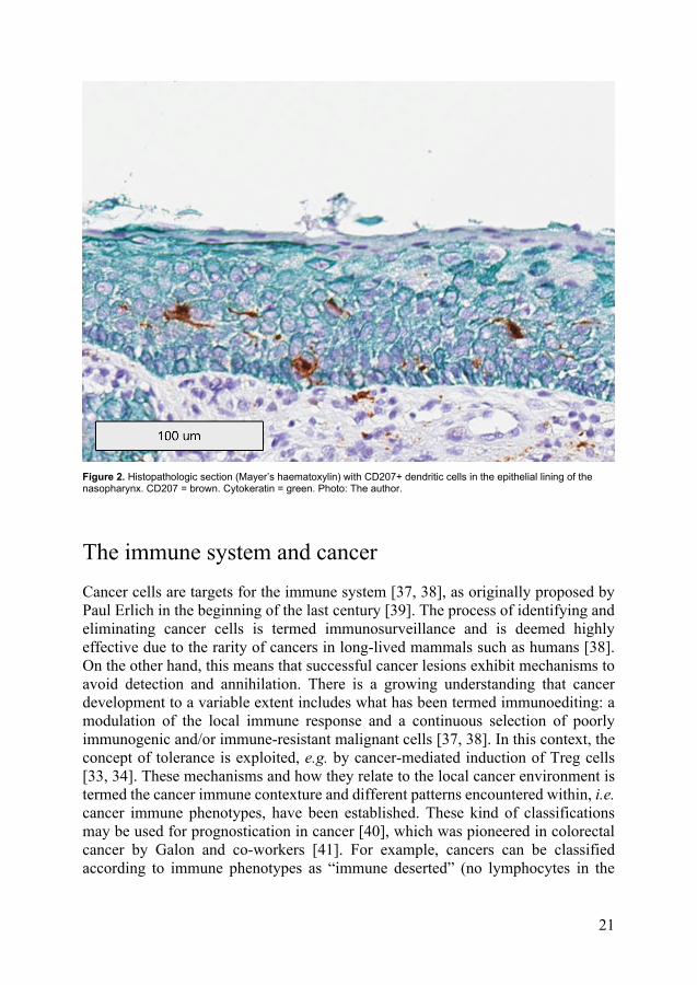
Figure 2. Histopathologic section (Mayer’s haematoxylin) with CD207+ dendritic cells in the epithelial lining of the nasopharynx. CD207 = brown. Cytokeratin = green. Photo: The author.
The immune system and cancer Cancer cells are targets for the immune system [37, 38], as originally proposed by Paul Erlich in the beginning of the last century [39]. The process of identifying and eliminating cancer cells is termed immunosurveillance and is deemed highly effective due to the rarity of cancers in long-lived mammals such as humans [38]. On the other hand, this means that successful cancer lesions exhibit mechanisms to avoid detection and annihilation. There is a growing understanding that cancer development to a variable extent includes what has been termed immunoediting: a modulation of the local immune response and a continuous selection of poorly immunogenic and/or immune-resistant malignant cells [37, 38]. In this context, the concept of tolerance is exploited, e.g. by cancer-mediated induction of Treg cells [33, 34]. These mechanisms and how they relate to the local cancer environment is termed the cancer immune contexture and different patterns encountered within, i.e. cancer immune phenotypes, have been established. These kind of classifications may be used for prognostication in cancer [40], which was pioneered in colorectal cancer by Galon and co-workers [41]. For example, cancers can be classified according to immune phenotypes as “immune deserted” (no lymphocytes in the
22
tumour), “immune excluded” (lymphocytes present but not infiltrating the tumour cells), and “immune inflamed” (lymphocytes infiltrate the tumour cells) [42].
Lymphocyte-based immune phenotypes in cancer also directly link to the concept of tumour-infiltrating lymphocytes (TILs); lymphocytes in direct cell-to-cell contact with tumour cells. These intralesional cells may be present in abundance in certain cancers, and the density of TILs has been suggested to be a positive prognostic factor in several types of cancer [43, 44]. Accordingly, clinical applications have been discussed, for example in breast cancer [45]. The presence of TILs is however obviously not enough to cure the cancer, stressing that other tumour strategies than avoiding the immune cells are also in play, such as creating an immunosuppressive environment. In accordance, numerous strategies of tumour cells to escape immunosurveillance have been described [46]. Examples of such are expression of PD-L1 on tumour cells [47] and induction of PD-1 expression on TILs [48] leading to impaired T-cell activation [46]. T-cell immune phenotype patterns arguably correlate to such different immune-evasive actions. This in turn may imply that apart from their prognostic value, these patterns may aid in choosing treatment strategy. In addition, they may point to possible strategies where certain phenotypes are transformed into ones more accessible to effective therapy [42]. For instance, “immune deserted” tumours do not respond well to certain types of immunotherapy [49]. On the other hand, there are strategies for shifting the immune phenotype pattern within a specific tumour, e.g. from “immune deserted” to “immune inflamed” [50]. This could make the tumour eligible to e.g. checkpoint inhibitor therapy. Arguably, immune phenotypes may also be associated with differences in intralesional patterns of other immune cells, such as DCs, which in turn may also be important in prognostication and selection of therapy.
The presence and function of tumour-infiltrating DCs have to date not been as thoroughly elucidated as that of TILs. Studies on different cancers [51, 52] including head and neck cancers [53] suggest that DCs infiltrate solid tumours, but that the suppressive environment created by the tumour prevents their maturation [54, 55]. In line with this, a study by Broz et al. [56] where intratumoural DC populations were delineated across species and cancers showed that DC presence in tumours were similar to that in normal tissues. Their conclusion was that efforts should be made to target those extremely rare subsets still capable of CTL activation. An alternative conclusion could instead be that the DC subsets are indeed present, but have to be activated, for example by PRR-ligands/adjuvants. This stresses that detailed knowledge of DC subsets, including their PRR repertoires, is of uttermost importance in specific cancers. In this context, it is interesting that DC phenotype patterns in tumour subsets may also be prognostic, as suggested in a study of breast cancer [57], possibly reflecting various tumour suppression patterns and further stressing the importance of DCs.
23
Immunotherapy in cancer The numerous interactions between a cancer lesion and the immune system and their relation to prognosis suggest that keys to successful cancer treatment might be found in immune modulating procedures, i.e. immunotherapy. This field have exploded in the last decade. To block how cancer cells take advantage of tolerance, i.e. unresponsiveness to antigen, is at present the most widely utilised immune modulating pathway in cancer therapy. Cancer cells can express PD-L1, which inhibits anti-cancer activity through engagement of PD-1 on effector T-cells [58]. Accordingly, inhibitors (monoclonal antibodies) of both PD-1 and PD-L1 have been developed. Inhibitors of CTLA-4 are blockers utilizing a different but in some ways similar mechanism, blocking the inhibitory APC-binding CTLA-4 checkpoint protein. PD-1, PD-L1 and CTLA-4 inhibitors are all examples of this new class of drugs termed immune checkpoint inhibitors [59]. These are currently introduced in various cancer treatment protocols, following encouraging results in for example melanoma treatment [60, 61].
Immunotherapy can be dependent on presence of tumour antigen or not, i.e. it can be antigen-specific or non-specific. Immunotherapy can also be categorised as adoptive or active, where adoptive immunotherapy alludes to procedures where effector cells are transferred to the recipient without activating the host immune system, whereas active immunotherapy relates to methods of steering the immune system into desired responses. Checkpoint inhibitor therapy is thus an example of non-specific active immunotherapy. It seems that a type-1 response is the most relevant mechanism for anti-cancer effects in an immune response, and part of the modulation exhibited by successful cancers are set to skew these kind of responses from an anti-tumour type-1 to a pro-tumour type-2 pattern [46, 62]. In accordance, an immunotherapeutic approach that has emerged is T-cell therapy, an adoptive strategy where modified type-1 effector cells, specifically CTLs, are used as therapeutic agents. A specific variety of T-cell therapy is chimeric antigen receptor T-cell therapy (CAR-T therapy), utilizing harvested and genetically altered T-cells expressing tumour-binding receptors. Chimeric in this sense means that both antigen-binding and T-cell activation functions are combined within a single receptor. However, despite impressive responses in certain cases, efficacy is yet low, especially for solid cancers [63].
In vaccination, i.e. antigen-specific active immunotherapy, originally pioneered by Jenner [64-67] and further developed by Pasteur [68-70], the immune system is taught to react to a specific antigen. This includes the induction of a memory. Such a memory-based immune response is by definition more rapid, larger and also qualitatively more efficient compared to a primary one. The concept of vaccination is foremost associated with prevention of infectious diseases, the most notable effort being the eradication of smallpox [71], but can also be applied to cancer treatment. The response is often augmented with an adjuvant: a compound that initiates, directs
24
and amplifies a facilitating immune response. The most widely used are the aluminium compounds. However, their major disadvantage is their incapacity to evoke a sufficient type 1-response [72], rendering them unsuitable in cancer therapy. An important group of adjuvants act through PRRs and some of these evoke the type-1 response desired in cancer therapy. Examples of such ligands are lipid A analogues, detoxified derivates of LPS that act as ligands for TLR4 [73], and glycan ligands that bind to the C-lectin receptor CD207 [18, 19].
To obtain an efficient antigen-specific active immunotherapy in e.g. cancer, a sufficient activation of effector cells is vital. The obvious targets to achieve this, considering their ability to cross-present antigen and elicit a desired type-1 response, are the DCs. Accordingly, DC-based cancer immunotherapies are of interest. These can be adoptive, utilising autologous DCs primed and directed ex vivo in a manner similar to T-cell therapy, but also active, eliciting the DC response in vivo through direct targeting of antigens [74]. There are several advantages with active immunotherapy in comparison to adoptive strategies in cancer therapy. In general, the response is considered more robust and effect duration may be long-lasting and even life-long [75]. The involvement of DCs implies the possibility of a broad immune response directed at several target antigens simultaneously and also provides the prerequisite for activation and expansion of all the different effector cells of cell-mediated resistance [76]. Furthermore, practical advantages exist. Costs are lower, larger vaccine quantities can be produced with higher consistency and reproducibility and administration is easier. Furthermore, side effects are milder in comparison to the sometimes severe toxicities of the cancer immunotherapies utilised at present [63, 77, 78]. Thus, establishing desired effects through the body’s own immune system, and in that context presumably more controlled such effects, arguably would be beneficial.
Nasopharyngeal cancer Nasopharyngeal cancer (NPC) is a squamous cell carcinoma (SCC) of the nasopharynx (Figure 3). The histopathological appearance is often distinct and tend to differ from other SCCs [79]. There are also clinical differences, for example patients tend be younger (including childhood cases) [80, 81] and the metastatic pattern is different [82]. NPC is separately classified within the TNM cancer stage system [83], though there is a rising opinion that the current TNM system is insufficient for predicting prognosis and stratifying patients to appropriate treatment in this disease [84].

25
Figure 3. Positron emission tomography-computed tomography (sagittal view) visualising a nasopharyngeal cancer. The hypermetabolic tumour is denoted with a red arrow. With kind permission by the patient.
Worldwide, NPC accounts for >50000 deaths annually. The incidence is highly diverse, with a slight male predominance of approximately 2-3:1 [85], and with low rates seen in e.g. Northern Europe, Latin America and Japan [86]. As an example, the annual incidence in Sweden is 0.2-0.5 per 100000 over the past 50 years [87]. In contrast, high risk areas such as North Africa and parts of Southeast Asia exhibit
26
more than 50-fold higher rates, with the highest encountered in southern China and especially Hong Kong, the latter with an annual incidence for men of >20 per 100000. High rates are also seen among certain ethnic groups, including the Inuits of Greenland and Alaska. [85]. A large majority of all new cases in the world are diagnosed in Southeast Asia (71% in 2012) [88]. Interestingly, incidence rates, at least in endemic areas, have been steadily declining over the last decades, arguably reflecting lifestyle and environmental changes [84].
The reason for the large variation in NPC incidence is not fully understood, but it is believed to be multi-factorial and to include genetic and environmental factors such as diet. These predisposing factors include certain risk HLA alleles and intake of preserved foods [89-92]. In addition, rare constitutional variants in the MST1R gene, involved in the innate immune response, have been shown to be strongly associated with early-age onset [93]. However, the most important, although not mandatory, etiological factor of NPC is previous infection with EBV [92, 94]. It is well-established that EBV-driven disease is the by far most encountered form of NPC in endemic areas [88], and it is likely that the frequency of EBV negative NPC is approximately the same worldwide. EBV positive NPC is frequently associated with a non-keratinising histopathologic subtype and with prominent infiltrates of lymphoid cells [95]. Furthermore, clinical associations exist with EBV-associated NPC more often presenting with spread disease, yet with a better prognosis than the locally more aggressive EBV negative NPC [88].
Epstein-Barr virus and cancer In 1964, Michael Epstein, Yvonne Barr and Bert Achong discovered the herpes virus later named EBV and showed that the virus was the causative agent of the endemic childhood lymphoma termed Burkitt’s lymphoma [96]. This was the first discovery of an oncogenic virus in humans. Following that, EBV was found to be the causative agent of the infectious disease mononucleosis [97], a disease afflicting up to 90% of the worldwide population [98]. Subsequently, other lymphomas were associated with EBV, predominantly B-cell lymphomas, but also epithelial cancers [99]. The worldwide total burden of EBV-associated cancers per year is approximately 200000 cases, representing 1% of all cancers [100].
27
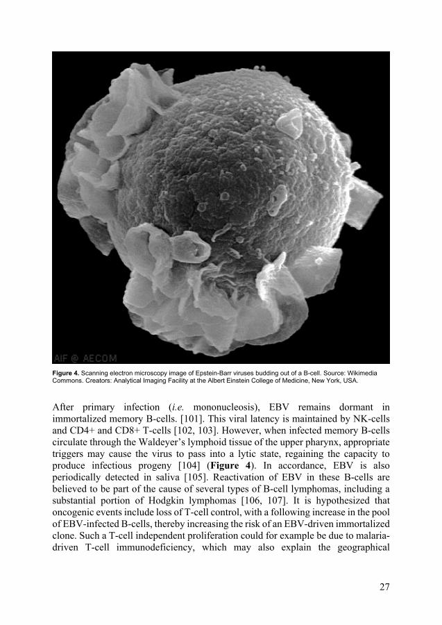
Figure 4. Scanning electron microscopy image of Epstein-Barr viruses budding out of a B-cell. Source: Wikimedia Commons. Creators: Analytical Imaging Facility at the Albert Einstein College of Medicine, New York, USA.
After primary infection (i.e. mononucleosis), EBV remains dormant in immortalized memory B-cells. [101]. This viral latency is maintained by NK-cells and CD4+ and CD8+ T-cells [102, 103]. However, when infected memory B-cells circulate through the Waldeyer’s lymphoid tissue of the upper pharynx, appropriate triggers may cause the virus to pass into a lytic state, regaining the capacity to produce infectious progeny [104] (Figure 4). In accordance, EBV is also periodically detected in saliva [105]. Reactivation of EBV in these B-cells are believed to be part of the cause of several types of B-cell lymphomas, including a substantial portion of Hodgkin lymphomas [106, 107]. It is hypothesized that oncogenic events include loss of T-cell control, with a following increase in the pool of EBV-infected B-cells, thereby increasing the risk of an EBV-driven immortalized clone. Such a T-cell independent proliferation could for example be due to malaria-driven T-cell immunodeficiency, which may also explain the geographical
28
distribution of Burkitt’s lymphoma, or due to HIV-driven T-helper cell depletion, explaining the association between HIV and EBV-associated B-cell lymphomas [108-110]. The importance of T-cell control is further stressed by the susceptibility of individuals with specific deficiencies in T-cell mediated immunity to fatal primary EBV infections [111, 112].
Epstein-Barr virus in nasopharyngeal cancer In addition to lymphomas, EBV is also the cause of certain epithelial cancers, predominantly NPC. The connection between EBV and NPC was made in the late 1960s and early 1970s by Georg Klein and co-workers [113, 114]. The underlying mechanism in epithelial cancers differs from that of B-cell lymphoma formation. There is no latent EBV-infection in epithelial cells. Instead, other pre-malignant changes, due to factors such as genetic susceptibility and environment, are thought to occur prior to a subsequent EBV-infection leading to the final cancer transformation [92, 115]. As opposed to the receptor-mediated EBV-infection of B-cells, the infection of epithelial cells is believed to occur through a cell-to-cell infection pathway [116-118].
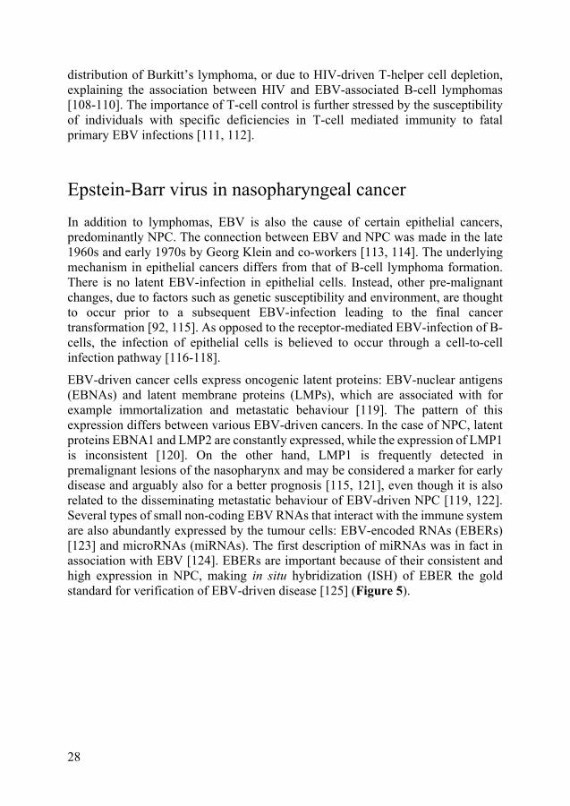
EBV-driven cancer cells express oncogenic latent proteins: EBV-nuclear antigens (EBNAs) and latent membrane proteins (LMPs), which are associated with for example immortalization and metastatic behaviour [119]. The pattern of this expression differs between various EBV-driven cancers. In the case of NPC, latent proteins EBNA1 and LMP2 are constantly expressed, while the expression of LMP1 is inconsistent [120]. On the other hand, LMP1 is frequently detected in premalignant lesions of the nasopharynx and may be considered a marker for early disease and arguably also for a better prognosis [115, 121], even though it is also related to the disseminating metastatic behaviour of EBV-driven NPC [119, 122]. Several types of small non-coding EBV RNAs that interact with the immune system are also abundantly expressed by the tumour cells: EBV-encoded RNAs (EBERs) [123] and microRNAs (miRNAs). The first description of miRNAs was in fact in association with EBV [124]. EBERs are important because of their consistent and high expression in NPC, making in situ hybridization (ISH) of EBER the gold standard for verification of EBV-driven disease [125] (Figure 5).
29
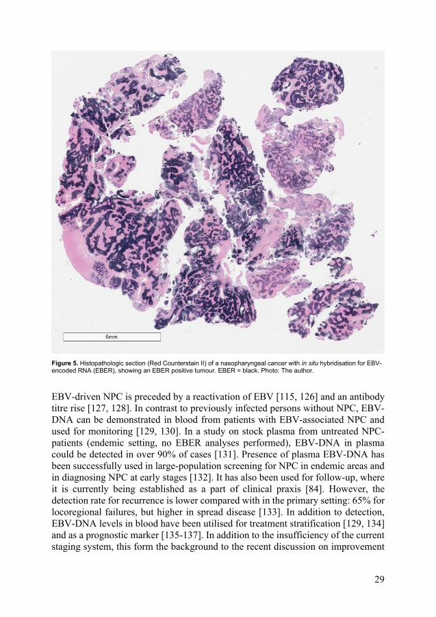
Figure 5. Histopathologic section (Red Counterstain II) of a nasopharyngeal cancer with in situ hybridisation for EBV-encoded RNA (EBER), showing an EBER positive tumour. EBER = black. Photo: The author.
EBV-driven NPC is preceded by a reactivation of EBV [115, 126] and an antibody titre rise [127, 128]. In contrast to previously infected persons without NPC, EBV-DNA can be demonstrated in blood from patients with EBV-associated NPC and used for monitoring [129, 130]. In a study on stock plasma from untreated NPC-patients (endemic setting, no EBER analyses performed), EBV-DNA in plasma could be detected in over 90% of cases [131]. Presence of plasma EBV-DNA has been successfully used in large-population screening for NPC in endemic areas and in diagnosing NPC at early stages [132]. It has also been used for follow-up, where it is currently being established as a part of clinical praxis [84]. However, the detection rate for recurrence is lower compared with in the primary setting: 65% for locoregional failures, but higher in spread disease [133]. In addition to detection, EBV-DNA levels in blood have been utilised for treatment stratification [129, 134] and as a prognostic marker [135-137]. In addition to the insufficiency of the current staging system, this form the background to the recent discussion on improvement
30
of staging procedures for NPC [84], suggesting inclusion of biomarkers in order to achieve more accurate prognostic prediction and support in choice of treatment [138-142].
EBV-DNA in nasopharyngeal brush samples has also been advocated as a tool for NPC diagnosis [143-145]. Furthermore, in one of these reports, Zheng et al. [145] showed that DNA-load in brush samples could reflect tumour stage. However, these EBV-DNA samplings must still be regarded as “extralesional” and judged on that basis. Intralesional EBV-DNA is much less explored. Present studies on truly intralesional EBV-DNA in NPC have focused on specificity and sensitivity in relation to diagnosis [146-148], while data on intralesional EBV-DNA load are extremely limited [149], albeit such information may be valuable. For example, EBV-DNA load as a marker for the potential of antigen presence and quantity, of interest in antigen-specific immunotherapy and in relation to prognosis, may be useful, similar to what has been shown for another oncogenic virus, human papilloma virus (HPV), and its relation to oropharyngeal cancer [150, 151] (Aim II).
Human papilloma virus in nasopharyngeal cancer In addition to the well-established relation with oropharyngeal cancer [152], HPV is linked to NPC [146, 153-162]. Compared to EBV positive disease, HPV positive NPC is associated with worse outcome in terms of local control and survival, while distant failures seem to be less common [154, 159]. Furthermore, HPV positivity correlates to increased survival compared to virus-negative NPC (neither EBV nor HPV), according to a recent nationwide study from Finland [158]. In contrast, two recent large studies – a study involving data on 956 patients [161] and a meta-analysis [160] – failed to correlate HPV positivity to survival. However, it should be noted that EBV-status was not established in these studies, thus the HPV negative cases likely included EBV positive patients with better prognosis and EBV negative patients with worse prognosis, in unknown proportions, making these data very hard to interpret. In all, the role of HPV as an oncogenic driver and prognostic marker in NPC is unclear, but it may be argued that HPV should be analysed in addition to EBV when evaluating NPC. In this context, it is worth considering that p16 overexpression, a well-established surrogate marker for HPV [163], and also suggested as such in NPC [158], is also frequently encountered in EBV-driven NPC [164].
31
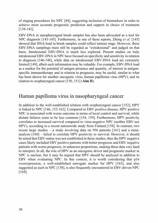
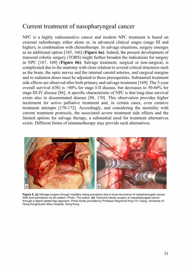
Current treatment of nasopharyngeal cancer NPC is a highly radiosensitive cancer and modern NPC treatment is based on external radiotherapy either alone or, in advanced clinical stages (stage III and higher), in combination with chemotherapy. In salvage situations, surgery emerges as an additional option [165, 166] (Figure 6a). Indeed, the present development of transoral robotic surgery (TORS) might further broaden the indications for surgery in NPC [167, 168] (Figure 6b). Salvage treatment, surgical or non-surgical, is complicated due to the anatomy with close relation to several critical structures such as the brain, the optic nerves and the internal carotid arteries, and surgical margins and re-radiation doses must be adjusted to these prerequisites. Substantial treatment side effects are observed after both primary and salvage treatment [169]. The 5-year overall survival (OS) is >80% for stage I-II disease, but decreases to 50-60% for stage III-IV disease [86]. A specific characteristic of NPC is that long time survival exists also in disseminated disease [88, 170]. This observation provides higher incitement for active palliative treatment and, in certain cases, even curative treatment attempts [170-172]. Accordingly, and considering the mortality with current treatment protocols, the associated severe treatment side effects and the limited options for salvage therapy, a substantial need for treatment alternatives exists. Different forms of immunotherapy may provide such alternatives.
Figure 6. (a) Salvage surgery through maxillary swing procedure due to local recurrence of nasopharyngeal cancer. With kind permission by the patient. Photo: The author. (b) Transoral robotic surgery of nasopharyngeal cancer through a lateral palatal flap approach. Photo kindly provided by Professor Raymond King-Yin Tsang, University of Hong Kong/Queen Mary Hospital, Hong Kong.
32
The introduction of immunotherapy in nasopharyngeal cancer In conformity with recent developments of cancer treatments, there is an increasing interest in checkpoint inhibitor therapy for head and neck cancer, including NPC. Furthermore, the finding that PD-L1 is frequently expressed in NPC [173, 174] stresses a potential benefit of checkpoint inhibitor therapy. It is presently unclear whether PD-1/PD-L1 expression in NPC is associated with worse survival [175-177] or not [173, 178-181]. Notably, Wang et al. [181] recently presented a specific immune signature, based on nine immune checkpoint markers including PD-L1, which could predict survival in patients eligible for curative treatment. Specifically, high expression of PD-L1 was associated with increased OS. Several trials have been conducted in NPC utilising PD-1 inhibition, specifically nivolumab [182-184] and pembrolizumab [185]. Furthermore, there are ongoing trials utilising PD-1 inhibitors: nivolumab (NCT03984357), pembrolizumab (NCT04227509), sintilimab (NCT04072107), toripalimab (NCT03907826, NCT03930498), and also the PD-L1 inhibitor durvalumab (NCT04231864), and nivolumab combined with the CTLA-4 inhibitor ipilimumab (NCT02834013). In addition, the PD-1 inhibitor camrelizumab has recently gained interest as an alternative for NPC, due to a beneficial toxicity profile in this particular diagnosis [186, 187], and trials are ongoing (NCT04143984, NCT04221516). However, only 20-30% of NPC patients seem to respond to current checkpoint inhibitor therapy [185, 186]. This is in agreement with findings for others cancers [188], warranting treatment adjustments and better patient selection, but also alternative treatments.
Antigen-specific immunotherapy in nasopharyngeal cancer The fact that EBV-driven disease is the major subgroup of NPC makes it reasonable to explore EBV-associated treatment options. The latent proteins expressed by EBV-related NPC are oncogenic, and drugs targeting these proteins are investigated [189, 190], including a present clinical trial on an EBNA-1-inhibitor in NPC (NCT03682055). Virus activation, with the underlying motive to make EBV visible to the immune system, and thereby susceptible to antiviral therapy [191], has gained interest as has gene therapy [192]. Furthermore, attempts have been made to create a prophylactic vaccine [193]. However, concerns have been raised that eradication of EBV would lead to a less effective immune protection against bacteria [194], a notion based on the hypothesis that the long co-evolution between EBV and humans has led to benefits also for the human counterpart [195]. Most importantly however,
33
EBV-associated NPC constantly expresses non-human EBV-associated targets, i.e. antigen, implicating a capacity for antigen-specific immunomodulation. In combination with the fact that this type of cancer is considered a highly immunoactive cancer, with often an abundance of intralesional immune cells [196] and with numerous immune regulatory molecules as well as CD4+ and CD8+ T-cell targets expressed on cancer cells [197-199], NPC should make an excellent candidate for antigen-specific immunotherapy.
Antigen-specific adoptive immunotherapy Adoptive immunotherapy, with transfer of either ex vivo activated CTLs or genetically altered T-cells (CAR-T) that bypass the antigen-presenting procedure, has been utilised in NPC. The underlying mechanisms of CTL transfer effects in NPC have been explored [200], and several clinical trials have been conducted in advanced NPC, for both salvage treatment [201-210] and first line treatment [211, 212]. The results, albeit promising, have been difficult to interpret due to parallel chemotherapy in the majority of trials. At present, there are several CD8+ T-cell transfer patient trials ongoing (NCT00516087, NCT00706316, NCT03925896, NCT03044743, NCT02421640). Attempts have also been made to utilize CAR-T therapy in NPC, with effects shown on a xenograft model [213], and patient trials are under way (NCT02915445, NCT03648697, NCT04107142). A variant of autologous immune cell transfer is the utilisation of an NK-cell based strategy, theoretically safer due to the very short life span of these effector cells. There are still no published data available, but two studies are ongoing (NCT02507154, NCT03007836). Further strategies for NK-cell mediated killing of NPC have been proposed based on in vitro trials, suggesting a combination with IFN-β and checkpoint inhibitor therapy [214]. Finally, patient trials with transfer of autologous EBV-activated DCs, an approach mimicking active immunotherapy, have shown partial tumour response [215-217], and further such trials are ongoing (NCT03282617, NCT03047525; the latter combined with NK cell transfer).
However, all the above mentioned procedures have limitations. Toxicities may be substantial, such as described for CAR-T therapy [63]. Importantly, treatment is frequently delayed due to the lengthy ex vivo expansion process (12-16 weeks for CTL transfer), during which patients might turn ineligible for treatment. To avoid this problem, Li et al. [212] took an interesting approach in their CTL transfer trial and used a rapid expansion protocol (4-5 weeks) that utilised TILs to hasten the process. However, further important limitations of adoptive transfers are that they are complicated, expensive and that no immunological memory is produced. Altogether, this renders the active DC targeting variety of antigen-specific immunotherapy interesting.
34
Antigen-specific active immunotherapy Trials exploring DC-targeting antigen-specific active immunotherapy in NPC are scarce. MVA-EL, a recombinant vaccinia virus encoding a functionally inactive EBNA1/LMP2 fusion protein, has been advocated as a mean to evoke a bodily-specific immune response, i.e. a vaccination response. Hui et al. [218] conducted a phase I-study comprising 18 locally advanced cases of endemic NPC in remission. The vaccine was well-tolerated and produced a substantial increase in circulating EBNA1- and LMP2-specific T-cell levels. Due to the endemic origin of the EBV strains used, a similar trial was conducted in a separate, non-endemic population showing a similar effect [219]. Further trials with MVA-EL are underway, with a phase Ib trial (NCT01800071) completed but not published and a phase II trial (NCT01094405) planned to be finished at the end of 2021. An alternative viral vector utilised is the adenovirus-based rAd5, carrying LMP2 (rAd5-EBV-LMP2). This was also reported to be well-tolerated in a phase I-trial [220]. In conclusion, experiences with active immunotherapy for NPC are promising but limited.
It has been suggested that antigen-specific active immunotherapy will be the future standard treatment for NPC, tentatively in combination with checkpoint inhibitors [88]. This places high demands on detailed knowledge of immunoactive cells and the immune environment in NPC. In contrast, especially regarding DCs, studies in NPC are few, and include, apart from early morphological characterizations, only occasional studies on intralesional DC subtypes [221-227]. In addition, data on the potential influence of DCs on prognostication are scarce and conflicting [199, 225]. In conclusion, data on DCs in NPC are limited and further studies are warranted, since the ability of adequate antigen presentation is fundamental in antigen-specific active immunotherapeutic interventions. Which intralesional DC subtypes that are present, and in which proportions, as well as their expression of relevant PRRs such as the C-lectin receptor CD207 need to be established. Sufficient data on intralesional DCs as a clinical and prognostic factor are also lacking, and arguably such studies should be related to EBV-status (Aim I and III).
If antigen-specific active immunotherapy in cancer is to be considered as a future treatment alternative, also the vaccination route is of interest. Apart from subcutaneous and systemic injections, currently there is an interest in direct vaccination into the tumour [228] or into adjacent lymph nodes [229]. Furthermore, the concept of mucosal vaccination is intriguing with arguably reduced adverse effects and a better local response, the latter of interest in mucosal cancers such as NPC, and benefits such as a simplified storage process and easier administration [230]. Other key features of the vaccination process is how to deliver the antigen to the appropriate cells, i.e. the DCs, and how to elicit an effective adjuvant activation leading to a type-1 response.
35
Intralesional CD8+ T-cells in nasopharyngeal cancer For an efficient immune reaction targeting cancer, a type-1 response is required [46, 62]. Accordingly, for effective antigen-specific active immunotherapy in cancers such as NPC, in-depth knowledge on type-1 effector cells is required, specifically at the tumour site. Compared to DCs, more data exist on intralesional lymphocytes, including CD8+ T-cells [227, 231-233]. Data also stem from trials with transfer of autologous TILs in NPC treatment [212]. The importance of TIL presence is indirectly supported by the finding that a favourable prognosis is associated with TIL density for many types of cancer [43], including head and neck cancer; CD4+ TILs [234] and CD3+ TILs [235]. Specifically, this has also been demonstrated for CD8+ TILs in NPC [180, 236], including in a recent large patient cohort where Wang et al. [237] also advocated TILs (with a broad definition) as an independent positive prognostic biomarker in NPC. This is in contrast to findings of Lu et al. [227], who characterized the intralesional density of several inflammatory cells including CD8+ T-cells in NPC, though without exploring the corresponding immune phenotype T-cell patterns. They associated high intralesional CD8+ T-cell density with worse survival, an observation that they explained by functional inactivation of TILs described in NPC [238], with the suggestion that successful cancer immunoediting could manifest as high load of harmless immune cells. The density of TILs may on the other hand also be viewed upon as a reflection of the immunological capacity of NPC. This perspective is further developed with the concept of lymphocyte-based immune phenotypes as a prognostic and therapy-deciding tool in cancer [42]. These are hitherto not evaluated in NPC, but the need to evaluate the cancer immune phenotype concept as a clinical and prognostic marker also for this disease is obvious. In this context, it is natural to expand on the phenotype concept into correlating the findings with TIL quantitates, at first hand CD8+ T-cells, and to evaluate these for prognostic value that could be therapy-directing with regards to e.g. immunotherapy. These analyses, as always in NPC, should be performed with knowledge of EBV-status (Aim III).
Nanoparticles Nano is the SI-prefix for 10-9 (0.000000001) [239] and nanotechnology operates within the nanoscale (1-100 nm). The nanoscale is synonymous with the molecular scale; to exemplify, the smallest atoms are approximately a quarter of a nanometre in diameter. In comparison, the smallest known cellular life forms, the mycoplasma bacteria, measure approximately 200 nm. The nanoscale is also the scale wherein the movement, behaviour and surface of solitary particles start to affect the system they exist in, thus the classical laws of physics are abandoned in favour of the laws of quantum physics [240]. Nanoparticles (NPs) are particle units defined by size,
36
generally 10-1000 nm [241] (Figure 7). Nanomedicine is the part of nanotechnology that is associated with medical applications. For example, NPs can be used for intracellular access, they may have effect propensities (nanodrugs) and they might act as carriers [242].
Figure 7. A nanoparticle (actual size) measuring 200nm. It would take 5000 of these nanoparticles to cover the distance of 1 mm. Figure: The author.
Nanoparticles as carriers and adjuvants in antigen-specific active immunotherapy In cancer therapy, NPs are of substantial interest for example in direct targeting of cancer cells [243] and in intracellular delivery of chemotherapeutics [244], but also as facilitators of antigen-specific active immunotherapy [245]. Biodegradable polymeric NPs have gained interest due to their ability to act as carriers of peptide vaccines. They are easily manufactured and can be modified according to which antigen to carry. Some of them can also act as presenters of antigen to APCs and also function as adjuvants [246]. The most widely investigated NPs in this context are manufactured from poly(lactic-co-glycolic) acid (PLGA) polymers, approved for use in humans by the European Medicine Agency (EMA) and the United States Food and Drug Administration (FDA) [247, 248]. However, these NPs show several disadvantages, with low encapsulation efficiency of water-soluble proteins and instability at formulation, freeze-drying and storage. An alternative might be nanoparticles formed by the naturally occurring polymer poly(γ-glutamic acid) (γ-PGA).
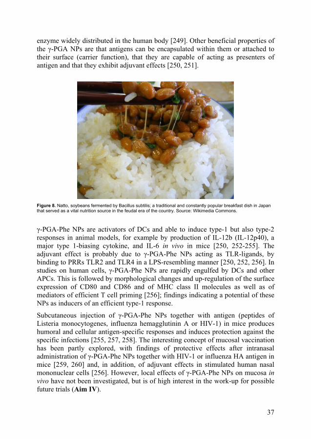
γ–PGA are polymers produced by Bacillus subtilis [249], a bacteria species most renowned for its use in the production of natto (Figure 8). Typically they are hydrophobically modified with for example L-phenylalanine ethylester to gain an amphiphilic nature. This enables them to spontaneously form NPs, γ-PGA graft-L-phenylalanine ethylester NPs (γ-PGA-Phe NPs), 30-400 nm in diameter, in aqueous solutions. These NPs are fully biodegradable by g-glutamyl transpeptidase, an
37
enzyme widely distributed in the human body [249]. Other beneficial properties of the γ-PGA NPs are that antigens can be encapsulated within them or attached to their surface (carrier function), that they are capable of acting as presenters of antigen and that they exhibit adjuvant effects [250, 251].
Figure 8. Natto, soybeans fermented by Bacillus subtilis; a traditional and constantly popular breakfast dish in Japan that served as a vital nutrition source in the feudal era of the country. Source: Wikimedia Commons.
γ-PGA-Phe NPs are activators of DCs and able to induce type-1 but also type-2 responses in animal models, for example by production of IL-12b (IL-12p40), a major type 1-biasing cytokine, and IL-6 in vivo in mice [250, 252-255]. The adjuvant effect is probably due to γ-PGA-Phe NPs acting as TLR-ligands, by binding to PRRs TLR2 and TLR4 in a LPS-resembling manner [250, 252, 256]. In studies on human cells, γ-PGA-Phe NPs are rapidly engulfed by DCs and other APCs. This is followed by morphological changes and up-regulation of the surface expression of CD80 and CD86 and of MHC class II molecules as well as of mediators of efficient T cell priming [256]; findings indicating a potential of these NPs as inducers of an efficient type-1 response.
Subcutaneous injection of γ-PGA-Phe NPs together with antigen (peptides of Listeria monocytogenes, influenza hemagglutinin A or HIV-1) in mice produces humoral and cellular antigen-specific responses and induces protection against the specific infections [255, 257, 258]. The interesting concept of mucosal vaccination has been partly explored, with findings of protective effects after intranasal administration of γ-PGA-Phe NPs together with HIV-1 or influenza HA antigen in mice [259, 260] and, in addition, of adjuvant effects in stimulated human nasal mononuclear cells [256]. However, local effects of γ-PGA-Phe NPs on mucosa in vivo have not been investigated, but is of high interest in the work-up for possible future trials (Aim IV).
38
Certain prerequisites for antigen-specific active immunotherapy in nasopharyngeal cancer In summary, detailed knowledge on intralesional DCs and their receptor repertoire is mandatory in designing an efficient antigen-specific active immune response to NPC, but at present data are scarce. Arguably, an analysis of intralesional DC-subtypes and their receptors, especially such receptors that may promote antigen presentation and facilitate a cell-mediated effector response, is of importance. The presence and distribution of effector cells must also be elucidated and the tumour-driven immunosuppression analysed. With regards to the latter, knowledge on the cancer immune phenotypes present, previously not explored in NPC, could be utilised for case selection.
In addition, EBV-status is important in the adaption of this concept. Verification of EBV antigen is fundamental, but also their localisation and immunogenicity. Arguably, also antigen quantity matters, which may be reflected by intralesional viral DNA load. Knowledge of the latter is extremely limited.
To establish a desired activation pattern directed at specific antigens, there is, among other prerequisites, a need for correct delivery to, and presentation for, the correct DC subsets. This includes proper adjuvant co-stimulation; specific nanoparticles may fill this role. γ-PGA-Phe NPs are possible candidates, but knowledge is lacking of their effect on target tissue in vivo, such as the mucosa of epithelial cancers, e.g. NPC.
Finally, the present staging system for NPC is insufficient. Virus-driven disease seems to have a better prognosis, and virus status must reasonably be evaluated and included in this context. Furthermore, in NPC, plasma load of EBV-DNA is promising as a prognostic factor, while intralesional load has not been elucidated. Also, histopathological cancer immune phenotyping, suggested as a means for selection for immunotherapeutic interventions, has shown promise as a prognostic factor for other cancers, while the concept has not been evaluated in NPC. In this context, the intralesional presence and quantity of specific immune cells may serve as marker for certain immune phenotypes, and themselves be utilised as prognostic and therapy-directing features. In summary, better prognostic tools would help selecting the correct patients for more or less aggressive therapy, including selecting the correct patients for possible future antigen-specific active immunotherapy.
39
Materials and methods
I. In this study, five patients with untreated NPC were recruited. Tumour biopsies were obtained and half of each sample was sent for routine histopathology. A single-cell suspension was prepared from the remaining half and subsequently treated with an antibody panel for leucocyte subset selection and CD207. Samples were analysed with a multi-colour flow-cytometry. A gating strategy was utilised to identify mDCs and pDCs based on expression of CD11c and CD123, respectively, and mDCs were then further subdivided into CD141+, CD1c+ and CD1c-CD141- cells. In addition, cell surface expression of CD207 was assessed with a similar gating strategy.
The study protocol was approved by the Regional Ethical Review Board at Lund University (2014/115) and written informed consents from all participants were obtained.
II. This study was of a retrospective design. It involved an analysis of 48 patients diagnosed with NPC between 2001 and 2015. A re-evaluation of patient data was performed and the primary (pre-treatment) tumour biopsies were re-examined focusing on histopathology and EBER expression. In addition, presence of high-risk HPV was investigated as well as p16-expression for HPV positive samples. Furthermore, formalin-fixated paraffin-embedded (FFPE) primary biopsy material was retrieved, prepared and DNA was extracted. EBV-DNA was then quantitated utilizing the single copy gene encoding EBNA1 as marker. Clinical and prognostic features such as cancer stage and survival could then be assessed in relation to EBV-DNA load and HPV-status.
Ethical approval (II-III) was granted by the Regional Ethical Review Board at Lund University (2014/117). In accordance with the approval, informed consent was not required due to the use of historical biopsy material. Prior to effectuation the study design was advertised in selected printed media, with the possibility to opt-out, as specified in the approval.
40
III. In this study, the patient cohort in the previous article (II) was revisited. New slides were prepared using FFPE primary biopsy material and stained for cytokeratin (all slides) and CD8 or CD207, respectively. All slides were digitally scanned. One case was excluded due to insufficient material for the analyses; this was incidentally also the only patient lacking follow-up, consequently 47 cases were analysed. Based on presence and distribution of lymphocytes, notably CD8+ T-cells, and blinded to EBER-status, the tumours were classified into immune phenotypes: “immune inflamed” (lymphocytes infiltrate tumour cells), “immune excluded” (lymphocyte presence in the cancer environment but no infiltration among tumour cells), and “immune deserted” (no lymphocytes). Furthermore, CD8+ and CD207+ cells in the whole biopsies as well as in selected areas were subjected to microdissection utilising a quantitative digital image technique generating a frequency of tissue area for each marker in comparison to the tissue area in total. The phenotype patterns and the quantitate frequencies were analysed in relation to each other and to intralesional EBV-DNA as well as to clinical stage and survival. Separately, gene expression data of 31 NPCs and 10 controls were retrieved from a public database and analysed in silico with focus on specific inflammatory signatures and on specific immune cell markers.
IV. In this blinded randomized sham-controlled study, biodegradable amphiphilic γ-PGA-Phe NPs were synthesized. Following transcervical dissection, 46 rats were subjected to superfusion of the right middle ear mucosa with either low-dose (n=17) or high-dose (n=9) of γ-PGA-Phe NPs, or sham (n=20). After four or twelve hours, according to a predetermined protocol, a transmyringial lavage was performed. After sacrifice, the heads were dissected and prepared for histology. The subsequent procedures were performed blinded to previous treatment. Cytokine concentrations (IL-1α, IL-1β, IL-6, IL-10, MIP-1α, MCP-1α, TNF-α and IFN-γ) in ear lavage fluids were assessed. The temporal bones were stained for tissue morphology, the amount of fluid in the middle ear cavity was semi-quantitated, and the thickness of the middle ear mucosa as well as of the round window membrane and the tympanic membrane was measured. Results of cytokine analyses and histopathological changes were assessed with regards to given treatment.
The study was approved by the Regional Ethical Review Board at Lund University (M240-2007) and animal experiments were performed in accordance with this as well as with local protocol (Department of Medical Microbiology, Lund University).
41
Results
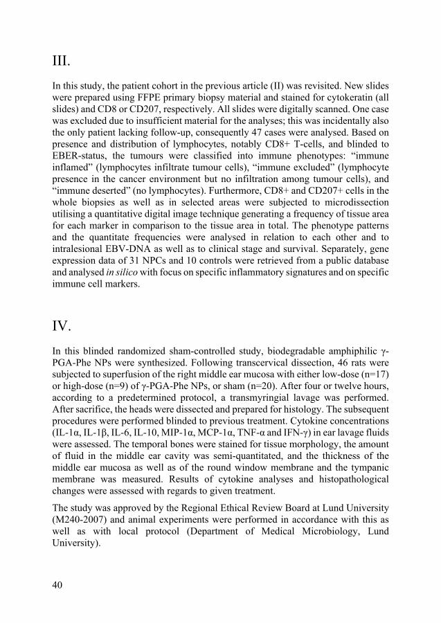
I. DCs are present in NPC lesions, constituting on average 0.78% of CD45+ leukocytes in situ. Subsets of DCs were detected: CD123+ pDCs, CD1c+ mDCs, CD141+ mDCs and CD1c-CD141- mDCs (Figure 9). In addition, CD207 expression was examined in two patients; results suggesting a higher expression on CD1c+ mDCs compared to the other DC subsets.
Figure 9. Dendritic cell (DC) subsets grouped by their subset markers shown as percentage out of the total numbers of intralesional DCs in individual nasopharyngeal cancer samples. Data are presented in a scatter plot with mean ± standard deviation. Reprinted with permission from Spandidos Publications.

42
II. Of the 48 NPC-patients, 36 (75%) featured tumours that were positive for EBER and 40 (83%) for EBV-DNA, the latter group including all EBER positive cases. Seven tumours (15%) were positive for high-risk HPV subtypes, of which five were p16 positive. Presence of HPV was highly associated with negative EBV-DNA (p=0.0019) as well as with negative EBER (p<0.0001). EBV-DNA load varied greatly: range 0.0005-94617 copies/cell (median 58.8, interquartile range 7.78-259) (Figure 10). The cases with lowest load were identical with the EBER negative/EBV-DNA positive cases (n=4).
Figure 10. Nasopharyngeal cancer cases per Epstein-Barr virus-DNA (EBV-DNA) load group. Reprinted with permission from Springer Nature.
43
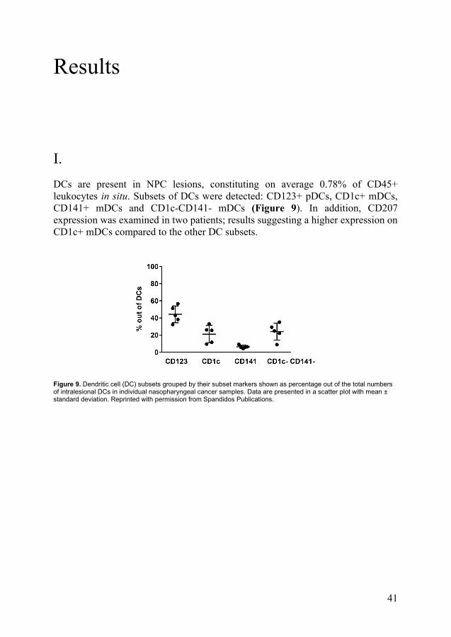
For EBV-DNA positive patients treated with curative intent, an EBV-DNA load of >70 copies/cell was associated with a favourable 7-year disease-free survival (DFS) (p=0.046) (Figure 11). This association remained when all EBER negative cases were omitted (p=0.050).
Figure 11. Kaplan-Meier estimate of 7-year disease-free survival for Epstein-Barr virus-DNA (EBV-DNA) positive nasopharyngeal cancer cases grouped according to EBV-DNA cell copy number. Vertical lines mark events (residual or recurrent cancer) and crosses mark end of follow-up before 7 years. Reprinted with permission from Springer Nature.
44
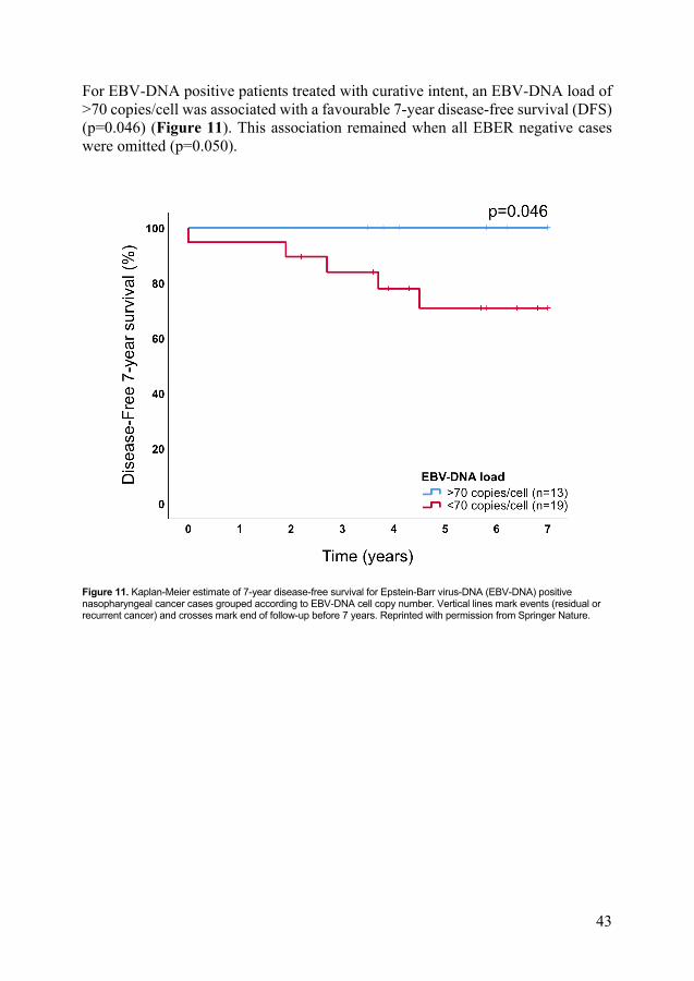
III. A total of 47 cases of NPC were examined. Overall, heterogeneous patterns of EBER, cytokeratin and immune cells were observed. Immune phenotypes based on lymphocyte distribution (notably CD8+ T-cells) could be demonstrated. The “inflamed” subtype constituted 61.7% of cases while 29.8% were “immune excluded” and 8.5% “immune deserted”. CD8+ cells were demonstrated in cancer cell areas and in the surrounding stroma whereas CD207+ cells were observed largely in cancer cell areas, where the ratio was 8-fold greater than within the tumour-adjacent surrounding stroma (Figure 12).
Figure 12. Ratios (%) for CD207+ cells (compared to all cells) in subareas of nasopharyngeal cancer presented as boxplots (median and interquartile range, with whiskers denoting 1.5 interquartile range and outliers as circles). There was a difference between CD207 ratios between cancer cell areas and cancer stroma (p<0.0001). Unpublished data.
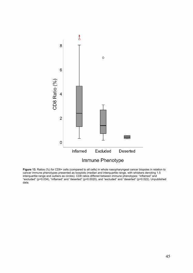
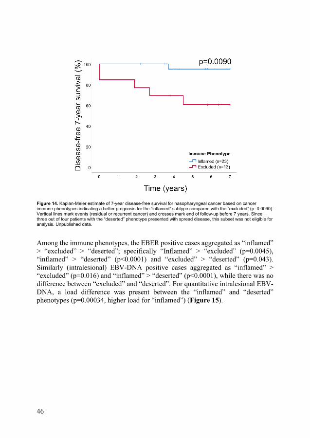
There was no correlation between CD8 and CD207 ratios. Among immune phenotypes, the CD8 densities aggregated as “inflamed” > “excluded” > “deserted” (Figure 13). In contrast, no such differences were observed for CD207 ratios. Differences in stage at presentation (I-III vs. IV) were observed among phenotypes, with the “inflamed” phenotype being associated with low-stage disease and the “deserted” with advanced. No difference in OS between immune phenotypes was shown, while DFS differed in favour of the “inflamed” over the “excluded” immune phenotype (p=0.0090) (Figure 14). The “deserted” phenotype was not eligible for DFS analysis, as all cases but one featured disseminated disease.
45
Figure 13. Ratios (%) for CD8+ cells (compared to all cells) in whole nasopharyngeal cancer biopsies in relation to cancer immune phenotypes presented as boxplots (median and interquartile range, with whiskers denoting 1.5 interquartile range and outliers as circles). CD8 ratios differed between immune phenotypes: “inflamed” and “excluded” (p=0.034), “inflamed” and “deserted” (p=0.0020), and “excluded” and “deserted” (p=0.022). Unpublished data.
46
Figure 14. Kaplan-Meier estimate of 7-year disease-free survival for nasopharyngeal cancer based on cancer immune phenotypes indicating a better prognosis for the “inflamed” subtype compared with the “excluded” (p=0.0090). Vertical lines mark events (residual or recurrent cancer) and crosses mark end of follow-up before 7 years. Since three out of four patients with the “deserted” phenotype presented with spread disease, this subset was not eligible for analysis. Unpublished data.
Among the immune phenotypes, the EBER positive cases aggregated as “inflamed” > “excluded” > “deserted”; specifically “Inflamed” > “excluded” (p=0.0045), “inflamed” > “deserted” (p<0.0001) and “excluded” > “deserted” (p=0.043). Similarly (intralesional) EBV-DNA positive cases aggregated as “inflamed” > “excluded” (p=0.016) and “inflamed” > “deserted” (p<0.0001), while there was no difference between “excluded” and “deserted”. For quantitative intralesional EBV-DNA, a load difference was present between the “inflamed” and “deserted” phenotypes (p=0.00034, higher load for “inflamed”) (Figure 15).
47
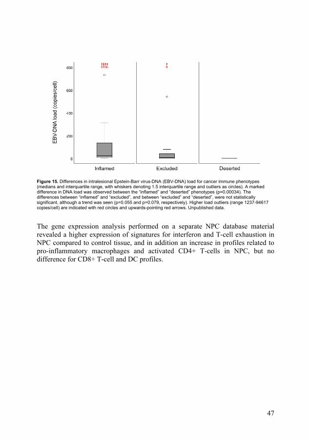
Figure 15. Differences in intralesional Epstein-Barr virus-DNA (EBV-DNA) load for cancer immune phenotypes (medians and interquartile range, with whiskers denoting 1.5 interquartile range and outliers as circles). A marked difference in DNA load was observed between the “inflamed” and “deserted” phenotypes (p=0.00034). The differences between “inflamed” and “excluded”, and between “excluded” and “deserted”, were not statistically significant, although a trend was seen (p=0.055 and p=0.079, respectively). Higher load outliers (range 1237-94617 copies/cell) are indicated with red circles and upwards-pointing red arrows. Unpublished data.
The gene expression analysis performed on a separate NPC database material revealed a higher expression of signatures for interferon and T-cell exhaustion in NPC compared to control tissue, and in addition an increase in profiles related to pro-inflammatory macrophages and activated CD4+ T-cells in NPC, but no difference for CD8+ T-cell and DC profiles.

48
IV. Topical mucosal administration of γ-PGA-Phe NPs on rat middle ear mucosa produced a dose- and time-dependent local inflammatory response characterized by generation of pro-inflammatory type 1 cytokines IL-1α, IL-1β, IL-6, MIP-1α and TNF-α (Figure 16). In addition, histopathological changes compatible with inflammation was demonstrated.
Figure 16. Cytokine levels in lavage fluid 4 and 12 hours after administration of specific nanoparticles (NPs): 0.5 mg and 2.5 mg poly(γ-glutamic acid) graft-L-phenylalanine ethylester NPs (γ–PGA-Phe NPs) to rat middle ear mucosa (mean ± standard error of the mean). After 4 hours, administration of the high NP dose (2.5 mg) resulted in increased levels of TNF-α, IL-6, and MIP-1α compared with sham, while no significant changes were observed for the low NP dose (0.5 mg) at this time point. After 12 hours, administration of the low NP dose resulted in increased levels of TNF-α, IL-1α, IL-1β, and MIP-1α, compared with sham. Reprinted with permission from Taylor & Francis. * denotes p < 0.05; *** denotes p < 0.001. Cytokines: TNF-α = Tumour necrosis factor-α, IL-1α = Interleukin-1α, IL-1β = Interleukin-1β, IL-6 = Interleukin-6, MIP-1α = Macrophage inflammatory protein-1α.
49
Discussion
Dendritic cells in nasopharyngeal cancer A presence of intralesional DCs in NPC has been suggested by previous morphological reports [221-227], and this was confirmed and expanded upon in the present studies (I, III). Accordingly, a detailed analysis of DC subsets according to present standards has been lacking until now. In study I, the specific DC subsets CD123+ pDCs, CD1c+ mDCs, CD141+ mDCs, and CD1c-CD141- mDCs, previously characterized in blood [10], were demonstrated intralesionally in NPC for the first time. These findings of specific subsets implicate a potential for modulation of the immune response, with the CD1c+ and CD141+ mDC subset being of particular interest due to its ability to cross-present antigen and initiate an antigen-specific cell-mediated response [261]. Furthermore, in the first study (I), a notable expression of CD207, a C-lectin receptor that facilitates cell-mediated response through antigen cross-presentation [18-20], was suggested on CD1c+ mDCs. This is in agreement with recent findings by Askmyr et al. (unpublished) where high levels of CD207 were detected on CD1c+ mDCs in human tonsils and lymph nodes. In study III, intralesional CD207+ DCs, presumably with CD1c+ DCs as the dominating subset, were identified and quantitated in NPC by means of digital microdissection. These intralesional CD207+ DCs were present in all tumours. Furthermore, they were predominantly distributed among cancer cells to an 8-fold higher degree compared to cancer cell-adjacent stroma.
DCs represent treatment possibilities, which warrant attempts to outline PRR expression of the different intralesional DC subsets. Arguably, focus should be on PRRs known to facilitate cell-mediated antigen-specific responses, including TLR2, TLR4, Dectin-1, Dectin-2, DEC205, CLEC9A, DC-SIGN (CD209) and CD207 [18-21]. Consequently, Askmyr et al. (unpublished) examined five PRRs (TLR2, TLR4, DC-SIGN, CD207 and CD206) and showed that their expression was subset-specific, suggesting that characteristics of different DC subsets might be attributed to their PRR profiles. Taken together, identification and characterisation of particular PRRs on distinct DC subsets in tumours such as NPC open up for active antigen-specific immunomodulation. While the present studies provide information on CD207, a further characterization on PRRs on DCs in NPC is warranted. Interestingly, the value of such background knowledge is further acknowledged by
50
a currently recruiting chemotherapy trial on advanced NPC (NCT03196869), where treatment effects on DC subsets in blood is the primary endpoint.
In conclusion, specific intralesional DC subsets can be characterised in NPC and the C-lectin receptor CD207, involved in antigen cross-presentation aimed at type-1 response, is frequently expressed by these. In addition, CD207+ cells, presumably DCs, are constantly encountered among cancer cells in NPC, suggesting CD207 as a possible target in future antigen-specific active immunotherapy in NPC.
Epstein-Barr virus in nasopharyngeal cancer In NPC, it is known that EBV positive disease has a favourable prognosis compared to EBV negative cases and it has also been suggested that the rarer HPV positive NPC, also a virus-driven NPC, places itself in between these two entities in terms of prognosis [88, 159]. In addition, among EBV positive cases, quantitates of EBV-DNA in peripheral blood have shown to be of prognostic value [135-137], whereas intralesional load has not been previously evaluated in that context. In study II, quantitates of intralesional EBV-DNA in pre-treatment NPC tissue were determined and related to stage and survival. Previous findings that EBV negative NPC features a poorer prognosis than EBV positive disease were confirmed. It has also been suggested that the clinical presentation of NPC depends on viral status, but findings have been diverging [158, 159]. No difference in stage between EBV positive and negative patients was detected in the present study (II), except that stage IV cancer was associated with EBV negative cancer when only HPV negative cases were evaluated.
The variation of EBV-DNA among positive cases observed in this study (II) was large, ranging from 0.0005 to 94617 copies/cell (median 58.8, interquartile range 7.78-259). In the only similar study to date, Shao et al. [149] reported a median EBV-DNA load of 27.8 copies/β-actin gene (interquartile range 6.35-279, range not specified) in a material of similar size. Even though similar, these numbers are difficult to compare. Apart from different normalisation strategies, the primers used for EBV-DNA detection in the study by Shao et al. were aimed at the BAMHI-W gene, which is known to be present in variable copy numbers, resulting in less precise quantification [262]. In a similar setting to EBV-DNA load in NPC, HPV-DNA load in oropharyngeal cancer, a wide range in DNA load have also been reported (0.003 to 1079 copies per cell) [151]. It may be speculated that differences in viral DNA load affect the intralesional immune milieu and have implications for clinical characteristics and prognosis. Indeed, high intralesional HPV-DNA load in oropharyngeal cancer have been associated with improved survival [150, 151]. In agreement, the present study (II) showed a better 7-year DFS for EBV-DNA positive patients with a high load (>70 copies/cell). Furthermore, none of the
51
patients with exceptionally high EBV-DNA load (1237-94617 copies per cell) featured distant metastases or residual or recurrent disease (5.8–12.3 years follow-up). This finding, that high levels of intralesional EBV-DNA are associated with a favourable prognosis, does not contradict previous observations that high EBV-DNA load in blood and in “extralesional” brush samples are associated with worse outcome in NPC [135-137, 143-145]. Instead, EBV-DNA load in blood may be viewed as a marker of the cancer burden [263, 264], whereas quantitates of intralesional EBV-DNA may reflect the degree of EBV presence. Studies are warranted to examine the relationship between these two features.
A subgroup of NPC is HPV-driven [146, 153-162] and HPV-testing should thus be included when evaluating viral association in NPC. In the present study (II), 15% of cases (n=7) were positive for high-risk type HPV-DNA, which is in agreement with recent data from Finland [158]. An additional p16-analysis was positive in 5/7 of these cases, supporting HPV as the true oncogenic driver in these cases, but such assumptions should be made with care considering that EBV-driven NPC may also be p16 positive [164].
In conclusion, in addition to previous knowledge that an EBV association in NPC is favourable for the prognosis, a 7-year survival benefit was shown for EBV-DNA positive patients with high intralesional EBV-DNA load (>70 copies/cell). Accordingly, intralesional EBV-DNA load may be of interest for prognostication, but also as a biomarker for antigen density and thus potentially for accessibility to antigen-specific active immunotherapy in NPC.
Cancer immune phenotypes and CD8+ T-cells in nasopharyngeal cancer In the third study (III), pre-treatment NPCs were categorised into specific immune phenotype subsets. These were based on presence and distribution of intralesional lymphocytes, notably CD8+ T-cells, with the majority of cases constituting an immune “inflamed” phenotype. The results of the phenotype classification were to some extent verified by quantification of intralesional CD8+ T-cells. It was further shown that all cases exhibiting the “deserted” phenotype presented with stage IV disease (n=4), and all cases presenting with stage I disease (n=4) were of the “inflamed” phenotype. Accordingly, an association could be discerned between phenotype and stage at presentation, with a pattern of less lymphocyte infiltration associated with more advance disease.
A pronounced difference in DFS was demonstrated between the “inflamed” and the “excluded” subtype, with the former presenting the more favourable outcome. Cases presenting with the “deserted” phenotype were excluded from the DFS analysis due
52
to a high frequency of spread disease among the few cases; arguably the prognosis was poorest within this group. Similar patterns were observed for OS, but failed to reach statistical significance. These findings are in agreement with those of other types of cancers, where intralesional lymphocyte infiltration has been reported to correlate with increased survival [43, 44]. Immune phenotypes should arguably be further evaluated for clinical application in NPC, for example as prognostic markers and for treatment selection. The latter could refer to whether or not to offer chemotherapy in addition to radiotherapy even for low-stage NPC if presenting with a “deserted” pattern and to offer cases with the “inflamed” phenotype, more likely to respond to checkpoint inhibitor therapy [49, 265], the addition of such adjuvant immunotherapy.
The gene expression analysis of an EBV positive NPC cohort [266-268] (study III) revealed a gene signature associated with up-regulation of interferon. Accordingly, NPC may feature immune subsets, likely corresponding to specific immune phenotypes, characterized by up-regulation of type-1 response markers including γ-interferon. Apart from interpreting this as an up-regulation of anti-viral mechanisms, it is tempting to consider such a feature as a beneficial anti-tumour response [269]. However, recent data point to a dual effect of γ-interferon, suggesting also pro-tumorigenic features of this cytokine, e.g. in the establishment of an immunosuppressive tumour microenvironment [270, 271]. In this context, a γ-interferon profile may be linked to the “inflamed” cancer immune phenotype [272]. To further explore this possibility in NPC, it would be of interest to compare the gene expression profile with the cancer immune phenotype in the sample cohort. In addition, it may be speculated that a virus-associated immune response also induces α- and β-interferons and, subsequently, CD207-expression [31]. Such stimulation may explain the relatively high density of CD207+ DCs among cancer cells in NPC described in study III.
In conclusion, lymphocyte-based immune phenotypes can be demonstrated in NPC, and these findings can be objectively confirmed by CD8+ T-cell quantification. These phenotypes can be used for prognostication. The findings further suggest that immune features of NPC, notably CD8+ T-cell presence, depend on EBV presence and it is reasonable to advocate that EBV analyses, in some form, should be included whenever immune features of NPC are examined.
Prognostic factors in nasopharyngeal cancer At histopathological examination (study III), a marked heterogeneity was noted in and between cases with regards to cancer growth patterns as well as to immune cell distribution and cytokeratin- and EBER-presence. These were patterns that could not be solely attributed to EBV presence or not. Furthermore, the gene expression
53
analysis (III) revealed a heterogeneity also for immune-related signatures. Thus, even a seemingly distinct entity such as EBV positive NPC can manifest itself in a variety of forms. This heterogeneity is further emphasised by a recent single cell RNA-sequencing study of three NPC tissue samples [273]. Arguably, such a heterogeneity may play a role in the proposed insufficiency of the current NPC staging system for prognostication of the disease and for treatment selection [84, 138-142]. The studies performed add candidates for incorporation into an improved staging system for NPC: intralesional EBV-DNA load (study II) and lymphocyte-based immune phenotyping (study III) as well as HPV diagnostics.
Intralesional EBV-DNA load correlated to survival (II), which has not been previously reported. This finding supports the notion that viral malignancies show a less aggressive phenotype in comparison with non-viral malignancies, as proposed for NPC [88]. A related observation is that by Shao et al. [149], where intralesional EBV-DNA load in NPC was not correlated to stage. This is in agreement with the lack of stage-related observations in study II, and not in contrast to the present findings on survival. Stage and survival may not be expected to correlate strongly in NPC, considering the insufficiency of the current staging system for NPC [84, 138-142]. In addition, HPV-status should arguably be routinely assessed, although present data diverge on its prognostic value in NPC [154, 158-161]. In the present study (II), the number of HPV cases was too small to admit a detailed analysis. However, it was noted that stage IV disease was associated with EBV negative cancer only when HPV positive cases, likely masking a worse prognosis among “true” EBV negative patients, were omitted from the analysis.
Intralesional DCs have been suggested as a prognostic marker in NPC, but data are scarce and diverging [199, 225]. In the present material (III), the density of CD207+ cells, presumably DCs, was not related to prognosis. Accordingly, CD207 ratios were also not related to lymphocyte-based immune phenotype patterns or EBV-markers, both demonstrated as prognosticators (III).
The strongest prognostic predictors of survival in NPC in the studies performed were the lymphocyte-based cancer immune phenotypes (III). The concept of TIL density as a biomarker in cancer can be directly linked to this, which also expands on previous observations on TILs in NPC and their prognostic potential [237]. Considering that EBV positive cases aggregated among immune phenotypes as “inflamed” > “excluded” > “deserted”, that CD8+ density was markedly higher in EBV positive compared to negative cases and that findings extended beyond presence or not, with high intralesional EBV-DNA loads associated with the “inflamed” phenotype and low loads with the “deserted” phenotype (study III), it is reasonable to associate the survival outcome among phenotypes with that of intralesional EBV-DNA (II). It thus seems likely that an “inflamed” phenotype and high intralesional load of EBV-DNA are interconnected with a favourable prognosis. These associations add to previous observations that links EBV to prognostic outcome.
54
In conclusion, NPCs are heterogeneous lesions in relation to presence and distribution of both cancer cells and immune cells. This, and the likely variable presence of EBV-associated antigen as well as the possible interference of HPV, must be taken into account when immunotherapeutic alternatives including antigen-specific therapy for NPC are explored. In the present studies, efforts have been made to overcome these obstacles by inclusion of HPV-DNA and p16 expression in the analysis (study II) and by digital image-based microdissection and quantitative digital analysis (study III). With a growing understanding of the value of sufficient background information and how to exploit it, emerging data will also allow better prognostication. In the performed studies, survival was predicted by both intralesional EBV-DNA load (study II) and lymphocyte-based immune phenotyping (study III). In addition, the latter measure adds the major advantage of being relatively simple to perform and thus arguably being easily incorporated into clinical routine praxis.
The potential role for γ-PGA-Phe nanoparticles in antigen-specific active immunotherapy In order for antigen-specific active immunotherapy to be successful, suitable antigens must be delivered to the tumour site, they must be presented to APCs, preferably DCs, and relevant PRRs must be activated. Furthermore, in the context of mucosal cancers such as NPC, mucosal administration may be beneficial.
The utilisation of NPs, specifically γ-PGA-Phe NPs, as mucosal adjuvants have been previously demonstrated [259, 260], but the local effects in vivo on a targeted mucosa have not been investigated. In the fourth study (IV), a rat model was used to evaluate the local immune response to topically applied γ-PGA-Phe NPs. An increase of pro-inflammatory cytokines IL-1α, IL-1β, IL-6, MIP-1α and TNF-α was observed, and the induced inflammation was emphasised by histopathological changes. Furthermore, the cytokine increase observed was dose- and time-dependent. The cytokine findings confirmed previous in vitro results demonstrating that stimulation of nasal DCs with γ-PGA-Phe NPs led to upregulated mRNA and protein levels of IL-1β, IL-6, MIP-1α and TNF-α as well as upregulated mRNA levels of IL-1α [256]. To summarise, the demonstrated pattern is compatible with that of a type-1 response – the desired pattern in a cancer treatment setting – where cancer immunoediting otherwise is expected to skew responses towards a pro-tumour type-2 pattern [46, 62].
The utilised NPs have previously been shown capable of effective antigen loading and delivery [250, 251] as well as of exhibition of adjuvant properties, probably through binding to TLR2 and TLR4 [256]. This emphasises their potential as vehicles for antigen-specific active immunotherapy in treatment of cancer such as
55
NPC. In accordance, subsequent studies, performed after study IV, have further elaborated upon these abilities, such as on antigen-specific DC induction and maturation by a NP complex including γ-PGA-Phe NPs [274] and on how different formulations of γ-PGA-Phe NPs and other γ-PGA NPs can be used to select a dominating type 1- or type 2-response pattern [275]. These advances combined have led to further studies aiming to develop a commercially available clinical drug-delivery system [276, 277]. In addition, variants of these NPs are being developed, such as synthetic antigen-carrying γ-PGA NPs capable of inducing antitumor immunity [278].
In conclusion, γ-PGA-Phe NPs, previously known to be able to act as antigen carriers and known to possess adjuvant properties, induce a local response in targeted mucosa compatible with a type-1 response. This further highlights these NPs as promising tools for antigen-specific active immunotherapy in cancers, e.g. NPC.
Methodological considerations
Patient material NPC is a rare cancer in Sweden, which is reflected by the low numbers of fresh samples in study I. However, with the main objective of this study being to describe presence of intralesional DCs and DC subsets, the sample size was sufficient. In study II-III, the retrospective setup enabled inclusion of a larger cohort. Furthermore, a particular strength gained from the long inclusion period needed to reach sufficient numbers was that an extensive follow-up was acquired. A particular concern to note when evaluating an NPC material is that EBV negative cases may be misdiagnosed cases of oropharyngeal cancer. In the current studies however, all patient cases were re-evaluated according to clinical presentation, radiology and histopathology, and cases not deemed as true NPC were excluded.
Intralesional dendritic cells in nasopharyngeal cancer DC subsets expressing CD207 are normally present in epithelium, i.e. Langerhans DCs, and since NPC is an epithelial cancer, intralesional quantification of CD207+ cells may partly reflect this population. However, the digital microdissection performed for all cases excluded all normal epithelium. Furthermore, at quantification the detected CD207+ DCs were mainly present among the cancer cells. Since there was no control material, the findings could not be compared to normal nasopharyngeal tissue, however, it has been stated that DC presence does not differ in cancers compared to their normal counterparts [56]. This may also be
56
reflected in the gene expression analysis of study III, where no increase in DC signatures was shown in NPC compared to control tissue.
Epstein-Barr virus markers EBER detection has long been considered the gold standard for verification of EBV-driven cancer [125]. In the second study (II), among the EBV-DNA positive cases (n=40), four EBER negative cases were found. These four cases were the cases with the lowest viral DNA load, allowing two possible interpretations: that these were cases with low EBV presence, which were missed with EBER diagnostics, or that they were false positive cases. The observation that two of these cases were p16 positive and high-risk HPV-DNA positive, thus suggesting an alternative genesis, supports the latter supposition. However, irrespective of which, EBV-DNA load was still prognostic even when the EBER negative/EBV-DNA positive cases were omitted from the analysis.
A particular concern when performing measurements on intralesional markers in cancers is that results are dependent on the size of the biopsies, the relative presence of cancer cells and the possible presence of tumour markers in non-cancer cells. In addition, when the marker is incorporated viral DNA, such as EBV-DNA, the tumour genome must also be taken into account. In the present study (II), the β-globin gene was used to normalize for cell numbers. Because NPC is generally believed to be diploid and as there are no reports of recurrent gains or deletions involving the β-globin gene locus [279, 280], this seems a reasonable strategy, even if further insights into the NPC genome in the future likely will present even better candidates for normalisation. On the other hand, the problem with cell heterogeneity, i.e. the relative presence of cancer cells within the tumour, remains a confounding factor. In addition, specifically NPC, as demonstrated in study III, is well known for a marked infiltration of non-cancer cells that may affect the results. However, given the logarithmic scale range of EBV-DNA revealed, the present results should not be substantially influenced. The objection based on hypothetically chronically EBV-infected lymphocytes independently present within the tumour could reasonably be similarly addressed, specifically with regards to that the latent virus pool is believed to be situated within the relatively rare memory B-cells [101]. A related issue is the potential presence of intralesional EBV-DNA in non-cancer nasopharyngeal tissue. However, reports on EBV-status in healthy nasopharyngeal tissue, and specifically based on EBV-DNA, are sparse and have demonstrated exceedingly low quantitates [149, 281].
57
Gene expression analysis The third study (III) included a gene expression data analysis. A disadvantage with the material available was that it was solely EBV positive and consisted only of low-stage cases. A heterogeneity was observed, stressing the difficulty to draw conclusions from such database analyses. However, this finding was in accordance with the morphological assessment of the NPC material in study III. The gene expression analyses did not show an increase in CD8+ T-cell profiles, as would have been expected from the morphological data (III), which could tentatively be due to insufficient control material.
Nanoparticles and the rat model In study IV, the effects of topical mucosal administration of γ-PGA-Phe NPs were examined. The model utilised was an established rat model developed for studies of middle ear infections [282]. An objection to the usage of this model is that the procedure itself is associated with a certain trauma, potentially producing an inflammatory response. To overcome this, the study was performed in a sham-controlled design. A further notion is the possible difficulties encountered when administering high-concentration NP solution, due to increased viscosity, in terms of higher demands on surgical-technical performance and potentially also with the risk of limited interaction with the target mucosa. However, the dose-dependent outcome speaks in favour for that these obstacles were overcome.

58
59
Conclusions
I. Intralesional DCs were present in NPC. These comprised specific subsets: CD123+ pDCs, CD1c+ mDCs, CD141+ mDCs and CD1c-CD141- mDCs, previously not described in NPC. CD207, a C-lectin receptor with cross-presentation facilitating properties, was found at a high frequency among CD1c+ mDCs.
II. Intralesional EBV-DNA was demonstrated in untreated NPC. In the patient cohort, 83% were positive for EBV-DNA, including all EBER positive cases. High-risk HPV subtypes were detected in 15% of cases and were highly associated with EBV negative disease. The EBV-DNA levels exhibited a wide range (0.0005 to 94617 copies/cell). An EBV-DNA load of >70 copies per cell for EBV-DNA positive cases treated with curative intent was associated with a favourable 7-year DFS.
III. Lymphocyte-based cancer immune phenotypes were demonstrated in NPC. The dominating pattern was the “inflamed” subtype (61.7%), which was supported by quantification of CD8+ T-cells. The “inflamed” phenotype was associated with better 7-year DFS. Intralesional CD207+ DCs were observed predominantly within areas of cancer cells. EBV-markers (intralesional EBV-DNA and EBER) were related to phenotype and higher EBV-DNA load was associated with the “inflamed” phenotype.
IV. Topical administration of γ-PGA-Phe NPs on middle ear mucosa in rats produced a dose- and time-dependent cytokine response compatible with a type 1-effect. This pro-inflammatory pattern was supported by histopathological changes.

60
61
Future perspectives and closing remarks
This thesis contributes with a few yet important steps toward future antigen-specific active immunotherapy for NPC. The observations highlight salient intralesional immune features that may be exploited, but additional information is needed. For example, a more detailed profiling of intralesional DCs as well as functional studies focusing on DC and T-cell interactions in the presence of antigen and treatment candidates including PRR ligands. Ligands to CD207 may be candidates in this context, such as recently synthesised high-affinity ligands [32]. Importantly, prior to any use in patients, accompanying diagnostics must be further developed, potentially including facets such as antigen presence and immune phenotypes, possibly incorporated in an improved NPC staging procedure.
Considering such subsequent studies, it is worth to keep in mind the eminent prerequisites at hand: NPC being a cancer frequently exhibiting non-self antigens and with a presentation signalling a vast immunological potential. By this, the potential gain of present and future findings may extend beyond this cancer type, utilising NPC as a comprehensive model for development of cancer treatments.

62
63
Sammanfattning på svenska
Nasofarynxcancer, i Sverige även känd under namnet epifarynxcancer, är en form av skivepitelcancer i nässvalget (nasofarynx). Det är en ovanlig cancerform (ca 30-50 fall per år i Sverige) men den vanligaste på just den här anatomiska platsen. Den klassifikation (stadie-indelning) vid nasofarynxcancer som används för ge en prognos och för att välja behandling anses av allt fler som otillräcklig; det finns ett behov av förbättring för att kunna ge rätt behandling till varje enskild patient.
Epstein-Barr virus (EBV), som bland annat också är orsaken till den mycket vanliga sjukdomen körtelfeber (mononukleos), är ofta men inte alltid en bakomliggande faktor vid nasofarynxcancer. Med tanke på att över 90% av jordens befolkning någon gång under livet smittas av EBV, och sedan fortsatt bär med sig viruset (som ligger vilande i specifika immunceller), så är dock risken för att en EBV-infektion ska leda till cancer väldigt liten. Utöver sin koppling till EBV karakteriseras också nasofarynxcancer ofta av en väldigt stor andel immunceller inne i tumören. Detta gör cancerformen extra intressant med avseende på så kallad immunterapi, behandlingsmetoder som syftar till att modulera kroppens immunsystem till att ge upphov till önskvärda reaktioner, till exempel en aktivering av immuncellstyper kapabla att döda cancerceller. En immunterapi som på det sättet använder sig av kroppens eget immunsystem kallas aktiv immunterapi. Dessutom ger kopplingen till EBV vid nasofarynxcancer, och den därmed frekventa närvaron av EBV-markörer på cancercellerna, en möjlighet att rikta en sådan immunreaktion mot dessa markörer, som i ett sådant sammanhang kallas för antigen. Det som gör detta koncept så tilltalande vid EBV-associerad cancer är att dessa antigen då är icke-kroppsegna, det vill säga reaktionen kan riktas specifikt mot cancercellerna och skona kroppens övriga celler.
När det handlar om antigen är de dendritiska cellerna (DCs) nyckelspelare i immunsystemet. Dessa kroppens mest potenta antigenpresenterade celler fångar upp och presenterar antigen för immunsystemets effektorceller och styr sedan också svaret. Detta gäller dels aktiv respons, som till exempel aktivering av immuncellstyper som kan förstöra andra celler eller ett antikroppssvar, och dels motsatsen, det som kallas tolerans. Sistnämnda är en viktig funktion hos immunförsvaret, det vill säga att också kunna lära sig att inte reagera på specifika främmande antigen, till exempel under en graviditet. Tolerans är också något som framgångsrika cancerformer utnyttjar, och att bryta sådan tolerans är en del av konceptet immunterapi.
64
Den här avhandlingen har undersökt olika förutsättningar för att kunna utveckla en framtida antigen-specifik aktiv immunterapi vid nasofarynxcancer och också prognostiska förutsättningar vid denna sjukdom.
I det första delarbetet (I) undersöktes förekomsten av DCs i färska tumörprover. Det kunde dels påvisas att DCs förekommer i nasofarynxcancer och dels att det rör sig om specifika subtyper, tidigare inte beskrivna i denna cancer: CD123+ pDCs, CD1c+ mDCs, CD141+ mDCs och CD1c-CD141- mDCs. Dessutom kunde CD207, som är en DC-receptor kopplad till responsmönster som är önskvärda vid cancerbehandling, identifieras i hög frekvens på CD1c+ mDCs.
I det andra delarbetet (II) undersöktes tumörprover från patienter med väldefinierad sjukhistoria och lång uppföljningstid. Arvsmassa från EBV (EBV-DNA) mättes i tumörerna och relaterades till cellantal. Samtidigt undersöktes också förekomsten av ett annat tumördrivande virus, humant papillomvirus (HPV). EBV-DNA påvisades i tumörprov från 83% av patienterna. Nivåerna av EBV-DNA uppvisade en väldigt stor variation (0,0005-94617 kopior/cell). Det kunde visas att nivåer av EBV-DNA överstigande 70 kopior/cell var förenligt med förlängd cancerfri överlevnad hos patienterna som hade en EBV-associerad cancer.
I det tredje delarbetet (III) undersöktes tumörprover från samma patientgrupp som i delarbete II. Dessa prover scannades in digitalt efter infärgningar för att sedan kvantifieras med hjälp av ett bildanalysprogram. Först utfördes en visuell bedömning av cancerns växtsätt i förhållande till immuncellerna i den. Tumörerna delades upp i så kallade immunfenotyper: ”inflamed” (infiltrerande immunceller bland cancercellerna), ”excluded” (immunceller i nära anslutning till cancercellerna, men inte bland dem), och ”deserted” (påtagligt få immunceller). Den dominerande fenotypen (61,7%) utgjordes av ”inflamed” medan ”excluded” utgjorde 29,8% och ”deserted” de sista 8,5%. En datorstödd kvantifiering av CD8+ T-lymfocyter i tumörerna, immunceller med kapacitet att döda cancerceller, genomfördes och deras fördelning i tumörerna understödde den visuella analysen. Det kunde visas att ”inflamed” fenotyp var kopplad till förlängd cancerfri överlevnad jämfört med ”excluded”. Även CD207+ DCs kvantifierades i tumörerna. Det fanns ingen koppling till de övriga fynden, men det kunde demonstreras att dessa celler fanns, framför allt i direkt anslutning till tumörcellerna. Slutligen relaterades också fynden till EBV. Fenotyperna kunde bindas till EBV-förekomst, på samma sätt som CD8+ T-celler, och det kunde också visas att höga nivåer av EBV-DNA var kopplat till ”inflamed”-fenotypen.
I det fjärde delarbetet (IV) undersöktes en annan förutsättning för antigen-specifik aktiv immunterapi. Närvaro av rätt sorts immunceller är otillräckligt om inte antigen kan levereras till dem, och om de inte samtidigt aktiveras på korrekt sätt (till exempel via särskilda receptorer såsom CD207). Specifika nanopartiklar (NPs), mycket små partikelenheter, kan potentiellt användas för detta. I arbete IV undersöktes effekten av γ-PGA-Phe NPs på slemhinna i en djurmodell, och ett
65
inflammatoriskt svar kunde påvisas. Detta uppvisade karakteristika överensstämmande med det som ses vid inducering av ett cellmedierat immunsvar, en respons som till exempel inkluderar stimulering av CD8+ T-celler, det vill säga det önskvärda vid en cancerbehandling. Det kunde dessutom visas att detta svar gav upphov till förändringar i slemhinnan förenliga med en inflammatorisk reaktion.
De ingående delarbetena visar därmed på ett antal specifika förutsättningar för antigen-specifik aktiv immunterapi vid nasofarynxcancer. Inventeringen av DCs i denna cancerform påvisade flera subtyper varav vissa kan vara lämpliga att utnyttja vid cancerbehandling. Specifikt påvisades förekomst av CD207+ DCs i tumörerna. CD207 är intressant som målreceptor vid aktivering av en relevant immunreaktion mot cancer, och det kunde också visas att CD207+ DCs befann sig i direkt anslutning till cancercellerna. Vidare kunde nivåer av EBV-DNA, som kan tänkas svara mot potentiella antigennivåer, mätas med ett brett utfall. Specifika immunfenotyper, mönster svarande mot interaktion mellan cancer och immunceller, kunde påvisas. Såväl nivåer av EBV-DNA som immunfenotypmönster kunde kopplas till överlevnad, och i enlighet med detta kunde också nivåer av EBV-DNA och immunfenotyper associeras. Dessa utfall skulle kunna användas för förbättrade prognosverktyg och för att välja behandling. Slutligen visade undersökningar av specifika nanopartiklar (γ-PGA-Phe NPs), potentiellt aktuella som bärare av antigen och aktivatorer av DCs, att de framkallar ett vid cancerinriktad immunterapi relevant svar i slemhinna. Detta skulle kunna vara av särskilt intresse vid behandling av cancerformer utgående från slemhinna, till exempel nasofarynxcancer.
Sammanfattningsvis visar genomförda arbeten på förutsättningar för antigen-specifik aktiv immunterapi vid nasofarynxcancer. Ytterligare studier, till exempel med avseende på fördjupad karakterisering av immunceller, krävs dock innan behandlingsförsök kan bli aktuella. En förbättrad stadie-indelning av dessa tumörer, lämpligtvis baserad även på EBV-status och immunstatus, kan bidra till att välja ut vilka tumörer som är lämpliga för sådan behandling.

66

67
Acknowledgements
A lot of people have contributed to this thesis, for which I am immensely grateful. To name a few:
Lennart Greiff. My supervisor, among a lot of other imposing possible epithets, friend being another one of them. You prefer (rimligen) the short version: THANK YOU!
Malin Lindstedt. My co-supervisor, who helped me navigate in the incomprehensible immunonanobiotechnological galaxy far, far away beyond the clinic. Lucky me!
Anders Cervin and Sven Lindberg. My much-appreciated former supervisors, in that project that crashed in every possible and impossible way. Those were frustrating years, but in the after-version I learned a lot, “karaktärsdanande”, as we say in Swedish. And in addition: Sven! Thank you so much for helping me to nail it in the end! And for our shared concerns for the lilywhites!
All my co-authors. Kristina Lundberg, always a big support. Sissela, Milad and Aastha, for all the efforts, not only with the hands-on laboratory work but also for the enthusiastic help in manuscript preparations. Ann Hermansson, you could have been acclaimed in so many different paragraphs in this section, and among other claims for the fame you were the one who taught me to operate on both humans and rats. Ola and Jonas, the positive ones, for bringing your various expertise to the studies and your efforts to force it inside of my head. Fredrik, to whom I have paid so many visits to “just quickly show a histopathological slide” and gain consent, that has been so appreciated. And Per, Akagi-san and Akashi-san, early associates who I unfortunately never met. Thank you all for your support!
David Askmyr. Never a co-author but continuously a big co-support in the project, as well as my back-up for the rare stuff at the clinic when I faded out in the end. I am prepared to repay!
All the rest who helped me with the articles and the thesis, then and now, in different ways. Lena and Charlotte. Frida Rydnert. Margareta Nilsson. Cristina Ciornei-Karlsson. Tommy Schyman and Jonas Björk. Johan Wennerberg. Daisy. Prem. Sabine. Maria Värendh. And all you others.
The University – for letting me do this. Måns – for persuading me to do it.

68
Patienterna. Tack för era osjälviska bidrag som möjliggjorde de genomförda studierna. Och tack till Laryngfonden/Mun- & Halscancerförbundet, det är fantastiskt med en patientförening som stöttar forskning på det sättet ni gör. Och ett särskilt tack till patienten som lät mig ha med bilder på honom i den här avhandlingen!
ÖNH: Mina eviga och högt värderade rumskompisar Marie och Johanna och min ständige sms-kompis Magnus. Mitt HH-gäng. Min mentor Anna Stjernquist-Desatnik, ständigt med. Johan Wennerberg och Peter White, som fick mig att checka in. De klinikledningar som följt. Anna Hafström (och innan henne samme Peter), som fått ihop schemat. Och alla ni andra, läkare, sjuksköterskor, undersköterskor, sekreterare, logopeder, audionomer, ingenjörer, fysioterapeuter, kuratorer, och dietister, som får Klinikernas Klinik att fortsätta att existera i högsta välmåga trots att den inte längre finns. You can check out any time you like, but you can never leave!
Alla mina vänner. Badminton-Magnus som jag diskuterat den här boken med en gång i veckan sedan någon gång förra seklet. Resten av Rauhrackel-gänget (det riktiga): Erik & Mårten. Kevin, du känns alltid nära (fast du flyttade till Uppsala). Annika och Robert. Och alla andra!
Anders, Ingrid, Kristin och nykomlingen Torgeir, och resten av släkten i den vida bemärkelsen, med det ingifta Lundagänget, och med familjen Folkesson! Anders, jag har alltid varit väldigt glad för att du blev min svåger. Mattias, du hamnade här som bror istället för i vän-paragrafen. Eva, en svärmor som du skulle alla ha.
Mina kära nära systrar Anna och Stina. Och vår mor, vad synd att du missade detta, du hade gått igång så fullkomligt på det!
Magda och Iris – Livet är så mycket roligare med er! Äntligen får jag sparka fotboll, lösa deckargåtor i Kalle Anka och högläsa Mumintrollen. Jag älskar er!
Kajsa – De sa att de bästa var tagna men de ljög, de ljög, för vi fanns ju kvar! Vad bra vi har det! Jag älskar dig innerligt! (Jag funderade på att skriva något om tack för all hjälp med den här boken också, genetik och korrektur och massor av annat, men jag tänkte att jag fokuserar på det viktigaste, kärleken! I know the thoughts that surround you, 'cause I can look inside your head!)
69
Funding
The original research presented in this thesis was made possible by funding from the following contributors:
Laryngfonden, the Acta Oto-Laryngologica foundation, Region Skåne (ALF, regionalt forskningsstöd), Lund University (doktorandmånader), Vinnova (Multidisciplinary Bio), the Alfred Österlund Foundation, the Japan Science and Technology Agency (Strategic International Cooperative Program and Core Research for Evolutional Science and Technology: CREST), the Japan Society for the Promotion of Science (Bilateral Joint Research Projects), Ida & Henning Persson’s Research Foundation, the Marie Sklodowska-Curie programme and Berta Kamprad’s Research Foundation.

70
71
References
[1] Abbas AL, Lichtman AH, Pillai S. Cellular and molecular immunology: Saunders, Elsevier; 2010. [2] Bettelli E, Carrier Y, Gao W, Korn T, Strom TB, Oukka M, Weiner HL, Kuchroo VK. Reciprocal developmental pathways for the generation of pathogenic effector TH17 and regulatory T cells. Nature. 2006;441:235-8. [3] Hori S, Nomura T, Sakaguchi S. Control of regulatory T cell development by the transcription factor Foxp3. Science. 2003;299:1057-61. [4] Shevach EM, DiPaolo RA, Andersson J, Zhao DM, Stephens GL, Thornton AM. The lifestyle of naturally occurring CD4+ CD25+ Foxp3+ regulatory T cells. Immunol Rev. 2006;212:60-73. [5] Kumar H, Kawai T, Akira S. Pathogen recognition by the innate immune system. Int Rev Immunol. 2011;30:16-34. [6] Venereau E, Ceriotti C, Bianchi ME. DAMPs from cell death to new life. Front Immunol. 2015;6:422. [7] Blum JS, Wearsch PA, Cresswell P. Pathways of antigen processing. Annu Rev Immunol. 2013;31:443-73. [8] Steinman RM, Cohn ZA. Identification of a novel cell type in peripheral lymphoid organs of mice. I. Morphology, quantitation, tissue distribution. J Exp Med. 1973;137:1142-62. [9] Joffre OP, Segura E, Savina A, Amigorena S. Cross-presentation by dendritic cells. Nat Rev Immunol. 2012;12:557-69. [10] Lundberg K, Rydnert F, Greiff L, Lindstedt M. Human blood dendritic cell subsets exhibit discriminative pattern recognition receptor profiles. Immunology. 2014;142:279-88. [11] Abolhalaj M, Askmyr D, Sakellariou CA, Lundberg K, Greiff L, Lindstedt M. Profiling dendritic cell subsets in head and neck squamous cell tonsillar cancer and benign tonsils. Sci Rep. 2018;8:8030. [12] Villani A-C, Satija R, Reynolds G, Sarkizova S, Shekhar K, Fletcher J, Griesbeck M, Butler A, Zheng S, Lazo S, Jardine L, Dixon D, Stephenson E, Nilsson E, Grundberg I, McDonald D, Filby A, Li W, De Jager PL, Rozenblatt-Rosen O, Lane AA, Haniffa M, Regev A, Hacohen N. Single-cell RNA-seq reveals new types of human blood dendritic cells, monocytes, and progenitors. Science. 2017;356. [13] Schreibelt G, Tel J, Sliepen KH, Benitez-Ribas D, Figdor CG, Adema GJ, de Vries IJ. Toll-like receptor expression and function in human dendritic cell subsets: implications for
72
dendritic cell-based anti-cancer immunotherapy. Cancer Immunol Immunother. 2010;59:1573-82. [14] Netea MG, Van der Meer JW, Sutmuller RP, Adema GJ, Kullberg BJ. From the Th1/Th2 paradigm towards a Toll-like receptor/T-helper bias. Antimicrob Agents Chemother. 2005;49:3991-6. [15] Alloatti A, Kotsias F, Magalhaes JG, Amigorena S. Dendritic cell maturation and cross-presentation: timing matters! Immunol Rev. 2016;272:97-108. [16] Jongbloed SL, Kassianos AJ, McDonald KJ, Clark GJ, Ju X, Angel CE, Chen CJ, Dunbar PR, Wadley RB, Jeet V, Vulink AJ, Hart DN, Radford KJ. Human CD141+ (BDCA-3)+ dendritic cells (DCs) represent a unique myeloid DC subset that cross-presents necrotic cell antigens. J Exp Med. 2010;207:1247-60. [17] Reynolds G, Haniffa M. Human and mouse mononuclear phagocyte networks: a tale of two species? Front Immunol. 2015;6:330. [18] Fehres CM, Duinkerken S, Bruijns SC, Kalay H, van Vliet SJ, Ambrosini M, de Gruijl TD, Unger WW, Garcia-Vallejo JJ, van Kooyk Y. Langerin-mediated internalization of a modified peptide routes antigens to early endosomes and enhances cross-presentation by human Langerhans cells. Cell Mol Immunol. 2017;14:360-70. [19] van den Berg LM, Cardinaud S, van der Aar AM, Sprokholt JK, de Jong MA, Zijlstra-Willems EM, Moris A, Geijtenbeek TB. Langerhans cell-dendritic cell cross-talk via langerin and hyaluronic acid mediates antigen transfer and cross-presentation of HIV-1. J Immunol. 2015;195:1763-73. [20] Idoyaga J, Cheong C, Suda K, Suda N, Kim JY, Lee H, Park CG, Steinman RM. Cutting edge: langerin/CD207 receptor on dendritic cells mediates efficient antigen presentation on MHC I and II products in vivo. J Immunol. 2008;180:3647-50. [21] Kreutz M, Tacken PJ, Figdor CG. Targeting dendritic cells - why bother? Blood. 2013;121:2836-44. [22] De Monte A, Olivieri CV, Vitale S, Bailleux S, Castillo L, Giordanengo V, Maryanski JL, Segura E, Doglio A. CD1c-related DCs that express CD207/langerin, but are distinguishable from Langerhans cells, are consistently present in human tonsils. Front Immunol. 2016;7:197. [23] Valladeau J, Ravel O, Dezutter-Dambuyant C, Moore K, Kleijmeer M, Liu Y, Duvert-Frances V, Vincent C, Schmitt D, Davoust J, Caux C, Lebecque S, Saeland S. Langerin, a novel C-type lectin specific to Langerhans cells, is an endocytic receptor that induces the formation of Birbeck granules. Immunity. 2000;12:71-81. [24] Chabrol E, Nurisso A, Daina A, Vassal-Stermann E, Thepaut M, Girard E, Vives RR, Fieschi F. Glycosaminoglycans are interactants of Langerin: comparison with gp120 highlights an unexpected calcium-independent binding mode. PLoS One. 2012;7:e50722. [25] Galustian C, Park CG, Chai W, Kiso M, Bruening SA, Kang YS, Steinman RM, Feizi T. High and low affinity carbohydrate ligands revealed for murine SIGN-R1 by carbohydrate array and cell binding approaches, and differing specificities for SIGN-R3 and langerin. Int Immunol. 2004;16:853-66. [26] Munoz-Garcia JC, Chabrol E, Vives RR, Thomas A, de Paz JL, Rojo J, Imberty A, Fieschi F, Nieto PM, Angulo J. Langerin-heparin interaction: two binding sites for small
73
and large ligands as revealed by a combination of NMR spectroscopy and cross-linking mapping experiments. J Am Chem Soc. 2015;137:4100-10. [27] Stambach NS, Taylor ME. Characterization of carbohydrate recognition by langerin, a C-type lectin of Langerhans cells. Glycobiology. 2003;13:401-10. [28] Tateno H, Ohnishi K, Yabe R, Hayatsu N, Sato T, Takeya M, Narimatsu H, Hirabayashi J. Dual specificity of Langerin to sulfated and mannosylated glycans via a single C-type carbohydrate recognition domain. J Biol Chem. 2010;285:6390-400. [29] de Jong MA, Vriend LE, Theelen B, Taylor ME, Fluitsma D, Boekhout T, Geijtenbeek TB. C-type lectin Langerin is a beta-glucan receptor on human Langerhans cells that recognizes opportunistic and pathogenic fungi. Mol Immunol. 2010;47:1216-25. [30] de Witte L, Nabatov A, Pion M, Fluitsma D, de Jong MA, de Gruijl T, Piguet V, van Kooyk Y, Geijtenbeek TB. Langerin is a natural barrier to HIV-1 transmission by Langerhans cells. Nat Med. 2007;13:367-71. [31] Maarifi G, Czubala MA, Lagisquet J, Ivory MO, Fuchs K, Papin L, Birchall JC, Nisole S, Piguet V, Blanchet FP. Langerin (CD207) represents a novel interferon-stimulated gene in Langerhans cells. Cell Mol Immunol. 2019. [32] Ota F, Hirayama T, Kizuka Y, Yamaguchi Y, Fujinawa R, Nagata M, Ismanto HS, Lepenies B, Aretz J, Rademacher C, Seeberger PH, Angata T, Kitazume S, Yoshida K, Betsuyaku T, Kida K, Yamasaki S, Taniguchi N. High affinity sugar ligands of C-type lectin receptor langerin. Biochim Biophys Acta Gen Subj. 2018;1862:1592-601. [33] Ghiringhelli F, Puig PE, Roux S, Parcellier A, Schmitt E, Solary E, Kroemer G, Martin F, Chauffert B, Zitvogel L. Tumor cells convert immature myeloid dendritic cells into TGF-β-secreting cells inducing CD4+CD25+ regulatory T cell proliferation. J Exp Med. 2005;202:919-29. [34] Wei S, Kryczek I, Zou L, Daniel B, Cheng P, Mottram P, Curiel T, Lange A, Zou W. Plasmacytoid dendritic cells induce CD8+ regulatory T cells in human ovarian carcinoma. Cancer Res. 2005;65:5020-6. [35] Barzaghi F, Amaya Hernandez LC, Neven B, Ricci S, Kucuk ZY, Bleesing JJ, Nademi Z, Slatter MA, Ulloa ER, Shcherbina A, Roppelt A, Worth A, Silva J, Aiuti A, Murguia-Favela L, Speckmann C, Carneiro-Sampaio M, Fernandes JF, Baris S, Ozen A, Karakoc-Aydiner E, Kiykim A, Schulz A, Steinmann S, Notarangelo LD, Gambineri E, Lionetti P, Shearer WT, Forbes LR, Martinez C, Moshous D, Blanche S, Fisher A, Ruemmele FM, Tissandier C, Ouachee-Chardin M, Rieux-Laucat F, Cavazzana M, Qasim W, Lucarelli B, Albert MH, Kobayashi I, Alonso L, Diaz De Heredia C, Kanegane H, Lawitschka A, Seo JJ, Gonzalez-Vicent M, Diaz MA, Goyal RK, Sauer MG, Yesilipek A, Kim M, Yilmaz-Demirdag Y, Bhatia M, Khlevner J, Richmond Padilla EJ, Martino S, Montin D, Neth O, Molinos-Quintana A, Valverde-Fernandez J, Broides A, Pinsk V, Ballauf A, Haerynck F, Bordon V, Dhooge C, Garcia-Lloret ML, Bredius RG, Kalwak K, Haddad E, Seidel MG, Duckers G, Pai SY, Dvorak CC, Ehl S, Locatelli F, Goldman F, Gennery AR, Cowan MJ, Roncarolo MG, Bacchetta R. Long-term follow-up of IPEX syndrome patients after different therapeutic strategies: An international multicenter retrospective study. J Allergy Clin Immunol. 2018;141:1036-49.e5. [36] Boussiotis VA. Molecular and biochemical aspects of the PD-1 checkpoint pathway. N Engl J Med. 2016;375:1767-78.
74
[37] Dunn GP, Bruce AT, Ikeda H, Old LJ, Schreiber RD. Cancer immunoediting: from immunosurveillance to tumor escape. Nat Immunol. 2002;3:991-8. [38] Vesely MD, Kershaw MH, Schreiber RD, Smyth MJ. Natural innate and adaptive immunity to cancer. Annu Rev Immunol. 2011;29:235-71. [39] Ehrlich P. Über den jetzigen Stand der Karzinomforschung. Nederlandsch Tijdschrift voor Geneeskunde. 1909;5:273-90. [40] Mlecnik B, Bindea G, Pages F, Galon J. Tumor immunosurveillance in human cancers. Cancer Metastasis Rev. 2011;30:5-12. [41] Galon J, Costes A, Sanchez-Cabo F, Kirilovsky A, Mlecnik B, Lagorce-Pages C, Tosolini M, Camus M, Berger A, Wind P, Zinzindohoue F, Bruneval P, Cugnenc PH, Trajanoski Z, Fridman WH, Pages F. Type, density, and location of immune cells within human colorectal tumors predict clinical outcome. Science. 2006;313:1960-4. [42] Chen DS, Mellman I. Elements of cancer immunity and the cancer-immune set point. Nature. 2017;541:321-30. [43] Pages F, Galon J, Dieu-Nosjean MC, Tartour E, Sautes-Fridman C, Fridman WH. Immune infiltration in human tumors: a prognostic factor that should not be ignored. Oncogene. 2010;29:1093-102. [44] Zhang L, Conejo-Garcia JR, Katsaros D, Gimotty PA, Massobrio M, Regnani G, Makrigiannakis A, Gray H, Schlienger K, Liebman MN, Rubin SC, Coukos G. Intratumoral T cells, recurrence, and survival in epithelial ovarian cancer. N Engl J Med. 2003;348:203-13. [45] Salgado R, Denkert C, Demaria S, Sirtaine N, Klauschen F, Pruneri G, Wienert S, Van den Eynden G, Baehner FL, Penault-Llorca F, Perez EA, Thompson EA, Symmans WF, Richardson AL, Brock J, Criscitiello C, Bailey H, Ignatiadis M, Floris G, Sparano J, Kos Z, Nielsen T, Rimm DL, Allison KH, Reis-Filho JS, Loibl S, Sotiriou C, Viale G, Badve S, Adams S, Willard-Gallo K, Loi S. The evaluation of tumor-infiltrating lymphocytes (TILs) in breast cancer: recommendations by an International TILs Working Group 2014. Ann Oncol. 2015;26:259-71. [46] Burkholder B, Huang R-Y, Burgess R, Luo S, Jones VS, Zhang W, Lv Z-Q, Gao C-Y, Wang B-L, Zhang Y-M, Huang R-P. Tumor-induced perturbations of cytokines and immune cell networks. Biochim Biophys Acta. 2014;1845:182-201. [47] Patel SP, Kurzrock R. PD-L1 expression as a predictive biomarker in cancer immunotherapy. Mol Cancer Ther. 2015;14:847-56. [48] Ahmadzadeh M, Johnson LA, Heemskerk B, Wunderlich JR, Dudley ME, White DE, Rosenberg SA. Tumor antigen-specific CD8 T cells infiltrating the tumor express high levels of PD-1 and are functionally impaired. Blood. 2009;114:1537-44. [49] Herbst RS, Soria JC, Kowanetz M, Fine GD, Hamid O, Gordon MS, Sosman JA, McDermott DF, Powderly JD, Gettinger SN, Kohrt HE, Horn L, Lawrence DP, Rost S, Leabman M, Xiao Y, Mokatrin A, Koeppen H, Hegde PS, Mellman I, Chen DS, Hodi FS. Predictive correlates of response to the anti-PD-L1 antibody MPDL3280A in cancer patients. Nature. 2014;515:563-7. [50] Bulgarelli J, Tazzari M, Granato AM, Ridolfi L, Maiocchi S, de Rosa F, Petrini M, Pancisi E, Gentili G, Vergani B, Piccinini F, Carbonaro A, Leone BE, Foschi G, Ancarani
75
V, Framarini M, Guidoboni M. Dendritic cell vaccination in metastatic melanoma turns "non-T cell inflamed" into "T-cell inflamed" tumors. Front Immunol. 2019;10:2353. [51] Treilleux I, Blay JY, Bendriss-Vermare N, Ray-Coquard I, Bachelot T, Guastalla JP, Bremond A, Goddard S, Pin JJ, Barthelemy-Dubois C, Lebecque S. Dendritic cell infiltration and prognosis of early stage breast cancer. Clin Cancer Res. 2004;10:7466-74. [52] Zou W, Machelon V, Coulomb-L'Hermin A, Borvak J, Nome F, Isaeva T, Wei S, Krzysiek R, Durand-Gasselin I, Gordon A, Pustilnik T, Curiel DT, Galanaud P, Capron F, Emilie D, Curiel TJ. Stromal-derived factor-1 in human tumors recruits and alters the function of plasmacytoid precursor dendritic cells. Nat Med. 2001;7:1339-46. [53] Hartmann E, Wollenberg B, Rothenfusser S, Wagner M, Wellisch D, Mack B, Giese T, Gires O, Endres S, Hartmann G. Identification and functional analysis of tumor-infiltrating plasmacytoid dendritic cells in head and neck cancer. Cancer Res. 2003;63:6478-87. [54] Bell D, Chomarat P, Broyles D, Netto G, Harb GM, Lebecque S, Valladeau J, Davoust J, Palucka KA, Banchereau J. In breast carcinoma tissue, immature dendritic cells reside within the tumor, whereas mature dendritic cells are located in peritumoral areas. J Exp Med. 1999;190:1417-26. [55] Gabrilovich D. Mechanisms and functional significance of tumour-induced dendritic-cell defects. Nat Rev Immunol. 2004;4:941-52. [56] Broz ML, Binnewies M, Boldajipour B, Nelson AE, Pollack JL, Erle DJ, Barczak A, Rosenblum MD, Daud A, Barber DL, Amigorena S, Van't Veer LJ, Sperling AI, Wolf DM, Krummel MF. Dissecting the tumor myeloid compartment reveals rare activating antigen-presenting cells critical for T cell immunity. Cancer Cell. 2014;26:638-52. [57] Michea P, Noel F, Zakine E, Czerwinska U, Sirven P, Abouzid O, Goudot C, Scholer-Dahirel A, Vincent-Salomon A, Reyal F, Amigorena S, Guillot-Delost M, Segura E, Soumelis V. Adjustment of dendritic cells to the breast-cancer microenvironment is subset specific. Nat Immunol. 2018;19:885-97. [58] Iwai Y, Ishida M, Tanaka Y, Okazaki T, Honjo T, Minato N. Involvement of PD-L1 on tumor cells in the escape from host immune system and tumor immunotherapy by PD-L1 blockade. Proc Natl Acad Sci U S A. 2002;99:12293-7. [59] Park Y-J, Kuen D-S, Chung Y. Future prospects of immune checkpoint blockade in cancer: from response prediction to overcoming resistance. Exp Mol Med. 2018;50:109. [60] Larkin J, Chiarion-Sileni V, Gonzalez R, Grob JJ, Cowey CL, Lao CD, Schadendorf D, Dummer R, Smylie M, Rutkowski P, Ferrucci PF, Hill A, Wagstaff J, Carlino MS, Haanen JB, Maio M, Marquez-Rodas I, McArthur GA, Ascierto PA, Long GV, Callahan MK, Postow MA, Grossmann K, Sznol M, Dreno B, Bastholt L, Yang A, Rollin LM, Horak C, Hodi FS, Wolchok JD. Combined Nivolumab and Ipilimumab or monotherapy in untreated melanoma. N Engl J Med. 2015;373:23-34. [61] Robert C, Long GV, Brady B, Dutriaux C, Maio M, Mortier L, Hassel JC, Rutkowski P, McNeil C, Kalinka-Warzocha E, Savage KJ, Hernberg MM, Lebbe C, Charles J, Mihalcioiu C, Chiarion-Sileni V, Mauch C, Cognetti F, Arance A, Schmidt H, Schadendorf D, Gogas H, Lundgren-Eriksson L, Horak C, Sharkey B, Waxman IM,
76
Atkinson V, Ascierto PA. Nivolumab in previously untreated melanoma without BRAF mutation. N Engl J Med. 2015;372:320-30. [62] Haabeth OA, Bogen B, Corthay A. A model for cancer-suppressive inflammation. Oncoimmunology. 2012;1:1146-55. [63] June CH, O'Connor RS, Kawalekar OU, Ghassemi S, Milone MC. CAR T cell immunotherapy for human cancer. Science. 2018;359:1361-5. [64] Jenner E. An inquiry into the causes and effects of the variolæ vaccinæ. 1798. [65] Jenner E. Further observations on the variolæ vaccinæ, or cow-pox. 1799. [66] Jenner E. A continuation of facts and observations relative to the variolæ vaccinæ. 1800. [67] Jenner E. The origin of the vaccine inoculation. 1801. [68] Pasteur L. Compte rendu sommaire des experiences faites a Pouilly-Le-Fort, pres de Melun, sur la vaccination charbonneuse. C R Acad Sci. 1881;92:1378–83. [69] Pasteur L. De l’attenuation des virus et de leur retour a la virulence. C R Acad Sci. 1881;92:430–5. [70] Pasteur L. Methode pour prevenir la rage apres morsure. C R Acad Sci. 1885;101:765–74. [71] Fenner F. The eradication of smallpox. Prog Med Virol. 1977;23:1-21. [72] Brewer JM. (How) do aluminium adjuvants work? Immunol Lett. 2006;102:10-5. [73] Coffman RL, Sher A, Seder RA. Vaccine adjuvants: putting innate immunity to work. Immunity. 2010;33:492-503. [74] Sabado RL, Balan S, Bhardwaj N. Dendritic cell-based immunotherapy. Cell Res. 2017;27:74-95. [75] Hammarlund E, Lewis MW, Hansen SG, Strelow LI, Nelson JA, Sexton GJ, Hanifin JM, Slifka MK. Duration of antiviral immunity after smallpox vaccination. Nat Med. 2003;9:1131-7. [76] Steinman RM, Banchereau J. Taking dendritic cells into medicine. Nature. 2007;449:419-26. [77] Weber JS, Yang JC, Atkins MB, Disis ML. Toxicities of immunotherapy for the practitioner. J Clin Oncol. 2015;33:2092-9. [78] Yang L, Yu H, Dong S, Zhong Y, Hu S. Recognizing and managing on toxicities in cancer immunotherapy. Tumour Biol. 2017;39:1010428317694542. [79] El-Naggar A, Chan J, Grandis J, Takata T, Slootweg P. WHO classification of head and neck tumours. 4th ed. Lyon, France: IARC Publications; 2017. [80] Ayan I, Kaytan E, Ayan N. Childhood nasopharyngeal carcinoma: from biology to treatment. Lancet Oncol. 2003;4:13-21. [81] Gioacchini FM, Tulli M, Kaleci S, Magliulo G, Re M. Prognostic aspects in the treatment of juvenile nasopharyngeal carcinoma: a systematic review. Eur Arch Otorhinolaryngol. 2017;274:1205-14.
77
[82] Ng S-H, Chang JT-C, Chan S-C, Ko S-F, Wang H-M, Liao C-T, Chang Y-C, Yen T-C. Nodal metastases of nasopharyngeal carcinoma: patterns of disease on MRI and FDG PET. Eur J Nucl Med Mol Imaging. 2004;31:1073-80. [83] UICC: TNM classification of malignant tumours 8th ed: John Wiley & Sons, Ltd; 2016. [84] Chen Y-P, Chan ATC, Le Q-T, Blanchard P, Sun Y, Ma J. Nasopharyngeal carcinoma. Lancet. 2019;394:64-80. [85] Yu MC, Yuan J-M. Epidemiology of nasopharyngeal carcinoma. Semin Cancer Biol. 2002;12:421-9. [86] Carioli G, Negri E, Kawakita D, Garavello W, La Vecchia C, Malvezzi M. Global trends in nasopharyngeal cancer mortality since 1970 and predictions for 2020: Focus on low-risk areas. Int J Cancer. 2017;140:2256-64. [87] The statistical database of The Swedish National Board of Health and Welfare 1970-2018. 2020. https://sdb.socialstyrelsen.se/if_can/val_eng.aspx [88] Chua MLK, Wee JTS, Hui EP, Chan ATC. Nasopharyngeal carcinoma. Lancet. 2016;387:1012-24. [89] Lu C-C, Chen J-C, Tsai S-T, Jin Y-T, Tsai J-C, Chan S-H, Su I-J. Nasopharyngeal carcinoma-susceptibility locus is localized to a 132 kb segment containing HLA-A using high-resolution microsatellite mapping. Int J Cancer. 2005;115:742-6. [90] Simons MJ, Wee GB, Day NE, Morris PJ, Shanmugaratnam K, De-The GB. Immunogenetic aspects of nasopharyngeal carcinoma: I. Differences in HL-A antigen profiles between patients and control groups. Int J Cancer. 1974;13:122-34. [91] Yuan J-M, Wang X-L, Xiang Y-B, Gao Y-T, Ross RK, Yu MC. Preserved foods in relation to risk of nasopharyngeal carcinoma in Shanghai, China. Int J Cancer. 2000;85:358-63. [92] Tsao SW, Yip YL, Tsang CM, Pang PS, Lau VM, Zhang G, Lo KW. Etiological factors of nasopharyngeal carcinoma. Oral Oncol. 2014;50:330-8. [93] Dai W, Zheng H, Cheung AKL, Tang CS-m, Ko JMY, Wong BWY, Leong MML, Sham PC, Cheung F, Kwong DL-W, Ngan RKC, Ng WT, Yau CC, Pan J, Peng X, Tung S, Zhang Z, Ji M, Chiang AK-S, Lee AW-M, Lee VH-f, Lam K-O, Au KH, Cheng HC, Yiu HH-Y, Lung ML. Whole-exome sequencing identifies MST1R as a genetic susceptibility gene in nasopharyngeal carcinoma. Proc Natl Acad Sci U S A. 2016;113:3317-22. [94] Nakanishi Y, Wakisaka N, Kondo S, Endo K, Sugimoto H, Hatano M, Ueno T, Ishikawa K, Yoshizaki T. Progression of understanding for the role of Epstein-Barr virus and management of nasopharyngeal carcinoma. Cancer Metastasis Rev. 2017;36:435-47. [95] Niedobitek G, Hansmann ML, Herbst H, Young LS, Dienemann D, Hartmann CA, Finn T, Pitteroff S, Welt A, Anagnostopoulos I, et al. Epstein-Barr virus and carcinomas: undifferentiated carcinomas but not squamous cell carcinomas of the nasopharynx are regularly associated with the virus. J Pathol. 1991;165:17-24. [96] Epstein MA, Achong BG, Barr YM. Virus particles in cultured lymphoblasts from Burkitt's Lymphoma. Lancet. 1964;1:702-3.
78
[97] Henle G, Henle W, Diehl V. Relation of Burkitt's tumor-associated herpes-type virus to infectious mononucleosis. Proc Natl Acad Sci U S A. 1968;59:94-101. [98] Hislop AD. Early virological and immunological events in Epstein-Barr virus infection. Curr Opin Virol. 2015;15:75-9. [99] Shannon-Lowe C, Rickinson A. The global landscape of EBV-associated tumors. Front Oncol. 2019;9:713. [100] Parkin DM. The global health burden of infection-associated cancers in the year 2002. Int J Cancer. 2006;118:3030-44. [101] Babcock GJ, Decker LL, Volk M, Thorley-Lawson DA. EBV persistence in memory B cells in vivo. Immunity. 1998;9:395-404. [102] Munz C. Role of human natural killer cells during Epstein-Barr virus infection. Crit Rev Immunol. 2014;34:501-7. [103] Taylor GS, Long HM, Brooks JM, Rickinson AB, Hislop AD. The immunology of Epstein-Barr virus-induced disease. Annu Rev Immunol. 2015;33:787-821. [104] Long HM, Meckiff BJ, Taylor GS. The T-cell response to Epstein-Barr virus - new tricks from an old dog. Front Immunol. 2019;10:2193. [105] Johnson KH, Webb CH, Schmeling DO, Brundage RC, Balfour HH, Jr. Epstein-Barr virus dynamics in asymptomatic immunocompetent adults: an intensive 6-month study. Clin Transl Immunology. 2016;5:e81. [106] Marques-Piubelli ML, Salas YI, Pachas C, Becker-Hecker R, Vega F, Miranda RN. Epstein-Barr virus-associated B-cell lymphoproliferative disorders and lymphomas: a review. Pathology. 2020;52:40-52. [107] Piris MA, Medeiros LJ, Chang KC. Hodgkin lymphoma: a review of pathological features and recent advances in pathogenesis. Pathology. 2020;52:154-65. [108] Moormann AM, Snider CJ, Chelimo K. The company malaria keeps: how co-infection with Epstein-Barr virus leads to endemic Burkitt lymphoma. Curr Opin Infect Dis. 2011;24:435-41. [109] Whittle HC, Brown J, Marsh K, Greenwood BM, Seidelin P, Tighe H, Wedderburn L. T-cell control of Epstein-Barr virus-infected B cells is lost during P. falciparum malaria. Nature. 1984;312:449-50. [110] Grewal R, Irimie A, Naidoo N, Mohamed N, Petrushev B, Chetty M, Tomuleasa C, Abayomi EA. Hodgkin's lymphoma and its association with EBV and HIV infection. Crit Rev Clin Lab Sci. 2018;55:102-14. [111] Hislop AD, Palendira U, Leese AM, Arkwright PD, Rohrlich PS, Tangye SG, Gaspar HB, Lankester AC, Moretta A, Rickinson AB. Impaired Epstein-Barr virus-specific CD8+ T-cell function in X-linked lymphoproliferative disease is restricted to SLAM family-positive B-cell targets. Blood. 2010;116:3249-57. [112] Sullivan JL, Byron KS, Brewster FE, Baker SM, Ochs HD. X-linked lymphoproliferative syndrome. Natural history of the immunodeficiency. J Clin Invest. 1983;71:1765-78.
79
[113] de Schryver A, Friberg S, Jr., Klein G, Henle W, Henle G, De-The G, Clifford P, Ho HC. Epstein-Barr virus-associated antibody patterns in carcinoma of the post-nasal space. Clin Exp Immunol. 1969;5:443-59. [114] Henle W, Henle G, Ho HC, Burtin P, Cachin Y, Clifford P, de Schryver A, de-The G, Diehl V, Klein G. Antibodies to Epstein-Barr virus in nasopharyngeal carcinoma, other head and neck neoplasms, and control groups. J Natl Cancer Inst. 1970;44:225-31. [115] Pathmanathan R, Prasad U, Sadler R, Flynn K, Raab-Traub N. Clonal proliferations of cells infected with Epstein-Barr virus in preinvasive lesions related to nasopharyngeal carcinoma. N Engl J Med. 1995;333:693-8. [116] Kuppers R. B cells under influence: transformation of B cells by Epstein-Barr virus. Nat Rev Immunol. 2003;3:801-12. [117] Lin C-T, Lin C-R, Tan GK, Chen W, Dee AN, Chan W-Y. The mechanism of Epstein-Barr virus infection in nasopharyngeal carcinoma cells. Am J Pathol. 1997;150:1745-56. [118] Young LS, Dawson CW, Brown KW, Rickinson AB. Identification of a human epithelial cell surface protein sharing an epitope with the C3d/Epstein-Barr virus receptor molecule of B lymphocytes. Int J Cancer. 1989;43:786-94. [119] Elgui de Oliveira D, Muller-Coan BG, Pagano JS. Viral carcinogenesis beyond malignant transformation: EBV in the progression of human cancers. Trends Microbiol. 2016;24:649-64. [120] Fahraeus R, Fu HL, Ernberg I, Finke J, Rowe M, Klein G, Falk K, Nilsson E, Yadav M, Busson P, et al. Expression of Epstein-Barr virus-encoded proteins in nasopharyngeal carcinoma. Int J Cancer. 1988;42:329-38. [121] Kondo S, Wakisaka N, Muramatsu M, Zen Y, Endo K, Murono S, Sugimoto H, Yamaoka S, Pagano JS, Yoshizaki T. Epstein-Barr virus latent membrane protein 1 induces cancer stem/progenitor-like cells in nasopharyngeal epithelial cell lines. J Virol. 2011;85:11255-64. [122] Yoshizaki T, Kondo S, Wakisaka N, Murono S, Endo K, Sugimoto H, Nakanishi S, Tsuji A, Ito M. Pathogenic role of Epstein-Barr virus latent membrane protein-1 in the development of nasopharyngeal carcinoma. Cancer Lett. 2013;337:1-7. [123] Glickman JN, Howe JG, Steitz JA. Structural analyses of EBER1 and EBER2 ribonucleoprotein particles present in Epstein-Barr virus-infected cells. J Virol. 1988;62:902-11. [124] Pfeffer S, Zavolan M, Grasser FA, Chien M, Russo JJ, Ju J, John B, Enright AJ, Marks D, Sander C, Tuschl T. Identification of virus-encoded microRNAs. Science. 2004;304:734-6. [125] Khan G, Coates PJ, Kangro HO, Slavin G. Epstein Barr virus (EBV) encoded small RNAs: targets for detection by in situ hybridisation with oligonucleotide probes. J Clin Pathol. 1992;45:616-20. [126] Chan AS, To KF, Lo KW, Mak KF, Pak W, Chiu B, Tse GM, Ding M, Li X, Lee JC, Huang DP. High frequency of chromosome 3p deletion in histologically normal nasopharyngeal epithelia from southern Chinese. Cancer Res. 2000;60:5365-70.
80
[127] Levine PH, Pearson GR, Armstrong M, Bengali Z, Berenberg J, Easton J, Goepfert H, Henle G, Henle W, Heffner D, Huang A, Hyams VJ, Lanier A, Neel HB, Pilch B, Pointek N, Taylor W, Terebelo H, Weiland L. The reliability of IgA antibody to Epstein-Barr virus (EBV) capsid antigen as a test for the diagnosis of nasopharyngeal carcinoma (NPC). Cancer Detect Prev. 1981;4:307-12. [128] Zeng Y, Zhang LG, Wu YC, Huang YS, Huang NQ, Li JY, Wang YB, Jiang MK, Fang Z, Meng NN. Prospective studies on nasopharyngeal carcinoma in Epstein-Barr virus IgA/VCA antibody-positive persons in Wuzhou City, China. Int J Cancer. 1985;36:545-7. [129] Yip TTC, Ngan RKC, Fong AHW, Law SCK. Application of circulating plasma/serum EBV DNA in the clinical management of nasopharyngeal carcinoma. Oral Oncol. 2014;50:527-38. [130] Zhang W, Chen Y, Chen L, Guo R, Zhou G, Tang L, Mao Y, Li W, Liu X, Du X, Sun Y, Ma J. The clinical utility of plasma Epstein-Barr virus DNA assays in nasopharyngeal carcinoma: the dawn of a new era? A systematic review and meta-analysis of 7836 cases. Medicine (Baltimore). 2015;94:e845. [131] Lin J-C, Wang W-Y, Liang W-M, Chou H-Y, Jan J-S, Jiang R-S, Wang J-Y, Twu C-W, Liang K-L, Chao J, Shen W-C. Long-term prognostic effects of plasma epstein-barr virus DNA by minor groove binder-probe real-time quantitative PCR on nasopharyngeal carcinoma patients receiving concurrent chemoradiotherapy. Int J Radiat Oncol Biol Phys. 2007;68:1342-8. [132] Chan KCA, Woo JKS, King A, Zee BCY, Lam WKJ, Chan SL, Chu SWI, Mak C, Tse IOL, Leung SYM, Chan G, Hui EP, Ma BBY, Chiu RWK, Leung SF, van Hasselt AC, Chan ATC, Lo YMD. Analysis of plasma Epstein-Barr virus DNA to screen for nasopharyngeal cancer. N Engl J Med. 2017;377:513-22. [133] Hong R-L, Lin C-Y, Ting L-L, Ko J-Y, Hsu M-M. Comparison of clinical and molecular surveillance in patients with advanced nasopharyngeal carcinoma after primary therapy: the potential role of quantitative analysis of circulating Epstein-Barr virus DNA. Cancer. 2004;100:1429-37. [134] Hara W, Le QT. Individualizing treatment for patients with nasopharyngeal cancer. Cancer. 2015;121:2671-3. [135] Alfieri S, Iacovelli NA, Marceglia S, Lasorsa I, Resteghini C, Taverna F, Mazzocchi A, Orlandi E, Guzzo M, Bianchi R, Fanti D, Pala L, Racca S, Dvir R, Quattrone P, Gloghini A, Volpi CC, Granata R, Bergamini C, Locati L, Licitra L, Bossi P. Circulating pre-treatment Epstein-Barr virus DNA as prognostic factor in locally-advanced nasopharyngeal cancer in a non-endemic area. Oncotarget. 2017;8:47780-9. [136] Lo YMD, Chan LYS, Chan ATC, Leung S-F, Lo K-W, Zhang J, Lee JCK, Hjelm NM, Johnson PJ, Huang DP. Quantitative and temporal correlation between circulating cell-free Epstein-Barr virus DNA and tumor recurrence in nasopharyngeal carcinoma. Cancer Res. 1999;59:5452-5. [137] Wang W-Y, Twu C-W, Chen H-H, Jiang R-S, Wu C-T, Liang K-L, Shih Y-T, Chen C-C, Lin P-J, Liu Y-C, Lin J-C. Long-term survival analysis of nasopharyngeal carcinoma by plasma Epstein-Barr virus DNA levels. Cancer. 2013;119:963-70.
81
[138] Tang LQ, Li CF, Li J, Chen WH, Chen QY, Yuan LX, Lai XP, He Y, Xu YX, Hu DP, Wen SH, Peng YT, Zhang L, Guo SS, Liu LT, Guo L, Wu YS, Luo DH, Huang PY, Mo HY, Xiang YQ, Sun R, Chen MY, Hua YJ, Lv X, Wang L, Zhao C, Cao KJ, Qian CN, Guo X, Zeng YX, Mai HQ, Zeng MS. Establishment and validation of prognostic nomograms for endemic nasopharyngeal carcinoma. J Natl Cancer Inst. 2016;108. [139] Xu C, Chen Y-P, Liu X, Li W-F, Chen L, Mao Y-P, Zhang Y, Guo R, Zhou G-Q, Tang L-L, Lin A-H, Sun Y, Ma J. Establishing and applying nomograms based on the 8th edition of the UICC/AJCC staging system to select patients with nasopharyngeal carcinoma who benefit from induction chemotherapy plus concurrent chemoradiotherapy. Oral Oncol. 2017;69:99-107. [140] Lee VH-F, Kwong DL-W, Leung T-W, Choi C-W, O'Sullivan B, Lam K-O, Lai V, Khong P-L, Chan S-K, Ng C-Y, Tong C-C, Ho PP-Y, Chan W-L, Wong L-S, Leung DK-C, Chan S-Y, So T-H, Luk M-Y, Lee AW-M. The addition of pretreatment plasma Epstein-Barr virus DNA into the eighth edition of nasopharyngeal cancer TNM stage classification. Int J Cancer. 2019;144:1713-22. [141] Guo R, Tang L-L, Mao Y-P, Du X-J, Chen L, Zhang Z-C, Liu L-Z, Tian L, Luo X-T, Xie Y-B, Ren J, Sun Y, Ma J. Proposed modifications and incorporation of plasma Epstein-Barr virus DNA improve the TNM staging system for Epstein-Barr virus-related nasopharyngeal carcinoma. Cancer. 2019;125:79-89. [142] Sun X-S, Xiao B-B, Lin C, Liu S-L, Chen Q-Y, Tang L-Q, Mai H-Q. Establishment and validation of two nomograms to predict the benefit of concurrent chemotherapy in stage II-IVa nasopharyngeal carcinoma patients with different risk factors: Analysis based on a large cohort. Cancer Med. 2020. [143] Adham M, Greijer AE, Verkuijlen SA, Juwana H, Fleig S, Rachmadi L, Malik O, Kurniawan AN, Roezin A, Gondhowiardjo S, Atmakusumah D, Stevens SJ, Hermani B, Tan IB, Middeldorp JM. Epstein-Barr virus DNA load in nasopharyngeal brushings and whole blood in nasopharyngeal carcinoma patients before and after treatment. Clin Cancer Res. 2013;19:2175-86. [144] Tong JHM, Tsang RKY, Lo K-W, Woo JKS, Kwong J, Chan MWY, Chang AR, van Hasselt CA, Huang DP, To K-F. Quantitative Epstein-Barr virus DNA analysis and detection of gene promoter hypermethylation in nasopharyngeal (NP) brushing samples from patients with NP carcinoma. Clin Cancer Res. 2002;8:2612-9. [145] Zheng X-H, Lu L-X, Li X-Z, Jia W-H. Quantification of Epstein-Barr virus DNA load in nasopharyngeal brushing samples in the diagnosis of nasopharyngeal carcinoma in southern China. Cancer Sci. 2015;106:1196-201. [146] Asante DB, Asmah RH, Adjei AA, Kyei F, Simpong DL, Brown CA, Gyasi RK. Detection of human papillomavirus genotypes and Epstein-Barr virus in nasopharyngeal carcinomas at the Korle-Bu Teaching Hospital, Ghana. Scientific World Journal. 2017:2721367. [147] Shahani T, Makvandi M, Samarbafzadeh A, Teimoori A, Ranjbar N, saki N, Nikakhlagh S, Neisi N, Hosseini Z, Pourrezaei S, Shabani A, Radmehr H, Mehravaran H, Kiani H, Haghi A. Frequency of Epstein Barr virus type 1 among nasopharyngeal carcinomas in Iranian patients. Asian Pac J Cancer Prev. 2017;18:327-31.
82
[148] Tsai ST, Jin YT, Mann RB, Ambinder RF. Epstein-Barr virus detection in nasopharyngeal tissues of patients with suspected nasopharyngeal carcinoma. Cancer. 1998;82:1449-53. [149] Shao J-Y, Zhang Y, Li Y-H, Gao H-Y, Feng H-X, Wu Q-L, Cui N-J, Cheng G, Hu B, Hu L-F, Ernberg I, Zeng Y-X. Comparison of Epstein-Barr virus DNA level in plasma, peripheral blood cell and tumor tissue in nasopharyngeal carcinoma. Anticancer Res. 2004;24:4059-66. [150] Cohen MA, Basha SR, Reichenbach DK, Robertson E, Sewell DA. Increased viral load correlates with improved survival in HPV-16-associated tonsil carcinoma patients. Acta Otolaryngol. 2008;128:583-9. [151] Faust H, Eldenhed Alwan E, Roslin A, Wennerberg J, Forslund O. Prevalence of human papillomavirus types, viral load and physical status of HPV16 in head and neck squamous cell carcinoma from the South Swedish Health Care Region. J Gen Virol. 2016;97:2949-56. [152] Stein AP, Saha S, Kraninger JL, Swick AD, Yu M, Lambert PF, Kimple RJ. Prevalence of human papillomavirus in oropharyngeal cancer: a systematic review. Cancer J. 2015;21:138-46. [153] Chan Y-H, Lo C-M, Lau H-Y, Lam T-H. Vertically transmitted nasopharyngeal infection of the human papillomavirus: does it play an aetiological role in nasopharyngeal cancer? Oral Oncol. 2014;50:326-9. [154] Huang WB, Chan JYW, Liu DL. Human papillomavirus and World Health Organization type III nasopharyngeal carcinoma: Multicenter study from an endemic area in Southern China. Cancer. 2018;124:530-6. [155] Kano M, Kondo S, Wakisaka N, Moriyama-Kita M, Nakanishi Y, Endo K, Murono S, Nakamura H, Yoshizaki T. The influence of human papillomavirus on nasopharyngeal carcinoma in Japan. Auris Nasus Larynx. 2017;44:327-32. [156] Lin Z, Khong B, Kwok S, Cao H, West RB, Le QT, Kong CS. Human papillomavirus 16 detected in nasopharyngeal carcinomas in white Americans but not in endemic Southern Chinese patients. Head Neck. 2014;36:709-14. [157] Maxwell JH, Kumar B, Feng FY, McHugh JB, Cordell KG, Eisbruch A, Worden FP, Wolf GT, Prince ME, Moyer JS, Teknos TN, Chepeha DB, Stoerker J, Walline H, Carey TE, Bradford CR. HPV-positive/p16-positive/EBV-negative nasopharyngeal carcinoma in white North Americans. Head Neck. 2010;32:562-7. [158] Ruuskanen M, Irjala H, Minn H, Vahlberg T, Randen-Brady R, Hagström J, Syrjanen S, Leivo I. Epstein-Barr virus and human papillomaviruses as favorable prognostic factors in nasopharyngeal carcinoma: A nationwide study in Finland. Head Neck. 2019;41:349-57. [159] Stenmark MH, McHugh JB, Schipper M, Walline HM, Komarck C, Feng FY, Worden FP, Wolf GT, Chepeha DB, Prince ME, Bradford CR, Mukherji SK, Eisbruch A, Carey TE. Nonendemic HPV-positive nasopharyngeal carcinoma: association with poor prognosis. Int J Radiat Oncol Biol Phys. 2014;88:580-8.
83
[160] Tham T, Teegala S, Bardash Y, Herman SW, Costantino P. Is human papillomavirus and p16 expression associated with survival outcomes in nasopharyngeal cancer?: A systematic review and meta-analysis. Am J Otolaryngol. 2018;39:764-70. [161] Verma V, Simone CBn, Lin C. Human papillomavirus and nasopharyngeal cancer. Head Neck. 2018;40:696-706. [162] Dogan S, Hedberg ML, Ferris RL, Rath TJ, Assaad AM, Chiosea SI. Human papillomavirus and Epstein-Barr virus in nasopharyngeal carcinoma in a low-incidence population. Head Neck. 2014;36:511-6. [163] Loizou C, Laurell G, Lindquist D, Öfverman C, Stefansson K, Nylander K, Olofsson K. Incidence of tonsillar cancer in northern Sweden: Impact of human papilloma virus. Oncol Lett. 2015;10:3565-72. [164] Jiang W, Chamberlain PD, Garden AS, Kim BY, Ma D, Lo EJ, Bell D, Gunn GB, Fuller CD, Rosenthal DI, Beadle BM, Frank SJ, Morrison WH, El-Naggar AK, Glisson BS, Sturgis EM, Phan J. Prognostic value of p16 expression in Epstein-Barr virus-positive nasopharyngeal carcinomas. Head Neck. 2016;38 Suppl 1:E1459-66. [165] Al-Sarraf M, LeBlanc M, Giri PG, Fu KK, Cooper J, Vuong T, Forastiere AA, Adams G, Sakr WA, Schuller DE, Ensley JF. Chemoradiotherapy versus radiotherapy in patients with advanced nasopharyngeal cancer: phase III randomized Intergroup study 0099. J Clin Oncol. 1998;16:1310-7. [166] Wei WI, Sham JS. Nasopharyngeal carcinoma. Lancet. 2005;365:2041-54. [167] Tsang RK, Holsinger FC. Transoral endoscopic nasopharyngectomy with a flexible next-generation robotic surgical system. Laryngoscope. 2016;126:2257-62. [168] Tsang RK, To VS, Ho AC, Ho W-k, Chan JY, Wei WI. Early results of robotic assisted nasopharyngectomy for recurrent nasopharyngeal carcinoma. Head Neck. 2015;37:788-93. [169] Zackrisson B, Mercke C, Strander H, Wennerberg J, Cavallin-Stahl E. A systematic overview of radiation therapy effects in head and neck cancer. Acta Oncol. 2003;42:443-61. [170] Tian Y-h, Zou W-h, Xiao W-w, Zeng L, Yuan X, Bai L, Lu T, Tian Y, Han F. Oligometastases in AJCC stage IVc nasopharyngeal carcinoma: A subset with better overall survival. Head Neck. 2016;38:1152-7. [171] Le QT, Colevas AD, O'Sullivan B, Lee AWM, Lee N, Ma B, Siu LL, Waldron J, Lim C-M, Riaz N, Lynn J, Malik S. Current treatment landscape of nasopharyngeal carcinoma and potential trials evaluating the value of immunotherapy. J Natl Cancer Inst. 2019. [172] Chee J, Liu X, Eu D, Loh T, Ho F, Wong LC, Tham I, Tan CS, Goh BC, Lim CM. Defining a cohort of oligometastatic nasopharyngeal carcinoma patients with improved clinical outcomes. Head Neck. 2020. [173] Larbcharoensub N, Mahaprom K, Jiarpinitnun C, Trachu N, Tubthong N, Pattaranutaporn P, Sirachainan E, Ngamphaiboon N. Characterization of PD-L1 and PD-1 expression and CD8+ tumor-infiltrating lymphocyte in Epstein-Barr virus-associated nasopharyngeal carcinoma. Am J Clin Oncol. 2018;41:1204-10.

84
[174] Gondhowiardjo SA, Handoko, Adham M, Rachmadi L, Kodrat H, Tobing DL, Haryoga IM, Dwiyono AG, Kristian YA, Mayang Permata TB. Tumor microenvironment predicts local tumor extensiveness in PD-L1 positive nasopharyngeal cancer. PLoS One. 2020;15:e0230449. [175] Fang W, Zhang J, Hong S, Zhan J, Chen N, Qin T, Tang Y, Zhang Y, Kang S, Zhou T, Wu X, Liang W, Hu Z, Ma Y, Zhao Y, Tian Y, Yang Y, Xue C, Yan Y, Hou X, Huang P, Huang Y, Zhao H, Zhang L. EBV-driven LMP1 and IFN-γ up-regulate PD-L1 in nasopharyngeal carcinoma: Implications for oncotargeted therapy. Oncotarget. 2014;5:12189-202. [176] Jiang F, Yu W, Zeng F, Cheng G, Xu J, Yang S, Shui Y, Wu D, Yu XF, Wei Q. PD-1 high expression predicts lower local disease control in stage IV M0 nasopharyngeal carcinoma. BMC Cancer. 2019;19:503. [177] Zhang J, Fang W, Qin T, Yang Y, Hong S, Liang W, Ma Y, Zhao H, Huang Y, Xue C, Huang P, Hu Z, Zhao Y, Zhang L. Co-expression of PD-1 and PD-L1 predicts poor outcome in nasopharyngeal carcinoma. Med Oncol. 2015;32:86. [178] Zhu Q, Cai M-Y, Chen C-L, Hu H, Lin H-X, Li M, Weng D-S, Zhao J-J, Guo L, Xia J-C. Tumor cells PD-L1 expression as a favorable prognosis factor in nasopharyngeal carcinoma patients with pre-existing intratumor-infiltrating lymphocytes. Oncoimmunology. 2017;6:e1312240. [179] Ono T, Azuma K, Kawahara A, Sasada T, Matsuo N, Kakuma T, Kamimura H, Maeda R, Hattori C, On K, Nagata K, Sato F, Chitose SI, Shin B, Aso T, Akiba J, Umeno H. Prognostic stratification of patients with nasopharyngeal carcinoma based on tumor immune microenvironment. Head Neck. 2018;40:2007-19. [180] Chan OSH, Kowanetz M, Ng WT, Koeppen H, Chan LK, Yeung RMW, Wu H, Amler L, Mancao C. Characterization of PD-L1 expression and immune cell infiltration in nasopharyngeal cancer. Oral Oncol. 2017;67:52-60. [181] Wang Y-Q, Zhang Y, Jiang W, Chen Y-P, Xu S-Y, Liu N, Zhao Y, Li L, Lei Y, Hong X-H, Liang Y-L, Li J-Y, Zhang L-L, Yun J-P, Sun Y, Li Y-Q, Ma J. Development and validation of an immune checkpoint-based signature to predict prognosis in nasopharyngeal carcinoma using computational pathology analysis. J Immunother Cancer. 2019;7:298. [182] Cabezas-Camarero S, Puebla F, Subhi-Issa AI, Sanz-Ortega J, Perez-Segura P. Durable response to first-line nivolumab in a patient with oligometastatic PD-L1 positive nasopharyngeal cancer. Oral Oncol. 2018;87:204-6. [183] Ma BBY, Lim WT, Goh BC, Hui EP, Lo KW, Pettinger A, Foster NR, Riess JW, Agulnik M, Chang AYC, Chopra A, Kish JA, Chung CH, Adkins DR, Cullen KJ, Gitlitz BJ, Lim DW, To KF, Chan KCA, Lo YMD, King AD, Erlichman C, Yin J, Costello BA, Chan ATC. Antitumor activity of nivolumab in recurrent and metastatic nasopharyngeal carcinoma: an international, multicenter study of the Mayo Clinic Phase 2 Consortium (NCI-9742). J Clin Oncol. 2018;36:1412-8. [184] Ma Y, Fang W, Zhang Y, Yang Y, Hong S, Zhao Y, Tendolkar A, Chen L, Xu D, Sheng J, Zhao H, Zhang L. A phase I/II open-label study of Nivolumab in previously treated advanced or recurrent nasopharyngeal carcinoma and other solid tumors. Oncologist. 2019;24:891-e431.
85
[185] Hsu C, Lee SH, Ejadi S, Even C, Cohen RB, Le Tourneau C, Mehnert JM, Algazi A, van Brummelen EMJ, Saraf S, Thanigaimani P, Cheng JD, Hansen AR. Safety and antitumor activity of Pembrolizumab in patients with programmed death-ligand 1-positive nasopharyngeal carcinoma: results of the KEYNOTE-028 study. J Clin Oncol. 2017;35:4050-6. [186] Fang W, Yang Y, Ma Y, Hong S, Lin L, He X, Xiong J, Li P, Zhao H, Huang Y, Zhang Y, Chen L, Zhou N, Zhao Y, Hou X, Yang Q, Zhang L. Camrelizumab (SHR-1210) alone or in combination with gemcitabine plus cisplatin for nasopharyngeal carcinoma: results from two single-arm, phase 1 trials. Lancet Oncol. 2018;19:1338-50. [187] Lv J-W, Li J-Y, Luo L-N, Wang Z-X, Chen Y-P. Comparative safety and efficacy of anti-PD-1 monotherapy, chemotherapy alone, and their combination therapy in advanced nasopharyngeal carcinoma: findings from recent advances in landmark trials. J Immunother Cancer. 2019;7:159. [188] Sharma P, Allison JP. The future of immune checkpoint therapy. Science. 2015;348:56-61. [189] Gianti E, Messick TE, Lieberman PM, Zauhar RJ. Computational analysis of EBNA1 "druggability" suggests novel insights for Epstein-Barr virus inhibitor design. J Comput Aided Mol Des. 2016;30:285-303. [190] Lee EK, Kim SY, Noh K-W, Joo EH, Zhao B, Kieff E, Kang M-S. Small molecule inhibition of Epstein-Barr virus nuclear antigen-1 DNA binding activity interferes with replication and persistence of the viral genome. Antiviral Res. 2014;104:73-83. [191] Wildeman MA, Novalic Z, Verkuijlen SA, Juwana H, Huitema AD, Tan IB, Middeldorp JM, de Boer JP, Greijer AE. Cytolytic virus activation therapy for Epstein-Barr virus-driven tumors. Clin Cancer Res. 2012;18:5061-70. [192] Li JH, Shi W, Chia M, Sanchez-Sweatman O, Siatskas C, Huang D, Busson P, Klamut H, Yeh WC, Richardson C, O'Sullivan B, Gullane P, Neligan P, Medin J, Liu FF. Efficacy of targeted FasL in nasopharyngeal carcinoma. Mol Ther. 2003;8:964-73. [193] Kanekiyo M, Bu W, Joyce MG, Meng G, Whittle JR, Baxa U, Yamamoto T, Narpala S, Todd J-P, Rao SS, McDermott AB, Koup RA, Rossmann MG, Mascola JR, Graham BS, Cohen JI, Nabel GJ. Rational design of an Epstein-Barr virus vaccine targeting the receptor-binding site. Cell. 2015;162:1090-100. [194] Barton ES, White DW, Cathelyn JS, Brett-McClellan KA, Engle M, Diamond MS, Miller VL, Virgin HWt. Herpesvirus latency confers symbiotic protection from bacterial infection. Nature. 2007;447:326-9. [195] Muhe J, Wang F. Non-human primate lymphocryptoviruses: past, present, and future. Curr Top Microbiol Immunol. 2015;391:385-405. [196] Gourzones C, Barjon C, Busson P. Host-tumor interactions in nasopharyngeal carcinomas. Semin Cancer Biol. 2012;22:127-36. [197] Agathanggelou A, Niedobitek G, Chen R, Nicholls J, Yin W, Young LS. Expression of immune regulatory molecules in Epstein-Barr virus-associated nasopharyngeal carcinomas with prominent lymphoid stroma. Evidence for a functional interaction between epithelial tumor cells and infiltrating lymphoid cells. Am J Pathol. 1995;147:1152-60.
86
[198] Blake N, Lee S, Redchenko I, Thomas W, Steven N, Leese A, Steigerwald-Mullen P, Kurilla MG, Frappier L, Rickinson A. Human CD8+ T cell responses to EBV EBNA1: HLA class I presentation of the (Gly-Ala)-containing protein requires exogenous processing. Immunity. 1997;7:791-802. [199] Chang C-S, Chang JH, Hsu NC, Lin H-Y, Chung C-Y. Expression of CD80 and CD86 costimulatory molecules are potential markers for better survival in nasopharyngeal carcinoma. BMC Cancer. 2007;7:88. [200] Ge Y, Zhou Z, Wang X, Zhou Y, Liu W, Teng Z, Zeng Y. In vitro evaluation of the therapeutic effectiveness of EBV-LMP2 recombinant adenovirus vaccine in nasopharyngeal carcinoma. Biomed Pharmacother. 2019;121:109626. [201] Chua D, Huang J, Zheng B, Lau SY, Luk W, Kwong DL, Sham JS, Moss D, Yuen KY, Im SW, Ng MH. Adoptive transfer of autologous Epstein-Barr virus-specific cytotoxic T cells for nasopharyngeal carcinoma. Int J Cancer. 2001;94:73-80. [202] Comoli P, Pedrazzoli P, Maccario R, Basso S, Carminati O, Labirio M, Schiavo R, Secondino S, Frasson C, Perotti C, Moroni M, Locatelli F, Siena S. Cell therapy of stage IV nasopharyngeal carcinoma with autologous Epstein-Barr virus-targeted cytotoxic T lymphocytes. J Clin Oncol. 2005;23:8942-9. [203] Huang J, Fogg M, Wirth LJ, Daley H, Ritz J, Posner MR, Wang FC, Lorch JH. Epstein-Barr virus-specific adoptive immunotherapy for recurrent, metastatic nasopharyngeal carcinoma. Cancer. 2017;123:2642-50. [204] Louis CU, Straathof K, Bollard CM, Ennamuri S, Gerken C, Lopez TT, Huls MH, Sheehan A, Wu MF, Liu H, Gee A, Brenner MK, Rooney CM, Heslop HE, Gottschalk S. Adoptive transfer of EBV-specific T cells results in sustained clinical responses in patients with locoregional nasopharyngeal carcinoma. J Immunother. 2010;33:983-90. [205] Louis CU, Straathof K, Bollard CM, Gerken C, Huls MH, Gresik MV, Wu MF, Weiss HL, Gee AP, Brenner MK, Rooney CM, Heslop HE, Gottschalk S. Enhancing the in vivo expansion of adoptively transferred EBV-specific CTL with lymphodepleting CD45 monoclonal antibodies in NPC patients. Blood. 2009;113:2442-50. [206] Lutzky VP, Crooks P, Morrison L, Stevens N, Davis JE, Corban M, Hall D, Panizza B, Coman WB, Coman S, Moss DJ. Cytotoxic T cell adoptive immunotherapy as a treatment for nasopharyngeal carcinoma. Clin Vaccine Immunol. 2014;21:256-9. [207] Secondino S, Zecca M, Licitra L, Gurrado A, Schiavetto I, Bossi P, Locati L, Schiavo R, Basso S, Baldanti F, Maccario R, Locatelli F, Siena S, Pedrazzoli P, Comoli P. T-cell therapy for EBV-associated nasopharyngeal carcinoma: preparative lymphodepleting chemotherapy does not improve clinical results. Ann Oncol. 2012;23:435-41. [208] Smith C, Lee V, Schuessler A, Beagley L, Rehan S, Tsang J, Li V, Tiu R, Smith D, Neller MA, Matthews KK, Gostick E, Price DA, Burrows J, Boyle GM, Chua D, Panizza B, Porceddu SV, Nicholls J, Kwong D, Khanna R. Pre-emptive and therapeutic adoptive immunotherapy for nasopharyngeal carcinoma: Phenotype and effector function of T cells impact on clinical response. Oncoimmunology. 2017;6:e1273311. [209] Smith C, Tsang J, Beagley L, Chua D, Lee V, Li V, Moss DJ, Coman W, Chan KH, Nicholls J, Kwong D, Khanna R. Effective treatment of metastatic forms of Epstein-Barr
87
virus-associated nasopharyngeal carcinoma with a novel adenovirus-based adoptive immunotherapy. Cancer Res. 2012;72:1116-25. [210] Straathof KC, Bollard CM, Popat U, Huls MH, Lopez T, Morriss MC, Gresik MV, Gee AP, Russell HV, Brenner MK, Rooney CM, Heslop HE. Treatment of nasopharyngeal carcinoma with Epstein-Barr virus-specific T lymphocytes. Blood. 2005;105:1898-904. [211] Chia W-K, Teo M, Wang W-W, Lee B, Ang S-F, Tai W-M, Chee C-L, Ng J, Kan R, Lim W-T, Tan S-H, Ong W-S, Cheung Y-B, Tan E-H, Connolly JE, Gottschalk S, Toh H-C. Adoptive T-cell transfer and chemotherapy in the first-line treatment of metastatic and/or locally recurrent nasopharyngeal carcinoma. Mol Ther. 2014;22:132-9. [212] Li J, Chen Q-Y, He J, Li Z-L, Tang X-F, Chen S-P, Xie C-M, Li Y-Q, Huang L-X, Ye S-b, Ke M, Tang L-Q, Liu H, Zhang L, Guo S-S, Xia J-C, Zhang X-S, Zheng L-M, Guo X, Qian C-N, Mai H-Q, Zeng Y-X. Phase I trial of adoptively transferred tumor-infiltrating lymphocyte immunotherapy following concurrent chemoradiotherapy in patients with locoregionally advanced nasopharyngeal carcinoma. Oncoimmunology. 2015;4:e976507. [213] Tang X, Zhou Y, Li W, Tang Q, Chen R, Zhu J, Feng Z. T cells expressing a LMP1-specific chimeric antigen receptor mediate antitumor effects against LMP1-positive nasopharyngeal carcinoma cells in vitro and in vivo. J Biomed Res. 2014;28:468-75. [214] Makowska A, Braunschweig T, Denecke B, Shen L, Baloche V, Busson P, Kontny U. Interferon β and anti-PD-1/PD-L1 checkpoint blockade cooperate in NK cell-mediated killing of nasopharyngeal carcinoma cells. Transl Oncol. 2019;12:1237-56. [215] Chia WK, Wang WW, Teo M, Tai WM, Lim WT, Tan EH, Leong SS, Sun L, Chen JJ, Gottschalk S, Toh HC. A phase II study evaluating the safety and efficacy of an adenovirus-ΔLMP1-LMP2 transduced dendritic cell vaccine in patients with advanced metastatic nasopharyngeal carcinoma. Ann Oncol. 2012;23:997-1005. [216] Li F, Song D, Lu Y, Zhu H, Chen Z, He X. Delayed-type hypersensitivity (DTH) immune response related with EBV-DNA in nasopharyngeal carcinoma treated with autologous dendritic cell vaccination after radiotherapy. J Immunother. 2013;36:208-14. [217] Lin C-L, Lo W-F, Lee T-H, Ren Y, Hwang S-L, Cheng Y-F, Chen C-L, Chang Y-S, Lee SP, Rickinson AB, Tam PKH. Immunization with Epstein-Barr Virus (EBV) peptide-pulsed dendritic cells induces functional CD8+ T-cell immunity and may lead to tumor regression in patients with EBV-positive nasopharyngeal carcinoma. Cancer Res. 2002;62:6952-8. [218] Hui EP, Taylor GS, Jia H, Ma BBY, Chan SL, Ho R, Wong WL, Wilson S, Johnson BF, Edwards C, Stocken DD, Rickinson AB, Steven NM, Chan ATC. Phase I trial of recombinant modified vaccinia ankara encoding Epstein-Barr viral tumor antigens in nasopharyngeal carcinoma patients. Cancer Res. 2013;73:1676-88. [219] Taylor GS, Jia H, Harrington K, Lee LW, Turner J, Ladell K, Price DA, Tanday M, Matthews J, Roberts C, Edwards C, McGuigan L, Hartley A, Wilson S, Hui EP, Chan AT, Rickinson AB, Steven NM. A recombinant modified vaccinia ankara vaccine encoding Epstein-Barr Virus (EBV) target antigens: a phase I trial in UK patients with EBV-positive cancer. Clin Cancer Res. 2014;20:5009-22.
88
[220] Si Y, Deng Z, Lan G, Du H, Wang Y, Si J, Wei J, Weng J, Qin Y, Huang B, Yang Y, Qin Y. The safety and immunological effects of rAd5-EBV-LMP2 vaccine in nasopharyngeal carcinoma patients: a Phase I clinical trial and two-year follow-up. Chem Pharm Bull. 2016;64:1118-23. [221] Hammar S, Bockus D, Remington F, Bartha M. The widespread distribution of Langerhans cells in pathologic tissues: an ultrastructural and immunohistochemical study. Hum Pathol. 1986;17:894-905. [222] Lauriola L, Michetti F, Sentinelli S, Cocchia D. Detection of S-100 labelled cells in nasopharyngeal carcinoma. J Clin Pathol. 1984;37:1235-8. [223] Ma CX, Jia TC, Li XR, Zhand ZF, Yiao CB. Langerhans cells in nasopharyngeal carcinoma in relation to prognosis. In Vivo. 1995;9:225-9. [224] Thomas JA, Iliescu V, Crawford DH, Ellouz R, Cammoun M, de-The G. Expression of HLA-DR antigens in nasopharyngeal carcinoma: an immunohistological analysis of the tumour cells and infiltrating lymphocytes. Int J Cancer. 1984;33:813-9. [225] Zong Y-s, Zhang C-q, Zhang F, Ruan J-b, Chen M-y, Feng K-t, Yu Z-f. Infiltrating lymphocytes and accessory cells in nasopharyngeal carcinoma. Jpn J Cancer Res. 1993;84:900-5. [226] Braz-Silva PH, Vitale S, Butori C, Guevara N, Santini J, Magalhaes M, Hofman P, Doglio A. Specific infiltration of langerin-positive dendritic cells in EBV-infected tonsil, Hodgkin lymphoma and nasopharyngeal carcinoma. Int J Cancer. 2011;128:2501-8. [227] Lu J, Chen X-M, Huang H-R, Zhao F-P, Wang F, Liu X, Li X-P. Detailed analysis of inflammatory cell infiltration and the prognostic impact on nasopharyngeal carcinoma. Head Neck. 2018;40:1245-53. [228] Li H, Yu J, Wu Y, Shao B, Wei X. In situ antitumor vaccination: Targeting the tumor microenvironment. J Cell Physiol. 2020. [229] Jiang H, Wang Q, Sun X. Lymph node targeting strategies to improve vaccination efficacy. J Control Release. 2017;267:47-56. [230] O'Hagan DT, Valiante NM. Recent advances in the discovery and delivery of vaccine adjuvants. Nat Rev Drug Discov. 2003;2:727-35. [231] Hsu M-C, Hsiao J-R, Chang K-C, Wu Y-H, Su I-J, Jin Y-T, Chang Y. Increase of programmed death-1-expressing intratumoral CD8 T cells predicts a poor prognosis for nasopharyngeal carcinoma. Mod Pathol. 2010;23:1393-403. [232] Oudejans JJ, Harijadi H, Kummer JA, Tan IB, Bloemena E, Middeldorp JM, Bladergroen B, Dukers DF, Vos W, Meijer CJ. High numbers of granzyme B/CD8-positive tumour-infiltrating lymphocytes in nasopharyngeal carcinoma biopsies predict rapid fatal outcome in patients treated with curative intent. J Pathol. 2002;198:468-75. [233] Zhang Y-L, Li J, Mo H-Y, Qiu F, Zheng L-M, Qian C-N, Zeng Y-X. Different subsets of tumor infiltrating lymphocytes correlate with NPC progression in different ways. Mol Cancer. 2010;9:4. [234] Badoual C, Hans S, Rodriguez J, Peyrard S, Klein C, Agueznay Nel H, Mosseri V, Laccourreye O, Bruneval P, Fridman WH, Brasnu DF, Tartour E. Prognostic value of
89
tumor-infiltrating CD4+ T-cell subpopulations in head and neck cancers. Clin Cancer Res. 2006;12:465-72. [235] Shibuya TY, Nugyen N, McLaren CE, Li K-T, Wei W-Z, Kim S, Yoo GH, Rogowski A, Ensley J, Sakr W. Clinical significance of poor CD3 response in head and neck cancer. Clin Cancer Res. 2002;8:745-51. [236] Ooft ML, van Ipenburg JA, Braunius WW, Zuur CI, Koljenovic S, Willems SM. Prognostic role of tumor infiltrating lymphocytes in EBV positive and EBV negative nasopharyngeal carcinoma. Oral Oncol. 2017;71:16-25. [237] Wang Y-Q, Chen Y-P, Zhang Y, Jiang W, Liu N, Yun J-P, Sun Y, He Q-M, Tang X-R, Wen X, Yang X-J, Zhang P-P, Zhang J, Lei Y, Li Y-Q, Ma J. Prognostic significance of tumor-infiltrating lymphocytes in nondisseminated nasopharyngeal carcinoma: A large-scale cohort study. Int J Cancer. 2018;142:2558-66. [238] Li J, Zeng X-h, Mo H-y, Rolén U, Gao Y-f, Zhang X-s, Chen Q-y, Zhang L, Zeng M-s, Li M-z, Huang W-l, Wang X-n, Zeng Y-X, Masucci MG. Functional inactivation of EBV-specific T-lymphocytes in nasopharyngeal carcinoma: implications for tumor immunotherapy. PLoS One. 2007;2:e1122. [239] Bureau international des poids et mesures. 2020. https://www.bipm.org [240] United States National Nanotechnology Initiative. 2020. https://www.nano.gov/ [241] Soppimath KS, Aminabhavi TM, Kulkarni AR, Rudzinski WE. Biodegradable polymeric nanoparticles as drug delivery devices. J Control Release. 2001;70:1-20. [242] Wiwanitkit V. Biodegradable nanoparticles for drug delivery and targeting. In: Pathak Y, editor. Surface modification of nanoparticles for targeted drug delivery: Springer, Cham; 2019. [243] Wu PH, Opadele AE, Onodera Y, Nam JM. Targeting integrins in cancer nanomedicine: applications in cancer diagnosis and therapy. Cancers (Basel). 2019;11. [244] Cerqueira BBS, Lasham A, Shelling AN, Al-Kassas R. Development of biodegradable PLGA nanoparticles surface engineered with hyaluronic acid for targeted delivery of paclitaxel to triple negative breast cancer cells. Mater Sci Eng C Mater Biol Appl. 2017;76:593-600. [245] Surendran SP, Moon MJ, Park R, Jeong YY. Bioactive nanoparticles for cancer immunotherapy. Int J Mol Sci. 2018;19. [246] Salvador A, Igartua M, Hernandez RM, Pedraz JL. An overview on the field of micro- and nanotechnologies for synthetic peptide-based vaccines. J Drug Deliv. 2011:181646. [247] Danhier F, Ansorena E, Silva JM, Coco R, Le Breton A, Preat V. PLGA-based nanoparticles: an overview of biomedical applications. J Control Release. 2012;161:505-22. [248] Makadia HK, Siegel SJ. Poly lactic-co-glycolic acid (PLGA) as biodegradable controlled drug delivery carrier. Polymers (Basel). 2011;3:1377-97. [249] Akagi T, Kaneko T, Kida T, Akashi M. Preparation and characterization of biodegradable nanoparticles based on poly(γ-glutamic acid) with l-phenylalanine as a protein carrier. J Control Release. 2005;108:226-36.
90
[250] Akagi T, Wang X, Uto T, Baba M, Akashi M. Protein direct delivery to dendritic cells using nanoparticles based on amphiphilic poly(amino acid) derivatives. Biomaterials. 2007;28:3427-36. [251] Akagi T, Kaneko T, Kida T, Akashi M. Multifunctional conjugation of proteins on/into bio-nanoparticles prepared by amphiphilic poly(γ-glutamic acid). J Biomater Sci Polym Ed. 2006;17:875-92. [252] Hamasaki T, Uto T, Akagi T, Akashi M, Baba M. Modulation of gene expression related to Toll-like receptor signaling in dendritic cells by poly(γ-glutamic acid) nanoparticles. Clin Vaccine Immunol. 2010;17:748-56. [253] Uto T, Akagi T, Hamasaki T, Akashi M, Baba M. Modulation of innate and adaptive immunity by biodegradable nanoparticles. Immunol Lett. 2009;125:46-52. [254] Uto T, Wang X, Akagi T, Zenkyu R, Akashi M, Baba M. Improvement of adaptive immunity by antigen-carrying biodegradable nanoparticles. Biochem Biophys Res Commun. 2009;379:600-4. [255] Uto T, Wang X, Sato K, Haraguchi M, Akagi T, Akashi M, Baba M. Targeting of antigen to dendritic cells with poly(γ-glutamic acid) nanoparticles induces antigen-specific humoral and cellular immunity. J Immunol. 2007;178:2979-86. [256] Broos S, Lundberg K, Akagi T, Kadowaki K, Akashi M, Greiff L, Borrebaeck CA, Lindstedt M. Immunomodulatory nanoparticles as adjuvants and allergen-delivery system to human dendritic cells: Implications for specific immunotherapy. Vaccine. 2010;28:5075-85. [257] Okamoto S, Yoshii H, Akagi T, Akashi M, Ishikawa T, Okuno Y, Takahashi M, Yamanishi K, Mori Y. Influenza hemagglutinin vaccine with poly(γ-glutamic acid) nanoparticles enhances the protection against influenza virus infection through both humoral and cell-mediated immunity. Vaccine. 2007;25:8270-8. [258] Wang X, Uto T, Akagi T, Akashi M, Baba M. Poly(γ-glutamic acid) nanoparticles as an efficient antigen delivery and adjuvant system: potential for an AIDS vaccine. J Med Virol. 2008;80:11-9. [259] Okamoto S, Matsuura M, Akagi T, Akashi M, Tanimoto T, Ishikawa T, Takahashi M, Yamanishi K, Mori Y. Poly(γ-glutamic acid) nano-particles combined with mucosal influenza virus hemagglutinin vaccine protects against influenza virus infection in mice. Vaccine. 2009;27:5896-905. [260] Wang X, Uto T, Akagi T, Akashi M, Baba M. Induction of potent CD8+ T-cell responses by novel biodegradable nanoparticles carrying human immunodeficiency virus type 1 gp120. J Virol. 2007;81:10009-16. [261] Segura E, Durand M, Amigorena S. Similar antigen cross-presentation capacity and phagocytic functions in all freshly isolated human lymphoid organ-resident dendritic cells. J Exp Med. 2013;210:1035-47. [262] Sanosyan A, Fayd'herbe de Maudave A, Bollore K, Zimmermann V, Foulongne V, Van de Perre P, Tuaillon E. The impact of targeting repetitive BamHI-W sequences on the sensitivity and precision of EBV DNA quantification. PLoS One. 2017;12:e0183856. [263] Li YQ, Khin NS, Chua MLK. The evolution of Epstein-Barr virus detection in nasopharyngeal carcinoma. Cancer Biol Med. 2018;15:1-5.
91
[264] Ma BBY, King A, Lo YMD, Yau YY, Zee B, Hui EP, Leung SF, Mo F, Kam MK, Ahuja A, Kwan WH, Chan ATC. Relationship between pretreatment level of plasma Epstein-Barr virus DNA, tumor burden, and metabolic activity in advanced nasopharyngeal carcinoma. Int J Radiat Oncol Biol Phys. 2006;66:714-20. [265] Tumeh PC, Harview CL, Yearley JH, Shintaku IP, Taylor EJ, Robert L, Chmielowski B, Spasic M, Henry G, Ciobanu V, West AN, Carmona M, Kivork C, Seja E, Cherry G, Gutierrez AJ, Grogan TR, Mateus C, Tomasic G, Glaspy JA, Emerson RO, Robins H, Pierce RH, Elashoff DA, Robert C, Ribas A. PD-1 blockade induces responses by inhibiting adaptive immune resistance. Nature. 2014;515:568-71. [266] Dodd LE, Sengupta S, Chen IH, den Boon JA, Cheng YJ, Westra W, Newton MA, Mittl BF, McShane L, Chen CJ, Ahlquist P, Hildesheim A. Genes involved in DNA repair and nitrosamine metabolism and those located on chromosome 14q32 are dysregulated in nasopharyngeal carcinoma. Cancer Epidemiol Biomarkers Prev. 2006;15:2216-25. [267] Hsu W-L, Tse K-P, Liang S, Chien Y-C, Su W-H, Yu KJ, Cheng Y-J, Tsang N-M, Hsu M-M, Chang K-P, Chen I-H, Chen T-I, Yang C-S, Goldstein AM, Chen C-J, Chang Y-S, Hildesheim A. Evaluation of human leukocyte antigen-A (HLA-A), other non-HLA markers on chromosome 6p21 and risk of nasopharyngeal carcinoma. PLoS One. 2012;7:e42767. [268] Sengupta S, den Boon JA, Chen IH, Newton MA, Dahl DB, Chen M, Cheng YJ, Westra WH, Chen CJ, Hildesheim A, Sugden B, Ahlquist P. Genome-wide expression profiling reveals EBV-associated inhibition of MHC class I expression in nasopharyngeal carcinoma. Cancer Res. 2006;66:7999-8006. [269] Young HA, Hardy KJ. Role of interferon-γ in immune cell regulation. J Leukoc Biol. 1995;58:373-81. [270] Mojic M, Takeda K, Hayakawa Y. The dark side of IFN-γ: its role in promoting cancer immunoevasion. Int J Mol Sci. 2017;19. [271] Zaidi MR. The Interferon-Gamma Paradox in Cancer. J Interferon Cytokine Res. 2019;39:30-8. [272] Ayers M, Lunceford J, Nebozhyn M, Murphy E, Loboda A, Kaufman DR, Albright A, Cheng JD, Kang SP, Shankaran V, Piha-Paul SA, Yearley J, Seiwert TY, Ribas A, McClanahan TK. IFN-γ-related mRNA profile predicts clinical response to PD-1 blockade. J Clin Invest. 2017;127:2930-40. [273] Zhao J, Guo C, Xiong F, Yu J, Ge J, Wang H, Liao Q, Zhou Y, Gong Q, Xiang B, Zhou M, Li X, Li G, Xiong W, Fang J, Zeng Z. Single cell RNA-seq reveals the landscape of tumor and infiltrating immune cells in nasopharyngeal cancer. Cancer Lett. 2020. [274] Uto T, Akagi T, Akashi M, Baba M. Induction of potent adaptive immunity by the novel polyion complex nanoparticles. Clin Vaccine Immunol. 2015;22:578-85. [275] Shima F, Akagi T, Akashi M. Effect of hydrophobic side chains in the induction of immune responses by nanoparticle adjuvants consisting of amphiphilic poly(γ-glutamic acid). Bioconjug Chem. 2015;26:890-8. [276] Ikeda M, Akagi T, Nagao M, Akashi M. Development of analytical methods for evaluating the quality of dissociated and associated amphiphilic poly(γ-glutamic acid) nanoparticles. Anal Bioanal Chem. 2018;410:4445-57.
92
[277] Ikeda M, Akagi T, Yasuoka T, Nagao M, Akashi M. Characterization and analytical development for amphiphilic poly(γ-glutamic acid) as raw material of nanoparticle adjuvants. J Pharm Biomed Anal. 2018;150:460-8. [278] Kim S-Y, Noh Y-W, Kang TH, Kim J-E, Kim S, Um SH, Oh D-B, Park Y-M, Lim YT. Synthetic vaccine nanoparticles target to lymph node triggering enhanced innate and adaptive antitumor immunity. Biomaterials. 2017;130:56-66. [279] Lin D-C, Meng X, Hazawa M, Nagata Y, Varela AM, Xu L, Sato Y, Liu L-Z, Ding L-W, Sharma A, Goh BC, Lee SC, Petersson BF, Yu FG, Macary P, Oo MZ, Ha CS, Yang H, Ogawa S, Loh KS, Koeffler HP. The genomic landscape of nasopharyngeal carcinoma. Nat Genet. 2014;46:866-71. [280] Zhang L, MacIsaac KD, Zhou T, Huang P-Y, Xin C, Dobson JR, Yu K, Chiang DY, Fan Y, Pelletier M, Wang Y, Jaeger S, Krishnamurthy Radhakrishnan V, JeBailey L, Skewes-Cox P, Zhang J, Fang W, Huang Y, Zhao H, Zhao Y, Li E, Peng B, Huang A, Dranoff G, Hammerman PS, Engelman J, Bitter H, Zeng Y-X, Yao Y. Genomic analysis of nasopharyngeal carcinoma reveals TME-based subtypes. Mol Cancer Res. 2017;15:1722-32. [281] Seishima N, Kondo S, Wakisaka N, Kobayashi E, Imoto T, Moriyama-Kita M, Nakanishi Y, Endo K, Murono S, Sugimoto H, Hatano M, Ueno T, Yoshizaki T. EBV infection is prevalent in the adenoid and palatine tonsils in adults. J Med Virol. 2017;89:1088-95. [282] Salomonsen RL, Hermansson A, Caye-Thomasen P. Ossicular bone modeling in acute otitis media. Otol Neurotol. 2010;31:1109-14.
Information on ongoing clinical trials was obtained through the NIH database ClinicalTrials.gov (https://clinicaltrials.gov/) as per March 27th, 2020.