Embed Size (px)
Citation preview
Division of General Internal Medicine and Geriatrics
Hospital Medicine
Revised 2015
1. Identify clinical presentation and etiologies of acute
pancreatitis
2. Recognize the importance of severity of pancreatitis in determining management and outcomes
3. Understand the indications for imaging and antibiotics in acute pancreatitis
Objectives
Abdominal pain, distention
Nausea/vomiting (90%)
Fever, tachycardia
Dyspnea/Resp failure
diaphragmatic irritation < pleural effusions < ARDS
Jaundice
Grey-Turner’s/Cullen’s 1% - intra-abdominal hemorrhage
shock, coma
Clinical presentation
Gallstones- 40-70% (increased ALT >150 is 50% sensitive and 96% specific) *women
Alcohol – 30% *men Usually will have a h/o heavy (>50g daily) EtOH use x >5 yrs
Idiopathic -15-20%
Hypertriglyceridemia (TG >1000, 1-4%)
Hypercalcemia
Post-ERCP -3% of diagnostic, 25% if SOD studies
Drugs – direct toxic, immunologic, ischemic causes – cocaine, 6-MP, Imuran, DDI, 5-ASA
Genetic mutations (SPINK, CFTR)
Trauma
Infection – Viruses (mumps, hep B, CMV, HIV), Bacteria (mycoplasma, legionella, salmonella), fungi, parasites
Autoimmune (IgG4)
Scorpion bite
SOD dysfunction? Pancreatic divisum?--controversial
Etiologies
Requires 2 of the following 3 features
1. Characteristic abdominal pain Epigastric, band-like radiating to back, assoc n/v
2. Amylase and/or lipase > 3 times the upper normal limit Amylase rises in 6-12 hrs, elevated 3-5 days
Lipase sensitivity 85-100%; more specific than amylase, stays elevated longer than amylase
3. Characteristic CECT (contrast enhanced CT) findings (or MRI/US) Edema, peripancreatic fat stranding, necrosis, calcifications,
pancreatic heterogeneity
Diagnosis
Plain XR - may have “sentinel loop” (localized ileus) or “colon
cutoff sign” in severe disease CXR - pleural effusion suggests increased risk of complications Abd US - Recommended for all pts with AP; useful to assess for
gallstones, visualization of the pancreas is usually limited by overlying bowel
Contrast CT scan –reserved for pts in whom the diagnosis is unclear, who fail to improve clinically within the first 48 – 72h of admission, or to evaluate complications >90% sensitivity and specificity for diagnosis
MRCP – better to eval for choledocholithiasis, panc duct disruptions
Imaging
1. Interstitial edematous
Acute inflammation of pancreatic parenchyma and peripancreatic tissue
2. Necrotizing Acute Pancreatitis
5-10% of patients
Pancreatic or peripancreatic necrosis
Appears as non-enhancing area
Early CECT may underestimate (wait 48-72h)
Classification of Acute Pancreatitis
Mild Acute
No organ failure, local or systemic complications. Symptoms improving, able to eat by 48h after admission.
Moderately Severe
Transient organ failure (OF) <48h AND/OR
Local or systemic complications.
Severe Acute (15-20%)
Persistent organ failure (>48h)
Mortality ~36-50%; higher w infected necrosis
Severity of Pancreatitis
Assess hemodynamic status early and frequently thereafter
Scoring systems are based on patient-specific conditions that increase mortality: age, obesity, comorbid conditions, signs of hypovolemia (BUN, Hct), SIRS, pulmonary effusions/infiltrate
APACHE II most widely used scoring system– score >8 = severe
BISAP – simple, can be done early
BUN, AMS, SIRS, Age>60, pleural effusion
>3 points indicates increased risk of death
Ranson’s criteria
Determining Severity
FLUIDS FLUIDS FLUIDS –Early, aggressive hydration with NS or LR
@250-500cc/hr unless cardiac/renal comorbid conditions preclude this
IVF are most beneficial for the first 12-24h; benefit beyond this time is less clear
Frequent reassessment of fluid needs for the first 24-48h
Determine severity and send severe to ICU
Determine etiology – history, LFTs, abdominal US, +/- TGs, Ca
Monitor BUN/Cr, Hct, lytes, LFTs (if initially elevated)
No need for daily lipase; no correlation with severity
Pain management
Nutrition – start oral feeds when pain improving, no ileus in mild dz
Management
In mild AP, early feeding = shorter hospital stay and
starting a low fat, soft diet is as safe as a clear liquid diet
In severe AP, early enteral nutrition (at 24-48 hrs) reduces mortality, multi-system organ failure, infections and need for operative interventions compared to TPN
Maintains intestinal barrier, prevents translocation
NG feedings have been found to be as safe as NJ feedings
Nutrition
ACG – prophylactic antibiotics not recommended
AGA – prophylactic abx should be restricted to pts with >30% pancreatic necrosis by CT and should be used for no more than 14 days
Meropenem or imipenem are drugs of choice
If infected pancreatic necrosis is suspected (pts who deteriorate or fail to improve by 7-10 days), CT-guided FNA with culture can be obtained vs empiric Abx and blood cultures
Antibiotics
Pancreatic necrosis – becomes infected in about 30%; usually
monomicrobial (Ecoli, Pseudomonas, Kleb) Asymptomatic, sterile necrosis does not require intervention If surgical intervention is required, preferable to defer for 4 weeks
until necrosis is walled off
Abscesses Pseudocysts If asymptomatic, need no further intervention Drainage prior to maturation (6 wks) can lead to complications
Splenic vein thrombosis (up to 19% of pts) Anticoagulation may be needed
Abdominal compartment syndrome ARDS, shock, renal failure, GI bleeding
Complications
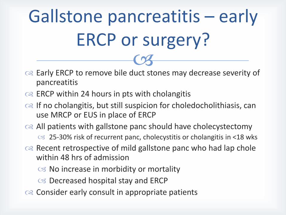
Early ERCP to remove bile duct stones may decrease severity of
pancreatitis
ERCP within 24 hours in pts with cholangitis
If no cholangitis, but still suspicion for choledocholithiasis, can use MRCP or EUS in place of ERCP
All patients with gallstone panc should have cholecystectomy 25-30% risk of recurrent panc, cholecystitis or cholangitis in <18 wks
Recent retrospective of mild gallstone panc who had lap chole within 48 hrs of admission
No increase in morbidity or mortality
Decreased hospital stay and ERCP
Consider early consult in appropriate patients
Gallstone pancreatitis – early ERCP or surgery?
Early aggressive hydration is critical in the management of
acute pancreatitis
Imaging with contrasted CT or MRI is indicated at 48-72 hrs in pts who are not improving or decompensating
Early enteral nutrition improves outcomes in pts with severe pancreatitis
Key Messages
Banks P et al. Practice Guidelines in Acute Pancreatitis. Am J Gastroenterol 2006;
101:2379-2400 Tenner S et al. Management of Acute Pancreatitis. Am J Gastroenterol 2013;
108:1400–1415 Banks P et al. Classification of acute pancreatitis-2012:revision of the Atlanta
classification and definitions by international consensus. Gut 2013; 62: 102-111. Al-Omran M et al. Enteral versus Parenteral Nutrition for Acute Pancreatitis
(Review). Cochrane Database of Systematic Reviews 2010, Issue1 Falor et al. Early Laparoscopic Cholecystectomy for Mild Gallstone Pancreatitis.
Arch Surg 147: Nov 2012 Van Santvoort et al. Early Endoscopic Retrograde Cholangiopancreatography in
Predicted Severe Acute Biliary Pancreatitis: A Prospective Multicenter Study. Annals of Surgery. 250 (1); July 2009
Wu, Bechien. Prognosis in Acute Pancreatitis. CMAJ: 183 (6); April 2011
References
Original Version: Ashley Duckett, MD and Theresa Cuoco,
MD 2013
Revised 6/11/2015: Stefanie Erway, MD
Revision History