Embed Size (px)
Citation preview

Journal of Microbiological Methods 90 (2012) 46–52
Contents lists available at SciVerse ScienceDirect
Journal of Microbiological Methods
j ourna l homepage: www.e lsev ie r .com/ locate / jmicmeth
DNA binding site analysis of Burkholderia thailandensis response regulators
Kristy L. Nowak-Lovato a, Alexana J. Hickmott a, Tuhin S. Maity a, Martha L. Bulyk b,c,d,John Dunbar a, Elizabeth Hong-Geller a,⁎a Bioscience Division, Los Alamos National Laboratory, Los Alamos, NM 87545, United Statesb Division of Genetics, Department of Medicine, Brigham & Women's Hospital and Harvard Medical School, Boston, MA 02115, United Statesc Department of Pathology, Brigham & Women's Hospital and Harvard Medical School, Boston, MA 02115, United Statesd Harvard-MIT Division of Health Sciences and Technology, Harvard Medical School, Boston, MA 02115, United States
⁎ Corresponding author. Tel.: +1 505 665 2465; fax:E-mail address: [email protected] (E. Hong-Geller).
0167-7012/$ – see front matter. Published by Elsevier Bdoi:10.1016/j.mimet.2012.03.019
a b s t r a c t
a r t i c l e i n f oArticle history:Received 19 March 2012Received in revised form 27 March 2012Accepted 27 March 2012Available online 13 April 2012
Keywords:Response regulatorProtein-binding microarrayPromoter binding siteBurkholderiaTwo component systemsGene regulation
Bacterial response regulators (RR) that function as transcription factors in two component signaling path-ways are crucial for ensuring tight regulation and coordinated expression of the genome. Currently, consen-sus DNA binding sites in the promoter for very few bacterial RRs have been identified. A systematic method tocharacterize these DNA binding sites for RRs would enable prediction of specific gene expression patterns inresponse to extracellular stimuli. To identify RR DNA binding sites, we functionally activated RRs usingberyllofluoride and applied them to a protein-binding microarray (PBM) to discover DNA binding motifsfor RRs expressed in Burkholderia, a Gram-negative bacterial genus. We identified DNA binding motifs forconserved RRs in Burkholderia thailandensis, including KdpE, RisA, and NarL, as well as for a previouslyuncharacterized RR at locus BTH_II2335 and its ortholog in the human pathogen Burkholderia pseudomalleiat locus BPSS2315. We further demonstrate RR binding of predicted genomic targets for the two orthologsusing gel shift assays and reveal a pattern of RR regulation of expression of self and other two component sys-tems. Our studies illustrate the use of PBMs to identify DNA binding specificities for bacterial RRs and enableprediction of gene regulatory networks in response to two component signaling.
Published by Elsevier B.V.
1. Introduction
Bacteria employ two-component signaling systems to couple thesensing of stress signals to adaptive changes in gene expression,thus ensuring tight regulation and coordinated expression of the ge-nome in response to the environment (Beier and Gross, 2006;Cheung and Hendrickson, 2010; Laub and Goulian, 2007). Two-component systems represent the single largest paralogous familyof signaling proteins in the bacterial kingdom and regulate diversecellular processes, including chemotaxis, osmoregulation, metabo-lism, and transport. As the name implies, the prototypical two-component system is composed of two parts. First, a histidine kinasecatalyzes autophosphorylation on a conserved histidine residue uponsensing changes in growth conditions and then transfers the phos-phoryl group to the receiver domain of the second component, a re-sponse regulator (RR), which functions as a downstream effectorprotein, often as a transcription factor that regulates gene expression(Bourret et al., 1991; Pao and Saier, 1995). For example, under K+
limitation, the histidine kinase KdpD activates the response regulatorKdpE, which in its phosphorylated state, induces expression of thekdpFABC operon via increased affinity for a 23 base pair sequence in
+1 505 665 3024.
.V.
the kdpFABC promoter. The kdpFABC operon, which lies adjacent tothe kdpDE operon, encodes an inducible high-affinity K+ uptake sys-tem that scavenges K+ to maintain ionic homeostasis in the cell(Gasell and Altendorf, 2001).
The rapid sequencing of bacterial genomes in the last several yearshas revealed a diversity of RRs with undefined regulatory functions.From 1123 distinct bacterial genomes, ~39,000 two-component pro-teins adjacent in the genome have been identified (Ulrich andZhulin, 2010). The majority of RRs with DNA binding capability fallinto three major families based on the structural similarity of their ef-fector domains, (1) OmpR/PhoB family, winged helix-turn-helix motif(Kenney, 2002), (2) NarL family, helix-turn-helix motif (Baikalovet al., 1996), and (3) NtrC family, ATPase domain (Yang et al., 2004).Although the target genes of some RRs can be predicted based on ge-nomic organization, such as KdpE control of kdpFABC, RRs can regulatemultiple target genes scattered throughout a bacterial genome. Thecompletion of sequenced bacterial genomes has enabled bioinformat-ics searches using consensus sequence motifs to predict DNA bindingsites for specific RRs. Thus far, experimental confirmation of DNAbinding sites for RRs has been limited. Aside from KdpE, DNA bindingsites have been determined for the Escherichia coli RRs OmpR (Prattand Silhavy, 1995), NarL (Baikalov et al., 1996; Maris et al., 2002),and PhoB (Makino et al., 1988), which regulate osmolarity, nitrate re-sponse, and phosphate availability, respectively. However, the greatmajority of bacterial RRs has been identified based only on sequence

47K.L. Nowak-Lovato et al. / Journal of Microbiological Methods 90 (2012) 46–52
homology, and their target DNA binding sites remain unknown orpoorly characterized. Further experimental identification of targetDNA binding sites and the cognate genes regulated by specific RRscan link extracellular inputs (e.g. nutrient deprivation, ion concentra-tion, pH change) to a regulatory gene network and better define themolecular mechanisms activated in response to two-component sig-naling pathways.
We have chosen Gram-negative bacteria Burkholderia spp. as themodel organism for discovery of RRDNA binding sites. The Burkholderiagenus encompasses ~60 species, which exhibit a wide range of biologi-cal functions, including pathogenicity, bioremediation, and nitrogen fix-ation. The two best-characterized species, Burkholderia pseudomalleiand Burkholderia mallei, the causative agents of human melioidosisand equine glanders, respectively, are categorized as Category B bio-threat agents by the CDC. We have employed protein-binding microar-ray (PBM) technology to determine the DNA binding specificities of RRsexpressed in Burkholderia thailandensis, a closely-related species to B.pseudomallei that is non-pathogenic in humans. The PBM is a rapidmethodology to simultaneously screen all sequence variants of a de-fined length and obtain comprehensive binding site measurements ofDNA–protein interactions in vitro (Berger and Bulyk, 2009; Bergeret al., 2006; Mukherjee et al., 2004). PBMs have been successfullyused to analyze transcription factor binding specificities in a wide vari-ety of organisms, including Saccharomyces cerevisiae, Caenorhabditiselegans, mice, and humans (Berger et al., 2008, 2006; Robasky andBulyk, 2011). To date, use of PBMs in bacterial systems has been limitedto a specific quorum sensing RR, LuxR, in the marine bacterium Vibrioharveyi (Pompeani et al., 2008), two nucleoid-associated proteins, H-NS and Lsr2, from Salmonella enterica and Mycobacterium tuberculosis(Gordon et al., 2011), and several TetR and MarR transcription factorsin Burkholderia xenovorans (Maity et al., 2011). In this study, we demon-strate the successful application of PBMs to both known and previously-uncharacterized Burkholderia RRs, as a broadly applicable method toidentify bacterial transcription factor binding sites for analysis of generegulation in a wide range of bacterial species. We also perform com-parative PBM analysis between a pair of RR orthologs in B. thailandensisand B. pseudomallei to investigate the overlap of DNA binding specific-ities in different Burkholderia species. We expect that identification ofRR DNA binding sites in Burkholderia can provide molecular insightsinto how two-component systemsmonitor different environmental pa-rameters and allow for prediction of cellular behavior across bacterialspecies.
2. Materials and methods
2.1. Cloning and expression of GST fusions to RRs
The RR genes were amplified from Burkholderia genomic DNAusing sequence-specific primers by PCR in 50 μl reactions [1 μl100 μM primer 1, 1 μl 100 μM primer 2, 50 ng genomic DNA isolatedfrom B. thailandensis E264 or B. pseudomallei K96243, 5 μl 10× Pfureaction buffer, 1 μl 100 mM dNTPs, 2.5 μl DMSO, 2.5 U of PfuUltraDNA polymerase (Agilent, Santa Clara, CA), and distilled H2O forthe remaining volume] using the following conditions, (1) 94 °C,3 min, (2) 94 °C, 1 min; 50 °C, 1 min, 72 °C, 1 min for 30 cycles,and (3) 94 °C, 1 min; 50 °C, 1 min, 72 °C, 10 min. The primers intro-duced 5′ BamHI and 3′ HindIII restriction sites for cloning.The primer sets used were: (1) KdpE (BTH_I1025) (F) GATCG-GATCCGAATGCCCATGAGTGAACCGACCGTCACC and (R) GATCAAGC-TTTCAGCCCGCGCCGACGAGCCGGTAGCC, (2) PhoB (BTH_I1267) (F)GATCGGATCCATGCCCAGCAACATTCTCGTCATCGAA and (R) GAT-CAAGCTTTTACGCGTGTTTCGCGAGCCGGTA, (3) OmpR (BTH_I2094)(F) GATCGGATCCATGGAAACGAAAAACCCCTCCAAG, and (R) GAT-CAAGCTTTCAGGCCGCGCCGTCGGGGATGAA (4) NarL (BTH_I1849)(F) GATCGGATCCATGACCATACGCGTACTGTTGATCGAC, and (R) GA-TCAAGCTTTCAGGCCTCGGCCGGATGCGGCGC, (5) RisA (BTH_I2094)
(F) GATCGGATCCATGGAAACGAAAAACCCCTCCAAG and (R) GAT-CAAGCTTTCAGGCCGCGCCGTCGGGGATGAA, (6) BTH_II2335 (F)GATCGGATCCATGACCACCGTTTCTTCCACGCCCCGC and (R) GATCAAG-CTTCTACCGCCTGCGATGCTCCACCGCGAA, and (7) BPSS2315 (F) GATC-GGATCCATGACTCCTGCCTCTTCCACGCCCCGC and (R) GATCAAGCTTC-TACCGCCTGCGATGCTCGACCGCGAA.
The RR genes were cloned as N-terminal GST fusions into thepGEX-KG vector using T4 ligase (NEB, Ipswich, MA), transformedinto BL21 E. coli competent cells, and induced for protein expressionwith 1 mM IPTG for 4 h. Cells were lysed with 1 mg ml−1 lysozymeon ice for 30 min, followed by treatment with 10 μg ml−1 DNaseand 10 mM MgCl2 for an additional 30 min, and centrifugation at40,000 rpm for 1 h. GST fusion proteins were purified from thecleared supernatants by incubation with agarose beads cross-linkedto glutathione for 1 h and eluted with 50 mM Tris–Cl (pH 8), 10 mMreduced glutathione. Protein samples were then dialyzed using a Sli-dealyzer cassette (Thermo Scientific, Rockford, Il.) with a 10,000 MWcut-off to remove free glutathione, quantified using the BCA proteinassay (Thermo Scientific), and stored at −80 °C in a final concentra-tion of 30% glycerol.
2.2. Gel shift assays
To demonstrate BeFx-mediated enhancement of RR binding, the pstSand nar promoter regions were PCR-amplified for use as target DNA ingel shift assays. The following primers were used for PCR: (1) pstS pro-moter (F) ATCGGCCGGACAGGCCGG and (R) GAGACCTCCAGTGTGTGAand (2) nar promoter (F) GATCGGATCCCGACATCGTGAGACGAAGCCGand (R) GATCAAGCTTGACGATTCTCTCGAGACGAGG. For the cstA(BTH_II2252−156,−130) promoter and internal histidine kinase(BTH_II2334+447,+468) gel shift assays, each set of complementaryoligonucleotides, (1) cstA (F) TGCTACGTAGCGGCCATACGTAGTTCCand (R) GGAACTACGTATGGCCGCTACGTAGCA, (2) BTH_II2334 (F)GGCTACGTGCGCTACGTCTGG and (R) CCAGACGTAGCGCACGTAGCC,and (3) non-specific oligos, (F) CGAGGGAGAATGATCGTTCTACCCTTand (R) AAGGGTAGAACGATCATTCTCCCTCG, was placed in a heatblock at 95 °C for 5 min followed by removal of the heat block to thebenchtop. The temperature of the heat block was allowed to decreaseto room temperature to allow for oligonucleotide annealing.
Binding reactions (20 μl) containing indicated concentrations ofGST–RR fusion proteins, 1 μM of target DNA sequences, 100 μMBeCl2, 10 mM NaF, and 2 μl of 10× binding buffer (20 mM Tris–ClpH 7.5, 0.5 mM EDTA, 5% glycerol, 1 mM DTT, 0.005% Triton X-100,50 mM NaCl, 5 mM MgCl2, and 2.5 mM CaCl2), were incubated for30 min at room temperature. Binding reactions were separated on8% non-denaturing polyacrylamide gels run in 0.5× TBE buffer and vi-sualized with Sybr Green DNA stain (Life Technologies, Grand Island,NY) using a ChemiDoc gel documentation system (Bio-Rad, Hercules,CA).
2.3. Protein-binding microarrays
Aminimum of two PBMs were performed for each RR as previous-ly described (Berger and Bulyk, 2009) with modifications. Briefly,microarrays were obtained from Agilent Technologies in a 4×44 Kformat, AMADID #015681 and #016060 (cat # G2514F). We per-formed primer extension from a universal 24-mer region to generatea double-stranded microarray platform. GST fusion proteins were di-luted to a final concentration of 125 nM in a volume of 175 μl (PBS, 2%milk, 200 μg ml−1 BSA, 0.3 μg ml−1 salmon testes DNA) in individualchambers of a four chamber gasket coverslip. In addition, we included2 μM BeCl2, 200 μM NaF and 1× binding buffer in all incubation andbuffer washing steps to maintain activation of the RRs and an opti-mized ionic environment during protein binding to the microarray.Microarrays were scanned (GenePix Pro 4200A, Axon Instruments,Sunnyvale, CA) to detect specific DNA–RR interactions at multiple
nar promoter
pstS promoter
GST-phoB (µM) 2 046024600+ + + +− − − − −− BeF
x
1 2 3 4 5 6 7 8 9 10
Bound
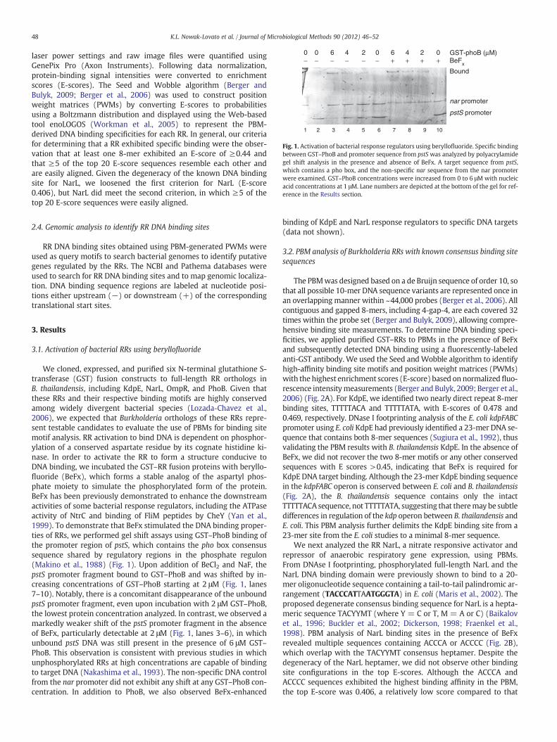
Fig. 1. Activation of bacterial response regulators using beryllofluoride. Specific bindingbetween GST–PhoB and promoter sequence from pstS was analyzed by polyacrylamidegel shift analysis in the presence and absence of BeFx. A target sequence from pstS,which contains a pho box, and the non-specific nar sequence from the nar promoterwere examined. GST–PhoB concentrations were increased from 0 to 6 μM with nucleicacid concentrations at 1 μM. Lane numbers are depicted at the bottom of the gel for ref-erence in the Results section.
48 K.L. Nowak-Lovato et al. / Journal of Microbiological Methods 90 (2012) 46–52
laser power settings and raw image files were quantified usingGenePix Pro (Axon Instruments). Following data normalization,protein-binding signal intensities were converted to enrichmentscores (E-scores). The Seed and Wobble algorithm (Berger andBulyk, 2009; Berger et al., 2006) was used to construct positionweight matrices (PWMs) by converting E-scores to probabilitiesusing a Boltzmann distribution and displayed using the Web-basedtool enoLOGOS (Workman et al., 2005) to represent the PBM-derived DNA binding specificities for each RR. In general, our criteriafor determining that a RR exhibited specific binding were the obser-vation that at least one 8-mer exhibited an E-score of ≥0.44 andthat ≥5 of the top 20 E-score sequences resemble each other andare easily aligned. Given the degeneracy of the known DNA bindingsite for NarL, we loosened the first criterion for NarL (E-score0.406), but NarL did meet the second criterion, in which ≥5 of thetop 20 E-score sequences were easily aligned.
2.4. Genomic analysis to identify RR DNA binding sites
RR DNA binding sites obtained using PBM-generated PWMs wereused as query motifs to search bacterial genomes to identify putativegenes regulated by the RRs. The NCBI and Pathema databases wereused to search for RR DNA binding sites and to map genomic localiza-tion. DNA binding sequence regions are labeled at nucleotide posi-tions either upstream (−) or downstream (+) of the correspondingtranslational start sites.
3. Results
3.1. Activation of bacterial RRs using beryllofluoride
We cloned, expressed, and purified six N-terminal glutathione S-transferase (GST) fusion constructs to full-length RR orthologs inB. thailandensis, including KdpE, NarL, OmpR, and PhoB. Given thatthese RRs and their respective binding motifs are highly conservedamong widely divergent bacterial species (Lozada-Chavez et al.,2006), we expected that Burkholderia orthologs of these RRs repre-sent testable candidates to evaluate the use of PBMs for binding sitemotif analysis. RR activation to bind DNA is dependent on phosphor-ylation of a conserved aspartate residue by its cognate histidine ki-nase. In order to activate the RR to form a structure conducive toDNA binding, we incubated the GST–RR fusion proteins with beryllo-fluoride (BeFx), which forms a stable analog of the aspartyl phos-phate moiety to simulate the phosphorylated form of the protein.BeFx has been previously demonstrated to enhance the downstreamactivities of some bacterial response regulators, including the ATPaseactivity of NtrC and binding of FliM peptides by CheY (Yan et al.,1999). To demonstrate that BeFx stimulated the DNA binding proper-ties of RRs, we performed gel shift assays using GST–PhoB binding ofthe promoter region of pstS, which contains the pho box consensussequence shared by regulatory regions in the phosphate regulon(Makino et al., 1988) (Fig. 1). Upon addition of BeCl2 and NaF, thepstS promoter fragment bound to GST–PhoB and was shifted by in-creasing concentrations of GST–PhoB starting at 2 μM (Fig. 1, lanes7–10). Notably, there is a concomitant disappearance of the unboundpstS promoter fragment, even upon incubation with 2 μM GST–PhoB,the lowest protein concentration analyzed. In contrast, we observed amarkedly weaker shift of the pstS promoter fragment in the absenceof BeFx, particularly detectable at 2 μM (Fig. 1, lanes 3–6), in whichunbound pstS DNA was still present in the presence of 6 μM GST–PhoB. This observation is consistent with previous studies in whichunphosphorylated RRs at high concentrations are capable of bindingto target DNA (Nakashima et al., 1993). The non-specific DNA controlfrom the nar promoter did not exhibit any shift at any GST–PhoB con-centration. In addition to PhoB, we also observed BeFx-enhanced
binding of KdpE and NarL response regulators to specific DNA targets(data not shown).
3.2. PBM analysis of Burkholderia RRs with known consensus binding sitesequences
The PBMwas designed based on a de Bruijn sequence of order 10, sothat all possible 10-mer DNA sequence variants are represented once inan overlapping manner within ~44,000 probes (Berger et al., 2006). Allcontiguous and gapped 8-mers, including 4-gap-4, are each covered 32times within the probe set (Berger and Bulyk, 2009), allowing compre-hensive binding site measurements. To determine DNA binding speci-ficities, we applied purified GST–RRs to PBMs in the presence of BeFxand subsequently detected DNA binding using a fluorescently-labeledanti-GST antibody. We used the Seed andWobble algorithm to identifyhigh-affinity binding site motifs and position weight matrices (PWMs)with the highest enrichment scores (E-score) based on normalizedfluo-rescence intensitymeasurements (Berger and Bulyk, 2009; Berger et al.,2006) (Fig. 2A). For KdpE, we identified two nearly direct repeat 8-merbinding sites, TTTTTACA and TTTTTATA, with E-scores of 0.478 and0.469, respectively. DNase I footprinting analysis of the E. coli kdpFABCpromoter using E. coli KdpE had previously identified a 23-mer DNA se-quence that contains both 8-mer sequences (Sugiura et al., 1992), thusvalidating the PBM results with B. thailandensis KdpE. In the absence ofBeFx, we did not recover the two 8-mer motifs or any other conservedsequences with E scores >0.45, indicating that BeFx is required forKdpE DNA target binding. Although the 23-mer KdpE binding sequencein the kdpFABC operon is conserved between E. coli and B. thailandensis(Fig. 2A), the B. thailandensis sequence contains only the intactTTTTTACA sequence, not TTTTTATA, suggesting that theremay be subtledifferences in regulation of the kdp operon between B. thailandensis andE. coli. This PBM analysis further delimits the KdpE binding site from a23-mer site from the E. coli studies to a minimal 8-mer sequence.
We next analyzed the RR NarL, a nitrate responsive activator andrepressor of anaerobic respiratory gene expression, using PBMs.From DNAse I footprinting, phosphorylated full-length NarL and theNarL DNA binding domain were previously shown to bind to a 20-mer oligonucleotide sequence containing a tail-to-tail palindromic ar-rangement (TACCCATTAATGGGTA) in E. coli (Maris et al., 2002). Theproposed degenerate consensus binding sequence for NarL is a hepta-meric sequence TACYYMT (where Y = C or T, M = A or C) (Baikalovet al., 1996; Buckler et al., 2002; Dickerson, 1998; Fraenkel et al.,1998). PBM analysis of NarL binding sites in the presence of BeFxrevealed multiple sequences containing ACCCA or ACCCC (Fig. 2B),which overlap with the TACYYMT consensus heptamer. Despite thedegeneracy of the NarL heptamer, we did not observe other bindingsite configurations in the top E-scores. Although the ACCCA andACCCC sequences exhibited the highest binding affinity in the PBM,the top E-score was 0.406, a relatively low score compared to that

-0.4
-0.2
0
0.2
0.4
A
B
A
C
G
T
T T T T AT
E-s
core
NarL (BTH_I1849)
NarL consensus binding site: TACYYMTY=C or T and M=C or A
KdpE (BTH_I1025)
E-score
0.478
0.469
E-score
0.406
C/TA
Fig. 2. PBM analysis of response regulators with known consensus binding sites.(A) The E-scores from PBM analysis of KdpE were averaged between two independentexperiments and the resultant top two PWMs are shown using enoLOGOS. Use of theSeed-and-Wobble method to determine positional nucleotide preference based onthe top 8-mer as a seed is displayed as a histogram. TheKdpE promoter binding site is con-served between E. coli and B. thailandensis upstream of kdpFABC. Nucleotide positions aremarked relative to the translational start sites of the adjacent gene. (B) The E-scores fromPBM analysis of NarL were averaged between two independent experiments and the re-sultant top PWM is shown using enoLOGOS. The degenerate consensus sequence forNarL binding is shown for comparison.
49K.L. Nowak-Lovato et al. / Journal of Microbiological Methods 90 (2012) 46–52
of KdpE. Nevertheless, the ACCCA and ACCCC sequences clearlyexhibited the highest binding affinity among the possible NarL bind-ing site configurations and were preferred in the limited 10-mer se-quence space of this PBM design.
We also used PBMs to analyze PhoB and OmpR binding in the pres-ence of BeFx. However, we did not recover the expected canonical PhoB(CTGTAC/CTGTCA) (Okamura et al., 2000) or OmpR (GXXAC) bindingmotifs (Kenney, 2002). E-scores for these two RRs were less than 0.4,with very little consensus among the top 10-mer sequences (data notshown). GST–PhoB protein had exhibited specific DNA binding to itstarget sequence by gel shift (Fig. 1), indicating that PhoB was properlyfolded and functionally active. Response regulators in the OmpR andPhoB families have been shown to dimerize upon activation and bindto direct repeat nucleotide segments with four or five spacer length nu-cleotides (de Been et al., 2008). It is possible that dimerization or coop-erativity in PhoB and OmpR is required for high-affinity DNA bindingand thus, RR–DNA binding could not be easily detected in the 10-merPBM format.
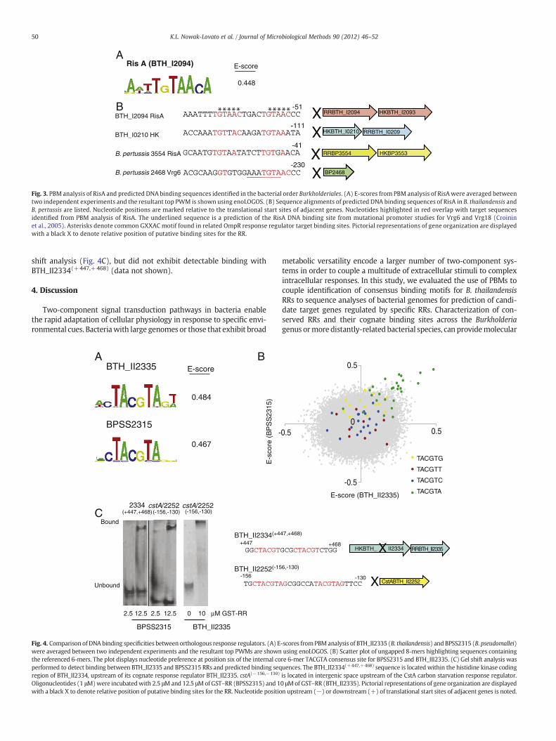
3.3. Identification of RisA DNA binding site motifs within Burkholderiales
Although PBM analysis of OmpR did not lead to identification ofthe GXXAC consensus sequence, we examined the binding site
sequences of RisA (BTH_I2094), a structurally-related response regu-lator to OmpR with 70% nucleotide homology (Stenson et al., 2005).PBM analysis of RisA revealed TGTAACA and TGTTACA binding sitemotifs with a top E-score of 0.448 (Fig. 3A). Interestingly, theGXXAC motif occurs in both of these binding sites. To identify poten-tial genes regulated by RisA, we examined intergenic sequences in thegenomes of bacterial order Burkholderiales using the two high-affinitybinding site motifs as the query sequences. We identified a direct re-peat TGTAAC upstream of RisA itself in B. thailandensis, separated byfour nucleotides, indicating the possibility of self-regulation by RisA(Fig. 3B). We also identified a degenerate direct TGTT/AA repeat up-stream of an independent two-component histidine kinase and RRpair (BTH_I0210/BTH_I0209). An imperfect TGTA/GA repeat up-stream of a RisA ortholog was also found in Bordetella pertussis, amember of the Burkholderiales order. The B. pertussis RisA had previ-ously been shown to regulate gene expression of the vrg virulence re-sponse genes. Mutation and gel shift analyses of the vrg6 and vrg18promoters had established a consensus sequence of AAATG/TTA forRisA binding (Croinin et al., 2005). Sequence analysis of the intergenicspace upstream of vrg6 revealed the sequence AAATGTAAC, in whichthe last six nucleotides overlap with the high-affinity binding se-quence identified using the PBM (Fig. 3B).
3.4. Comparison of DNA binding specificities between an orthologouspair of RRs in Burkholderia spp.
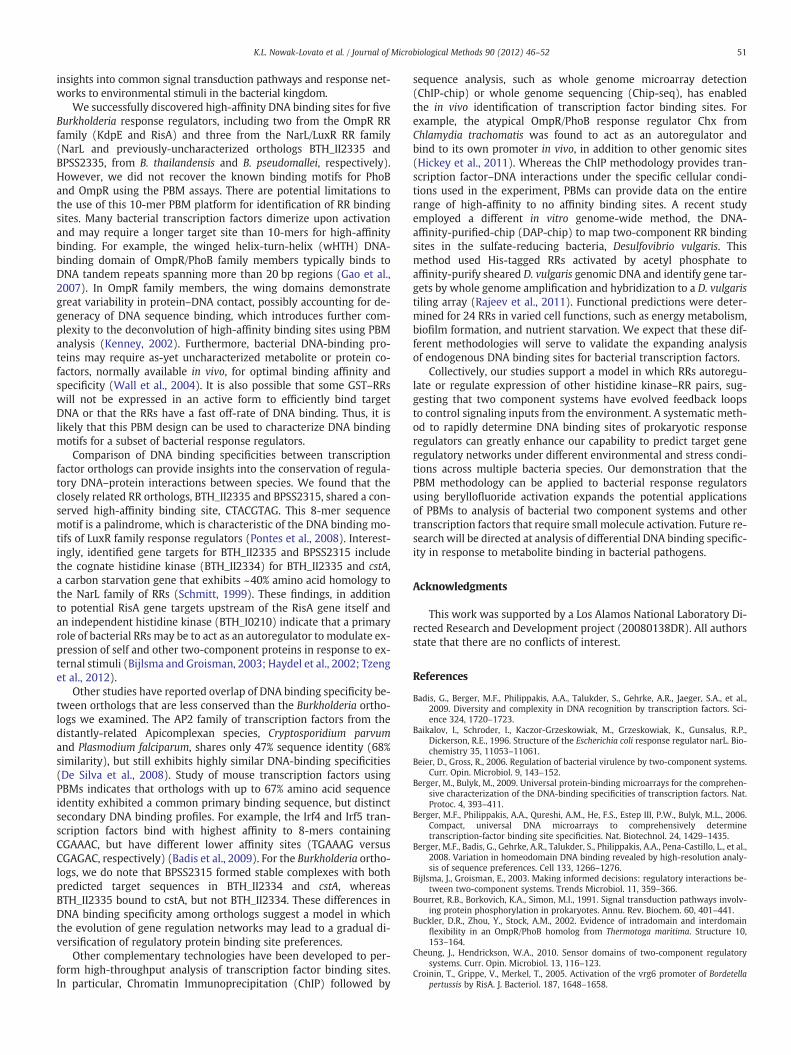
Given the success in identifying credible DNAbindingmotifs for Bur-kholderia orthologs of characterized RRs,we applied the PBMmethodol-ogy to an unknown RR from the NarL/LuxR family, at gene locusBTH_II2335. PBM analysis yielded a high-affinity palindromic bindingsequence CTACGTAG, E-score 0.484, in the presence of berylliumactiva-tion (Fig. 4A). To compare DNA binding specificities and provide insightinto conservation of gene regulation among Burkholderia species, weexamined BPSS2315 in B. pseudomallei, exhibiting ~96% nucleotideand amino acid identities to its B. thailandensis BTH_II2335 ortholog.Upon beryllium activation, the B. pseudomallei BPSS2315 ortholog alsodisplayed high binding affinity to the CTACGTAG sequence, with an E-score 0.467 (Fig. 4A). Analysis of overlapping sequenceswith the prima-ry CTACGTAG binding site revealed specific nucleotide and positionaldependencies. For example, the internal 6-mer sequence, TACGTA,appeared in the top 10 binding sequences for BPSS2315, whereas thefirst ‘C’ nucleotide and last ‘G’ nucleotide of the full 8-mer motif werepreferred in four of ten and seven of ten sequences, respectively. Com-parison of binding sequences for BPSS2315 and BTH_II2335 demon-strated a preference by both RRs to bind the 6-mer internal TACGTAsequence, with markedly decreased affinity upon changing any nucleo-tide of the internal core, for example the last A (Fig. 4B).
To identify genes potentially regulated by the BTHII2335 andBPSS2315 RRs, we searched the B. thailandensis genome for theCTACGTAG motif. We found putative binding sites at nucleotide posi-tions +449 and +458 within the cognate histidine kinase(BTH_II2334) adjacent to BTH_II2335, suggesting co-regulation ofthis histidine kinase–RR pair. The binding site within BTH_II2334 con-tains a direct repeat of CTACGT, with a spacer of three nucleotides.Putative sites were also found at −154 and −140 nucleotides up-stream of the translational start site of cstA, a carbon starvationgene (BTH_II2252), which exhibits ~40% amino acid homology toNarL (Schmitt, 1999) (Fig. 4C). The site upstream of the cstA genecontains an imperfect repeat of the CTACGTAG sequence separatedby 6 nucleotides. RRs have been shown to bind to direct or invertednucleotide repeat segments with a spacer region of 2 to 11 nucleo-tides (de Been et al., 2008). Gel shift analysis demonstrated that theBPSS2315 RR can specifically bind to both target sequences,BTH_II2334(+447,+468) and cstA(−156,−130) (Fig. 4C). TheBTH_II2335 RR also demonstrated binding to cstA(−156,−130) by gel
ARis A (BTH_I2094)
B
E-score
0.448
-51
-111
-41
-230
X
HKBTH_I2093RRBTH_I2094
HKBTH_I0210 RRBTH_I0209
X
X
HKBP3553RRBP3554
X
BP2468
BTH_I2094 RisA
BTH_I0210 HK
B. pertussis 3554 RisA
B. pertussis 2468 Vrg6
Fig. 3. PBM analysis of RisA and predicted DNA binding sequences identified in the bacterial order Burkholderiales. (A) E-scores from PBM analysis of RisAwere averaged betweentwo independent experiments and the resultant top PWM is shown using enoLOGOS. (B) Sequence alignments of predicted DNA binding sequences of RisA in B. thailandensis andB. pertussis are listed. Nucleotide positions are marked relative to the translational start sites of adjacent genes. Nucleotides highlighted in red overlap with target sequencesidentified from PBM analysis of RisA. The underlined sequence is a prediction of the RisA DNA binding site from mutational promoter studies for Vrg6 and Vrg18 (Croininet al., 2005). Asterisks denote common GXXAC motif found in related OmpR response regulator target binding sites. Pictorial representations of gene organization are displayedwith a black X to denote relative position of putative binding sites for the RR.
50 K.L. Nowak-Lovato et al. / Journal of Microbiological Methods 90 (2012) 46–52
shift analysis (Fig. 4C), but did not exhibit detectable binding withBTH_II2334(+447,+468) (data not shown).
4. Discussion
Two-component signal transduction pathways in bacteria enablethe rapid adaptation of cellular physiology in response to specific envi-ronmental cues. Bacteriawith large genomes or those that exhibit broad
BTH_II2335 A B
-
E-s
core
(B
PS
S23
15)
C
BTH_II2334(+4
+447
BTH_II2252(-15
-156
Bound
Unbound
2.5 12.5 2.5 12.5 0 10 µM GST-RR
BPSS2315 BTH_II2335
cstA/22522334(+447,+468) (-156,-130) (-156,-130)
BPSS2315
E-score
0.484
0.467
cstA/2252
Fig. 4. Comparison of DNA binding specificities between orthologous response regulators. (A) Ewere averaged between two independent experiments and the resultant top PWMs are shownthe referenced 6-mers. The plot displays nucleotide preference at position six of the internal coperformed to detect binding between BTH_II2335 and BPSS2315 RRs and predicted binding seqregion of BTH_II2334, upstream of its cognate response regulator BTH_II2335. cstA(−156,−130)
Oligonucleotides (1 μM)were incubatedwith 2.5 μMand 12.5 μMofGST–RR (BPSS2315) and 1with a black X to denote relative position of putative binding sites for the RR. Nucleotide positio
metabolic versatility encode a larger number of two-component sys-tems in order to couple a multitude of extracellular stimuli to complexintracellular responses. In this study, we evaluated the use of PBMs tocouple identification of consensus binding motifs for B. thailandensisRRs to sequence analyses of bacterial genomes for prediction of candi-date target genes regulated by specific RRs. Characterization of con-served RRs and their cognate binding sites across the Burkholderiagenus ormore distantly-related bacterial species, can providemolecular
X CstABTH_II2252
HKBTH_ II2334 RRBTH_II2335X
0.5
-0.5
0.50
0.5
E-score (BTH_II2335)
TACGTG
TACGTT
TACGTC
TACGTA
47,+468)
+468
6,-130)
-130
-scores fromPBM analysis of BTH_II2335 (B. thailandensis) and BPSS2315 (B. pseudomallei)using enoLOGOS. (B) Scatter plot of ungapped 8-mers highlighting sequences containingre 6-mer TACGTA consensus site for BPSS2315 and BTH_III2335. (C) Gel shift analysis wasuences. The BTH_II2334(+447,+468) sequence is located within the histidine kinase codingis located in intergenic space upstream of the CstA carbon starvation response regulator.0 μMof GST–RR (BTH_II2335). Pictorial representations of gene organization are displayedn upstream (−) or downstream (+) of translational start sites of adjacent genes is noted.
51K.L. Nowak-Lovato et al. / Journal of Microbiological Methods 90 (2012) 46–52
insights into common signal transduction pathways and response net-works to environmental stimuli in the bacterial kingdom.
We successfully discovered high-affinity DNA binding sites for fiveBurkholderia response regulators, including two from the OmpR RRfamily (KdpE and RisA) and three from the NarL/LuxR RR family(NarL and previously-uncharacterized orthologs BTH_II2335 andBPSS2335, from B. thailandensis and B. pseudomallei, respectively).However, we did not recover the known binding motifs for PhoBand OmpR using the PBM assays. There are potential limitations tothe use of this 10-mer PBM platform for identification of RR bindingsites. Many bacterial transcription factors dimerize upon activationand may require a longer target site than 10-mers for high-affinitybinding. For example, the winged helix-turn-helix (wHTH) DNA-binding domain of OmpR/PhoB family members typically binds toDNA tandem repeats spanning more than 20 bp regions (Gao et al.,2007). In OmpR family members, the wing domains demonstrategreat variability in protein–DNA contact, possibly accounting for de-generacy of DNA sequence binding, which introduces further com-plexity to the deconvolution of high-affinity binding sites using PBManalysis (Kenney, 2002). Furthermore, bacterial DNA-binding pro-teins may require as-yet uncharacterized metabolite or protein co-factors, normally available in vivo, for optimal binding affinity andspecificity (Wall et al., 2004). It is also possible that some GST–RRswill not be expressed in an active form to efficiently bind targetDNA or that the RRs have a fast off-rate of DNA binding. Thus, it islikely that this PBM design can be used to characterize DNA bindingmotifs for a subset of bacterial response regulators.
Comparison of DNA binding specificities between transcriptionfactor orthologs can provide insights into the conservation of regula-tory DNA–protein interactions between species. We found that theclosely related RR orthologs, BTH_II2335 and BPSS2315, shared a con-served high-affinity binding site, CTACGTAG. This 8-mer sequencemotif is a palindrome, which is characteristic of the DNA binding mo-tifs of LuxR family response regulators (Pontes et al., 2008). Interest-ingly, identified gene targets for BTH_II2335 and BPSS2315 includethe cognate histidine kinase (BTH_II2334) for BTH_II2335 and cstA,a carbon starvation gene that exhibits ~40% amino acid homology tothe NarL family of RRs (Schmitt, 1999). These findings, in additionto potential RisA gene targets upstream of the RisA gene itself andan independent histidine kinase (BTH_I0210) indicate that a primaryrole of bacterial RRs may be to act as an autoregulator to modulate ex-pression of self and other two-component proteins in response to ex-ternal stimuli (Bijlsma and Groisman, 2003; Haydel et al., 2002; Tzenget al., 2012).
Other studies have reported overlap of DNA binding specificity be-tween orthologs that are less conserved than the Burkholderia ortho-logs we examined. The AP2 family of transcription factors from thedistantly-related Apicomplexan species, Cryptosporidium parvumand Plasmodium falciparum, shares only 47% sequence identity (68%similarity), but still exhibits highly similar DNA-binding specificities(De Silva et al., 2008). Study of mouse transcription factors usingPBMs indicates that orthologs with up to 67% amino acid sequenceidentity exhibited a common primary binding sequence, but distinctsecondary DNA binding profiles. For example, the Irf4 and Irf5 tran-scription factors bind with highest affinity to 8-mers containingCGAAAC, but have different lower affinity sites (TGAAAG versusCGAGAC, respectively) (Badis et al., 2009). For the Burkholderia ortho-logs, we do note that BPSS2315 formed stable complexes with bothpredicted target sequences in BTH_II2334 and cstA, whereasBTH_II2335 bound to cstA, but not BTH_II2334. These differences inDNA binding specificity among orthologs suggest a model in whichthe evolution of gene regulation networks may lead to a gradual di-versification of regulatory protein binding site preferences.
Other complementary technologies have been developed to per-form high-throughput analysis of transcription factor binding sites.In particular, Chromatin Immunoprecipitation (ChIP) followed by
sequence analysis, such as whole genome microarray detection(ChIP-chip) or whole genome sequencing (Chip-seq), has enabledthe in vivo identification of transcription factor binding sites. Forexample, the atypical OmpR/PhoB response regulator Chx fromChlamydia trachomatis was found to act as an autoregulator andbind to its own promoter in vivo, in addition to other genomic sites(Hickey et al., 2011). Whereas the ChIP methodology provides tran-scription factor–DNA interactions under the specific cellular condi-tions used in the experiment, PBMs can provide data on the entirerange of high-affinity to no affinity binding sites. A recent studyemployed a different in vitro genome-wide method, the DNA-affinity-purified-chip (DAP-chip) to map two-component RR bindingsites in the sulfate-reducing bacteria, Desulfovibrio vulgaris. Thismethod used His-tagged RRs activated by acetyl phosphate toaffinity-purify sheared D. vulgaris genomic DNA and identify gene tar-gets by whole genome amplification and hybridization to a D. vulgaristiling array (Rajeev et al., 2011). Functional predictions were deter-mined for 24 RRs in varied cell functions, such as energy metabolism,biofilm formation, and nutrient starvation. We expect that these dif-ferent methodologies will serve to validate the expanding analysisof endogenous DNA binding sites for bacterial transcription factors.
Collectively, our studies support a model in which RRs autoregu-late or regulate expression of other histidine kinase–RR pairs, sug-gesting that two component systems have evolved feedback loopsto control signaling inputs from the environment. A systematic meth-od to rapidly determine DNA binding sites of prokaryotic responseregulators can greatly enhance our capability to predict target generegulatory networks under different environmental and stress condi-tions across multiple bacteria species. Our demonstration that thePBM methodology can be applied to bacterial response regulatorsusing beryllofluoride activation expands the potential applicationsof PBMs to analysis of bacterial two component systems and othertranscription factors that require small molecule activation. Future re-search will be directed at analysis of differential DNA binding specific-ity in response to metabolite binding in bacterial pathogens.
Acknowledgments
This work was supported by a Los Alamos National Laboratory Di-rected Research and Development project (20080138DR). All authorsstate that there are no conflicts of interest.
References
Badis, G., Berger, M.F., Philippakis, A.A., Talukder, S., Gehrke, A.R., Jaeger, S.A., et al.,2009. Diversity and complexity in DNA recognition by transcription factors. Sci-ence 324, 1720–1723.
Baikalov, I., Schroder, I., Kaczor-Grzeskowiak, M., Grzeskowiak, K., Gunsalus, R.P.,Dickerson, R.E., 1996. Structure of the Escherichia coli response regulator narL. Bio-chemistry 35, 11053–11061.
Beier, D., Gross, R., 2006. Regulation of bacterial virulence by two-component systems.Curr. Opin. Microbiol. 9, 143–152.
Berger, M., Bulyk, M., 2009. Universal protein-binding microarrays for the comprehen-sive characterization of the DNA-binding specificities of transcription factors. Nat.Protoc. 4, 393–411.
Berger, M.F., Philippakis, A.A., Qureshi, A.M., He, F.S., Estep III, P.W., Bulyk, M.L., 2006.Compact, universal DNA microarrays to comprehensively determinetranscription-factor binding site specificities. Nat. Biotechnol. 24, 1429–1435.
Berger, M.F., Badis, G., Gehrke, A.R., Talukder, S., Philippakis, A.A., Pena-Castillo, L., et al.,2008. Variation in homeodomain DNA binding revealed by high-resolution analy-sis of sequence preferences. Cell 133, 1266–1276.
Bijlsma, J., Groisman, E., 2003. Making informed decisions: regulatory interactions be-tween two-component systems. Trends Microbiol. 11, 359–366.
Bourret, R.B., Borkovich, K.A., Simon, M.I., 1991. Signal transduction pathways involv-ing protein phosphorylation in prokaryotes. Annu. Rev. Biochem. 60, 401–441.
Buckler, D.R., Zhou, Y., Stock, A.M., 2002. Evidence of intradomain and interdomainflexibility in an OmpR/PhoB homolog from Thermotoga maritima. Structure 10,153–164.
Cheung, J., Hendrickson, W.A., 2010. Sensor domains of two-component regulatorysystems. Curr. Opin. Microbiol. 13, 116–123.
Croinin, T., Grippe, V., Merkel, T., 2005. Activation of the vrg6 promoter of Bordetellapertussis by RisA. J. Bacteriol. 187, 1648–1658.

52 K.L. Nowak-Lovato et al. / Journal of Microbiological Methods 90 (2012) 46–52
de Been, M., Bart, M., Abee, J., Siezen, R., Francke, C., 2008. The identification of re-sponse regulator-specific binding sites reveals new roles of two-componentsystems in Bacillus cereus and closely related low-GC Gram-positives. Environ.Microbiol. 10, 2796–2809.
De Silva, E., Gehrke, A., Olszewski, K., Leon, I., Chahal, J., Bulyk, M., et al., 2008. SpecificDNA-binding by Apicomplexan AP2 transcription factors. Proc. Natl. Acad. Sci. U. S. A.105, 8393–8398.
Dickerson, R.E., 1998. DNA bending: the prevalence of kinkiness and the virtues of nor-mality. Nucleic Acids Res. 26, 1906–1926.
Fraenkel, E., Rould, M.A., Chambers, K.A., Pabo, C.O., 1998. Engrailed homeodomainDNA complex at 2.2 A resolution: a detailed view of the interface and comparisonwith other engrailed structures. J. Mol. Biol. 284, 351–361.
Gao, R., Mack, T., Stock, A., 2007. Bacterial response regulators: versatile regulatorystrategies from common domains. Trends Biochem. Sci. 32, 225–234.
Gasell, M., Altendorf, K., 2001. Analysis of KdpC of the K(+)-transporting KdpFABCcomplex of Escherichia coli. Eur. J. Biochem. 6, 1772–1781.
Gordon, B.R., Li, Y., Cote, A., Weirauch, M.T., Ding, P., Hughes, T.R., et al., 2011. Structuralbasis for recognition of AT-rich DNA by unrelated xenogeneic silencing proteins.Proc. Natl. Acad. Sci. U. S. A. 108, 10690–10695.
Haydel, S., Benjamin Jr., W., Dunlap, N., Clark-Curtiss, J., 2002. Expression, autoregula-tion, and DNA binding properties of the Mycobacterium tuberculosis TrcR responseregulator. J. Bacteriol. 184, 2192–2203.
Hickey, J.M., Weldon, L., Hefty, P.S., 2011. The atypical OmpR/PhoB response regulatorChxR from Chlamydia trachomatis forms homodimers in vivo and binds a direct re-peat of nucleotide sequences. J. Bacteriol. 193, 389–398.
Kenney, L., 2002. Structure/function relationships in OmpR and other winged-helixtranscription factors. Curr. Opin. Microbiol. 5, 135–141.
Laub, M.T., Goulian, M., 2007. Specificity in two-component signal transduction path-ways. Annu. Rev. Genet. 41, 121–145.
Lozada-Chavez, I., Janga, S.C., Collado-Vides, J., 2006. Bacterial regulatory networks areextremely flexible in evolution. Nucleic Acids Res. 34, 3434–3445.
Maity, T., Close, D., Valdez, Y., Nowak-Lovato, K., Marti-Arbona, R., Nguyen, T., et al.,2011. Discovery of DNA operators for TetR and MarR family transcription factorsfrom Burkholderia xenovorans. Microbiology 158, 571–582.
Makino, K., Shinagawa, H., Amemura, M., Kimura, S., Nakata, A., Ishihama, A., 1988.Regulation of the phosphate regulon of Escherichia coli. Activation of pstS tran-scription by PhoB protein in vitro. J. Mol. Biol. 203, 85–95.
Maris, A.E., Sawaya, M.R., Kaczor-Grzeskowiak, M., Jarvis, M.R., Bearson, S.M.D., Kopka,M.L., et al., 2002. Dimerization allows DNA target site recognition by the NarL re-sponse regulator. Nat. Struct. Biol. 9, 771–778.
Mukherjee, S., Berger, M.F., Jona, G., Wang, X.S., Muzzey, D., Snyder, M., et al., 2004.Rapid analysis of the DNA-binding specificities of transcription factors with DNAmicroarrays. Nat. Genet. 36, 1331–1339.
Nakashima, K., Sugiura, A., Kanamaru, K., Mizuno, T., 1993. Signal transduction be-tween the two regulatory components involved in the regulation of the kdpABCoperon in Escherichia coli: phosphorylation-dependent functioning of the positiveregulator, KdpE. Mol. Microbiol. 7, 109–116.
Okamura, H., Hanaoka, S., Nagadoi, A., Makino, K., Nishimura, Y., 2000. Structural com-parison of the PhoB and OmpR DNA-binding/transactivation domains and the ar-rangement of PhoB molecules on the phosphate box. J. Mol. Biol. 295, 1225–1236.
Pao, G.M., Saier Jr., M.H., 1995. Response regulators of bacterial signal transduction sys-tems: selective domain shuffling during evolution. J. Mol. Evol. 40, 136–154.
Pompeani, A., Irgon, J., Berger, M., Bulyk, M., Wingreen, N., Bassler, B.L., 2008. TheVibrio harveyi master quorum-sensing regulator, LuxR, a TetR-type protein is bothan activator and a repressor: DNA recognition and binding specificity at target pro-moters. Mol. Microbiol. 70, 76–88.
Pontes, M., Babst, M., Lochhead, R., Oakeson, K., Smith, K., Dale, C., 2008. Quorum sendingprimes the oxidative stress response in the insect endosymbiont, Sodalis glossinidius.PLoS One 3, e3541.
Pratt, L.A., Silhavy, T.J., 1995. Identification of base pairs important for OmpR–DNA in-teraction. Mol. Microbiol. 17, 565–573.
Rajeev, L., Luning, E., Dehal, P., Price, M., Arking, A., Mukhopadhyay, A., 2011. Systematicmapping of two component response regulators to gene targets in a model sulfatereducing bacterium. Genome Biol. 12.
Robasky, K., Bulyk, M.L., 2011. UniPROBE, update 2011: expanded content and searchtools in the online database of protein-binding microarray data on protein–DNAinteractions. Nucleic Acids Res. 39, D124–D128.
Schmitt, M., 1999. Identification of a two-component signal transduction system fromCorynebacterium diphtheriae that activates gene expression in response to the pres-ence of heme and hemoglobin. J. Bacteriol. 181, 5330–5340.
Stenson, T.H., Allen, A.G., al-Meer, J.A., Maskell, D., Peppler, M.S., 2005. Bordetella pertussisrisA, but not risS, is required for maximal expression of bvg-repressed genes. Infect.Immun. 73, 5995–6004.
Sugiura, A., Nakashima, K., Tanaka, K., Mizuno, T., 1992. Clarification of the structuraland functional features of the osmoregulated kdp operon of Escherichia coli. Mol.Microbiol. 6, 1769–1776.
Tzeng, Y., Zhou, X., Bao, S., Zhao, S., Noble, C., Stephens, D., 2012. Autoregulation of theMisR/MisS two-component signal transduction system in Neisseria meningitidis. J.Bacteriol. 188, 5055–5065.
Ulrich, L., Zhulin, I., 2010. The MiST2 database: a comprehensive genomics resource onmicrobial signal transduction. Nucleic Acids Res. 38, D401–D407.
Wall, M.E., Hlavacek, W.S., Savageau, M.A., 2004. Design of gene circuits: lessons frombacteria. Nat. Rev. Genet. 5, 34–42.
Workman, C., Yin, Y., Corcoran, D., Ideker, T., Stormo, G., Benos, P.V., 2005. enoLOGOS: aversatile web tool for energy normalized sequence logos. Nucleic Acids Res. 33,W389–W392.
Yan, D., Cho, H., Hastings, C., Igo, M., Lee, S., Pelton, J.G., et al., 1999. Beryllofluoridemimics phosphorylation of NtrC and other bacterial response regulators. ProcNatl Acad Sci USA 96, 14789–14794.
Yang, X.F., Ji, Y., Schneider, B.L., Reitzer, L., 2004. Phosphorylation-independent dimer–dimer interactions by the enhancer-binding activator NtrC of Escherichia coli: athird function for the C-terminal domain. J. Biol. Chem. 279, 36708–36714.