Embed Size (px)
Citation preview
E. Estrelles, J. Prieto, N. Fuentes & A. M. Ibars
Microstructure of seed coat in Genisteae (Fabaceae)
Abstract
Estrelles, E., Prieto J., Fuentes, N. & Ibars, A. M.: Microstructure of seed coat in Genisteae(Fabaceae). — Bocconea 19: 119-128. 2006. — ISSN 1120-4060.
Structural features of two regions of seeds of 15 species of Genisteae were investigated by scan-ning electron microscopy procedure. Six basic microstructures types were found whenanalysing surface upper ×1000 magnification. Differences between the studied areas are clear;therefore the area of observation is relevant in order to find comparable patterns among differ-ent taxa.
Introduction
It is acknowledged that morphologic features of different seed structures provide a wideresearch field; these characters play an important role on the identification of taxa (Vaughan1968) and have traditionally been used to solve systematic and phylogenetic problems.
The scanning electron microscopy (SEM) provides further insight where gross morphologydid not and allow us to analyse seed coat structure and surface sculpture. These traits are of agreat taxonomic value at generic and infra-generic status (Brisson & Peterson 1976, 1977).
Research on the Fabaceae proves that true. Studies on species of the tribe of Vicieae(Chernoff & al. 1992), the investigation on Genisteae (Saint-Martín 1986, Tahiri & al.1999), as well as the work of Newell & Hymowitz (1978) on Glycine Willd., Heyn &Herrnstadt (1977) and Gammar Ghrabi & al. (1997) on Lupinus L., or Cubas & Pardo(1988) on species of Ulex L., are good examples of the use of seed characters in Fabaceae.Studies on testa sculpture in Mediterranean species of genus Genista L. were developed byVilla (1989) and Azzioui & Es-Sgaouri (1999).
The present work revealed new data on the seed coat of 15 species included in the group ofGenisteae.
Materials and methods
Seed samples were taken either from Herbaria, Seed Banks or the wild. The origin ofthe material are listed below:
Genista dorycnifolia Font Quer (sect. Asterospartum) –Spain: Ibiza. Botanical Garden ofSoller (92/0607).
Genista legionensis (Pau) Lainz (sect. Erinacoides) – Spain: Asturias, Somiedo. (J.Fagundez collect.).
Genista tinctoria L. (sect. Genista) – Germany: NRW, Warstein Stillenbergskopf.Botanical Garden of Münster University (458).
Genista umbellata (L’Hér.) Poir. (sect. Lasiospartum) - Spain: Alicante, Hondón de lasNieves, prox. Canalosa Alta, 30S XH 7941, own collection (N. Fuentes).
Genista anglica L. (sect. Phyllospartum) - Portugal: Serra da Estrella. Botanical Garden ofCoimbra University.
Genista cinerea subsp. murcica (Coss.) Cantó & M. J. Sánchez (sect. Spartioides) – Spain:Alicante, Redovan, 30SXH82, own collection (J. Riera, F. Marco & E. Estrelles).
Genista florida subsp. polygaliphylla (Brot.) Cout. (sect. Spartioides) – Spain: León,Puerto Pajares, 30TTN7564, FCO 10206.
Genista obtusiramea Gay ex Spach (sect. Spartioides) – Spain: León, Panderrueda,30TUN37, JACA 446385.
Genista pilosa L. (sect. Spartioides) - Spain: Navarra, Garralda, 30TXN3241, JACA 33762.Genista ramosissima (Desf.) Poir. (sect. Spartioides) - Spain: Almería, Sorbas, 30SWG70,
ALME 7875.Genista aetnensis (Biv.) DC. (sect. Spartocarpus) – Italy: Sicily, Catania, Etna. Botanical
Garden of Berlin-Dahlem (2121).Genista tournefortii Spach (sect. Voglera) – Portugal: Serra do Boa Viagem, Figueira do
Fog. Botanical Garden of Coimbra University.Genista triacanthos Brot. (sect. Voglera) – Spain: La Coruña. Pico Sacro Boqueixon. (J.
Fagundez collect.).Chamaespartium sagittale (L.) P. Gibbs – Germany: Baden-Württemberg, Kreis Villingen-
Schwenningen, Niedereschach, Botanical Garden of Berlin-Dahlem (2124).Teline linifolia (L.) Webb & Berth. - Morocco: Maâmora Forest (northwest of Rabat), own
collection (J. Güemes). (From plants cultivated in the Botanical Garden of ValenciaUniversity).
Samples collected from the wild are distinguished by the name of the collector. Thematerial provided by Herbaria and Seed Banks are referred to their original herbarium oraccession number.
Seeds were submitted to the usual techniques of SEM. Three to four seeds of each sam-ple were mounted on metal stubs and were sputter-coated with a 100-200 Å thick layer ofgold and palladium by a Ion Sputter (Bio-rad SC-500). Seed surface analysis was per-formed at an accelerating voltage of 15 KV with a SEM Hitachi S-4100, file emission,from the SCSIE department, electronic microscopy section of the University of Valencia.
Two regions of the seed coat were systematically checked: perihilar and equatorial areasin order to verify the homogeneity of this character.
Features (seed coat surface patterns and epidermal cell structure) were described fol-lowing the terminology by Barthlott (1984), Stearn (1992) and Font Quer (1993).
120 Estrelles & al.: Microstructure of seed coat...
Bocconea 19 — 2006 121
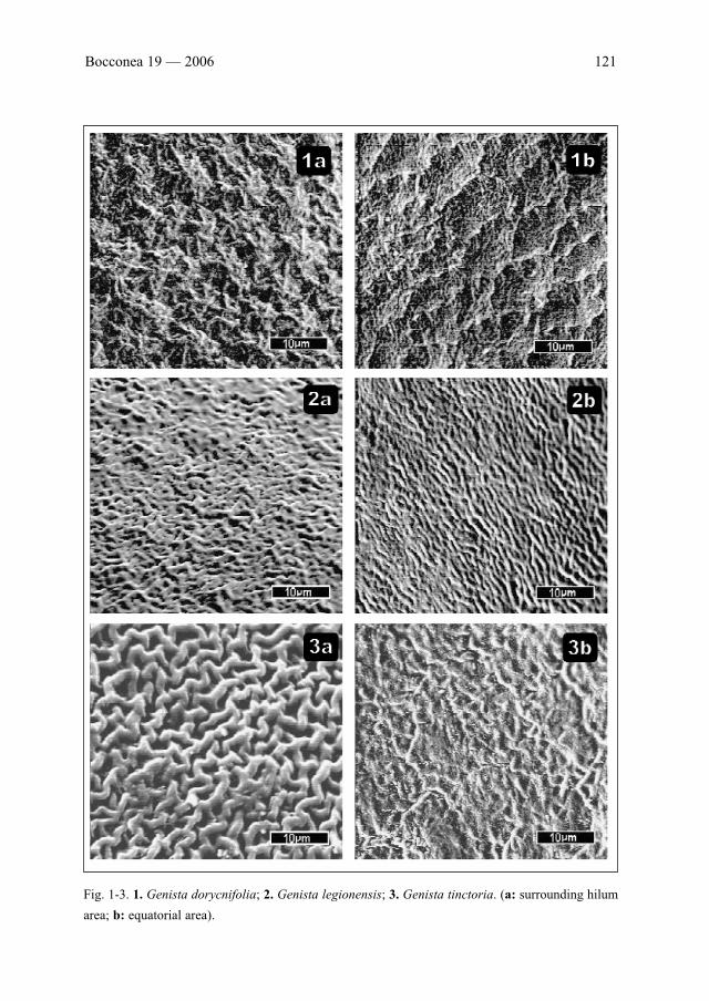
Fig. 1-3. 1. Genista dorycnifolia; 2. Genista legionensis; 3. Genista tinctoria. (a: surrounding hilum
area; b: equatorial area).
122 Estrelles & al.: Microstructure of seed coat...
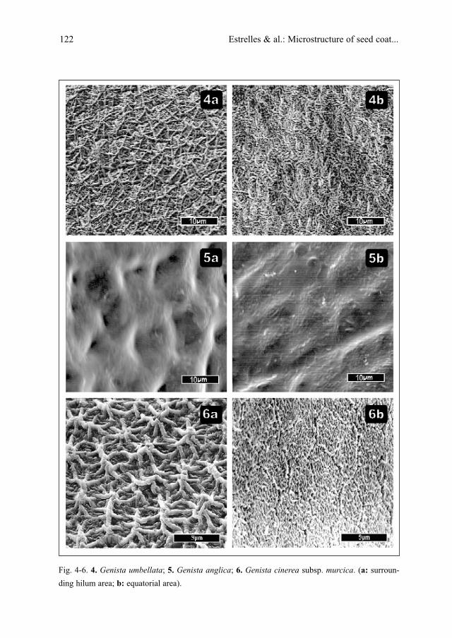
Fig. 4-6. 4. Genista umbellata; 5. Genista anglica; 6. Genista cinerea subsp. murcica. (a: surroun-
ding hilum area; b: equatorial area).
Bocconea 19 — 2006 123
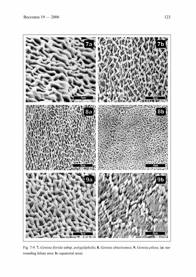
Fig. 7-9. 7. Genista florida subsp. polygaliphylla; 8. Genista obtusiramea; 9. Genista pilosa. (a: sur-
rounding hilum area; b: equatorial area).
124 Estrelles & al.: Microstructure of seed coat...
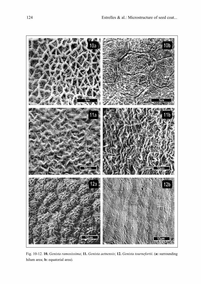
Fig. 10-12. 10. Genista ramosissima; 11. Genista aetnensis; 12. Genista tournefortii. (a: surrounding
hilum area; b: equatorial area).
Bocconea 19 — 2006 125
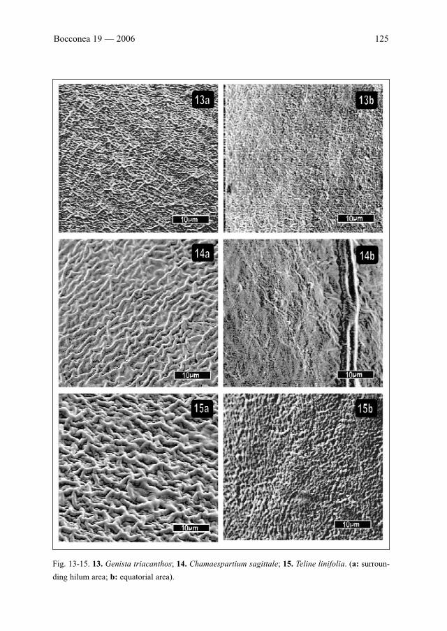
Fig. 13-15. 13. Genista triacanthos; 14. Chamaespartium sagittale; 15. Teline linifolia. (a: surroun-
ding hilum area; b: equatorial area).
126 Estrelles & al.: Microstructure of seed coat...
Results and discussion
Seed coat structure traits of the checked areas are shown as follows: in every picture theperihilar ornamentation has been set on the left side while the equatorial area is shown onthe right side.
Applying Barthlott terminology, the seeds show a primary ornament, characterised bythe presence or absence of hexa or pentagonal cells, and a typical secondary sculpture withbasically six microstructure types: I. Rugose-reticulate (wrinkled), II. Reticulate (networkpattern), in any cases microreticulate, III. Stellate, IV. Pappilate, V. Reticulate-annulate andVI. Foveate. Many different combinations of these characters are noticed. Type I and II arethe most frequent patterns in the studied taxa.
It seems that there are no clear patterns to separate taxonomic groups, species or sec-tions, within the genus Genista. These findings have been found in accordance to those byVilla (1989) and Azzioui & Es-Sgaouri (1999) while investigating another species belong-ing to the group of Genista.
It must be noted that our study reveals the occurrence of distinguishing features on bothchecked areas within the same seed. This fact should always be taken into account whenattempting investigations in this specific group.
Slight differences can be seen when comparing different populations of the samespecies stimulating subsequent studies on the issue.
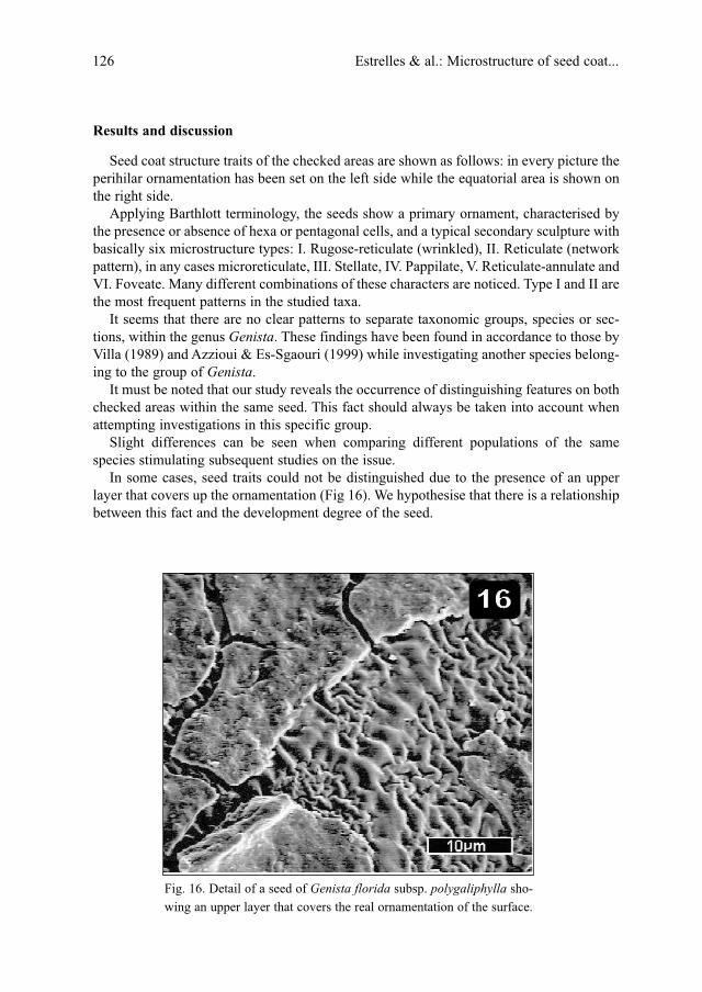
In some cases, seed traits could not be distinguished due to the presence of an upperlayer that covers up the ornamentation (Fig 16). We hypothesise that there is a relationshipbetween this fact and the development degree of the seed.
Fig. 16. Detail of a seed of Genista florida subsp. polygaliphylla sho-
wing an upper layer that covers the real ornamentation of the surface.
Conclusions
The results of our research, covering 15 species of Genisteae, seem not to be muchhelpful to the determination of species limits into this group. Given the complex grouptaxonomy, further studies including a larger number of species are needed.
Nevertheless, in the light of what it has been shown, it can be concluded that thematuration degree of the seed must be carefully chosen and the investigated seed coatarea must be indicated given the remarkable difference between the studied ones.
Acknowledgements
The authors gratefully acknowledge the technical assistance of Tomas Montan, Pilar Gomez-Garcia and Enrique Navarro in using the equipment of the electronic microscopy section of theSCSIE (“Servicio de Soporte a la Investigación Experimental”). Sincere thanks to all of them.
References
Azzioui, O. & Es-Sgaouri, A. 1999: Étude du tégument des graines du genre Genista L. (Fabaceae)au Maroc. – Acta Bot. Malacitana 24: 43-51.
Barthlott, W. 1984: Microstructural features of seed surfaces. – Pp. 95-105 in: Heywood V. H. & D.M. Moore (eds.), Current Concepts in Plant Taxonomy. – London.
Brisson, J. D. & Peterson, R. L. 1976: A critical review of the use of scanning electron microscopyin the study of the seed coat. – Proceedings of the workshop on plant science applications ofthe SEM-IIT Research Inst. Chicago, 7(2): 477-495.
— & — 1977: The scanning electron microscope and X-ray microanalysis in the study of seeds: abibliography covering the period of 1967-1976. – Scanning Electron Microscopy-IIT ResearchInst. Chicago, 2: 697-712.
Chernoff, M., Plitmann, U. & Kislev, M. E. 1992: Seed characters and testa texture in species of theVicieae: Their taxonomic significance. – Israel J. Plant Sci. 41: 167-186.
Cubas, P. & Pardo, C. 1988: Morfología de las semillas del género Ulex L. (Genisteae,Papilionoideae). – Lagascalia 15(Extra): 275-283.
Font Quer, P. 1993: Diccionario de Botánica.– Barcelona.Gammar Ghrabi, Z., Puech, S., Zouaghi, M. & Nabli, M. A. 1997: Le tégument des graines chez les
Lupinus (Fabaceae) de Tunisie. – Bot. Gallica 38: 5-21.Heyn, C. C. & Herrnstadt, I. 1977: Seed coat structure of Old World Lupinus species. – Bot. Not.
130: 427-435.Newell, C. A. & Hymowitz, T. 1978: Seed coat variation in Glycine Willd. subgenus Glycine
(Leguminosae) by SEM. – Brittonia 30: 76-88.Saint-Martin, M. 1986: Micromorphologie tégumentaire des graines de Papilionaceae. – Bull. Soc.
Bot. Fr., Lettres Bot. 133(2): 137-153.Stearn, W. T. 1992: Botanical Latin. – London. Tahiri, H., Ouyahya, A. & El Alaoui-Faris, F.-E. 1999: Étude du tégument des graines des genres
Cytisus L., Argyrocytisus (Maire) Rainaud, Chamaecytisus Link. et Genista L. (section TelineMedik.) (Fabaceae) au Maroc. – Acta Bot. Malacitana 24: 53-61.
Vaughan, F. G. 1968: Seed anatomy and taxonomy. – Proc. Linn. Soc. Lond. 179: 251-255.
Bocconea 19 — 2006 127

Villa, R. 1989: Studi sul rivestimento del seme in alcune specie del genere Genista L. – Webbia43(1): 41-49.
Address of the authors:Elena Estrelles, Josefa Prieto, Noemí Fuentes & Ana M. Ibars,Banco de Germoplasma, ICBiBE - Jardí Botànic, Universitat de València. Quart, 80, E-46008 València, Spain.
128 Estrelles & al.: Microstructure of seed coat...