Embed Size (px)
Citation preview
REVIEW
IntroductionDiagnosis of intrauterine fetal growth restriction (IUGR) is essentially an ultrasound diagnostic. If the fetal biom-etry assessed the fetal development and its correspondence to gestational age (adequate for gestational age – AGA or small for gestational age – SGA/IUGR), the Doppler exa-mination (power Doppler or color Doppler) is extremely useful in fetal functional assessment and allows the moni-toring of fetuses with IUGR during pregnancy (especially in the third trimester).
An optimal way to clinically approach the variety of fe-tal vessels that can be investigated by Doppler examination is to conceptualize the progressive nature of the pathologi-cal process of IUGR and grouping them into three com-partments related to the fetal heart [1]:
1. Postcardiac (arterial) Doppler:Umbilical artery; f
Middle cerebral artery. f
2. Cardiac Doppler:Outflow tract peak velocities; f
Mitral and tricuspid valve; f
Tricuspid regurgitation. f
3. Precardiac (venous) Doppler: Venous duct; f
Hepatic veins; f
Inferior vena cava; f
Hepatic and amniotic cavity umbilical vein. f
This classification follows the physiological adaptations of fetal circulation to the progressive abnormalities of the pla-cental vascular tree. Architectural changes of placental cir-culation in pregnancies with IUGR induce an increase of the vascular bed resistance, which can be detected by um-bilical artery and middle cerebral artery Doppler velocity. These are the early changes in fetuses with IUGR. As the fetus is still affected, there can be detected velocity changes
of the cardiac outflow tracts, as well as abnormal valvular flows. In later stages of suffering, Doppler changes are vis-ible precordially and in the venous system: ductus venosus, hepatic vein, inferior vena cava (IVC) and umbilical vein (intrahepatic and intraamniotic) [1,2,3].
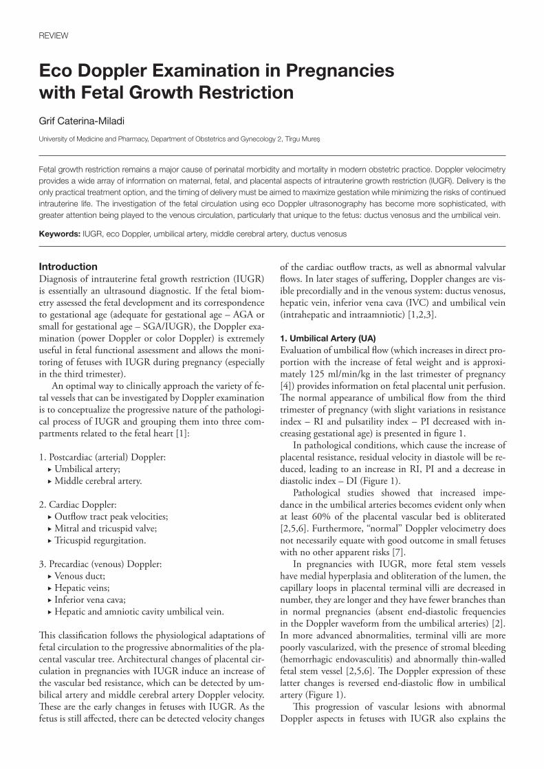
1. Umbilical Artery (UA)Evaluation of umbilical flow (which increases in direct pro-portion with the increase of fetal weight and is approxi-mately 125 ml/min/kg in the last trimester of pregnancy [4]) provides information on fetal placental unit perfusion. The normal appearance of umbilical flow from the third trimester of pregnancy (with slight variations in resistance index – RI and pulsatility index – PI decreased with in-creasing gestational age) is presented in figure 1.
In pathological conditions, which cause the increase of placental resistance, residual velocity in diastole will be re-duced, leading to an increase in RI, PI and a decrease in diastolic index – DI (Figure 1).
Pathological studies showed that increased impe- dance in the umbilical arteries becomes evident only when at least 60% of the placental vascular bed is obliterated [2,5,6]. Furthermore, ‘‘normal’’ Doppler velocimetry does not necessarily equate with good outcome in small fetuses with no other apparent risks [7].
In pregnancies with IUGR, more fetal stem vessels have medial hyperplasia and obliteration of the lumen, the capillary loops in placental terminal villi are decreased in number, they are longer and they have fewer branches than in normal pregnancies (absent end-diastolic frequencies in the Doppler waveform from the umbilical arteries) [2]. In more advanced abnormalities, terminal villi are more poorly vascularized, with the presence of stromal bleeding (hemorrhagic endovasculitis) and abnormally thin-walled fetal stem vessel [2,5,6]. The Doppler expression of these latter changes is reversed end-diastolic flow in umbilical artery (Figure 1).
This progression of vascular lesions with abnormal Doppler aspects in fetuses with IUGR also explains the
Eco Doppler Examination in Pregnancies with Fetal Growth RestrictionGrif Caterina-Miladi
University of Medicine and Pharmacy, Department of Obstetrics and Gynecology 2, Tîrgu Mureș
Fetal growth restriction remains a major cause of perinatal morbidity and mortality in modern obstetric practice. Doppler velocimetry provides a wide array of information on maternal, fetal, and placental aspects of intrauterine growth restriction (IUGR). Delivery is the only practical treatment option, and the timing of delivery must be aimed to maximize gestation while minimizing the risks of continued intrauterine life. The investigation of the fetal circulation using eco Doppler ultrasonography has become more sophisticated, with greater attention being played to the venous circulation, particularly that unique to the fetus: ductus venosus and the umbilical vein.
Keywords: IUGR, eco Doppler, umbilical artery, middle cerebral artery, ductus venosus
394 Grif Caterina-Miladi et al.
progressive worsening of placental dysfunction, assessed as mild, progressive and severe [8], as shown in Table I.
Several randomized control trials have evaluated the umbilical artery Doppler in pregnancies complicated with pre-eclampsia and/or SGA fetuses. It has been demonstrat-ed that increasing supervision focus on monitoring Dop-pler changes in the umbilical artery may lead to a 30% reduction in perinatal mortality, providing we can distin-guish between small but normal fetuses without vascular abnormalities, and fetuses affected by placental vascular conditions causing growth restriction secondary to placen-tal dysfunction, fetuses with an increased risk of intrapar-tum and perinatal death [9].
The appearance of umbilical RI is considered patholog-ical if its value is greater than 90 percentile on the standard reference curve, with a physiological fetal cardiac rate. The evolution index is also considered pathological if between two successive examinations at an interval of four weeks there is a stagnation or a decrease in diastolic flow [5].
The combination of small fetal abdominal circumfe-rence (AC), normal fetal anatomy, normal or decreased amniotic fluid volume and abnormal umbilical artery Doppler is highly suggestive for IUGR caused by placental insufficiency [10].
In extreme situations in IUGR, as noted above, may be an absent or a reverse end-diastolic flow in umbilical artery, suggesting a progressive and severe deterioration of fetal-placental haemodynamics with the risk of serious perinatal complications [1,5,11].
Doppler changes occur within a few days to several weeks (range varies from one pregnancy to another and is influ-enced by gestational age at time of first abnormal Doppler, the presence of maternal hypertension and pulsations in the umbilical vein) before clinical manifestations such as abnor-mal heart-rate pattern become evident in the fetus [6,11].
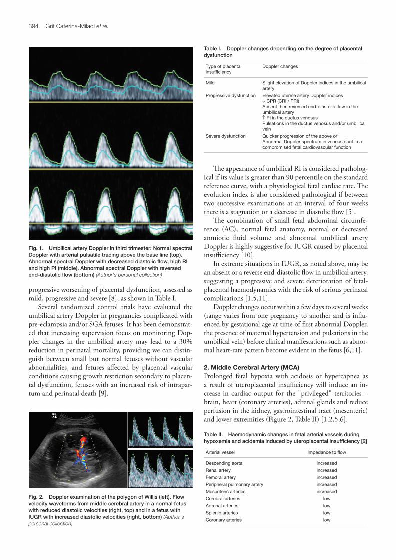
2. Middle Cerebral Artery (MCA)Prolonged fetal hypoxia with acidosis or hypercapnea as a result of uteroplacental insufficiency will induce an in-crease in cardiac output for the "privileged" territories – brain, heart (coronary arteries), adrenal glands and reduce perfusion in the kidney, gastrointestinal tract (mesenteric) and lower extremities (Figure 2, Table II) [1,2,5,6].
Table I. Doppler changes depending on the degree of placental dysfunction
Type of placental insufficiency
Doppler changes
Mild Slight elevation of Doppler indices in the umbilical artery
Progressive dysfunction Elevated uterine artery Doppler indices ↓ CPR (CRI / PRI)Absent then reversed end-diastolic flow in the umbilical artery↑ PI in the ductus venosus Pulsations in the ductus venosus and/or umbilical vein
Severe dysfunction Quicker progression of the above orAbnormal Doppler spectrum in venous duct in a compromised fetal cardiovascular function
Table II. Haemodynamic changes in fetal arterial vessels during hypoxemia and acidemia induced by uteroplacental insufficiency [2]
Arterial vessel Impedance to flow
Descending aorta increased
Renal artery increased
Femoral artery increased
Peripheral pulmonary artery increased
Mesenteric arteries increased
Cerebral arteries low
Adrenal arteries low
Splenic arteries low
Coronary arteries low
Fig. 1. Umbilical artery Doppler in third trimester: Normal spectral Doppler with arterial pulsatile tracing above the base line (top). Abnormal spectral Doppler with decreased diastolic flow, high RI and high PI (middle). Abnormal spectral Doppler with reversed end-diastolic flow (bottom) (Author's personal collection)
Fig. 2. Doppler examination of the polygon of Willis (left). Flow velocity waveforms from middle cerebral artery in a normal fetus with reduced diastolic velocities (right, top) and in a fetus with IUGR with increased diastolic velocities (right, bottom) (Author's personal collection)
395Eco Doppler Examination in Pregnancies with Fetal Growth Restriction
The most commonly used cerebral artery for Doppler assessment of the effect of fetal "brain sparing" or centra-lized circulation is the middle cerebral artery (MCA).
Velocimetric aspects of this compensatory vasodilation phenomenon are a decrease in vascular resistance within the brain and a consequent increase in diastolic blood flow. RI and PI values decrease and the DI values increase.
Vascular adaptation to hypoxemia is limited in time due to cerebral edema development, decreased cardiac con-tractility or by changes in smooth muscle cells and vascular endothelial cells, preceding by about two weeks the late deceleration of fetal heart rate [2].
There are some particular situations in the evolution of cerebral RI in pathological conditions:
If RI is below normal, but continues to fall gradually (its 1. curve follows a path relatively parallel to a normal fetus) the hypoxia is significant, but well compensated by a corresponding premature increase in brain perfusion. If an already diminished RI continues to fall rapidly be-2. coming divergent from the lower limit of normal (thus more pathologic), hypoxia is important. If an RI with clear pathological values returns to normal 3. it can be considered that the adaptive possibilities of redistributing blood flows were exceeded. Under these conditions the hypoxia is decompensated and the fetus is in acidosis [5].
The ratio between cerebral resistance index (CRI) and um-bilical or placental resistance index (PRI) determines the cerebroplacental ratio (Arbeille Index): CPR = CRI/PRI. Introduced in 1987, this parameter continues to be used in quantifying the degree of fetal distress, although according to some authors [12] this ratio did not appear to be cor-related significantly with perinatal outcome after 34 weeks. They consider the ratio between fetal PI of the thoracic aorta and PI of the middle cerebral artery to be more useful in the last trimester of pregnancy [12].
Beside the phenomenon of redistribution of fetal vascu-lar flow, the amplitude of diastolic velocity in the cerebral arteries is always lower than in the umbilical artery so that cerebral RI will be greater than umbilical RI and the value of CPR will be more than 1. On the contrary, the appea-rance of a redistribution of blood flow through cerebral va-sodilation territory, in conditions of chronic fetal distress, will cause a significant decrease in cerebral RI, and CPR will become subunit [5,13].
The main advantage of CPR is that it takes into ac-count both the placental vascular abnormality respon-sible for impaired maternal-fetal exchanges and the re-sponse of fetal cerebral haemodynamic to the occurrence of hypoxia [5].
The analysis of the four circumstances in which CPR is below 1 allows a discriminative evaluation in terms of the severity of chronic fetal distress:
When both the umbilical RI and cerebral RI are still 1. within normal limits, but CPR is below 1, chronic fetal distress is anticipated. When there is an increase in placental resistance (pa-2. thologic umbilical/placental RI) and the cerebral RI is normal, chronic fetal distress is less important. When the placental resistance is normal and the cere-3. bral resistance is reduced (only the cerebral RI is patho-logic), the chronic fetal distress appears in the context of fetal malformations, chromosomal aberrations or fetal-maternal infections. When there is an increase in placental resistance associ-4. ated with fetal hypoxia that is both cerebral and umbi-lical, RI are pathological and the chronic fetal distress is exacerbated [5].
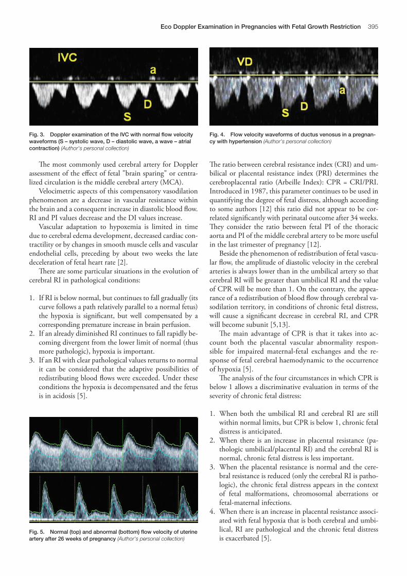
Fig. 3. Doppler examination of the IVC with normal flow velocity waveforms (S – systolic wave, D – diastolic wave, a wave – atrial contraction) (Author's personal collection)
Fig. 5. Normal (top) and abnormal (bottom) flow velocity of uterine artery after 26 weeks of pregnancy (Author's personal collection)
Fig. 4. Flow velocity waveforms of ductus venosus in a pregnan-cy with hypertension (Author's personal collection)
396 Grif Caterina-Miladi et al.
Some authors [14] have argued and demonstrated the im-portance of assessing a similar ratio between the umbilical artery PI and the middle cerebral artery PI, showing direct correlation with IUGR and the predictive value of this ra-tio for non-reactive fetal cardiotocographic tests and hos-pitalization period. A ratio above 1.26 predicted a duration of newborn hospitalization over 15 days (sensitivity 56%, specificity 92%) and many fetuses with intraventricular hemorrhage (n = 6) [14].
3. Fetal cardiac Doppler Cardiac Doppler studies in fetuses with IUGR mainly in-clude assessment of peak velocities of the outflow tracts, left and right ventricular cardiac output and flow ratios across the valves, as well as tricuspid valve regurgitation assessment [1,2].
Cardiac output is strongly influenced by the changes of arterial impedance to flow. Cerebral vasodilatation produces a decrease in left ventricle afterload, whereas increased pla-cental and systemic resistance produce increased right ven-tricle afterload.
Hypoxemia may lead to reduced cardiac contractility, decreased ventricular filling and decreased peak velocities in the aortic and pulmonary arteries. These intracardiac hemodynamic changes of the left ventricle in the early stages of placental insufficiency, lead to the growth of brain perfusion without altering oxygen and energy substrates supply [2].
As hypoxemia is emphasised by decreasing placental "reserve", the ventricular ejection force decreases gradually (in about a week) and cardiac filling is significantly reduced [2]. Tricuspid valve regurgitation is a late sign during fetal IUGR development [1].
4. Fetal venous Doppler Fetal venous Doppler studies are useful in monitoring fe-tuses with IUGR and fetal "brain sparing" or centralized circulation phenomena. Normal fetal venous flow suggests further compensation (most fetuses are with normal non-stress test – NST and biophysical profile), while abnor-mal venous flow indicates the "collapse" of haemodynamic compensatory mechanisms [1,2,3,5,6,8].
Doppler velocimetric changes in the fetal venous sys-tem shows an advanced placental dysfunction with worse fetal hypoxic condition, the evolution in these cases being accompanied by increased perinatal morbidity and morta-lity, with possible antepartum demise.
Normal flow velocity waveforms on Doppler examina-tion of the IVC are shown in figure 3.
Studies in growth-restricted human fetuses have de-monstrated that, in the inferior vena cava (IVC), an in-crease of reverse flow during atrial contraction (a wave) occurs with progressive fetal deterioration, suggesting a higher pressure gradient in the right atrium.
The next step of fetal hypoxemia is the extension of the abnormal reversed flow in the inferior vena cava to the
ductus venosus (DV), with an increase of systolic wave S/a ratio in particular, by lowering the a component of the ve-locimetric wave (Figure 4).
When DV is assessed during pregnancies with IUGR a succession of changes in the flow spectrum can be seen: (1) normal waves, (2) alternating normal and abnormal waves and (3) persistence of abnormal waves [15]. The same au-thor recommended a new spectrum analysis index of DV called SIA index (S/IVR + a), where S = systolic wave, IVR = isovolumetric relaxation between systole and diastole and a = atrial contraction, this index having a more precise pre-dictive value for fetal evolution compared with only that of the reversed flow during atrial contraction. An SIA index of 4.02 is already associated in many cases with perinatal fetal death [15].
Finally, increased venous pressure induces a reduction of end-diastolic velocity in the umbilical vein, causing end-diastolic pulses. There are two types of abnormal waves: a model with high pulsation and another with low, superfi-cial pulsation. Both types have a bad clinical significance and prognosis, but there is a more severe compromise of fetuses in the group with low pulsation of the umbilical vein [2]. Decreased contractility and reduced ventricular ejection with concomitant reduction of ventricular filling would be responsible for this type of pulsations [2].
Regular flow in the umbilical vein may also have undu-lations in physiological conditions during fetal breath, the distinction being important because of the different mea-nings (fetal breathing or fetal heart failure) [1,5].
5. Pathological aspects of uterine DopplerIn pathological obstetrical conditions that alter the process of trophoblastic invasion of utero-spiral arteries, a decrease of the maternal vessels' compliance has been observed, leading to a reduction in utero-placental vascularization. The examination of the uterine artery or its branches al-lows highlighting these anomalies translated by the follo-wing velocimetric aspects:
Change of velocimetric indices evolution (increased f
levels of RI, PI, systole-diastolic index SDI and decre-ased DI);Velocimetric spectrum presents the “notch”, the second f
downward slope changes direction (Figure 5).
This aspect is related to the persistence of the muscular elastic layer of the vessel wall, with a diastolic blood reflux. The notch may exist physiologically up to 26 weeks of ges-tation.
The detection of some pathological aspects of uterine Doppler in the second trimester of pregnancy may have long-term predictive value regarding the occurrence of maternal-fetal complications in the third trimester (preec-lampsia, eclampsia, IUGR, intrauterine fetal demise, retro-placental hematoma, premature birth) [5,16].
It must be emphasized that there is no correlation be-tween the intensity of velocimetric anomalies and the gra-
397Eco Doppler Examination in Pregnancies with Fetal Growth Restriction
vity of pathology developed in the third trimester of preg-nancy.
The significance of uterine arteries Doppler examina-tion increases when taking into account both the index va-lues and the presence of "notch" site [5,13]. If both is-sues are pathological, the risk of an acute maternal-fetal accident in the third trimester increases according to some authors [4] up to four times. Also important is the pres-ence of unilateral or bilateral "notch" site, uterine artery Doppler at 22–24 weeks gestation being a useful screening test for IUGR with bilateral uterine artery notches repre-senting a relative risk of 33.7 [13].
In large high-risk multiparous studies, the bilateral per-sistence of the notch with an average of RI ≥0.55 and a unilateral notch with RI ≥0.65 to 20 weeks of pregnancy, identify the vast majority of women who will subsequently deve-lop complications secondary to utero-placental insuf-ficiency [16].
Other prospective multicenter studies carried out be-tween 22–24 weeks of gestation in unselected women with monofetal pregnancies showed that the eco Doppler evalu-ation of the uterine artery is more effective in identifying pre-eclampsia, requiring preterm delivery rather than de-livery at term [17].
Since sensitivity and specificity of uterine artery Dop-pler measurements are relatively low, some authors do not include uterine artery velocimetric indices in the fetal sur-veillance routine protocol [18,19].
All Doppler changes occurring in IUGR fetuses play an important role in the diagnosis and management of these fetuses and based on these changes, some studies have pro-posed a staging system for IUGR fetuses that demonstrates its prognostic value [20].
ConclusionsIUGR is associated with increased perinatal morbidity and mortality. Doppler studies of the fetal circulation play an important role in the monitoring of pregnancies with IUGR. Abnormal umbilical artery studies rarely require delivery. Doppler assessment of the middle cerebral artery and venous side of the fetal circulation play a vital role in the monitoring and management of IUGR, a greater at-tention being played to the venous circulation, particularly that unique to the fetus: the umbilical vein and ductus venosus.
Delivery is the real treatment option for IUGR and should be tailored to maximize maturity. Timing of deli-very depends primarily on gestational age, estimated fetal weight and severity of the condition.
Further clinical experimental and epidemiological studies are required to identify the best time of delivery of growth-restricted fetuses, especially under 34 weeks of gestation.
The future studies should differentiate among the dif-ferent types of IUGR fetuses.
ReferencesKahn BF, Hobbins JC, Galan HL – Intrauterine Growth Restriction in Gibbs 1. RS, Karlan BY, Haney AF, Nygaard IE (eds): Danforth's Obstetrics and Gynecology. Lippincott Williams & Wilkins Publishers, Philadelphia, PA, 2008, 198–219
Nicolaides K, Rizzo G, Hecker K, Ximenes R – Doppler studies in fetal 2. hypoxemic hypoxia, in Doppler in Obstetrics 2002, produced at Centrus ® (http://www.centrus.com.br/)
Schwarze A, Gembruch U, Krapp M et al. – Qualitative venous Doppler 3. flow waveform analysis in preterm intrauterine growth-restricted fetuses with ARED flow in the umbilical artery-correlation with short-term outcome. Obstetrical & Gynecological Survey, 2005, 60(11): 710–711
Sutton MS, Theard MA, Bhatia SJ et al. – Changes in placental blood flow 4. in the normal human fetus with gestational age. Pediatr Res 1990, 28(4): 383–387
Mihu D – Examenul ecografic Doppler în trimestrele II și III de sarcină, 5. în Mihu D (ed): Ecografia Doppler în obstetrică și ginecologie. Editura Clusium, Cluj-Napoca, 2001, 78–214
Abramowicz JS, Sheiner E – Ultrasound of the placenta: a systematic 6. approach. Part II: Functional assessment (Doppler). Placenta 2008, 29: 921–929
Figueras F, Eixarch E, Gratacos E, Gardosi J – Predictiveness of antenatal 7. umbilical artery Doppler for adverse pregnancy outcome in small-for-gestational-age babies according to customised birthweight centiles: population based study. BJOG 2008, 115(5): 590–594
Turan OM, Turan S, Gungor S et al. – Progression of Doppler abnormalities 8. in intrauterine growth restriction. Ultrasound Obstet Gynecol 2008, 32(2): 160–167
Neilson JP, Alfirevic Z – Doppler ultrasound for fetal assessment in high-9. risk pregnancies. Cochrane Database Syst Rev 2000, 2: CD000073
Baschat AA – Arterial and venous Doppler in the diagnosis and 10. management of early onset fetal growth restriction. Early Hum Dev 2005, 81(11): 877–887
Baschat AA, Cosmi E, Bilardo CM, Wolf H, Berg C, et al. – Predictors of 11. neonatal outcome in early-onset placental dysfunction. Obstet Gynecol 2007, 109(2 Pt. 1): 253–261
Harrington K, Thompson MO, Carpenter RG, Nguyen M, Campbell S – 12. In third trimester fetuses the ratio in pulsatility index between the fetal descending thoracic aorta and the middle cerebral artery may be more useful. Doppler fetal circulation in pregnancies complicated by pre-eclampsia or delivery of a small for gestational age baby. 2. Longitudinal analysis. Br J Obstet Gynaecol 1999; 106: 453–466
Mousa HA, Loughna Pam. – Fetal growth restriction: investigation and 13. treatment. Obstetrics, Gynaecology and Reproductive Medicine 2008, 18(9): 247–252
Piazze J, Padula F,. Cerekja A, Cosmi EV, Anceschi MM – Prognostic 14. value of umbilical-middle cerebral artery pulsatility index ratio in fetuses with growth restriction. International Journal of Gynecology and Obstetrics 2005; 91: 233–237
Mari G – Doppler ultrasonography in obstetrics: from the diagnosis of fetal 15. anemia to the treatment of intrauterine growth-restricted fetuses. Am J Obstet Gynecol 2009; 200(6): 613–614
Harrington K, Fayyad A, Thakur V, Aquilina J – The value of uterine artery 16. Doppler in the prediction of uteroplacental complications in multiparous women. Ultrasound Obstet Gynecol 2004, 23(1): 50–55
Yu CK, Khouri O, Onwudiwe N, Spiliopoulos Y, Nicolaides KH, Fetal 17. Medicine Foundation Second-Trimester Screening Group – Prediction of pre-eclampsia by uterine artery Doppler imaging: relationship to gestational age at delivery and small-for-gestational age. Ultrasound Obstet Gynecol 2008, 31: 210–213
Harper T, Lam G – Fetal Growth Restriction, 2005, eMedicine Specialties, 18. eMedicine from WebMD
Ross MG, Mansano RZ – Fetal Growth Restriction, 2010, eMedicine 19. Specialties, Obstetrics and Gynecology, eMedicine from WebMD
Mari G, Picconi J – Doppler vascular changes in intrauterine growth 20. restriction. Semin Perinatol. 2008, 32(3): 182–189


![Early intrauterine development of mixed giant … · Early intrauterine development of mixed giant ... but with intrauterine death at 29 weeks [5]. Fetal . Early intrauterine development](https://img.pdfslide.net/doc/110x75/5b63022f7f8b9ade588b8aac/early-intrauterine-development-of-mixed-giant-early-intrauterine-development.jpg)


![Role of the Atg9a gene in intrauterine growth and survival of ......34 malnutrition, leading to fetal growth restriction (FGR) and intrauterine fetal death (IUFD) 35 [11-13]. Therefore,](https://img.pdfslide.net/doc/110x75/5fe3615f5637b735267b0386/role-of-the-atg9a-gene-in-intrauterine-growth-and-survival-of-34-malnutrition.jpg)