Embed Size (px)
Citation preview

Edinburgh Research Explorer
Cyanobacterial endobionts within a major marine, planktonic,calcifier (Globigerina bulloides, Foraminifera) revealed by 16SrRNA metabarcoding
Citation for published version:Bird, C, Darling, KF, Russell, AD, Davis, CV, Fehrenbacher, J, Free, A, Wyman, M & Ngwenya, BT 2017,'Cyanobacterial endobionts within a major marine, planktonic, calcifier (Globigerina bulloides, Foraminifera)revealed by 16S rRNA metabarcoding' Biogeosciences, vol. 14, no. 4, pp. 901-920. DOI: 10.5194/bg-14-901-2017, 10.5194/bg-14-901-2017
Digital Object Identifier (DOI):10.5194/bg-14-901-201710.5194/bg-14-901-2017
Link:Link to publication record in Edinburgh Research Explorer
Document Version:Publisher's PDF, also known as Version of record
Published In:Biogeosciences
Publisher Rights Statement:© Author(s) 2017. CC Attribution 3.0 License.
General rightsCopyright for the publications made accessible via the Edinburgh Research Explorer is retained by the author(s)and / or other copyright owners and it is a condition of accessing these publications that users recognise andabide by the legal requirements associated with these rights.
Take down policyThe University of Edinburgh has made every reasonable effort to ensure that Edinburgh Research Explorercontent complies with UK legislation. If you believe that the public display of this file breaches copyright pleasecontact [email protected] providing details, and we will remove access to the work immediately andinvestigate your claim.
Download date: 19. Jan. 2019

Biogeosciences, 14, 901–920, 2017www.biogeosciences.net/14/901/2017/doi:10.5194/bg-14-901-2017© Author(s) 2017. CC Attribution 3.0 License.
Cyanobacterial endobionts within a major marine planktoniccalcifier (Globigerina bulloides, Foraminifera) revealed by16S rRNA metabarcodingClare Bird1, Kate F. Darling1,2, Ann D. Russell3, Catherine V. Davis3, Jennifer Fehrenbacher3,4, Andrew Free5,Michael Wyman6, and Bryne T. Ngwenya1
1School of Geosciences, University of Edinburgh, Grant Institute, The King’s Buildings, James Hutton Road,Edinburgh, EH9 3FE, UK2School of Geography and Geosciences, University of St Andrews, North Street, St Andrews, KY16 9AL, UK3Earth and Planetary Sciences, University of California Davis, 2119 Earth and Physical Sciences, One Shields Avenue,Davis, CA 95616, USA4College of Earth, Ocean, and Atmospheric Sciences, Oregon State University, Corvallis, OR 97331, USA5School of Biological Sciences, University of Edinburgh, Roger Land Building, The King’s Buildings, Alexander CrumBrown Road, Edinburgh, EH9 3FF, UK6Biological and Environmental Sciences, Faculty of Natural Sciences, Cottrell Building, University of Stirling,Stirling, FK9 4LA, UK
Correspondence to: Clare Bird ([email protected])
Received: 23 September 2016 – Discussion started: 16 November 2016Revised: 26 January 2017 – Accepted: 8 February 2017 – Published: 28 February 2017
Abstract. We investigated the possibility of bacterial sym-biosis in Globigerina bulloides, a palaeoceanographicallyimportant, planktonic foraminifer. This marine protist iscommonly used in micropalaeontological investigations ofclimatically sensitive subpolar and temperate water massesas well as wind-driven upwelling regions of the world’soceans. G. bulloides is unusual because it lacks the protistalgal symbionts that are often found in other spinose species.In addition, it has a large offset in its stable carbon andoxygen isotopic compositions compared to other planktonicforaminifer species, and also that predicted from seawaterequilibrium. This is suggestive of novel differences in ecol-ogy and life history of G. bulloides, making it a good candi-date for investigating the potential for bacterial symbiosis asa contributory factor influencing shell calcification. Such in-formation is essential to evaluate fully the potential responseof G. bulloides to ocean acidification and climate change. Toinvestigate possible ecological interactions between G. bul-loides and marine bacteria, 18S rRNA gene sequencing, flu-orescence microscopy, 16S rRNA gene metabarcoding andtransmission electron microscopy (TEM) were performed
on individual specimens of G. bulloides (type IId) collectedfrom two locations in the California Current. IntracellularDNA extracted from five G. bulloides specimens was sub-jected to 16S rRNA gene metabarcoding and, remarkably,37–87 % of all 16S rRNA gene sequences recovered wereassigned to operational taxonomic units (OTUs) from thepicocyanobacterium Synechococcus. This finding was sup-ported by TEM observations of intact Synechococcus cellsin both the cytoplasm and vacuoles of G. bulloides. Theirconcentrations were up to 4 orders of magnitude greater in-side the foraminifera than those reported for the CaliforniaCurrent water column and approximately 5 % of the intra-cellular Synechococcus cells observed were undergoing celldivision. This suggests that Synechococcus is an endobiont ofG. bulloides type IId, which is the first report of a bacterialendobiont in the planktonic foraminifera. We consider thepotential roles of Synechococcus and G. bulloides within therelationship and the need to determine how widespread theassociation is within the widely distributed G. bulloides mor-phospecies. The possible influence of Synechococcus respi-ration on G. bulloides shell geochemistry is also explored.
Published by Copernicus Publications on behalf of the European Geosciences Union.

902 C. Bird et al.: Cyanobacterial endobionts within a major marine planktonic calcifier
1 Introduction
Mutualistic associations between organisms in marineecosystems can provide the partners involved with the ca-pacity to adapt to environmental stresses such as energy ornutrient limitation as well as provide robustness under thechallenges caused by climate change. For example, the closeassociation between photosynthetic microalgae and plank-tonic foraminifera supplies valuable fixed carbon and otherbenefits to the host and is a common feature of oligotrophicsurface waters (Decelle et al., 2015). Since Murray first pro-posed a symbiotic role for these intracellular phototrophsin 1897 (Murray, 1897), many cytological and ultrastruc-tural studies using light, fluorescence, and transmission elec-tron microscopy have confirmed the presence of intracellu-lar photosynthetic dinoflagellates or chrysophyte algae in awide range of planktonic foraminifera (see Gastrich, 1987;Hemleben et al., 1989). Symbiosis was demonstrated first byBé et al. (1982) in Orbulina universa by the elimination ofits dinoflagellate endobionts and their successful reinfection.Other experimental techniques focused on tracing radiola-belled C and N (Gastrich and Bartha, 1988), stable isotopeanalysis (Uhle et al., 1997, 1999), and microsensor studiesof the chemical microenvironment (Jørgensen et al., 1985;Rink et al., 1998). However, direct microscopic observationsremain an important first step in assessing potential symbi-otic associations.
Since not all planktonic foraminifera harbour protist mi-croalgae, there is obvious diversity in the requirement forthis type of mutualistic relationship. Technological advancesnow allow the investigation of the potential diversity andphysiological role of other organisms (e.g. bacteria) enter-ing into relationships with these abundant, calcifying pro-tists. Understanding and investigating the full range of theirmutualistic relationships is of great importance in plank-tonic foraminifera because of their considerable importancein Earth’s biogeochemical cycles. Their calcium carbon-ate shells play a significant role in marine carbon cyclingwithin the water column and ocean sediments. They con-tribute up to 40 % of the biogenic carbonate exported fromthe surface ocean (Schiebel, 2002; Schiebel et al., 2007),and shell dissolution at depth provides a significant bufferingof ocean carbonate chemistry and atmospheric CO2 (Holli-gan and Robertson, 1996; Iglesias-Rodriguez et al., 2002;Schiebel, 2002; Feely et al., 2004; Ridgwell and Zeebe,2005; Sarmiento and Gruber, 2006). However, calcificationrates of foraminifera are now known to be affected by thecontinued release of anthropogenic CO2, and are sensitive tothe associated changes in surface seawater pH (Ridgwell andZeebe, 2005; Manno et al., 2012). Assessment of the impactof climate change on planktonic foraminifera and the impli-cations for future biogenic carbonate production is hamperedcurrently by a lack of basic ecological information (Lombardet al., 2011; Roy et al., 2015), including information on thediversity of symbiont–foraminifera associations that might
allow different species to adapt to future environmental con-ditions. In addition to their role in marine carbon cycling, thegeochemistry of foraminiferal shells buried within the sed-iments provides a long-term repository of information thatreveals past changes in ocean conditions. Reconstructions ofpast seawater temperature, pH, dissolved inorganic carbon(DIC) concentrations and other environmental parametersbased on these shell geochemical proxies provide essentialconstraints for refining climate change projections (Kucera2007; Katz et al., 2010; Henderson, 2002). The underlyingrationale in the use of foraminiferal shell chemistry for thispurpose is that it reflects conditions in bulk seawater at thetime of deposition. Yet this chemical signal can be alteredby protist algal symbionts within some foraminifer species,complicating the interpretation of proxy records (Spero et al.,1991; Bemis et al., 1998; 2002; Anand et al., 2003; Russellet al., 2004). Important geochemical proxies such as δ18O,δ13C, and δ11B are influenced by the consumption or additionof CO2 to the calcifying microenvironment through photo-synthesis by algal symbionts and symbiont–host respiration(Mashiotta et al., 1997; Rink et al., 1998; Wolf-Gladrow etal., 1999; Hönisch et al., 2003; Eggins et al., 2004). The pres-ence of other symbiotic or endobiotic organisms like bacteriawithin a foraminiferal host could also potentially complicatethe interpretation of the proxy record via cryptic isotope frac-tionation through respiration or other metabolic processes.
Although the role and importance of the protist algal sym-bionts within planktonic foraminifera is widely recognisedand relatively well understood, the association of planktonicforaminifera with bacteria has received very little scientificattention. Apart from a few studies reporting the presenceof living bacteria inside benthic foraminifera from dysoxicsediments (Bernhard et al., 2000, 2006, 2012; Tsuchiya etal., 2015), there has been little consideration of specific en-dosymbioses between foraminifera and prokaryotes. Indeed,there are no reports of planktonic foraminiferal relationshipswith bacteria other than a single report observing the ex-ternal association of Globigerinella siphonifera type I withthe marine nitrogen-fixing, filamentous cyanobacterium Tri-chodesmium (Huber et al., 1997). This oversight is surpris-ing, since the occurrence of bacterial symbiosis within otherprotists is well established, as is their great potential for pro-viding highly specialised metabolic processes to their hosts(e.g. van Hoek et al., 2000; Schweikert and Meyer, 2001;Beier et al., 2002; Ashton et al., 2003; Foikin et al., 2003;Gast, 2009; Nowack et al., 2010; Orsi et al., 2012; Gilbert etal., 2012).
G. bulloides is a spinose planktonic foraminifer lackingprotist algal symbionts (Febvre-Chevalier, 1971; Spero andLea, 1996) that is abundant in the subpolar and temperate re-gions and also in the lower-latitude upwelling systems (Klei-jne et al., 1989; Naidu and Malmgren, 1996). In these cli-matically sensitive areas it dominates the downward flux offoraminiferal shells to the sea floor and, as a consequence,is of considerable importance for palaeoclimate reconstruc-
Biogeosciences, 14, 901–920, 2017 www.biogeosciences.net/14/901/2017/

C. Bird et al.: Cyanobacterial endobionts within a major marine planktonic calcifier 903
tions (Sautter and Thunell, 1991; Spero and Lea, 1996). Acomplication in using G. bulloides for palaeoclimate recon-struction, however, is that despite its lack of protist algalsymbionts, the shell secreted by G. bulloides is still out ofisotopic equilibrium with respect to both carbon and oxygenisotopes, deviating from predicted values by more than anyother extant, surface-dwelling species (Deuser et al., 1981;Kahn and Williams, 1981; Curry and Matthews, 1981; Kroonand Darling, 1995; Spero and Lea, 1996; Bijma et al., 1999).Such deviations are difficult to explain in the absence of pro-tist algal symbionts, although some of the disequilibrium hasbeen potentially linked to growth and ontogeny or even to G.bulloides respiration rates (Spero and Lea, 1996). The pres-ence of intracellular bacteria may provide an additional orcontributing explanation.
The study of G. bulloides is further complicated by ourcurrent inability to morphologically distinguish the numer-ous genotypes of G. bulloides identified within the morphos-pecies (Darling and Wade, 2008; Seears et al., 2012; Morardet al., 2013). The majority of the genotypes have been el-evated to species level status (Andre et al., 2014) and allare potentially ecologically distinct, though they are com-monly found in the same water column, where their adap-tive ranges overlap (Darling and Wade, 2008; Morard et al.,2013). Where this occurs, aggregation of two ecologicallydistinct G. bulloides species could introduce significant noiseinto palaeoclimate calibrations (Darling et al., 2000; Kuceraand Darling, 2002), particularly if they exhibit genotype-specific geochemical signatures as has recently been demon-strated in the Arabian Sea (Sadekov et al., 2016).
In this study we have focussed on the cool water lineageG. bulloides type IId. With the exception of a single speci-men of type IIa appearing off Santa Barbara in January (Dar-ling et al., 2003), type IId was the only genotype identifiedoff the coast of California throughout the year in both up-welling and non-upwelling hydrographic regimes (Darling etal., 2003; Darling and Wade, 2008). This is also the regionwhere the majority of experimental geochemical studies onthe G. bulloides morphospecies have been carried out, whichmeans that current calibrations for this area should be robust(e.g. Spero and Lea, 1996; Bemis et al., 1998, 2000, 2002),but may not be globally applicable since this genotype hasnot been found elsewhere to date (Darling and Wade, 2008;Morard et al., 2013).
We examined the intracellular bacterial population of in-dividual specimens of the planktonic foraminifer G. bul-loides using a multiphasic approach. We used 18S rRNAgene sequencing to identify the genotype of the host cellsand fluorescence microscopy, 16S rRNA gene metabarcod-ing via next-generation sequencing, transmission electronmicroscopy (TEM), and genetic characterisation using thepolymerase chain reaction (PCR) to investigate the distri-bution and taxonomic affiliations of the intracellular bac-teria. We demonstrate that intact, viable cells of the pic-ocyanobacterium Synechococcus spp. accumulate in large
numbers within the cytoplasm of G. bulloides type IId. Wepropose that these cells are likely to be taken up directlyfrom the surrounding water column and that Synechococcusshould be considered an endobiont of G. bulloides type IId.We go on to consider the nature of this association and its po-tential metabolic and geochemical implications. We also dis-cuss the power of the methodological approach adopted forimproving ecological knowledge of planktonic foraminifera.
2 Materials and methods
2.1 Oceanographic setting
Sampling was undertaken off the Californian coast in wa-ters influenced by two opposing currents. The CaliforniaCurrent flows equatorward from the North Pacific Current(∼ 50◦ N) to Baja California (∼ 15–25◦ N). Southerly along-shore winds drive upwelling of cold nutrient-rich waters inearly spring and summer in central California, and weakerbut more sustained upwelling further toward the south. Therelatively warm, saline Davidson Current and California Un-dercurrent flow poleward over the continental shelf. Dur-ing the southern California summer, the California Cur-rent moves farther offshore, and the Davidson Current pre-dominates near shore (Checkley and Barth, 2009). For thisstudy, samples were collected along the narrow central Cal-ifornia shelf ∼ 1 km off Bodega Head, California (38.3◦ N,123.0◦W), and in the Southern California Bight off SantaCatalina Island (33.4◦ N, 118.4◦W; Fig. 1). At both sites, lo-cal variation in foraminiferal abundances and species compo-sition is well understood (Thunell and Sautter, 1992; Field,2004, Davis et al., 2016), driven by periods of upwelling,relaxation or downwelling, and/or seasonal predominanceof the Davidson Current. The Santa Catalina Island site isclose to the San Pedro Ocean Time-series (SPOT; 33.55◦,118.4◦W) station, where the composition of the bacterial as-semblage in the water column has been monitored for over adecade (Chow et al., 2013; Cram et al., 2015).
2.2 Sample collection
The Bodega Head samples were collected during November2014, when there is a relaxation in the upwelling before thewinter storms, and also in April 2015 (Table 1). Normally,the southerly along-shore winds start to drive renewed up-welling in early spring (Garcia-Reyes and Largier, 2012) butthe samples for this study were collected prior to the delayedupwelling season of 2015, an unusual phenomenon possiblydue to the strong El Niño–Southern Oscillation (e.g. Jacox etal., 2015). Bodega Head samples were obtained from ver-tically integrated 150 µm mesh-size net tows, deployed toa maximum depth of 160 m, or to 10 m above the seafloorat shallower sites. Tow material was transferred to ambientsurface seawater and kept chilled during transit to shore atthe Bodega Marine Laboratory, where live foraminifera were
www.biogeosciences.net/14/901/2017/ Biogeosciences, 14, 901–920, 2017

904 C. Bird et al.: Cyanobacterial endobionts within a major marine planktonic calcifier
Figure 1. Map of the Californian coast and, at higher magnification,the region within the red box (insert) showing the Bodega Headand Santa Catalina Island sampling locations, (black circles) and theSPOT sampling site (white circle). The hydrography of the region isdescribed in Sect. 2.1, whilst the directions of flow of the two majorcoastal currents, the California Current and the Davidson Current,are indicated by the arrows.
wet picked. G. bulloides were then identified morphologi-cally to the morphospecies level, rinsed in 0.6 µm filteredsurface seawater and preserved in RNALater® (Ambion™).This reagent conserves cell integrity, inhibits nucleases atambient temperatures, and dissolves the calcite shell. TheSanta Catalina Island samples were collected by scuba divingor net tows during July/August 2013 towards the end of theweaker upwelling season off southern California. Collectedforaminifera were treated as at the Bodega Marine Labora-tory and transferred to RNALater® at the Wrigley MarineScience Center.
2.3 Decalcification and washing of samples
To remove the shell and shell-associated, external contam-inants, each individual specimen was decalcified by expo-sure to RNALater® (Ambion™). The cell was then washedin filter-sterilised, salt-adjusted phosphate-buffered saline(PBS) or sterile artificial seawater, transferred to a new ster-ile 1.5 mL tube and washed a further three times before beingtransferred to DOC DNA extraction buffer (Sect. 2.4; Holz-man and Pawlowski, 1996) for DNA analysis, or 4 % (w/v)paraformaldehyde in salt-adjusted PBS for microscopy. Pre-liminary experiments with the kleptoplastic foram Elphidiumspecies confirmed the efficacy of this treatment. The majority
of prokaryote shell contaminants (> 80 %) were removed inthe washing steps following dissolution in RNALater® (Birdet al., 2017).
2.4 Foram genotyping and Sanger DNA sequencing
DNA was extracted from individual foraminifer specimensusing the DOC extraction method to identify the specificgenotype (Holzman and Pawlowski, 1996). PCR amplifica-tion of the foraminiferal 18S rRNA gene was performed ac-cording to Seears et al. (2012). DNA sequencing was carriedout using the BigDye® Terminator v3.1 Cycle SequencingKit and an ABI 3730 DNA sequencer (both Applied Biosys-tems).
2.5 DAPI staining and fluorescence microscopy
Foraminifer cells were stained with 4′,6-diamadino-2-phenylindole (DAPI), which forms a highly fluorescentDAPI–DNA complex that allows the visualisation of bac-terial cells and eukaryotic cell nuclei under fluorescencemicroscopy. Individual decalcified and washed foraminiferawere fixed in 4 % (w/v) paraformaldehyde in salt-adjustedPBS for 4 h at 4 ◦C. Fixed cells were transferred to apolylysine-coated microscope slide and dehydrated throughan ethanol series of 70, 90 and 100 % ethanol. Cells werestained in 1 µg mL−1 DAPI (dilactate, Sigma-Aldrich) inPBS for 3 min and then rinsed with sterile deionised wa-ter. The stained preparations were mounted in AF1 mountantsolution (Citifluor) and bacteria and eukaryotic nuclei visu-alised using a Zeiss Axio Imager Fluorescence microscopeequipped with a DAPI filter set.
An unstained specimen of G. bulloides was also examinedby fluorescence microscopy to observe the background levelsof autofluorescence under the DAPI filter set to compare withthe appearance of DAPI stained individuals. A TRITC fil-ter set (excitation wavelength 540 nm, emission wavelength580 nm) was used also on unstained individuals to inves-tigate for the presence of autofluorescent, phycoerythrin-containing cyanobacterial cells.
2.6 DNA extraction, amplification and 16S rRNA genemetabarcoding
DNA for 16S rRNA gene metabarcoding of the bacterialpopulation within the foraminifera was extracted from de-calcified and washed planktonic foraminiferal cells by theDOC extraction method (Holzman and Pawlowski, 1996).The DNA from the six samples – three independent G. bul-loides isolates (BUL34, BUL36, BUL37) and a single non-spinose Neogloboquadrina dutertrei (DUT55) collected inJuly/August off Santa Catalina Island and two additionalG. bulloides cells collected in November off Bodega Head(BUL22, BUL23; Table 1) – was amplified alongside threereagent controls containing (i) no DNA template (two repli-cates) and (ii) DOC buffer only. The V4 region of the
Biogeosciences, 14, 901–920, 2017 www.biogeosciences.net/14/901/2017/

C. Bird et al.: Cyanobacterial endobionts within a major marine planktonic calcifier 905
Table 1. Sampling information and details of analyses performed for each planktonic foraminifer specimen collected.
Morphospecies Sample ID Sampling site Sampling date Co-ordinates Sea surface Analysistemperature
G. bulloides BUL21 Santa Catalina Island Jul/Aug 2013 33.4◦ N, 118.4◦W 18–21.5 ◦C Control for fluorescence microscopyG. bulloides BUL24 Santa Catalina Island Jul/Aug 2013 33.4◦ N, 118.4◦W 18–21.5 ◦C DAPI staining and fluorescence microscopyG. bulloides BUL25 Santa Catalina Island Jul/Aug 2013 33.4◦ N, 118.4◦W 18–21.5 ◦C DAPI staining and fluorescence microscopyG. bulloides BUL26 Santa Catalina Island Jul/Aug 2013 33.4◦ N, 118.4◦W 18–21.5 ◦C DAPI staining and fluorescence microscopyG. bulloides BUL28 Santa Catalina Island Jul/Aug 2013 33.4◦ N, 118.4◦W 18–21.5 ◦C DAPI staining and fluorescence microscopyG. bulloides BUL29 Santa Catalina Island Jul/Aug 2013 33.4◦ N, 118.4◦W 18–21.5 ◦C DAPI staining and fluorescence microscopyG. bulloides BUL30 Santa Catalina Island Jul/Aug 2013 33.4◦ N, 118.4◦W 18–21.5 ◦C DAPI staining and fluorescence microscopyG. bulloides BUL32 Santa Catalina Island July/Aug 2013 33.4◦ N, 118.4◦W 18–21.5 ◦C TEMG. bulloides BUL34 Santa Catalina Island Jul/Aug 2013 33.4◦ N, 118.4◦W 18–21.5 ◦C Metabarcoding, genotyping and Synechococcus 16S
and rbcL cloning and sequencingG. bulloides BUL36 Santa Catalina Island Jul/Aug 2013 33.4◦ N, 118.4◦W 18–21.5 ◦C MetabarcodingG. bulloides BUL37 Santa Catalina Island Jul/Aug 2013 33.4◦ N, 118.4◦W 18–21.5 ◦C MetabarcodingG. bulloides BUL39 Santa Catalina Island Jul/Aug 2013 33.4◦ N, 118.4◦W 18–21.5 ◦C TEMG. bulloides BUL69 Santa Catalina Island Jul/Aug 2013 33.4◦ N, 118.4◦W 18–21.5 ◦C TEMN. dutertrei DUT55 Santa Catalina Island Jul/Aug 2013 33.4◦ N, 118.4◦W 18–21.5 ◦C Metabarcoding and genotypingG. bulloides BUL04 Bodega Head Nov 2014 38.3◦ N, 123.0◦W 14–15 ◦C Synechococcus 16S and rbcL∗
G. bulloides BUL05 Bodega Head Nov 2014 38.3◦ N, 123.0◦W 14–15 ◦C Synechococcus 16S and rbcL∗
G. bulloides BUL13 Bodega Head Nov 2014 38.3◦ N, 123.0◦W 14–15 ◦C Synechococcus 16S and rbcL∗
G. bulloides BUL14 Bodega Head Nov 2014 38.3◦ N, 123.0◦W 14–15 ◦C Synechococcus 16S and rbcL∗
G. bulloides BUL15 Bodega Head Nov 2014 38.3◦ N, 123.0◦W 14–15 ◦C Synechococcus 16S and rbcL∗
G. bulloides BUL22 Bodega Head Nov 2014 38.3◦ N, 123.0◦W 14–15 ◦C MetabarcodingG. bulloides BUL23 Bodega Head Nov 2014 38.3◦ N, 123.0◦W 14–15 ◦C MetabarcodingG. bulloides BUL71 Bodega Head Apr 2015 38.3◦ N, 123.0◦W 10.5 ◦C TEMG. bulloides BUL73 Bodega Head Apr 2015 38.3◦ N, 123.0◦W 10.5 ◦C Synechococcus 16S and rbcL∗
G. bulloides BUL74 Bodega Head Apr 2015 38.3◦ N, 123.0◦W 10.5 ◦C Synechococcus 16S and rbcL∗
G. bulloides BUL82 Bodega Head Apr 2015 38.3◦ N, 123.0◦W 10.5 ◦C Synechococcus 16S and rbcL∗
G. bulloides BUL83 Bodega Head Apr 2015 38.3◦ N, 123.0◦W 10.5 ◦C Synechococcus 16S and rbcL∗
G. bulloides BUL84 Bodega Head Apr 2015 38.3◦ N, 123.0◦W 10.5 ◦C Synechococcus 16S and rbcL∗
G. bulloides BUL85 Bodega Head Apr 2015 38.3◦ N, 123.0◦W 10.5 ◦C Synechococcus 16S and rbcL∗
G. bulloides BUL86 Bodega Head Apr 2015 38.3◦ N, 123.0◦W 10.5 ◦C Synechococcus 16S and rbcL∗
G. bulloides BUL88 Bodega Head Apr 2015 38.3◦ N, 123.0◦W 10.5 ◦C Synechococcus 16S and rbcL∗
∗ PCR amplification of Synechococcus 16S rRNA gene and rbcL (RuBisCo large subunit).
16S rRNA gene was chosen for amplification using the 515Fforward primer and a barcoded 806R reverse primer series(Caporaso et al., 2012). This primer set is widely used bythe Earth Microbiome Project (Gilbert et al., 2010), andtherefore the amplification biases are known and well docu-mented. For example, there is a bias against amplification ofthe SAR11 group of marine Alphaproteobacteria and a biastowards over-amplification of Gammaproteobacteria (Apprillet al., 2015; Walters et al., 2016; Parada et al., 2016). EachDNA sample and control was PCR-amplified with a uniquebarcode tag that enabled demultiplexing of the samples af-ter being pooled for sequencing. The thermal cycling con-ditions are detailed by Caporaso et al. (2012). PCR reac-tions contained 1× Taq buffer plus additional MgCl2 (finalconcentration 2.5 mM), 0.2 mM of each dNTP, 0.25 µM ofeach primer, 1 µL of template DNA and 1.25 U of Taq DNApolymerase (Roche Applied Science), with the volume madeup to 25 µL with PCR-grade water (Sigma). All PCR reac-tions were set up in a PCR6 vertical laminar airflow cabinetwith UV sterilisation (Labcaire Systems, Bristol, UK) as de-scribed by Pagaling et al. (2014). Reaction tubes and PCRmixtures were treated for 15 min with 15 W UV light (wave-length= 254 nm) to destroy contaminating DNA, prior to ad-dition of dNTPs, Taq polymerase and template DNA (Padua
et al., 1999). The six functional and three control PCR reac-tions were run on a 1 % agarose gel and the products werepurified with the Wizard® SV Gel and PCR Clean-Up Sys-tem (Promega). The purified amplicons were quantified us-ing a Quant-iT PicoGreen ds-DNA assay kit (Life Technolo-gies) prior to pooling at equimolar concentrations for DNAsequencing. The total number of quality-filtered sequencingreads including controls was 862 954. DNA sequencing wasperformed at Edinburgh Genomics using an Illumina MiSeqv3 to generate 250 base pair (bp) paired-end reads.
2.6.1 Quality filtering and contaminant removal
The Quantitative Insights in Microbial Ecology (QIIME,v1.8.0; Caporaso et al., 2010) pipeline was used to assem-ble paired-end reads and quality-filter the sequences. Rawreads were paired with an overlap of 200 bp and quality-filtered with a minimum Phred score of 20 for maximumaccuracy (Kozich et al., 2013). Reads of less than 245 bp(i.e. short reads) were removed from the dataset with thePython script filter_short_reads.py from http://gist.github.com/walterst/7602058. Chimeras were detected using Use-arch v6.1.544 default settings (Edgar at al., 2011) and version13_8 of Greengenes 16S rRNA gene reference database (De-Santis et al., 2006). Given the low yield of endogenous bac-
www.biogeosciences.net/14/901/2017/ Biogeosciences, 14, 901–920, 2017

906 C. Bird et al.: Cyanobacterial endobionts within a major marine planktonic calcifier
terial DNA in these small-sized samples, we anticipated thatamplicon contamination from PCR amplification reagents,DNA extraction reagents, and the ultra-pure water systemwould contribute a significant number of DNA sequences andOTUs from contaminant genera to the sample set (Salter etal., 2014; Laurence et al., 2014). Operational taxonomic unitswith greater than 1000 sequences in any of the three controlsamples were considered to be potential contaminants andwere removed from the sample set. Two OTUs were removeddue to contamination in the two PCR controls: a Bradyrhi-zobiaceae of the class Alphaproteobacteria and an Acineto-bacter of the class Gammaproteobacteria. Twelve contam-inating OTUs were removed due to contamination via theDOC buffer, with eight of these of the class Alphaproteobac-teria. Seven of the eight were from the order Rhizobiales,five of which were classified to a higher taxonomic leveland included two of additional Bradyrhizobiaceae OTUs andone each of Methylobacterium, Mesorhizobium, Pedomicro-bium. The eighth OTU from the class Alphaproteobacteriawas a Sphingomonas of the order Sphingomonadales. The fi-nal four OTUs were Burkholdaria bryophila of the class Be-taproteobacteria; two Sediminibacterium OTUs of the phy-lum Bacteriodetes; and lastly an OTU identified as a Strepto-phyta chloroplast. A single Bradyrhizobiaceae OTU was byfar the largest contaminant, with a total of 284 636 sequencesfrom all samples and controls, and it is known to be a com-mon contaminant of next-generation sequencing data, alongwith other Alphaproteobacteria (Laurence et al., 2014).
2.6.2 Operational taxonomic unit (OTU) picking andtaxonomic assignment
The default QIIME pipeline was used for data analysis:OTU picking and taxonomic assignment. De novo picking(pick_de_novo_otus.py) clusters DNA sequences into OTUswith 97 % similarity with no external reference and selectsa representative sequence of each OTU for alignment andsubsequent assignment of taxonomy. This script keeps all di-versity, including unknowns, in the sample set. Closed refer-ence picking was also performed, which removes OTUs thatare not closely matched (< 97 %) with OTUs in the Green-genes database (pick_closed_reference_otus.py). This outputis required for normalisation by copy number (NBCN) usingthe online Galaxy tool (http://huttenhower.sph.harvard.edu/galaxy/). This corrects the abundance of each OTU to bet-ter reflect the true organism abundance by normalising pre-dicted 16S rRNA gene copy number for each OTU. In bothOTU picking methods, OTUs with fewer than 10 sequencesacross all samples were removed from the sample set (fil-ter_otus_from_otu_table.py).
2.6.3 Alpha-rarefaction and sequencing depth
In QIIME, the script alpha_rarefaction.py was used to as-sess whether the sequencing depth was adequate to detect
foraminiferal bacterial diversity. Samples were rarefied to thelowest sequencing depth observed across all samples (10 551in closed-reference picking in sample BUL22) and OTUrichness curves were generated, using the observed-speciesmetric, which counts the number of unique OTUs found in asample.
2.7 TEM
TEM was used to observe and document the structural rela-tionships between the endobiotic bacteria and foraminiferalcells. Decalcified G. bulloides were fixed in 3 % glutaralde-hyde in 0.1 M sodium cacodylate buffer, pH 7.3, for 2 h fol-lowed by three 10 min washes in 0.1 M sodium cacodylate.Specimens were then post-fixed in 1 % osmium tetroxidein 0.1 M sodium cacodylate for 45 min, followed by a fur-ther three 10 min washes in 0.1 M sodium cacodylate buffer.Specimens were then dehydrated in 50, 70, 90 and 100 %ethanol (X3) for 15 min each, then in two 10 min changes inpropylene oxide prior to being embedded in TAAB 812 resin.Sections (1 µm thick) were cut on a Leica Ultracut ultrami-crotome, stained with toluidine blue, and then viewed undera light microscope to select suitable specimen areas for in-vestigation. Ultrathin sections (60 nm thick) were cut fromselected areas, stained in uranyl acetate and lead citrate andthen viewed with a JEOL JEM-1400 Plus TEM.
2.8 Genetic identification of Synechococcus cellsidentified in G. bulloides
Synechococcus cells were found in large numbers inside G.bulloides and were genetically characterised. A 422 bp frag-ment of the Synechococcus 16S rRNA gene was amplifiedfrom total DNA extracted via the DOC method from indi-vidual specimen BUL34 (Table 1). This provided a larger,more informative fragment for phylogenetic analysis com-pared with the 253 bp generated by 16S rRNA metabarcod-ing. Cyanobacterial specific primers were used (CYA359f 5′-GGGGAATCYTTCCGCAATGGG-3′ and CYA781R a andb 5′-GACTACWGGGGTATCTAATCCCWTT-3′; Nübel etal., 1997) and thermocycler conditions were as follows:94 ◦C for 2 min followed by 30 cycles at 94 ◦C for 15 s, 55 ◦Cfor 15 s and 72 ◦C for 30 s, followed by a final extension at72 ◦C for 5 min. PCR reactions were performed with My-Taq REDDY mix (Bioline) and 0.25 µM of each primer and1 µL of template DNA, with the volume made up to 25 µLwith PCR-grade water (Sigma). The PCR product obtainedwas cloned (TOPO®-TA cloning kit, Invitrogen) and Sanger-sequenced.
Clone sequences were aligned with reference Synechococ-cus 16S rRNA gene sequences retrieved from the Gen-Bank database (NCBI) using ClustalW software within thepackage MEGA6 (Tamura et al., 2013). Phylogenetic trees(maximum likelihood, neighbour joining, minimum evolu-tion, UPGMA, maximum parsimony; Sect. 3.5) were gen-
Biogeosciences, 14, 901–920, 2017 www.biogeosciences.net/14/901/2017/
C. Bird et al.: Cyanobacterial endobionts within a major marine planktonic calcifier 907
erated using the default settings of MEGA6 with 500 boot-strap resamplings to determine the closest taxonomic af-filiations (i.e. clade designation sensu Fuller et al., 2003)of the G. bulloides-associated Synechococcus. Informedby this analysis, further primers were designed that tar-get other signature genes harboured by the Synechococcusclades identified, including that for rbcL. This phylogenet-ically informative gene encodes the large subunit of Ru-BisCO (ribulose-1,5-bisphosphate carboxylase/oxygenase),the primary CO2-fixing enzyme found in cyanobacteria.Primers (SynrbcL_For 5′-CGGCAACTTCTTCGATCAGG-3′; SynrbcL_Rev1 5′-ATGTCGCGGCTTTCTTTCTC-3′;SynrbcL_Rev2 5′-CCGGCTTCCATAAGGATGTC-3′) weredesigned with Primer 3 (http://primer3.ut.ee/) that target a252 bp fragment of rbcL from the most closely related Syne-chococcus spp. (i.e. strains CC9902, CC9311 and WH8120;see below).
Purified DNA from 14 G. bulloides specimens (Table 1)generated products of the correct size on PCR amplificationwith the rbcL primers. The product obtained from isolateBUL34 was selected and TA-cloned and DNA-sequenced asdescribed above. PCR reactions were performed in a Biome-tra Personal Thermocycler using MyTaq REDDY mix (Bio-line) and 0.25 µM of each primer and 1 µL of template DNA,with the volume made up to 25 µL with PCR-grade water(Sigma). Thermocycling conditions were 94 ◦C for 2 min fol-lowed by 30 cycles at 94 ◦C for 15 s, 56 ◦C for 15 s and 72 ◦Cfor 30 s, followed by a final extension at 72 ◦C for 5 min.
3 Results
In total, 29 individual specimens of G. bulloides collectedfrom waters off Santa Catalina Island and Bodega Head(Fig. 1) were investigated during this study. The samplinginformation and analyses performed on each specimen aredetailed in Table 1, and the sampling strategy and geneticcharacterisation are described in the methods.
3.1 Genotyping of foraminifera
Partial 18S rRNA (SSU) gene sequences amplified fromBUL34 and DUT55 have been submitted to Genbank (NCBI,accession numbers KX816046 and KX816047 respectively).G. bulloides specimen BUL34 is type IId and N. dutertreispecimen DUT55 is type Ic and it is the first time a∼ 1000 bpfragment has been amplified for this genotype. Both geno-types have been found routinely in the Southern CaliforniaBight (Darling et al., 2003).
3.2 Fluorescence microscopy and DAPI staining
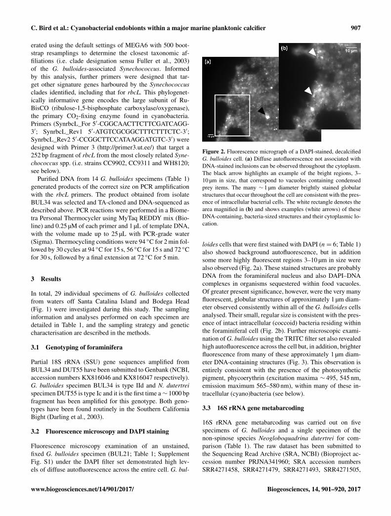
Fluorescence microscopy examination of an unstained,fixed G. bulloides specimen (BUL21; Table 1; SupplementFig. S1) under the DAPI filter set demonstrated high lev-els of diffuse autofluorescence across the entire cell. G. bul-
Figure 2. Fluorescence micrograph of a DAPI-stained, decalcifiedG. bulloides cell. (a) Diffuse autofluorescence not associated withDNA-stained inclusions can be observed throughout the cytoplasm.The black arrow highlights an example of the bright regions, 3–10 µm in size, that correspond to vacuoles containing condensedprey items. The many ∼ 1 µm diameter brightly stained globularstructures that occur throughout the cell are consistent with the pres-ence of intracellular bacterial cells. The white rectangle denotes thearea magnified in (b) and shows examples (white arrows) of theseDNA-containing, bacteria-sized structures and their cytoplasmic lo-cation.
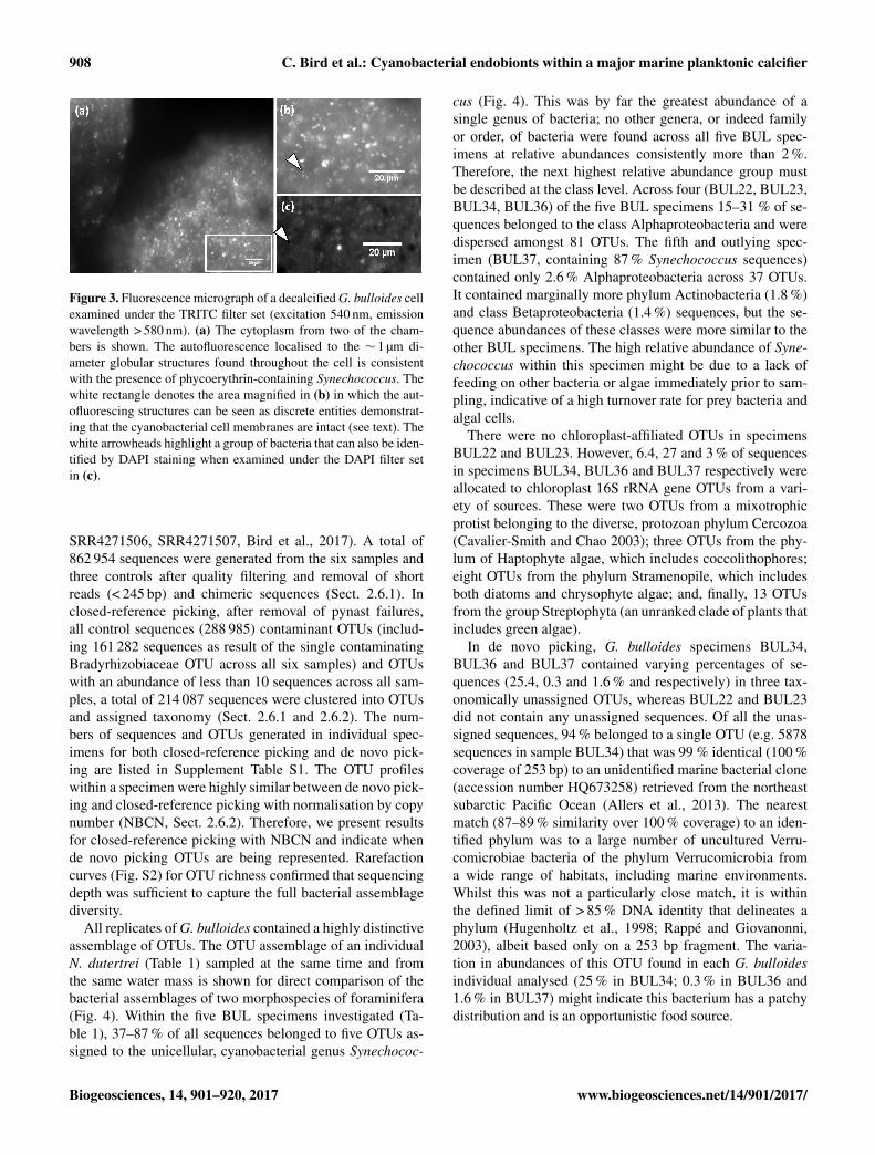
loides cells that were first stained with DAPI (n= 6; Table 1)also showed background autofluorescence, but in additionsome more highly fluorescent regions 3–10 µm in size werealso observed (Fig. 2a). These stained structures are probablyDNA from the foraminiferal nucleus and also DAPI–DNAcomplexes in organisms sequestered within food vacuoles.Of greater present significance, however, were the very manyfluorescent, globular structures of approximately 1 µm diam-eter observed consistently within all of the G. bulloides cellsanalysed. Their small, regular size is consistent with the pres-ence of intact intracellular (coccoid) bacteria residing withinthe foraminiferal cell (Fig. 2b). Further microscopic exami-nation of G. bulloides using the TRITC filter set also revealedhigh autofluorescence across the cell but, in addition, brighterfluorescence from many of these approximately 1 µm diam-eter DNA-containing structures (Fig. 3). This observation isentirely consistent with the presence of the photosyntheticpigment, phycoerythrin (excitation maxima ∼ 495, 545 nm,emission maximum 565–580 nm), within many of these in-tracellular (cyano)bacteria (see below).
3.3 16S rRNA gene metabarcoding
16S rRNA gene metabarcoding was carried out on fivespecimens of G. bulloides and a single specimen of thenon-spinose species Neogloboquadrina dutertrei for com-parison (Table 1). The raw dataset has been submitted tothe Sequencing Read Archive (SRA, NCBI) (Bioproject ac-cession number PRJNA341960; SRA accession numbersSRR4271458, SRR4271479, SRR4271493, SRR4271505,
www.biogeosciences.net/14/901/2017/ Biogeosciences, 14, 901–920, 2017
908 C. Bird et al.: Cyanobacterial endobionts within a major marine planktonic calcifier
Figure 3. Fluorescence micrograph of a decalcified G. bulloides cellexamined under the TRITC filter set (excitation 540 nm, emissionwavelength > 580 nm). (a) The cytoplasm from two of the cham-bers is shown. The autofluorescence localised to the ∼ 1 µm di-ameter globular structures found throughout the cell is consistentwith the presence of phycoerythrin-containing Synechococcus. Thewhite rectangle denotes the area magnified in (b) in which the aut-ofluorescing structures can be seen as discrete entities demonstrat-ing that the cyanobacterial cell membranes are intact (see text). Thewhite arrowheads highlight a group of bacteria that can also be iden-tified by DAPI staining when examined under the DAPI filter setin (c).
SRR4271506, SRR4271507, Bird et al., 2017). A total of862 954 sequences were generated from the six samples andthree controls after quality filtering and removal of shortreads (< 245 bp) and chimeric sequences (Sect. 2.6.1). Inclosed-reference picking, after removal of pynast failures,all control sequences (288 985) contaminant OTUs (includ-ing 161 282 sequences as result of the single contaminatingBradyrhizobiaceae OTU across all six samples) and OTUswith an abundance of less than 10 sequences across all sam-ples, a total of 214 087 sequences were clustered into OTUsand assigned taxonomy (Sect. 2.6.1 and 2.6.2). The num-bers of sequences and OTUs generated in individual spec-imens for both closed-reference picking and de novo pick-ing are listed in Supplement Table S1. The OTU profileswithin a specimen were highly similar between de novo pick-ing and closed-reference picking with normalisation by copynumber (NBCN, Sect. 2.6.2). Therefore, we present resultsfor closed-reference picking with NBCN and indicate whende novo picking OTUs are being represented. Rarefactioncurves (Fig. S2) for OTU richness confirmed that sequencingdepth was sufficient to capture the full bacterial assemblagediversity.
All replicates of G. bulloides contained a highly distinctiveassemblage of OTUs. The OTU assemblage of an individualN. dutertrei (Table 1) sampled at the same time and fromthe same water mass is shown for direct comparison of thebacterial assemblages of two morphospecies of foraminifera(Fig. 4). Within the five BUL specimens investigated (Ta-ble 1), 37–87 % of all sequences belonged to five OTUs as-signed to the unicellular, cyanobacterial genus Synechococ-
cus (Fig. 4). This was by far the greatest abundance of asingle genus of bacteria; no other genera, or indeed familyor order, of bacteria were found across all five BUL spec-imens at relative abundances consistently more than 2 %.Therefore, the next highest relative abundance group mustbe described at the class level. Across four (BUL22, BUL23,BUL34, BUL36) of the five BUL specimens 15–31 % of se-quences belonged to the class Alphaproteobacteria and weredispersed amongst 81 OTUs. The fifth and outlying spec-imen (BUL37, containing 87 % Synechococcus sequences)contained only 2.6 % Alphaproteobacteria across 37 OTUs.It contained marginally more phylum Actinobacteria (1.8 %)and class Betaproteobacteria (1.4 %) sequences, but the se-quence abundances of these classes were more similar to theother BUL specimens. The high relative abundance of Syne-chococcus within this specimen might be due to a lack offeeding on other bacteria or algae immediately prior to sam-pling, indicative of a high turnover rate for prey bacteria andalgal cells.
There were no chloroplast-affiliated OTUs in specimensBUL22 and BUL23. However, 6.4, 27 and 3 % of sequencesin specimens BUL34, BUL36 and BUL37 respectively wereallocated to chloroplast 16S rRNA gene OTUs from a vari-ety of sources. These were two OTUs from a mixotrophicprotist belonging to the diverse, protozoan phylum Cercozoa(Cavalier-Smith and Chao 2003); three OTUs from the phy-lum of Haptophyte algae, which includes coccolithophores;eight OTUs from the phylum Stramenopile, which includesboth diatoms and chrysophyte algae; and, finally, 13 OTUsfrom the group Streptophyta (an unranked clade of plants thatincludes green algae).
In de novo picking, G. bulloides specimens BUL34,BUL36 and BUL37 contained varying percentages of se-quences (25.4, 0.3 and 1.6 % and respectively) in three tax-onomically unassigned OTUs, whereas BUL22 and BUL23did not contain any unassigned sequences. Of all the unas-signed sequences, 94 % belonged to a single OTU (e.g. 5878sequences in sample BUL34) that was 99 % identical (100 %coverage of 253 bp) to an unidentified marine bacterial clone(accession number HQ673258) retrieved from the northeastsubarctic Pacific Ocean (Allers et al., 2013). The nearestmatch (87–89 % similarity over 100 % coverage) to an iden-tified phylum was to a large number of uncultured Verru-comicrobiae bacteria of the phylum Verrucomicrobia froma wide range of habitats, including marine environments.Whilst this was not a particularly close match, it is withinthe defined limit of > 85 % DNA identity that delineates aphylum (Hugenholtz et al., 1998; Rappé and Giovanonni,2003), albeit based only on a 253 bp fragment. The varia-tion in abundances of this OTU found in each G. bulloidesindividual analysed (25 % in BUL34; 0.3 % in BUL36 and1.6 % in BUL37) might indicate this bacterium has a patchydistribution and is an opportunistic food source.
Biogeosciences, 14, 901–920, 2017 www.biogeosciences.net/14/901/2017/

C. Bird et al.: Cyanobacterial endobionts within a major marine planktonic calcifier 909
0 %
10 %
20 %
30 %
40 %
50 %
60 %
70 %
80 %
90 %
100 %
BUL22 BUL23 BUL34 BUL36 BUL37 DUT55
Genus Synechococcus
Class Alphaproteobacteria; f amily Bradyrhizobiaceae
Class Alphaproteobacteria; other
Class Gammaproteobacteria
Class Betaproteobacteria
Classes Delta- and Epsilonproteobacteria
Phylum Planctomycetes
Phylum Firmicutes
Phylum Actinobacteria
Phylum Bacteriodetes
Other bacteria
Phylum Streptophyta chloroplast (green algae)
Phylum Stramenopile chloroplast (includes diatoms and chrysophyte algae)
Phylum Haptophyceae chloroplast (includes coccolithophores)
Phylum Cercozoa chloroplast (diverse protists)
Figure 4. Relative abundance of taxonomically assigned 16S rRNAgene sequences from bacteria and chloroplasts within the cytoplasmof six individual foraminifer specimens: five G. bulloides (BUL22,BUL23, BUL34, BUL36 and BUL37) and one N. dutertrei spec-imen (DUT55). Sequences are assigned to operational taxonomicunits (OTUs) grouped at different levels of taxonomic classification(see key). 16S rRNA gene sequences assigned to OTUs of the genusSynechococcus are the most abundant within G. bulloides and are atthe highest level of classification when compared to the other OTUsassigned to individual classes or phyla.
3.4 Transmission electron microscopy
Transmission electron microscopy (TEM) imaging was car-ried out on four G. bulloides specimens (BUL32, BUL39,BUL69, BUL71; Table 1, Fig. 5) to observe whether anyendobionts were present within the cell. No intracellulareukaryotic cells were observed, confirming a lack of algalsymbionts. However, numerous intact coccoid cells contain-ing carboxysomes (Fig. 5a) within the central cytoplasmsurrounded by thylakoid membranes, characteristic of thecyanobacterium Synechococcus, were observed throughoutthe cytoplasm and also in vacuoles of all individual G. bul-loides observed (Fig. 5b). Approximately 5 % of the ob-served Synechococcus cells were undergoing cell division(Fig. 5c).
To compare foraminiferal cellular Synechococcus concen-trations with those of the water column, the concentrationof Synechococcus cells per millilitre of foraminiferal cy-
Figure 5. Transmission electron microscope images of Synechococ-cus cells inside G. bulloides. (a) A Synechococcus cell with charac-teristic polyhedral carboxysomes in the central cytoplasmic region(white arrow) surrounded by thylakoid membranes. (b) NumerousSynechococcus cells within a G. bulloides cell are observed in boththe cytoplasm and vacuoles (black arrow). This is a region of cyto-plasm rich in fibrillar bodies (white arrow) found only in planktonicforaminifera, and whose function is unknown. (c) Synechococcuscell within a G. bulloides cell undergoing cell division as indicatedby the presence of a constriction at the cell midpoint (white arrow).
toplasm was calculated by assuming a conservative aver-age host cell diameter of 200 µm (Spero and Lea, 1996;Aldridge et al., 2012), a spheroid morphology (Geslin etal., 2011) and that the cytoplasm was equivalent to 75 %of the shell volume (Hannah et al., 1994). Based on aver-aged cell counts from the TEM images, the total numberof Synechococcus cells within G. bulloides occupied lessthan 2 % of the foraminiferal cell volume but was equivalentto 3.8× 10⁹ Synechococcus cells mL−1. This is far higherthan the well established range of concentrations of Syne-chococcus found throughout the global ocean that rangefrom 1× 10²–1.5× 10⁶ Synechococcus cells per millilitreof seawater (Partensky et al., 1999; Paerl et al., 2011). Inthe Southern California Bight, Synechococcus cell countsare generally fewer than 1.5× 10⁵ cells mL−1 but can reach6× 105 cells mL−1 during the blooms that are generally ob-served in late spring to early summer (Tai and Palenik, 2009;Tai et al., 2011). The concentration of Synechococcus in theG. bulloides cell was therefore up to 4 orders of magnitudegreater than peak bloom concentrations measured in the Cal-ifornia Bight. This suggests that Synechococcus cells accu-mulate within the cytoplasm of G. bulloides type IId.
3.5 Genetic characterisation of intracellularSynechococcus
Five Synechococcus OTUs were assigned in 16S rRNAgene metabarcoding with closed-reference picking. How-ever, more than 99 % of the BUL Synechococcus sequenceswere assigned to just one of these OTUs. The representa-tive nucleotide sequence (253 bp) of this OTU is a 100 %match to the coastal clade IV Synechococcus sp. strain
www.biogeosciences.net/14/901/2017/ Biogeosciences, 14, 901–920, 2017

910 C. Bird et al.: Cyanobacterial endobionts within a major marine planktonic calcifier
CC9902, originally isolated from the California Current.Two further OTUs were highly similar to this abundantOTU and were 99 % identical to Synechococcus sp. strainCC9902. The remaining two OTUs both had a nucleotidematch of 99 % with Synechococcus sp. strain WH8020, aclade I strain also found typically in coastal waters. In or-der to confirm these clade assignments, phylogenetic anal-ysis (Fig. S3) of a larger (422 bp) fragment of the Syne-chococcus 16S rRNA gene generated from BUL34 totalDNA was performed. Ten clones (GenBank accession num-bers KX815969–KX815978) clustered with the clade IVSynechococcus sp. strain CC9902 and two clones (Gen-Bank accession numbers KX815979 and KX815980) clus-tered with clade I strains CC9311 (another California Currentisolate) and WH8020, in agreement with the 16S rRNA genemetabarcoding data. The topologies of the phylogenetic treesproduced were all in overall agreement with well-establishedanalyses of Synechococcus 16S rRNA genes (Scanlan etal., 2009), confirming the phylogenetic resolution of the se-quence data included in the present study.
In addition, a 252 bp fragment of the SynechococcusrbcL gene was cloned and 230 bp of this clone was DNA-sequenced (GenBank accession number KX816048) fromBUL34 (Table 1). A GenBank BLAST search (NCBI)found 100 % nucleotide sequence identity with the RuBisColarge subunit coding region of Synechococcus sp. strainCC9902 and 92 % identity with Synechococcus sp. strainWH8020, confirming the presence of Synechococcus sp.strain CC9902, or a very closely related clade IV strain. TheDNA of 13 further G. bulloides specimens (Table 1) alsoyielded products of∼ 252 bp on amplification with the Syne-chococcus rbcL primers confirming the consistency of the as-sociation between G. bulloides type IId and Synechococcusstrains in the California Current year round.
4 Discussion
Our results highlight a novel endobiotic association betweenthe usually free-living, photoautotrophic picocyanobac-terium Synechococcus and its host, G. bulloides type IId,a genotype of a spinose planktonic foraminiferal morphos-pecies, barren of protist algal symbionts. Below, we dis-cuss the evidence for this endobiosis, and possible roles ofSynechococcus in G. bulloides host metabolism and its char-acteristic cytoplasm colouration. A better understanding ofG. bulloides genotype ecology will ultimately provide eco-logical information for modelling foraminiferal distribution,abundance and seasonality under different climate regimesand improve the accuracy of the palaeoceanographic proxyrecords.
4.1 Evidence for Synechococcus as an abundantendobiont of Globigerina bulloides type IId
G. bulloides has consistently been reported to be barren ofprotist algal symbionts (Febvre-Chevalier, 1971; Gastrich,1987; Hemleben et al., 1989; Spero and Lea, 1996). Thecurrent study supports this conclusion, since no intact al-gal cells were found in any of the G. bulloides cell sec-tions examined using TEM. However, we do have strong evi-dence that G. bulloides type IId contains large numbers of thephotoautotrophic picocyanobacterium Synechococcus. IntactSynechococcus cells accumulate within the host cytoplasmin abundances far greater than those found under bloom con-ditions in the California Bight or in any other foraminiferalspecies investigated. How this association occurs is unclear,but G. bulloides type IId is the only foraminiferal speciescurrently known to associate with Synechococcus, an obser-vation that implies a specific potentially mutualistic bene-fit, particularly since G. bulloides does not exploit protistalgal symbionts as in other spinose species. Based on theobservations discussed below, we propose that these pico-cyanobacteria are abundant, metabolically active endobiontsliving within the G. bulloides cell, rather than prey.
4.1.1 Synechococcus cells are intact and viable
DNA degradation in prey items limits the success of amplifi-cation of DNA sequences greater than ∼ 250 bp (Pompanonet al., 2012). In this study we targeted a 253 bp fragment ofthe 16S rRNA gene via metabarcoding, thus providing in-formation not only on intact, undigested bacteria but alsoon those bacteria phagocytosed for food. Subsequent TEMimaging has enabled us to distinguish between prey and en-dobiont. Indeed, TEM images have demonstrated that, of thediversity of bacteria identified in 16S rRNA gene metabar-coding, only Synechococcus cells were observable in theG. bulloides cell. The Synechococcus cell membranes werephysically intact (Fig. 5a) and, whilst some Synechococcuscells were observed within vacuoles, many were distributedthroughout the cytoplasm of G. bulloides (Fig. 5b), wheredigestion does not occur. As many as 5 % of the intracel-lular Synechococcus population were observed to be in theprocess of cell division (Fig. 5c), indicative of actively grow-ing, viable individuals (Campbell and Carpenter, 1986). Sig-nificantly, Bernhard et al. (2000) considered as few as 3 %dividing cells a substantial enough proportion of the popu-lation to suggest a symbiotic role for the intracellular bac-teria they observed in the benthic foraminifer Buliminellatenuata. Further, successful PCR amplification of a longer(422 bp) fragment of the Synechococcus 16S rRNA genesuggests that the Synechococcus DNA was more intact thanmight be expected if it were the DNA of a prey organism(i.e. > 250 bp; Pompanon et al., 2012). This and the ampli-fication of a second short fragment of the rbcL gene pro-vides additional evidence that Synechococcus DNA was not
Biogeosciences, 14, 901–920, 2017 www.biogeosciences.net/14/901/2017/

C. Bird et al.: Cyanobacterial endobionts within a major marine planktonic calcifier 911
grossly degraded by nucleases. In further confirmation ofthe intact nature of the intracellular Synechococcus popula-tion, autofluorescence in the orange/red spectral region aris-ing from the photosynthetic pigment phycoerythrin was read-ily detected within these DNA-containing endobionts withinG. bulloides (Fig. 3). Phycoerythrin, a water-soluble bilipro-tein found routinely in marine Synechococcus, rapidly dif-fuses into the aqueous surroundings if the cell membranesare compromised (Stewart and Farmer, 1984; Wyman, 1992).
4.1.2 Synechococcus are endobionts in marine protists
Whilst Synechococcus spp. are known primarily as free-living organisms (Waterbury et al., 1979; Richardson andJackson, 2007), an endobiotic lifestyle has also been ob-served in association with a number of different marine pro-tist groups. Synechococcus has been identified in the benthicforaminifer Fursenkoina rotundata, sampled from the ben-thos at 600 m using both fluorescence microscopy (identi-fied as cyanobacteria by Bernhard et al., 2000) and in TEMimaging (identified as Synechococcus; Buck and Bernhard,2006). At these depths, however, the Synechococcus endo-bionts would be unable to photosynthesise, which rules outthe most obvious functional metabolic role for this poten-tial symbiont. Synechococcus has also been found living em-bedded within the extracellular matrix surrounding a marinediatom (Buck and Bentham, 1998), and within a polycystineradiolarian (Yuasa et al., 2012). This study now confirms thatthey are also to be found within the living cells of at least onetype of planktonic foraminifer.
4.1.3 Synechococcus cells accumulate in the G.bulloides cytoplasm
Intact Synechococcus cells accumulate within the G. bul-loides cytoplasm at densities (∼ 3.8× 10⁹ cells mL−1) thatare 4 orders of magnitude more concentrated than those re-ported in the surrounding seawater (Tai and Palenik 2009;Tai et al., 2011). Whilst DNA sequences from other bacteriawere identified by 16S rRNA gene metabarcoding (Fig. 4),no bacterial cells lacking carboxysomes were observed byTEM, indicating that, unlike Synechococcus, other bacte-ria were rapidly digested once taken up. Quite how Syne-chococcus accumulate in the foraminiferal cell is yet to beestablished. For example, does the host or endobiont insti-gate the association? Are the cyanobacteria passed on viaparental gametes or is the association established via directuptake of Synechococcus from the water column? In the caseof planktonic foraminifera harbouring protist algae, a smallnumber of symbionts are taken up directly from the watercolumn (horizontal transmission) rather than being inheritedthrough vertical transmission via parental gametes (Hem-leben et al., 1989; Bijma et al., 1990). Juveniles with only2 to 3 chambers already have ∼ 3 to 5 symbionts, and it isassumed that they are taken up from the water column ex-
clusively since no protist symbionts (5–10 µm cell diame-ter) have been observed within the much smaller flagellatedgametes (∼ 2.5 µm; Hemleben et al., 1989). Although pic-ocyanobacteria such as Synechococcus are much smaller insize (∼ 1 µm diameter) than algal symbionts and could po-tentially be inherited via parental gametes, we favour the hy-pothesis that the Synechococcus population within G. bul-loides is similarly taken up from the water column, despiteevidence for both horizontal (Ashton et al., 2003) and ver-tical transmission (Schweikert and Meyer, 2001) of bacteriawithin protist hosts.
To investigate the potential mode of transmission of theSynechococcus, we compared the strain assemblages withinG. bulloides to those of the surrounding water column. If theSynechococcus endobionts were horizontally transferred toG. bulloides via uptake from the water column, we wouldexpect that the diversity of the internal strain assemblagewould mirror that of the surrounding waters closely. Alter-natively, if the endobionts were vertically transmitted, a de-gree of genetic drift would be expected between the internaland free-living strains of Synechococcus as the result of ge-netic isolation over time (Wernegreen, 2002; Bright and Bul-gheresi, 2010). Off the coast of California, the most preva-lent strains of Synechococcus are those belonging to clades Iand IV (see Fuller et al., 2003) that display seasonal popula-tion differences throughout the annual cycle (Tai and Palenik,2009; Tai, et al., 2011). The Synechococcus 16S rRNA genesequences cloned from a G. bulloides specimen collectedin July/August (Table 1) show that the strain compositionstrongly reflects the seasonal cladal distribution patterns thatare observed in the water column at that time of year (Taiand Palenik, 2009). Up to 100 % nucleotide identity wasfound for the 16S rRNA gene clones and the rbcL genesequences of the internal endobionts and those of the free-living clade IV Synechococcus sp. strain CC9902, originallyisolated from waters off the California coast. Whilst therecan be a high degree of diversity among strains seeminglyclosely related through rbcL and 16S rRNA gene phyloge-nies, this evidence supports a strategy of horizontal ratherthan vertical transmission for the G. bulloides endobionts.
4.1.4 Intracellular OTU relative abundances do notreflect those of the water column
The intracellular 16S rRNA gene OTU profiles of G. bul-loides were very different from those of the water columnassemblages, indicating uptake of specific bacteria from thegeneral microbial population. The foraminifer collection siteoff Santa Catalina Island in the San Pedro Channel is adja-cent to the SPOT sampling location, where seasonality andtrophic interactions within the microbial assemblages in thewater column have been studied routinely for over a decade(Chow et al., 2013; Cram et al., 2015). In both the surfacewaters and deep chlorophyll maximum layer, the microbialassemblage at the SPOT sampling site is dominated by OTUs
www.biogeosciences.net/14/901/2017/ Biogeosciences, 14, 901–920, 2017
912 C. Bird et al.: Cyanobacterial endobionts within a major marine planktonic calcifier
from the ubiquitous SAR11 group (Giovannoni 1990; Mor-ris et al., 2002) of marine Alphaproteobacteria, which rep-resent over 30 % of the assemblage. In addition, members ofthe Actinobacteria account for approximately 15 % of OTUs,while the picocyanobacteria represent just 2–5 % of the totalbacterioplankton. Of the latter, Prochlorococcus dominatesthe assemblage although Synechococcus is also present yearround (Chow et al., 2013). The remaining 50 % of the mi-crobial population comprises a series of OTUs from a va-riety of marine bacteria each representing less than 2 % ofthe assemblage (Chow et al., 2013). This water column as-semblage contrasts strongly with the intracellular 16S rRNAgene OTUs of G. bulloides, where between 37 and 87 %of the total number of sequences recovered belong to Syne-chococcus OTUs. It should be noted that no amplificationbias towards Synechococcus has been reported for this primerset and data from a number of marine locations supports this(Apprill et al., 2016). Strikingly, Prochlorococcus sequenceswere not identified in the three G. bulloides specimens col-lected close to the SPOT sampling location (BUL34, BUL36and BUL37), even though Prochlorococcus represents themajority of the picocyanobacteria in the water column in thisregion. Further, < 4.5 % of OTUs in the amplified G. bul-loides specimens were assigned to the Actinobacteria (com-pared to ∼ 15 % in the water column) and no OTUs of theubiquitous SAR11 group of Alphaproteobacteria were iden-tified in our sample set. However, in part this is likely to bea result of bias against SAR11 clades (Apprill et al., 2015;Walters et al., 2015) in the primer set used in this study (Ca-poraso et al., 2012).
The composition of the internal microbial population ofthe G. bulloides cells clearly does not mirror that of the sur-rounding water column, highlighting the genotype-specificnature of the OTU assemblages observed within the G. bul-loides cell. This observation is reinforced by the fact that theintracellular OTU assemblage within G. bulloides also dif-fers substantially from those identified within specimens ofthe non-spinose species N. dutertrei (e.g. DUT55; Fig. 4),collected at the same time and location. N. dutertrei contains∼ 2 % bacterial OTUs, with the majority of OTUs (> 97 %)being assigned to Stramenopiles (a group that includes di-atoms and chrysophyte algae; 53 %) and Cercozoa (a di-verse phylum of mixotrophic protists; 44.5 %). This high-lights again the morphospecies/genotype-specific nature ofthe G. bulloides intracellular OTU assemblage.
4.1.5 Unusual cytoplasm colouration of G. bulloides: arole for endobiotic Synechococcus
Living G. bulloides cells often exhibit a distinctive browncolouration in the specimens found off the coast of Cali-fornia (Spero and Lea, 1996) that is not a general featureof the other spinose species in the region. The discoveryof phycoerythrin-containing Synechococcus within the cyto-plasm of the foraminifera reported here provides a plausible
explanation for this unusual property. A number of Syne-chococcus strains isolated from the California Current arebrown in colour owing to the production of urobilin-rich phy-coerythrins (Toledo and Palenik, 1997). Many of the clade Iand IV strains with which the G. bulloides endobionts clus-ter in phylogenetic analysis are type IV chromatic adaptersthat exhibit elevated concentrations of this urobilin-rich phy-coerythrin (Six et al., 2007) as well as the photoprotectivecarotenoid zeaxanthin (Bidigare et al., 1989) under blue light(i.e. under the illumination conditions typical of the olig-otrophic waters off the California coast from which the sam-ples were obtained during the present study). The presence ofthese pigments within the Synechococcus endobionts there-fore probably contributes to the unusual cytoplasm coloura-tion observed in G. bulloides from this location.
4.2 Potential metabolic roles for the G. bulloidesendobionts
There are some obvious potential metabolic benefits toeach organism in a G. bulloides–Synechococcus partner-ship. Firstly, the foraminifer might benefit from a supplyof photosynthetically fixed carbon, as is the case with theforaminifera that harbour protist algal symbionts (Caron etal., 1995; Uhle et al., 1997, 1999). If this were the sole ben-efit, however, one would question why G. bulloides pref-erentially recruits Synechococcus for this purpose, ratherthan the more conventional algal symbionts found in otherspecies. One possible explanation is that G. bulloides inhab-its a wide range of depths that often extend below the photiczone and it is also common in unstable upwelling waters,where potential algal symbionts may not thrive. Synechococ-cus has been found alive in aphotic waters at depths of 600 m(Bernhard et al., 2000), and has been shown to assimilatecarbon mixotrophically (Paoli et al., 2008). It could there-fore augment phototrophy with carbon assimilated through(photo)heterotrophy, depending on the water column depthof the host. In addition, some Synechococcus strains withinclade I and those so far characterised in clade IV, as found inG. bulloides, are chromatic adapters, able to modify their pig-ment composition and absorption properties depending onthe underwater light field (Six et al., 2007). Such adaptabilitymight make Synechococcus a more compatible symbiont forthe G. bulloides lifestyle.
Alternatively, Synechococcus may have additional or quiteseparate functional roles in association with G. bulloidesbeside endobiotic photosynthetic activity within the photiczone. For example, approximately half of the nitrogen assim-ilated by the host cell in the Orbulina universa foraminifer–symbiont system is transferred via the algal symbionts, acontribution that increases further to ∼ 90–100 % in nitrate-depleted waters (Uhle et al., 1999). Synechococcus has avery high affinity for combined nitrogen (e.g. nitrate, ni-trite and ammonium) and accumulates expanded stores ofthis element within its light-harvesting phycobilisomes un-
Biogeosciences, 14, 901–920, 2017 www.biogeosciences.net/14/901/2017/

C. Bird et al.: Cyanobacterial endobionts within a major marine planktonic calcifier 913
der N-replete conditions (Wyman et al., 1985). Likewise,Synechococcus sequesters large stores of P within its cells aspolyphosphate, even under low external concentrations (Mar-tin et al., 2014). These nutrient reservoirs could be readilymobilised and exploited by the foraminiferal cell, particu-larly prior to gametogenesis, when planktonic foraminiferarequire extra elemental resources for DNA production (Hem-leben et al., 1989). For Synechococcus, being housed within aforaminiferal cell could protect it from grazers and the mul-titude of cyanophages present in the water column (Suttleand Chan, 1994; Mühling et al., 2005). Synechococcus mayalso benefit presumably from a supply of host metabolic by-products or from specific nutrients as products of prey diges-tion.
4.3 Feeding preferences and life strategy of G. bulloidestype IId
TEM in combination with 16S rRNA gene metabarcodingenables identification of both bacteria and eukaryotic chloro-plasts within the foraminiferal cell. This methodology doesnot amplify eukaryotic, nuclear-encoded (18S) rRNA genesand, as a result, does not provide any information about thenon-chloroplast-bearing zooplankton prey of G. bulloides.Observations of large numbers of freshly collected speci-mens of G. bulloides confirm that they feed on small zoo-plankton prey as well as phytoplankton (Spero and Lea,1996). Amongst the latter, a preference for some speciesof diatoms and chlorophytes over dinoflagellates or chrys-ophytes has been reported (Lee et al., 1966). Interestingly,however, two of the five G. bulloides specimens in thismetabarcoding study (BUL22, BUL23) did not contain anychloroplast DNA, indicating that they had not fed on phyto-plankton prior to sampling. However, these specimens weresampled in November (off Bodega Head) during the pe-riod of relaxation in upwelling, from vertically integratednet tows and may have been obtained from resident pop-ulations as deep as 150 m. In contrast, the three individu-als in which chloroplast 16S rRNA sequences were present(BUL34, BUL36 and BUL37) were sampled from shallowwater nets in July/August (off Santa Catalina Island) towardsthe end of the weak summer upwelling period. These differ-ences in OTU composition therefore could be as a result oflocation, depth or seasonal differences in available diet. Thethree G. bulloides with chloroplast sequences (6.4, 27 and3 %, respectively) were clearly feeding on a range of pho-tosynthesising eukaryotes (Sect. 3.3). OTUs indicate theseto be Cercozoa (mixotrophic protists), Streptophyta (whichincludes green algae), Haptophyta (which includes coccol-ithophores) and Stramenopiles (which includes both diatomsand chrysophyte algae).
Our data suggest that G. bulloides may also utilise bacte-ria as a significant food source. G. bulloides contained 33.7–62.5 % of non-Synechococcus bacterial sequences within thecell (BUL37 was an outlier with only 10 %, Fig. 4), corre-
sponding to a diverse assemblage of 200 OTUs. We assumethat these sequences are derived from prey species becauseno intact bacteria lacking the carboxysomes and thylakoidmembranes found in Synechococcus were observed in TEMimages of the G. bulloides cytoplasm or non-digestive vac-uoles. The most abundant group of sequences recovered (15–31 %, outlier BUL37 contained 2.6 %) comprise 81 OTUsbelonging to the class Alphaproteobacteria (including thoseOTUs of the family Bradyrhizobiaceae that were not ex-cluded as contaminants), perhaps indicating a preferential se-lection of specific members within this class. The remain-ing 17–47.5 % (outlier BUL37, 7.5 %) of sequences weremade up of a diverse collection representing other majorphyla of bacteria (Sect. 3.3; Fig. 4), including the Acidobac-teria, Actinobacteria, Bacteroidetes, Firmicutes and Plancto-mycetes and the classes Beta- and Gammaproteobacteria ofthe phylum Proteobacteria.
G. bulloides type IId is found throughout the year in theSouthern California Bight, where it is exposed to cool up-welling periods of high productivity and also to warmer pe-riods characterised by more stratified, less productive condi-tions (Darling et al., 2003; Darling and Wade, 2008). It has arelatively high growth rate, possibly reproducing within 2–3 weeks (Spero and Lea, 1996; Lombard et al., 2009). Incombination with our data, this suggests that G. bulloidestype IId is a generalist predator with an opportunistic feed-ing strategy, utilising bacterioplankton as well as phyto- andzooplankton, the proportions of which may be seasonal anddepth-dependent. Such opportunism may enable G. bulloidesto grow and reproduce rapidly within its diverse habitat.We propose two hypotheses for the life strategy of G. bul-loides type IId to survive the challenges presented withinthe broad seasonal changes in the region. The first is thatit is a mixotrophic feeder (Mitra et al., 2016) and that theSynechococcus endobionts are photosynthesising symbiontscontributing fixed carbon to the foraminiferal host. In thisscenario, they would be fulfilling a functional role similar tothat of algal symbionts in other spinose species, and in par-ticular could provide additional resources during the maxi-mum growth phase of the shallow-dwelling juveniles. Alter-natively, or concurrently, Synechococcus may be exploitedby G. bulloides type IId for its nutrient assimilation and stor-age capacity and then digested as an extra energy, nitrogenand phosphate source for DNA replication at reproduction.
4.4 The importance of genotype ecology
Since G. bulloides occurs in great abundance in cool, highlatitudes and mid- to lower-latitude upwelling systems (Klei-jne et al., 1989; Naidu and Malmgren, 1996), it is one of themost commonly used planktonic foraminifera for palaeocli-mate reconstruction (Sautter and Thunell, 1991; Spero andLea 1996). In order to reconstruct past changes in oceanicconditions using the shell geochemical data, it is impor-tant to obtain a thorough understanding of the relationship
www.biogeosciences.net/14/901/2017/ Biogeosciences, 14, 901–920, 2017

914 C. Bird et al.: Cyanobacterial endobionts within a major marine planktonic calcifier
between foraminiferal ecology and the geochemistry of itsshell. This relationship is based on the assumption that eachforaminiferal morphospecies represents a genetically con-tinuous species with a unique habitat preference. However,since G. bulloides inhabits such a wide range of differentecosystems, it is not surprising that several ecologically dis-tinct genotypes have been recognised (Darling et al., 1999;Kucera and Darling, 2002; Darling and Wade, 2008; Seearset al., 2012; Morard et al., 2013). Indeed, recent speciesdelineation studies support species status for several of theG. bulloides genotypes (André et al., 2014), including G.bulloides type IId. Such diversity could result in genotype-specific geochemical signatures across the morphospecies(Healy-Williams, 1985; Bijma et al., 1998; de Vargas, 2001;Kucera and Kennet, 2002; Sadekov et al., 2016). Both Kuceraand Darling (2002) and Morard et al. (2013) have demon-strated that, based on ecological knowledge, integrating G.bulloides genotypes into assemblage-based SST reconstruc-tions significantly improves resolution. This demonstratesthe value in understanding the ecology of genotypes within amorphospecies and the necessity of establishing whether theassociation between G. bulloides type IId and Synechococcusis universal across this foraminiferal morphospecies com-plex.
4.5 Implications for palaeoceanography
The discovery of intracellular bacteria within a palaeo-ceanographically significant foraminiferal host may lead toa significant improvement in our current understanding offoraminiferal shell geochemistry. The carbon isotopic com-position of planktonic foraminifera has the potential to helpreconstruct changes in the chemocline of the surface ocean,providing insights into changes in ocean circulation (Spero etal., 2003). However, the interpretation of δ13C data is oftencomplicated by poor understanding of the causes of offsetsbetween shell δ13C and the δ13C of the dissolved inorganiccarbon from which foraminifera build their shells. In partic-ular, the δ13C of G. bulloides shells deviates from predictedvalues more than that of any other extant species (Deuser etal., 1981; Kahn and Williams, 1981; Curry and Matthews,1981; Kroon and Darling, 1995; Spero and Lea, 1996; Bi-jma et al., 1999), implying consistent use of metabolic car-bon during calcification by this morphospecies (Deuser et al.,1981).
Symbiont photosynthesis as well as symbiont and host res-piration alters the chemical microenvironment surroundingthe host shell, which in turn influences their shell geochemi-cal signatures (Rink et al., 1998; Wolf-Gladrow et al., 1999;Eggins et al., 2004). In this G. bulloides/Synechococcus as-sociation, respiration of both endobiont and host would con-tribute 13C-depleted CO2 to the calcifying microenvironment(Spero and Lea, 1996), whilst Synechococcus photosynthesiswould counteract this by preferentially removing 12CO2 andhence elevating 13C / 12C ratios in the remaining dissolved
CO2, as occurs in protist algal symbiont-bearing planktonicforaminifera (Spero et al., 1997). The large offset towards13C-depleted values measured in G. bulloides suggests thatSynechococcus respiration dominates the shell geochemicalsignature, and implies that photosynthesis is not the pri-mary role of Synechococcus in this association (Sect. 4.2).The presence of metabolically active Synechococcus in G.bulloides type IId therefore may account for the unusualshell δ13C determined via culture-based studies conductedat Santa Catalina Island. Since G. bulloides type IId is abun-dant here, it is unlikely that variation in genotype has con-tributed to uncertainties in these calibrations, but applyingthese culture-based calibrations to other regions in whichdifferent genotypes dominate may produce erroneous results(Darling et al., 2003). It is therefore of particular importanceto determine whether the Synechococcus/G. bulloides associ-ation exists in other G. bulloides genotypes in order to gener-ate and apply genotype-specific palaeoclimate calibrations.
5 Conclusions
This is the first report of bacterial endobionts within a plank-tonic foraminiferal species. Our results show that the pic-ocyanobacteria Synechococcus is found in large numberswithin the protist algal symbiont-barren foraminifer, G. bul-loides. Synechococcus is taken up from the water columnby the host and lives and divides within the host cytoplasmat substantially higher concentrations (∼ 4 orders of mag-nitude) than those found in the surrounding seawater. Itsrole is not yet known, but its potential for both phototrophyand (photo)heterotrophy makes Synechococcus an ideal sym-biont for G. bulloides as it occupies water depths both withinand below the photic zone. Additionally, the ability of Syne-chococcus to store P as polyphosphate, and N within bilipro-teins under nitrogen-replete conditions, would be beneficialfor a foraminiferal host exhibiting fast reproductive turnover,with a high nutrient and energy demand at gametogenesis.Further experiments are required on the G. bulloides typeIId/Synechococcus association to elucidate the full relation-ship between the two organisms. More investigations are alsoneeded of the G. bulloides morphospecies globally in order todetermine how widespread the association is to improve un-derstanding and accuracy of this species as a palaeoclimateproxy.
In this study we have demonstrated that 16S rRNAgene metabarcoding of the intracellular DNA of planktonicforaminifera and TEM has the potential to provide new in-sights into the biological associations and seasonal feedingpreferences of ecologically distinct genotypes of planktonicforaminifera. With the addition of 18S rRNA gene metabar-coding to target protist and multicellular zooplankton, next-generation DNA sequencing technologies could transformthe usefulness and accuracy of planktonic foraminiferalglobal distribution and seasonality models by providing the
Biogeosciences, 14, 901–920, 2017 www.biogeosciences.net/14/901/2017/
C. Bird et al.: Cyanobacterial endobionts within a major marine planktonic calcifier 915
essential ecological information currently unavailable (Fraileet al., 2008; Lombard et al., 2011; Roy et al., 2015).
6 Data availability
Sanger DNA sequences are available from Gen-bank at http://www.ncbi.nlm.nih.gov, under acces-sion numbers KX816046, KX816047, KX815969-KX815980 and KX816048. The Illumina next-generation sequencing dataset is available at http://www.ncbi.nlm.nih.gov/bioproject/?term=PRJNA341960(Bird et al., 2017) in the Sequencing Read Archive (SRA);BioProject accession number PRJNA341960; SRA run ac-cession numbers SRR4271458, SRR4271479, SRR4271493,SRR4271505, SRR4271506 and SRR4271507 (Bird et al.,2017).
The Supplement related to this article is available onlineat doi:10.5194/bg-14-901-2017-supplement.
Author contributions. Clare Bird conceived and devised the projectand methods, carried out the lab work and prepared the manuscript.Kate F. Darling and Bryne T. Ngwenya advised on methodology.Ann D. Russell, Catherine V. Davis and Jennifer Fehrenbacher col-lected planktonic foraminifera. Andrew Free contributed to next-generation sequencing and analysis. Michael Wyman contributed toanalysis of Synechococcus DNA. Clare Bird and Kate F. Darlingwrote the manuscript with contributions from all co-authors.
Competing interests. The authors declare that they have no conflictof interest.
Acknowledgements. Clare Bird is a Daphne Jackson Fellowsponsored by NERC and the University of Edinburgh via theDaphne Jackson Trust. This work was also supported by a NERCaward to Michael Wyman (ref: NE/K015095/1). Field collectionswere supported by the NSF (grant number OCE-1261519). Theauthors would like to sincerely thank Bärbel Hönisch for collectingforaminifera for this study in July/August 2013. Thanks arealso extended to Edinburgh Genomics and Steve Mitchell andDave Kelly of Edinburgh University for microscopy assistance.
Edited by: H. KitazatoReviewed by: two anonymous referees
References
Anand, P., Elderfield, H., and Conte, M. H.: Calibrationof Mg /Ca thermometry in planktonic foraminifera froma sediment trap time series, Paleoceanography, 18, 1050doi:10.1029/2002PA000846, 2003.
Aldridge, D., Beer, C. J., and Purdie, D. A.: Calcification in theplanktonic foraminifera Globigerina bulloides linked to phos-phate concentrations in surface waters of the North AtlanticOcean, Biogeosciences, 9, 1725–1739, doi:10.5194/bg-9-1725-2012, 2012.
Allers, E., Wright, J. J., Konwar, K. M., Howes, C. G.,Beneze, E., Hallam, S. J., and Sullivan, M. B.: Diversityand population structure of Marine Group A bacteria inthe Northeast subarctic Pacific Ocean, ISME J., 7, 256–268,doi:10.1038/ismej.2012.108, 2012.
André, A., Quillévéré, F., Morard, R., Ujiié, Y., Escarguel, G.,de Vargas, C., de Garidel-Thoron, T., and Douady, C. J.: SSUrDNA Divergence in Planktonic Foraminifera: Molecular Tax-onomy and Biogeographic Implications, Plos One, 9, e104641,doi:10.1371/journal.pone.0104641, 2014.
Ashton, M., Rosado, W., Govind, N. S., and Tosteson, T. R.: Cul-turable and nonculturable bacterial symbionts in the toxic ben-thic dinoflagellate Ostreopsis lenticularis, Toxicon, 42, 419–424,doi:10.1016/S0041-0101(03)00174-0, 2003.
Apprill, A., McNally, S., Parsons, R., and Weber, L.: Minor revi-sion to V4 region SSU rRNA 806R gene primer greatly increasesdetection of SAR11 bacterioplankton, Aquat. Microb. Ecol., 75,129–137, doi:10.3354/ame01753, 2015.
Bé, A. W. H., Spero, H. J., and Anderson, O. R.: Effects of symbiontelimination and reinfection on the life processes of the plank-tonic foraminifer Globigerinoides sacculifer, Mar. Biol., 70, 73–86, doi:10.1007/BF00397298, 1982.
Beier, C. L., Horn, M., Michel, R., Schweikert, M., Görtz,H.-D., and Wagner, M.: The genus Caedibacter comprisesendosymbionts of Paramecium spp. related to the Rick-ettsiales (Alphaproteobacteria) and to Francisella tularensis(Gammaproteobacteria), Appl. Environ. Microb., 68, 6043–6050, doi:10.1128/AEM.68.12.6043-6050.2002, 2002.
Bemis, B. E., Spero, H. J., Bijma, J., and Lea, D. W.: Reevaluationof the oxygen isotopic composition of planktonic foraminifera:Experimental results and revised paleotemperature equations,Paleoceanography, 13, 150–160, doi:10.1029/98PA00070, 1998.
Bemis, B. E., Spero, H. J., Lea, D. W., and Bijma, J.: Temperatureinfluence on the carbon isotopic composition of Globigerina bul-loides and Orbulina universa (planktonic foraminifera), Mar. Mi-cropaleontol., 38, 213–228, doi:10.1016/S0377-8398(00)00006-2, 2000.
Bemis, B. E., Spero, H. J., and Thunell, R. C.: Using species-specific paleotemperature equations with foraminifera: a casestudy in the Southern California Bight, Mar. Micropaleontol., 46,405–430, doi:10.1016/S0377-8398(02)00083-X, 2002.
Bernhard, J. M., Buck, K. R., Farmer, M. A., and Bowser, S. S.:The Santa Barbara Basin is a symbiosis oasis, Nature, 403, 77–80, doi:10.1038/47476, 2000.
Bernhard, J., Habura, A., and Bowser, S.: An endobiont-bearing al-logromiid from the Santa Barbara Basin: Implications for theearly diversification of foraminifera, J. Geophys. Res., 111,G03002, doi:10.1029/2005JG000158, 2006.
Bernhard, J. M., Edgcomb, V. P., Casciotti, K. L., McIlvin, M.R., and Beaudoin, D. J.: Denitrification likely catalyzed by en-dobionts in an allogromiid foraminifer, ISME J., 6, 951–960,doi:10.1038/ismej.2011.171, 2012.
Bidigare R. R., Schofield, O., and Prézelin, B. B.: Influence ofzeaxanthin on quantum yield of photosynthesis of Synechococ-
www.biogeosciences.net/14/901/2017/ Biogeosciences, 14, 901–920, 2017

916 C. Bird et al.: Cyanobacterial endobionts within a major marine planktonic calcifier
cus clone WH7803 (DC2), Mar. Ecol. Prog. Ser., 56, 177–188,doi:10.3354/meps056177, 1989.
Bijma, J., Erez, J., and Hemleben, C.: Lunar and semi-lunar re-productive cycles in some spinose planktonic foraminifers, J.Formin. Res., 20, 117–127, doi:10.2113/gsjfr.20.2.117, 1990.
Bijma, J., Hemleben, C., Huber, B. T., Erlenkeuser, H., and Kroon,D.: Experimental determination of the ontogenetic stable isotopevariability in two morphotypes of Globigerinella siphonifera(d’Orbigny), Mar. Micropaleontol., 35, 141–160, 1998.
Bijma, J., Spero, H. J., and Lea, D. W.: Reassessing foraminiferalstable isotope geochemistry: Impact of the oceanic carbonatesystem (experimental results), in: Use of proxies in paleoceanog-raphy, edited by: Fischer, G. and Wefer, G., Springer-Verlag,Berlin Heidelberg New York, 489–512, 1999.
Bird, C., Darling, K. F., Russell, A. D., Davis, C. V., Fehrenbacher,J., Free, A., Wyman, M., and Ngwenya, B. T.: Sequencing ReadArchive data set, BioProject accession number PRJNA341960,Cyanobacterial endobionts wihtin a major marine planktoniccalcifier (Globigerina bulloides, foraminifera) revealed by 16SrRNA metabarcoding, available at: http://www.ncbi.nlm.nih.gov/bioproject/?term=PRJNA341960, last access: 23 February 2017.
Bright, M. and Bulgheresi, S.: A complex journey: transmis-sion of microbial symbionts, Nat. Rev. Microbiol., 8, 218–230,doi:10.1038/nrmicro2262, 2010.
Buck, K. R. and Bentham, W. N.: A novel symbiosis be-tween a cyanobacterium, Synechococcus sp., an aplastidicprotist, Solenicola setigera, and a diatom, Leptocylindrusmediterraneus, in the open ocean, Mar. Biol., 132, 349–355,doi:10.1007/s002270050401, 1998.
Buck, K. R. and Bernhard, J. M.: Protistan-Prokaryotic symbiosesin deep-sea sulfidic sediments, in: Symbiosis: mechanisms andmodel systems, edited by: Seckbach, J., Kluwer Academic Pub-lishers, doi:10.1007/0-306-48173-1, 2006.
Campbell, L. and Carpenter E. J.: Diel patterns of cell divisionin marine Synechococcus spp. (Cyanobacteria): use of the fre-quency of dividing cells technique to measure growth rate, Mar.Ecol. Prog. Ser., 32, 139–148, 1986
Caporaso, G., Kuczynski, J., Stombaugh, J., Bittinger, K., Bush-man, F., Costello, E., Fierer, N., Peña, A., Goodrich, J., Gor-don, J., Huttley, G., Kelley, S., Knights, D., Koenig, J., Ley, R.,Lozupone, C., McDonald, D., Muegge, B., Pirrung, M., Reeder,J., Sevinsky, J., Turnbaugh, P., Walters, W., Widmann, J., Yat-sunenko, T., Zaneveld, J., and Knight, R.: QIIME allows analysisof high-throughput community sequencing data, Nat. Methods,7, 335–336, doi:10.1038/nmeth.f.303, 2010.
Caporaso, G., Lauber, C., Walters, W., Berg-Lyons, D., Huntley, J.,Fierer, N., Owens, S., Betley, J., Fraser, L., Bauer, M., Gormley,N., Gilbert, J., Smith, G., and Knight, R.: Ultra-high-throughputmicrobial community analysis on the Illumina HiSeq and MiSeqplatforms, ISME J., 6, 1621–1624, doi:10.1038/ismej.2012.8,2012.
Caron, D. A., Michaels, A. F., and Swanberg, N. R.: Primaryproductivity by symbiont-bearing planktonic sarcodines (Acan-tharia, Radiolaria, Foraminifera) in surface waters near Bermuda,J. Plankton Res., 17, 103–129, doi:10.1093/plankt/17.1.103,1995.
Cavalier-Smith, T. and Chao, E. E.: Phylogeny and classifica-tion of phylum Cercozoa (Protozoa), Protist, 154, 341–58,doi:10.1078/143446103322454112, 2003.
Checkley, D. M. and Barth, J. A.: Patterns and processes inthe California Current System, Prog. Oceanogr., 83, 49–64,doi:10.1016/j.pocean.2009.07.028, 2009.
Chow, C.-E. T., Sachdeva, R., Cram, J. A., Steele, J. A., Needham,D. M., Patel, A., Parada, A. E., and Fuhrman, J. A.: Temporalvariability and coherence of euphotic zone bacterial communitiesover a decade in the Southern California Bight, ISME J., 7, 2259–2273, doi:10.1038/ismej.2013.122, 2013
Cram, J. A., Chow, C.-E. T., Sachdeva, R., Needham, D. M., Parada,A. E., Steele, J. A., and Fuhrman, J. A.: Seasonal and interannualvariability of the marine bacterioplankton community through-out the water column over ten years, ISME J., 9, 563–580,doi:10.1038/ismej.2014.153, 2015.
Curry, W. B. and Matthews, R. K.: Equilibrium 18O fractionationin small size fraction planktic foraminifera: Evidence from re-cent Indian Ocean sediments, Mar. Micropaleontol., 6, 327–337,1981.
Darling, K. F., Wade, C. M., Kroon, D., Leigh Brown, A. J., andBijma, J.: The diversity and distribution of modern plankticforaminiferal small subunit ribosomal RNA genotypes and theirpotential as tracers of present and past ocean circulations, Paleo-ceanography, 14, 3–12 doi:10.1029/1998PA900002, 1999.
Darling, K. F., Wade, C. M., Stewart, I. A., Kroon, D., Dingle,R., and Brown, A. J.: Molecular evidence for genetic mix-ing of Arctic and Antarctic subpolar populations of planktonicforaminifers, Nature, 405, 43–47, doi:10.1038/35011002, 2000.
Darling, K. F., Kucera, M., Wade, C. M., von Langen, P., andPak, D.: Seasonal distribution of genetic types of planktonicforaminifer morphospecies in the Santa Barbara Channel and itspaleoceanographic implications, Paleoceanography, 18, 1032–1043, doi:10.1029/2001PA000723, 2003.
Darling, K. F. and Wade, C.: The genetic diversity ofplanktic foraminifera and the global distribution of ribo-somal RNA genotypes, Mar. Micropaleontol., 67, 216–238,doi:10.1016/j.marmicro.2008.01.009, 2008.
Davis, C. V., Hill, T. M., Russell, A. D., Gaylord, B. P., and Jah-ncke, J. and: Seasonality in planktic foraminifera of the centralCalifornia coastal upwelling region, Biogeosciences, 13, 5139–5150, doi:10.5194/bg-13-5139-2016, 2016
Decelle, J., Colin, S., and Foster, R. A.: Photosymbiosis in marineplanktonic protists, in: Marine Protists: Diversity and Dynam-ics, edited by: Ohtsuka, S., Suzaki, T., Horiguchi, T., Suzuki, N.and Not, F., Springer Japan, doi:10.1007/978-4-431-55130-0_19,2015.
DeSantis, T. Z., Hugenholtz, P., Larsen, N., Rojas, M., Brodie, E.L., Keller, K., Huber, T., Dalevi, D., Hu, P., and Andersen, G. L.:Greengenes, a Chimera-Checked 16S rRNA Gene Database andWorkbench Compatible with ARB, Appl. Environ. Microb., 72,5069–5072, doi:10.1128/AEM.03006-05, 2006.
Deuser, W. G., Ross, E. H., Hemleben, C., and Spindler, M.: Sea-sonal changes in species composition, numbers, mass, size, andisotopic composition of planktonic foraminifera settling intothe deep Sargasso Sea, Palaeogeogr., Palaeocl., 33, 103–127,doi:10.1016/0031-0182(81)90034-1, 1981.
de Vargas, C., Renaud, S., Hilbrecht, H., and Pawlowski,J.: Pleistocene adaptive radiation in Globorotalia trun-catulinoides: genetic, morphologic, and environmentalevidence, Paleobiology, 27, 104–125, doi:10.1666/0094-8373(2001)027<0104:PARIGT>2.0.CO;2, 2001.
Biogeosciences, 14, 901–920, 2017 www.biogeosciences.net/14/901/2017/

C. Bird et al.: Cyanobacterial endobionts within a major marine planktonic calcifier 917
Edgar, R., Haas, B. J., Clemente, J. C., Quince, C., andKnight, R.: UCHIME improves sensitivity and speedof chimera detection, Bioinformatics, 27, 2194–2200,doi:10.1093/bioinformatics/btr381, 2011.
Eggins, S. M., Sadekov, A., and De Deckker, P.: Modulation anddaily banding of Mg/Ca in Orbulina universa tests by sym-biont photosynthesis and respiration: a complication for sea-water thermometry?, Earth Planet. Sc. Lett., 225, 411–419,doi:10.1016/j.epsl.2004.06.019, 2004.
Febvre-Chevalier, C.: Constitution ultrastructural de Globigerinabulloides d’ Orbigny (Rhizopoda-Foraminifera), Protistologica,7, 311–324, 1971.
Feely, R. A., Sabine, C. L., Lee, K., Berelson, W., Kleypas, J.,Fabry, V. J., and Millero, F. J.: Impact of anthropogenic CO2on the CaCO3 system in the oceans, Science, 305, 362–6,doi:10.1126/science.1097329, 2004.
Field, D. B.: Variability in vertical distributions of plank-tonic foraminifera in the California Current: Relationshipsto vertical ocean structure, Paleoceanography, 19, PA2014,doi:10.1029/2003PA000970, 2004.
Fokin, S. I.: Bacterial endocytobionts of ciliophora and their in-teractions with the host cell, Int. Rev. Cytol., 236, 181–249,doi:10.1016/S0074-7696(04)36005-5, 2004.
Fraile, I., Schulz, M., Mulitza, S., and Kucera, M.: Predicting theglobal distribution of planktonic foraminifera using a dynamicecosystem model, Biogeosciences, 5, 891–911, doi:10.5194/bg-5-891-2008, 2008.
Fuller, N. J., Marie, D., Partensky, F., Vaulot, D., Post, A.F., and Scanlan, D. J.: Clade-specific 16S ribosomal DNAoligonucleotides reveal the pre-dominance of a single ma-rine Synechococcus clade throughout a stratified water col-umn in the Red Sea, Appl. Environ. Microb., 69, 2430–2443,doi:10.1128/AEM.69.5.2430-2443.2003, 2003.
García-Reyes and Largier: Seasonality of coastal upwelling off cen-tral and northern California: New insights, including temporaland spatial variability, J. Geophys. Res.-Oceans, 117, C03028,doi:10.1029/2011jc007629, 2012.
Gast, R. J., Sanders, R. W., and Caron, D. A.: Ecologi-cal strategies of protists and their symbiotic relationshipswith prokaryotic microbes, Trends Microbiol., 17, 563–569,doi:10.1016/j.tim.2009.09.001, 2009.
Gastrich, M.: Ultrastructure of a new intracellular symbiotic algafound within planktonic foraminifera, J. Phycol., 23, 623–632,doi:10.1111/j.1529-8817.1987.tb04215.x, 1987.
Gastrich, M. and Bartha, R.: Primary productivity in the planktonicforaminifer Globigerinoides ruber (d’Orbigny), J. Foramin. Res.,18, 137–142, doi:10.2113/gsjfr.18.2.137, 1988.
Geslin, E., Risgaard-Petersen, N., Lombard, F., Metzger,E., Langlet, D., and Jorissen, F.: Oxygen respirationrates of benthic foraminifera as measured with oxygenmicrosensors, J. Exp. Mar. Biol. Ecol., 396, 108–114,doi:10.1016/j.jembe.2010.10.011, 2011.
Gilbert, J. A., Jansson, J. K., and Knight, R.: The Earth Micro-biome Project: successes and aspirations, BMC Biol., 12, 69–72,doi:10.1186/s12915-014-0069-1, 2014.
Gilbert, S. F., Sapp, J., and Tauber, A. I.: A symbiotic view oflife: we have never been individuals, Q. Rev. Biol., 87, 325–341doi:10.1086/668166, 2012.
Giovannoni, S. J., Britschgi, T. B., Moyer, C. L., and Field, K. G.:Genetic diversity in Sargasso Sea bacterioplankton, Nature, 345,60–63, doi:10.1038/345060a0, 1990.
Hannah, F., Rogerson, R., and laybourne-Parry, J.: Respira-tion rates and biovolumes of common benthic foraminifera(Protozoa), J. Mar. Biol. Assoc. UK, 74, 301–312,doi:10.1017/S0025315400039345, 1994.
Healy-Williams, N., Ehrlich, R., and Williams, D. F.: Morpho-metric and stable isotopic evidence for subpopulations ofGloborotalia truncatulinoides, J. Foramin. Res., 15, 242–253,doi:10.2113/gsjfr.15.4.242, 1985.
Hemleben, C., Spindler, M., and Anderson, O. R.: Modern plank-tonic foraminifera, Springer-Verlag, New York, 1989
Henderson, G. M.: New oceanic proxies for paleoclimate, EarthPlanet. Sc. Lett., 203, 1–13, doi:10.1016/S0012-821X(02)00809-9, 2002.
Holligan, P. M. and Robertson, J. E.: Significance of ocean carbon-ate budgets for the global carbon cycle, Glob. Change Biol., 2,85–95, doi:10.1111/j.1365-2486.1996.tb00053.x, 1996.
Holzmann, M. and Pawlowski, J.: Preservation of foraminifera forDNA extraction and PCR amplification, J. Foramin. Res., 26,264–267, doi:10.2113/gsjfr.26.3.264, 1996.
Hönisch, B., Bijma, J., Russell, A. D. and Spero, H. J, Palmer,M. R., Zeebe, R. E., and Eisenhauer, A.: The influenceof symbiont photosynthesis on the boron isotopic composi-tion of foraminifera shells, Mar. Micropaleontol., 49, 87–96,doi:10.1016/S0377-8398(03)00030-6, 2003.
Huber, B. T., Bijma, J., and Darling, K. F.: Cryptic spe-ciation in the living planktonic foraminifer Globiger-inella siphonifera (d’Orbigny), Paleobiology, 23, 33–62,doi:10.1017/S0094837300016638, 1997
Hugenholtz, P., Pitulle, C., Hershberger, K. L., and Pace, N. R.:Novel division level bacterial diversity in a Yellowstone hotspring, J. Bacteriol., 180, 366–376, 1998.
Iglesias-Rodriguez, M. D., Armstrong, R., Feely, R., Hood, R.,Kleypas, J., Milliman, J. D., Sabine, C., and Sarmiento,J.: Progress made in study of ocean’s calcium carbon-ate budget, Eos, Tran. Amer. Geophys. Un., 83, 365–375,doi:10.1029/2002eo000267, 2002.
Jacox, M., Fiechter, J., Moore, A., and Edwards, C.: ENSO and theCalifornia Current coastal upwelling response, J. Geophys. Res.-Oceans, 120, 1691–1702, doi:10.1002/2014jc010650, 2015.
Kahn, M. I. and Williams, D. F.: Oxygen and carbon iso-topic composition of living planktonic foraminifera from thenortheast Pacific Ocean, Palaeogeogr. Palaeocl., 33, 47–69,doi:10.1016/0031-0182(81)90032-8, 1981.
Katz, M. E., Cramer, B. S., Franzese, A., Hönisch, B., Miller, K.G., Rosenthal, Y., and Wright, J. D.: Traditional and emerginggeochemical proxies in foraminifera, J. Foramin. Res., 40, 165–192, doi:10.2113/gsjfr.40.2.165, 2010.
Kleijne, A., Kroon, D., and Zevenboom, W.: Phytoplanktonand foraminiferal frequencies in northern Indian Ocean andRed Sea surface waters, Neth. J. Sea Res., 24, 531–539,doi:10.1016/0077-7579(89)90131-2, 1989.
Kozich, J. J., Westcott, S. L., Baxter, N. T., Highlander, S. K.,and Schloss, P. D.: Development of a Dual-Index SequencingStrategy and Curation Pipeline for Analyzing Amplicon Se-quence Data on the MiSeq Illumina Sequencing Platform, Appl.
www.biogeosciences.net/14/901/2017/ Biogeosciences, 14, 901–920, 2017

918 C. Bird et al.: Cyanobacterial endobionts within a major marine planktonic calcifier
Environ. Microb., 79, 5112–5120, doi:10.1128/AEM.01043-13,2013.
Kroon, D. and Darling, K. F.: Size and upwelling con-trol of the stable isotope composition of Neogloboquad-rina dutertrei (d’Orbigny), Globigerinoides ruber (d’Orbigny)and Globigerina bulloides d’Orbigny: examples from thePanama Basin and Arabian Sea, J. Foramin. Res., 25, 39–52,doi:10.2113/gsjfr.25.1.39, 1995
Kucera, M. and Darling, K. F.: Cryptic species of plank-tonic foraminifera: their effect on palaeoceanographicreconstructions, Philos. T. Roy. Soc. A, 360, 695–718,doi:10.1098/rsta.2001.0962, 2002.
Kucera, M. and Kennett, J. P.: Causes and consequences of amiddle Pleistocene origin of the modern planktonic foraminiferNeogloboquadrina pachyderma sinistral, Geology, 30, 539–542,doi:10.1130/0091-7613(2002)030<0539:CACOAM>2.0.CO;2,2002.
Kucera, M.: Planktonic Foraminifera as Tracers of Past OceanicEnvironments, in: Developments in Marine Geology, edited by:Hillaire-Marcel, C. and De Vernal, A., Elsevier, 1, 213–262,doi:10.1016/S1572-5480(07)01011-1, 2007.
Laurence, M., Hatzis, C., and Brash, D. E.: Common con-taminants in next-generation sequencing that hinder discov-ery of low-abundance microbes, PLoS One, 9, e97876,doi:10.1371/journal.pone.0097876, 2014.
Lee, J. J., McEnery, M., Pierce, S., Freudenthal, H. D., and Muller,W. A.: Tracer Experiments in Feeding Littoral Foraminifera,J. Eukaryot. Microbiol., 13, 659–670, doi:10.1111/j.1550-7408.1966.tb01978.x, 1966.
Lombard, F., Erez, J., Michel, E., and Labeyrie, L.: Temperature ef-fect on respiration and photosynthesis of the symbiont-bearingplanktonic foraminifera Globigerinoides ruber, Orbulina uni-versa, and Globigerinella siphonifera, Limnol. Oceanogr., 54,210–218, doi:10.4319/lo.2009.54.1.0210, 2009.
Lombard, F., Labeyrie, L., Michel, E., Bopp, L., Cortijo, E.,Retailleau, S., Howa, H., and Jorissen, F.: Modelling plank-tic foraminifer growth and distribution using an ecophysio-logical multi-species approach, Biogeosciences, 8, 853–873,doi:10.5194/bg-8-853-2011, 2011.
Manno, C, Morato, N., and Bellerby, R.: Effect of ocean acidifi-cation and temperature increase on the planktonic foraminiferNeogloboquadrina pachyderma (sinstral), Polar Biol., 35, 1311–1319, doi:10.1007/s00300-012-1174-7, 2012.
Martin, P., Dyhrman, S. T., Lomas, M. W., Poulton, N. J., andVan Mooy, B. A. S.: Accumulation and enhanced cyclingof polyphosphate by Sargasso Sea plankton in response tolow phosphorus, P. Natl. Acad. Sci. USA, 111, 8089–8094,doi:10.1073/pnas.1321719111, 2014.
Mashiotta, T. A., Lea, D. W., and Spero, H. J.: Experimen-tal determination of cadmium uptake in shells of the plank-tonic foraminifera Orbulina universa and Globigerina bul-loides: Implications for surface water paleoreconstructions,Geochim. Cosmochim. Ac., 61, 4053–4065, doi:10.1016/S0016-7037(97)00206-8, 1997.
Mitra, A., Flynn, K. J., Tillmann, U., Raven, J. A., Caron, D.,Stoeker, D. K., Not, F., Hansen, P. J., Hallegraeff, G., Sanders,R., Wilken, S., MacManus, G., Johnson, M., Pitta, P., Våge, S.,Berge, T., Calbet, A., Thingstad, F., Jin Jeong, H., Burkholder, J.,Glibert, P. M., Granéli, E., and Lundgren, V.: Defining Planktonic
Protist Functional Groups on Mechanisms for Energy and Nu-trient Acquisition: Incorporation of Diverse Mixotrophic Strate-gies, Protist, 167, 106–120, doi:10.1016/l.protis.2016.01.003,2016.
Morard, R., Quillévéré, F., Escarguel, G., de Garidel-Thoron, T., deVargas, C., and Kucera, M.: Ecological modeling of the temper-ature dependence of cryptic species of planktonic Foraminiferain the Southern Hemisphere, Palaeogeogr. Palaeocl., 391, 13–33,doi:10.1016/l.palaeo.2013.05.011, 2013.
Morris, R. M, Rappé, M. S., Connon, S. A, Vergin, K. L., Siebold,W. A, Carlson, C. A., and Giovannoni, S. J.: SAR11 clade domi-nates ocean surface bacterioplankton communities, Nature, 420,806–810, doi:10.1038/nature01240, 2002.
Mühling, M., Fuller, N. J., Millard, A., Somerfield, P. J., Marie, D.,Wilson, W. H., Scanlan, D. J., Post, A. F., Joint, I., and Mann, N.H.: Genetic diversity of marine Synechococcus and co-occurringcyanophage communities: evidence for viral control of phyto-plankton, Environ. Microbiol., 7, 499–508, doi:10.1111/j.1462-2920.2005.00713.x, 2005.
Murray, J.: On the distribution of the pelagic foraminifera at thesurface and on the floor of the ocean, Nat. Sci., 11, 17–24, 1897.
Naidu, P. D. and Malmgren, B. A.: Relationship between late Qua-ternary upwelling history and coiling properties of Neoglobo-quadrina pachyderma and Globigerina bulloides in the Ara-bian Sea, J. Foramin. Res., 26, 64–70, doi:10.2113/gsjfr.26.1.64,1996.
Nowack, E. C. M. and Melkonian, M.: Endosymbiotic associa-tions within protists, Philos. T. Roy. Soc. B, 365, 699–712,doi:10.1098/rstb.2009.0188, 2010.
Nübel, U., Garcia-Pichel, F., and Muyzer, G.: PCR primers to am-plify 16S rRNA genes from cyanobacteria, Appl. Environ. Mi-crob., 63, 3327–32, 1997.
Orsi, W., Charvet, S., Vd’acný, P., Bernhard, J. M., and Edgcomb,V. P: Prevalence of partnerships between bacteria and ciliatesin oxygen-depleted marine water columns, Front. Microbiol., 3,341, doi:10.3389/fmicb.2012.00341, 2012.
Paerl, R. W., Johnson, K. S., Welsh, R. M., Worden, A. Z., Chavez,F. P., and Zehr, J. P.: Differential Distributions of SynechococcusSubgroups Across the California Current System, Front. Micro-biol., 2, 59–71, doi:10.3389/fmicb.2011.00059, 2011.
Padua, R. A., Parrado, A., Larghero, J., and Chomienne, C.: UVand clean air result in contamination-free PCR, Leukemia, 13,1898–1899, 1999.
Pagaling, E., Strathdee, F., Spears, B. M., Cates, M. E., Allen,R. J., and Free, A.: Community history affects the predictabil-ity of microbial ecosystem development, ISME J., 8, 19–30,doi:10.1038/ismej.2013.150, 2014.
Paoli, A., Celussi, M., Del Negro, P. Umani, S. F., and Talarico, L.:Ecological advantages from light adaptation and heterotrophic-like behavior in Synechococcus harvested from the Gulf of Tri-este (Northern Adriatic Sea), FEMS Microbiol. Ecol., 64, 153–327, doi:10.1111/j.1574-6941.2008.00459.x, 2008.
Parada, A., Needham, D., and Fuhrman, J.: Every base mat-ters: assessing small subunit rRNA primers for marine micro-biomes with mock communities, time series and global field sam-ples, Environ. Microbiol., 18, 1403–1414, doi:10.1111/1462-2920.13023, 2016.
Partensky, F., Blanchot, J., and Vaulot, D.: Differential distribu-tion and ecology of Prochlorococcus and Synechococcus in
Biogeosciences, 14, 901–920, 2017 www.biogeosciences.net/14/901/2017/

C. Bird et al.: Cyanobacterial endobionts within a major marine planktonic calcifier 919
oceanic waters: a review, Bulletin de l’Institut océanographique,available at: http://horizon.documentation.ird.fr/exl-doc/pleins_textes/divers15-02/010019788.pdf (last access: 22 February2017), 1999.
Rappé, M. S. and Giovannoni, S. J: The uncultured mi-crobial majority, Annu. Rev. Microbiol., 57, 369–394,doi:10.1146/annurev.micro.57.030502.090759, 2003.
Richardson, T. L. and Jackson, G. A.: Small phytoplankton andcarbon export from the surface ocean, Science, 315, 838–40,doi:10.1126/science.1133471, 2007.
Ridgwell, A. and Zeebe, R. E.: The role of the global carbonatecycle in the regulation and evolution of the Earth system, EarthPlanet. Sc. Lett., 234, 299–315, doi:10.1016/j.epsl.2005.03.006,2005.
Rink, S., Kühl, M., Bijma, J., and Spero, H. J.: Microsen-sor studies of photosynthesis and respiration in the symbi-otic foraminifer Orbulina universa, Mar. Biol., 131, 583–595,doi:10.1007/S002270050350, 1998.
Roy, T., Lombard, F., Bopp, L., and Gehlen, M.: Projected im-pacts of climate change and ocean acidification on the globalbiogeography of planktonic Foraminifera, Biogeosciences, 12,2873–2889, doi:10.5194/bg-12-2873-2015, 2015.
Russell, A. D., Hönisch, B., Spero, H. J., and Lea, D. W.: Effects ofseawater carbonate ion concentration and temperature on shellU, Mg, and Sr in cultured planktonic foraminifera, Geochim.Cosmochim. Ac. 68, 4347–4361, doi:10.1016/j.gca.2004.03.013,2004.
Sadekov, A., Darling, K. F., Ishimura, T., Wade, C., Kimoto,K., Singh, A., Anand, P., Kroon, D., Jung, S., Ganssen,G., Ganeshram, R., Tsunogai, U., and Elderfield, H.: Geo-chemical imprints of genotypic variants of Globigerina bul-loides in the Arabian Sea, Paleoceanography, 31, 1440–1452,doi:10.1002/2016pa002947, 2016.
Salter, S. J., Cox, M. J., Turek, E. M., Calus, S. T., Cookson, W. O.,Moffatt, M. F., Turner, P., Parkhill, J., Loman, N. J., and Walker,A. W.: Reagent and laboratory contamination can critically im-pact sequence-based microbiome analyses, BMC Biol., 12, 87–99, doi:10.1186/S12915-014-0087-Z, 2014.
Sarmiento, J. L. and Gruber, N. (Eds): Ocean Biogeochemical Dy-namics. Princeton University Press, New Jersey, 2005.
Sautter, L. R. and Thunell, R. C.: Planktonic foraminiferal responseto upwelling and seasonal hydrographic conditions; sedimenttrap results from San Pedro Basin, Southern California Bight,J. Foramin. Res., 21, 347–363, doi:10.2113/gsjfr.21.4.347, 1991.
Scanlan, D. J., Ostrowski, M., Mazard, S., Dufresne, A., Gar-czarek, L., Hess, W. R., Post, A. F., Hagemann, M., Paulsen,I., and Partensky, F.: Ecological genomics of marine pic-ocyanobacteria, Microbiol. Mol. Biol. Rev., 73, 249–299,doi:10.1128/MMBR.00035-08, 2009.
Schiebel, R.: Planktic foraminiferal sedimentation and the ma-rine calcite budget, Global Biogeochem. Cy., 16, 1–21,doi:10.1029/2001GB001459, 2002.
Schiebel, R., Barker, S., Lendt, R., Thomas, H., and Bollmann, J.:Planktic foraminiferal dissolution in the twilight zone, Deep-Sea.Res. Pt. II, 54, 676–686, doi:10.1016/j.dsr2.2007.01.009, 2007.
Schweikert, M. and Meyer, B.: Characterization of intracellular bac-teria in the freshwater dinoflagellate Peridinium cinctum, Proto-plasma, 217, 177–184, doi:10.1007/BF01283398, 2001.
Seears, H., Darling, K. F., and Wade, C. M.: Ecological partition-ing and diversity in tropical planktonic foraminifera, BMC Evol.Biol., 12, 54–69, doi:10.1186/1471-2148-12-54, 2012.
Six, C., Thomas, J.-C., Garczarek, L., Ostowski, M., Dufresne, A.,Blot, N., Scanlan, D. J., and Partensky, F.: Diversity and evolu-tion of phycobilisomes in marine Synechococcus spp.: a compar-ative genomics study, BMC Genomics, 8, R259, doi:10.1186/gb-2007-8-12-r259, 2007
Spero, H. J., Lerche, I., and Williams, D. F.: Opening the carbonisotope “vital effect” black box, 2, Quantitative model for inter-preting foraminiferal carbon isotope data, Paleoceanography, 6,639–655, doi:10.1029/91PA02022, 1991.
Spero, H. J. and Lea, D. W.: Experimental determination of stableisotope variability in Globigerina bulloides: implications for pa-leoceanographic reconstructions, Mar. Micropaleontol., 28, 231–246, doi:10.1016/0377-8398(96)00003-5, 1996
Spero, H. J., Bijma, J., Lea, D. W., and Bemis, B. E.: Effect of sea-water carbonate concentration on foraminiferal carbon and oxy-gen isotopes, Nature, 390, 497–500, doi:10.1038/37333, 1997.
Spero, H. J., Mielke, K. M., Kalve, E. M., Lea, D. W., andPak, D. Multispecies approach to reconstructing eastern equa-torial Pacific thermocline hydrography during the past 360 kyr,Paleoceanography, 18, 1022–1038, doi:10.1029/2002PA000814,2003.
Stewart, D. E. and Farmer, F. H.: Extraction, identifica-tion, and quantitation of phycobiliprotein pigments fromphototrophic plankton, Limnol. Oceanogr., 29, 392–397,doi:10.4319/lo.1984.29.2.0392, 1984
Suttle, C. A. and Chan, A. M.: Dynamics and distribution ofcyanophages and their effect on marine Synechococcus spp.,Appl. Environ. Microbiol., 60, 3167–3174, 1994.
Tai, V. and Palenik, B.: Temporal variation of Synechococcus cladesat a coastal Pacific Ocean monitoring site, ISME J., 3, 903–915,doi:10.1038/ismej.2009.35, 2009.
Tai, V., Burton, R. S., and Palenik, B.: Temporal and spatial distribu-tions of marine Synechococcus in the Southern California Bightassessed by hybridization to bead-arrays, Mar. Ecol. Prog. Ser.,426, 133–147, doi:10.3354/meps09030, 2011.
Tamura, K., Stecher, G., Peterson, D., Filipski, A., and Kumar, S.:MEGA6: molecular evolutionary genetics analysis version 6.0,Mol. Biol. Evol., 30, 2725–2729, doi:10.1093/molbev/mst197,2013.
Thunell, R. and Sautter, L. R.: Planktonic foraminiferal faunal andstable isotopic indices of upwelling: a sediment trap study in theSan Pedro Basin, Southern California Bight, Geol. Soc. Lond.Special Publ., 64, 77–91, doi:10.1144/GSL.SP.1992.064.01.05,1992.
Toledo, G. and Palenik, B.: Synechococcus diversity in the Califor-nia current as seen by RNA polymerase (rpoC1) gene sequencesof isolated strains, Appl. Environ. Microbiol., 63, 4298–4303,1997.
Tsuchiya, M., Toyofuku, T., Uematsu, K., Brüchert, V., Collen, J.,Yamamoto, H., and Kitazato, H.: Cytologic and Genetic Charac-teristics of Endobiotic Bacteria and Kleptoplasts of Virgulinellafragilis (Foraminifera), J. Eukaryot. Microbiol., 62, 454–469,doi:10.1111/jeu.12200, 2015.
Uhle, M. E., Macko, S. A., Spero, H. J., Engel, M. H., and Lea,D. W.: Sources of carbon and nitrogen in modern planktonicforaminifera: the role of algal symbionts as determined by bulk
www.biogeosciences.net/14/901/2017/ Biogeosciences, 14, 901–920, 2017

920 C. Bird et al.: Cyanobacterial endobionts within a major marine planktonic calcifier
and compound specific stable isotopic analyses, Org. Geochem.,27, 103–113, doi:10.1016/S0146-6380(97)00075-2, 1997.
Uhle, M. E., Macko, S. A., Spero, H. J., Lea, D. W., Ruddiman, W.F., and Engel, M. H.: The fate of nitrogen in the Orbulina uni-versa foraminifera–symbiont system determined by nitrogen iso-tope analyses of shell-bound organic matter, Limnol. Oceanogr.,44, 1968–1977, doi:10.4319/lo.1999.44.8.1968, 1999.
van Hoek, A. H. A. M., van Alen, T. A., Sprakel, V. S. I., Leunissen,J. A. M., Brigge, T., Vogels, G. D., and Hackstein, J. H. P.: Multi-ple acquisition of methanogenic archaeal symbionts by anaerobicciliates, Mol. Biol. Evol., 17, 251–258, 2000.
Walters, W., Hyde, E. R., Berg-Lyons, D., Ackermann, G.,Humphrey, G., Parada, A., Gilbert, J. A., Jansson, J. K., Capo-raso, J. G., Fuhrman, J. A., Apprill, A., and Knight, R.: ImprovedBacterial 16S rRNA Gene (V4 and V4-5) and Fungal InternalTranscribed Spacer Marker Gene Primers for Microbial Commu-nity Surveys, Systems, 1, e00009, doi:10.1128/mSystems.00009-15, 2015.
Waterbury, J. B., Watson, S. W., Guillard, R. R. L., and Brand, L.E.: Widespread occurrence of a unicellular, marine, planktonic,cyanobacterium, Nature, 277, 293–294, doi:10.1038/277293a0,1979.
Wernegreen, J. J.: Genome evolution in bacterial endosymbionts ofinsects, Nat. Rev. Genet., 3, 850–861, doi:10.1038/nrg931, 2002.
Wolf-Gladrow, D. A., Riebesell, U., Burkhardt, S., and Bijma,J.: Direct effects of CO2 concentration on growth and iso-topic composition of marine plankton, Tellus B, 51, 461–476,doi:10.1034/j.1600-0889.1999.00023.x, 1999.
Wyman, M., Gregory, R. P. F., and Carr, N. G.: Novel role for phyco-erythrin in a marine cyanobacterium, Synechococcus strain DC2,Science, 230, 818–820, doi:10.1126/science.230.4727.818,1985.
Wyman, M.: An in vivo method for the estimation of phy-coerythrin concentrations in marine cyanobacteria (Syne-chococcus spp.), Limnol. Oceanogr., 37, 1300–1306,doi:10.4319/lo.1992.37.6.1300, 1992.
Yuasa, T., Horiguchi, T., Mayama, S., Matsuoka, A., and Takahashi,O.: Ultrastructural and molecular characterization of cyanobacte-rial symbionts in Dictyocoryne profunda (polycystine radiolaria),Symbiosis, 57, 51–55, doi:10.1007/s13199-012-0174-2, 2012.
Biogeosciences, 14, 901–920, 2017 www.biogeosciences.net/14/901/2017/