Embed Size (px)
Citation preview
RESEARCH ARTICLE Open Access
Effect of aqueous extract of Tinospora cordifoliaon functions of peritoneal macrophages isolatedfrom CCl4 intoxicated male albino miceMahuya Sengupta1*, Gauri D Sharma2 and Biswajit Chakraborty1
Abstract
Background: The current practice of ingesting phytochemicals for supporting the immune system or fightinginfections is based on centuries-old tradition. Macrophages are involved at all the stages of an immune response.The present study focuses on the immunostimulant properties of Tinospora cordifolia extract that are exerted oncirculating macrophages isolated from CCl4 (0.5 ml/kg body weight) intoxicated male albino mice.
Methods: Apart from damaging the liver system, carbon tetrachloride also inhibits macrophage functions thus,creating an immunocompromised state, as is evident from the present study. Such cell functions include cellmorphology, adhesion property, phagocytosis, enzyme release (myeloperoxidase or MPO), nitric oxide (NO) release,intracellular survival of ingested bacteria and DNA fragmentation in peritoneal macrophages isolated from theseimmunocompromised mice. T. cordifolia extract was tested for acute toxicity at the given dose (150 mg/kg bodyweight) by lactate dehydrogenase (LDH) assay.
Results: The number of morphologically altered macrophages was increased in mice exposed to CCl4.Administration of CCl4 (i.p.) also reduced the phagocytosis, cell adhesion, MPO release, NO release properties ofcirculating macrophages of mice. The DNA fragmentation of peritoneal macrophages was observed to be higher inCCl4 intoxicated mice. The bacterial killing capacity of peritoneal macrophages was also adversely affected by CCl4.However oral administration of aqueous fraction of Tinospora cordifolia stem parts at a dose of 40 mg/kg bodyweight (in vivo) in CCl4 exposed mice ameliorated the effect of CCl4, as the percentage of morphologically alteredmacrophages, phagocytosis activity, cell adhesion, MPO release, NO release, DNA fragmentation and intracellularkilling capacity of CCl4 intoxicated peritoneal macrophages came closer to those of the control group. No acutetoxicity was identified in oral administration of the aqueous extract of Tinospora cordifolia at a dose of 150 mg/kgbody weight.
Conclusion: From our findings it can be suggested that, polar fractions of Tinospora cordifolia stem parts containmajor bioactive compounds, which directly act on peritoneal macrophages and have been found to boost thenon-specific host defenses of the immune system. However, the molecular mechanism of this activity of Tinosporacordifolia on immune functions needs to be elucidated.
BackgroundThe immune system is increasingly found to be chal-lenged by several chronic illnesses, for which allopathicmedicine has provided limited tools of treatment andespecially prevention. In that context, it appears worth-while to target the immune system in order to modulate
the risk of certain chronic illnesses. Meanwhile, naturalhealth products (NHPs) are generating renewed interest,particularly in the prevention and treatment of severalchronic diseases. Macrophages are armed with severalmechanisms to destroy microorganisms within phago-somes, which include nutrient limitation, toxic oxygenand nitrogen intermediates, acidification of the compart-ment and fusion of phagosomes with lysosomes that arerich in hydrolytic enzymes. Herbal drugs are known toposses immunomodulatory properties and generally act
* Correspondence: [email protected] of Biotechnology, Assam University, Silchar-788 011, Assam;IndiaFull list of author information is available at the end of the article
Sengupta et al. BMC Complementary and Alternative Medicine 2011, 11:102http://www.biomedcentral.com/1472-6882/11/102
© 2011 Sengupta et al; licensee BioMed Central Ltd. This is an Open Access article distributed under the terms of the CreativeCommons Attribution License (http://creativecommons.org/licenses/by/2.0), which permits unrestricted use, distribution, andreproduction in any medium, provided the original work is properly cited.
by stimulating or suppressing both specific and non-spe-cific immunity.The herb Tinospora cordifolia (T. cordifolia, Menisper-
maceae) belongs to a group of medicinal plants thatgrows in the tropical and subtropical regions of India. Itis a large glabrous climber with succulent corky stem,subdeltoid cordate leaves, branches sending down, andpendulous fleshy roots. The herb is extensively used inthe Indian System of Medicine; the extract of differentparts of the herb has found wide use in variety of dis-eases. It is known for its immunomodulatory, antioxi-dant, and antibacterial properties [1-3]. The novel (1,4)-alpha-D-glucan from Tinospora cordifolia activates theimmune system through the activation of macrophagesthat occurs through TLR6 signaling, NF kappa B trans-location and cytokine production[4]. The hydro-metha-nolic extract of T. cordifolia (stem) possessesantibacterial and immunomodulatory properties [5]. Thewater soluble fraction of T. cordifolia leaf fraction hasimmunostimulatory and disease resistance propertiesand has potential to be used as an immunoprophylacticagent [6]. Although various literatures suggested that T.cordifolia has immunomodulatory properties very fewreporting was observed regarding the effect of T. cordi-folia in relation to non specific host response.It has been well reported that CCl4 causes liver
damage; as such, this model of hepatotoxicity [7] hasbeen widely used to study the protective effect of anyexogenous drug in an experimental animal model.Hepatotoxicity and its allied symptoms of jaundice areroutinely associated with immunosuppression and exis-tence of an immunocompromised state that leads toopportunistic infections. In our study for inducing hepa-totoxicity and associated immune dysfunction, carbontetrachloride was administered at 0.5 ml/kg b.w. for 7days. Animals were then fed an aqueous stem-extract ofT. cordifolia. Our results provide evidence of the adverseeffect of CCl4 on the liver functions [8] and associatedimmunosuppression. The objective of creating an immu-nocompromised state in mice having hepatic injury wasto mimic such condition occurring naturally in patholo-gical manifestation of jaundice.In a previous study by Bishayi et al [9], it was reported
that CCl4 - induced hepatotoxicity is ameliorated by T.cordifolia extract (100 mg/kg. body weight) as evidentfrom the serum enzyme levels viz., SGOT, SGPT andalkaline phosphatase. However the mechanism of CCl4-induced immunotoxicity and the deletion of such immu-nosuppressive effects by T. cordifolia have been fullyelucidated in the present study. Peritoneal macrophagemorphology and cell functions like, cell adhesion, pha-gocytosis, myeloperoxidase (MPO) release, nitric oxide(NO) release and intracellular killing were studied inthis present work. DNA fragmentation was estimated as
an indicator of cell death. The present study was aimedat evaluating the immunomodulatory properties of T.cordifolia on function of peritoneal macrophages iso-lated from CCl4 intoxicated male albino mice.
MethodsPreparation of ExtractsCollection of plantT. cordifolia samples were collected from niches of wildflora adjoining Assam University and neighboring areasof Silchar.Grinding of the selected plant materialsAfter drying at 37°C for 72 h the plant material wasground into powder. Exposure to sunlight was avoidedto prevent the loss of active components.
Extraction of selected plant material powder bymaceration methodOne liter of double distilled water was mixed with 100g of powdered T. cordifolia stem, filtered twice withWhatman no.1 and then with nitrocellulose membrane.The extracted liquid was subjected to water bath eva-poration to remove the water. For water bath evapora-tion, liquid extract material was be placed into abeaker and subjected to water bath evaporation at 60°C temperature for 7-10 h daily for 2-3 days until asemisolid state of extracted liquid is obtained. Thesemisolid extract produced was kept in the deep free-zer at -20°C overnight and then subjected to freezedrying. Extract obtained by this method was thenweighed and stored at 22°C in desiccators until furtheruse. The mice were fed with powdered plant materialextract mixed with sterile tap water. Phytochemicalscreening of the aqueous extract of T. cordifolia wasalso carried out (Table 1).
AnimalsTwenty Swiss male albino mice weighing approximately(20 ± 1.0 g) were taken and these mice were dividedinto four groups of five mice each. The first group wasadministered (i.p.) with 0.1 ml sterile isotonic saline andkept as control. The second group comprised of micetreated with CCl4 at a dose of 0.5 ml/kg b.w. (i.p.) admi-nistered in the last 7 days of the experiment. In thethird group, the mice were orally administered withextract of T. cordifolia at a concentration of 40 mg/kg b.w. by feeding needle for 15 days. In the fourth group,
Table 1 Phytochemical screening of aqueous extract of T.cordifolia stem parts
Alkaloids Glycosides Reducingsugar
Saponins Tannins Polyphenols
Present Present Present Present Present absent
Sengupta et al. BMC Complementary and Alternative Medicine 2011, 11:102http://www.biomedcentral.com/1472-6882/11/102
Page 2 of 9
the mice were administered with T. cordifolia extract(for 15 days, orally) and CCl4 (for last 7 days, i.p.). Ani-mal experiments were in accordance with the instruc-tions for the care and use provided by the institution atwhich the research was carried out. The study wasapproved by the Institutional Animal Ethics Committee(IAEC), Assam University, Silchar.
Isolation of peritoneal macrophagesOn day 13, all the mice were injected with 50 μl of 3%starch (i.p). After two days they were sacrificed by cervi-cal dislocation. Following this 5 ml of ice-cold RPMI-1640 were injected (i.p) in all the dead mice and theinjected area was lavaged softly. A small perforation wasmade to withdraw the RPMI-1640 containing peritonealfluid. The process was repeated to accumulate theremaining fluid by aspirating and collecting in plasticcentrifuge tubes. The samples were then centrifuged for30 min at 3500 rpm. All the samples were then washedin RPMI-1640 twice and the pellet were collected sepa-rately and incubated for 2 h at 37°C. The supernatantwas decanted and the adhered macrophages present inthe micro centrifuge tubes were resuspended in 1 mlRPMI-1640. A portion of entire cell samples (100 μl)isolated from all the mice, were then aspirated andsmeared on a glass slide and incubated for 1 h, fixedwith 4.1% formaldehyde, kept for 30 min before stainingwith Giemsa and all these assay were performed sepa-rately for each group of mice. After sometime the slideswere washed, air dried and observed under microscope.The other portions of cell samples were kept intact forfurther assays [10].
Morphological alteration assayA volume of 100 μl sample cells in HBSS-BSA from allmice was taken separately and fixed in an equal volumeof 2.5% glutaraldehyde in HBSS (Hank’s buffered saltsolution). After 10 min, cells were centrifuged at 2000rpm for 5 min. The supernatant was removed and thepellet was resuspended in HBSS. Smears of cells weredrawn on glass slides, air dried, fixed in methanol andstained with Giemsa. Cells were observed under oilimmersion microscope. Any cell having intensive roughsurface was scored as polarized and this was expressedas a percentage of the total number of cells counted[11].
In vitro cell adhesion assayCells were seeded separately for different groups in 96-well microtitre plates and allowed to adhere for differenttimes (0, 20, 40 and 60 min). In time, wells were washedwith HBSS, and then 100 μl of 0.5% crystal violet in 12%neutral formaldehyde, and 10% ethanol was added toeach well and incubated for 4 h to fix and stain the
cells. Wells were washed and air dried for 30 min crystalviolet was extracted from the macrophage adhered inthe wells by lysing with 0.1% SDS in HBSS. Absorbancewas measured spectrophotometrically at 570 nm. Celladhesion was measured at 0, 20, 40 and 60 min andexpressed as increased absorbance at 570 nm [12].
Phagocytosis assayA volume of 100 μl of cells from all mice was allowed toadhere separately on glass slides whereas non adherentcells were washed out with DPBS (1X). To the glassslides containing adhered macrophages 10% heat killedStaphylococcus aureus was added and incubated for 3 hat 37°C which were then washed with DPBS (1X) anddried. The cells were at last fixed in 50% methanol,stained with Giemsa, observed under oil immersionmicroscope and counted for number of bacterial cellsingested [13].
Myeloperoxidase release assayA volume of 200 μl of cell suspension from differentgroups was taken into micro centrifuge tubes and stimu-lated with LPS (100 ng/ml) for 1 h at 37°C and centri-fuged at 13000 rpm for 10 min. The supernatant thusobtained from different sets was recovered separatelyand kept at -20°C until further use. The cell free super-natant was used for assay of the partial MPO release fordifferent groups. The pellet that recovered from all thefour groups were lysed in 0.01% SDS and then centri-fuged again; the supernatant was recovered as before fortotal MPO release assay. Subsequently 100 μl of cell freesupernatant as well as from lysis of cells were reactedwith 100 μl substrate buffer (orthophenylenediamine)and kept at 37°C for 20 min, then the reaction wasstopped by adding 100 μl of 2(N) H2SO4 and absor-bance was measured at 492 nm [14].
Nitric oxide release assay100 μl of macrophage cells were isolated from respectivegroups from 106 cells/ml dilution and then suspended inDPBS-BSA. The cells were than stimulated with LPS(100 ng/ml) for 1 h at 37°C [15].
DNA fragmentation assayThe DPA reaction was performed according to themethod described elsewhere [16]. Perchloric acid (0.5M) was added to the pellets containing uncut DNA(resuspended in 200 μl of hypotonic lysis buffer) and tothe other half of the supernatant containing DNA frag-ments. Then 2 volumes of a solution containing 0.088M diphenylamine (DPA), 98% (vol/vol) glacial aceticacid, 1.5% (vol/vol) sulfuric acid, and a 0.5% (vol/vol)concentration of 1.6% acetaldehyde solution were added.The samples were stored at 4°C for 48 h. The
Sengupta et al. BMC Complementary and Alternative Medicine 2011, 11:102http://www.biomedcentral.com/1472-6882/11/102
Page 3 of 9
colorimetric reaction was quantified spectrophotometri-cally at 575 nm. The percent fragmentation was calcu-lated as the ratio of DNA in the supernatants to thetotal DNA [17].
Intracellular killing assayIn this procedure, 100 μl of bacterial cells were incu-bated separately for all groups with macrophages in atotal volume of 1 ml DPBS-BSA and kept in the shakerfor 20 min at 37°C. Non ingested bacteria (Staphylococ-cus aureus) were then removed by differential centrifu-gation for 10 min at 10000 rpm at 4°C. Following that,two washes were given with ice-cold DPBS-BSA. Thecells containing ingested bacteria is resuspended inDPBS-BSA containing 10% normal serum and incubatedat 37°C for 25 min. At 0 min and after 15, 30 and 45min time intervals 0.1 ml sample is removed each timeand treated with gentamycin. The above content wasthen plated onto nutrient agar petri plate and the num-ber of viable intracellular bacteria was determined bycounting the individual colony formed [18].
Statistical analysisA one-tailed Student’s t test as well as ANOVA was per-formed to compare the mean values of control and T.cordifolia extract group. The results are expressed asmean ± standard error mean and all the experiment wasdone in triplicates. (Note: P < 0.0001 as given as P =****, P < 0.001 = P***, P < 0.01 = P** and P < 0.05 = P*)
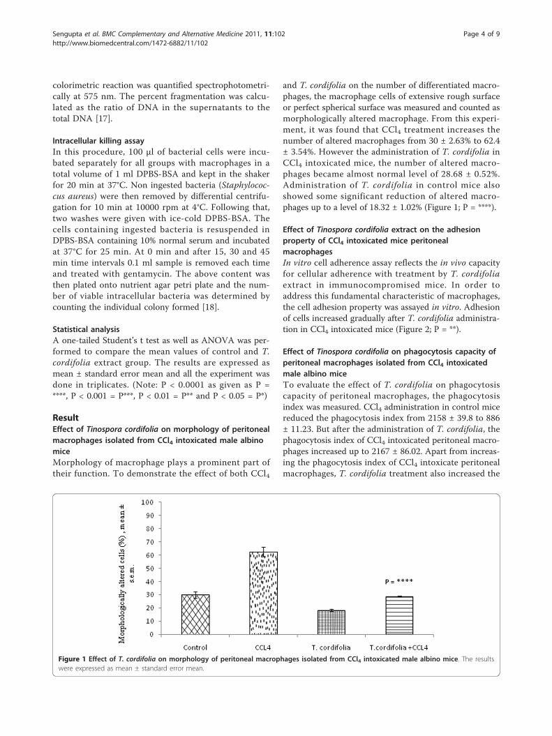
ResultEffect of Tinospora cordifolia on morphology of peritonealmacrophages isolated from CCl4 intoxicated male albinomiceMorphology of macrophage plays a prominent part oftheir function. To demonstrate the effect of both CCl4
and T. cordifolia on the number of differentiated macro-phages, the macrophage cells of extensive rough surfaceor perfect spherical surface was measured and counted asmorphologically altered macrophage. From this experi-ment, it was found that CCl4 treatment increases thenumber of altered macrophages from 30 ± 2.63% to 62.4± 3.54%. However the administration of T. cordifolia inCCl4 intoxicated mice, the number of altered macro-phages became almost normal level of 28.68 ± 0.52%.Administration of T. cordifolia in control mice alsoshowed some significant reduction of altered macro-phages up to a level of 18.32 ± 1.02% (Figure 1; P = ****).
Effect of Tinospora cordifolia extract on the adhesionproperty of CCl4 intoxicated mice peritonealmacrophagesIn vitro cell adherence assay reflects the in vivo capacityfor cellular adherence with treatment by T. cordifoliaextract in immunocompromised mice. In order toaddress this fundamental characteristic of macrophages,the cell adhesion property was assayed in vitro. Adhesionof cells increased gradually after T. cordifolia administra-tion in CCl4 intoxicated mice (Figure 2; P = **).
Effect of Tinospora cordifolia on phagocytosis capacity ofperitoneal macrophages isolated from CCl4 intoxicatedmale albino miceTo evaluate the effect of T. cordifolia on phagocytosiscapacity of peritoneal macrophages, the phagocytosisindex was measured. CCl4 administration in control micereduced the phagocytosis index from 2158 ± 39.8 to 886± 11.23. But after the administration of T. cordifolia, thephagocytosis index of CCl4 intoxicated peritoneal macro-phages increased up to 2167 ± 86.02. Apart from increas-ing the phagocytosis index of CCl4 intoxicate peritonealmacrophages, T. cordifolia treatment also increased the
Figure 1 Effect of T. cordifolia on morphology of peritoneal macrophages isolated from CCl4 intoxicated male albino mice. The resultswere expressed as mean ± standard error mean.
Sengupta et al. BMC Complementary and Alternative Medicine 2011, 11:102http://www.biomedcentral.com/1472-6882/11/102
Page 4 of 9
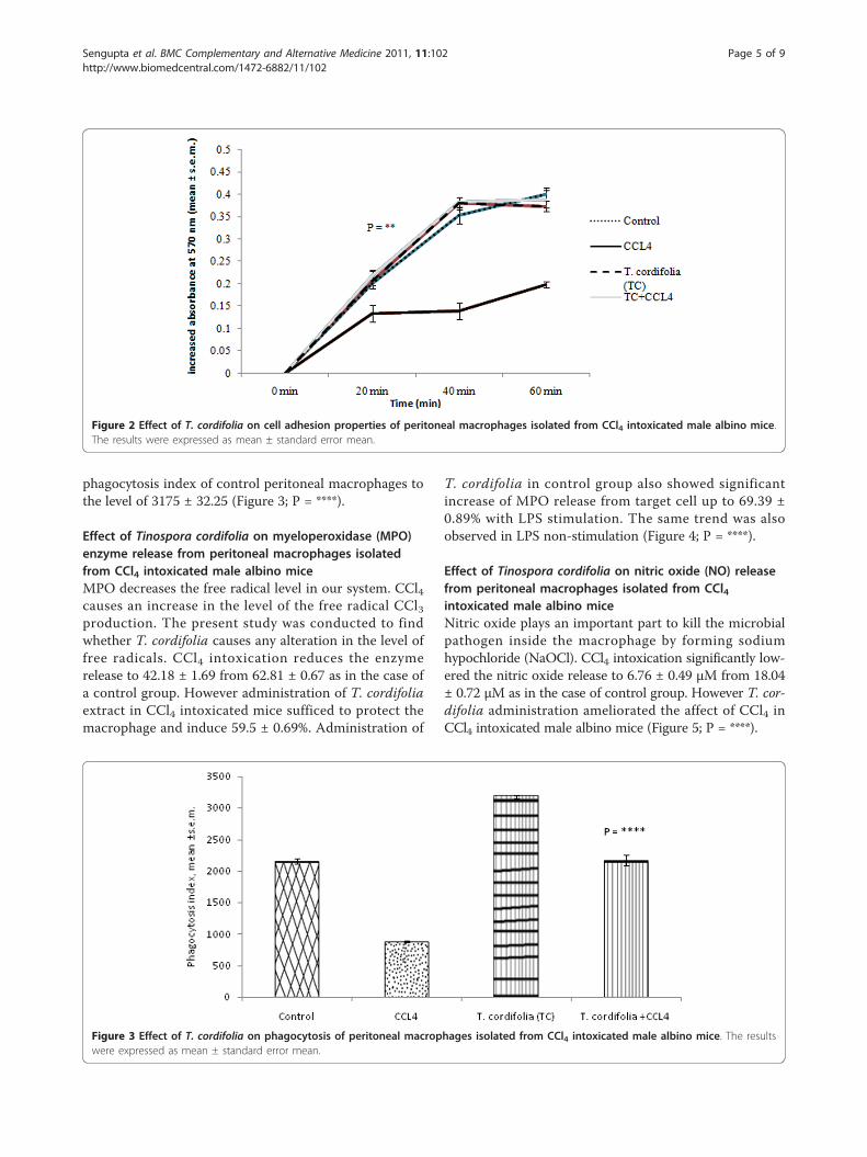
phagocytosis index of control peritoneal macrophages tothe level of 3175 ± 32.25 (Figure 3; P = ****).
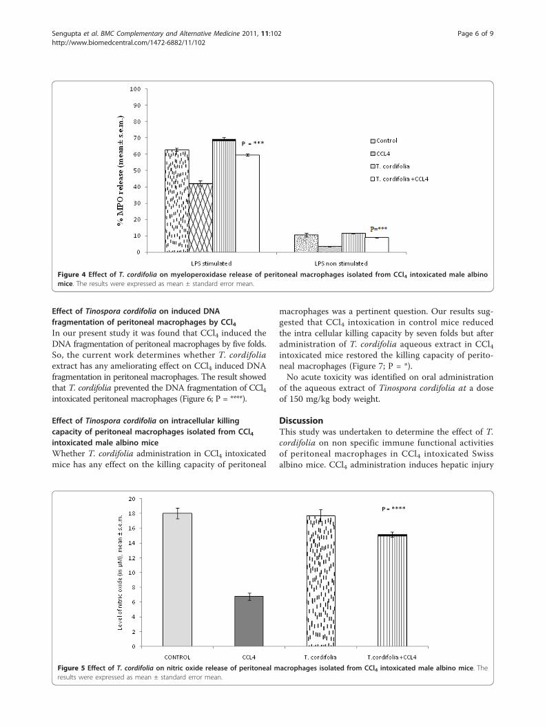
Effect of Tinospora cordifolia on myeloperoxidase (MPO)enzyme release from peritoneal macrophages isolatedfrom CCl4 intoxicated male albino miceMPO decreases the free radical level in our system. CCl4causes an increase in the level of the free radical CCl3production. The present study was conducted to findwhether T. cordifolia causes any alteration in the level offree radicals. CCl4 intoxication reduces the enzymerelease to 42.18 ± 1.69 from 62.81 ± 0.67 as in the case ofa control group. However administration of T. cordifoliaextract in CCl4 intoxicated mice sufficed to protect themacrophage and induce 59.5 ± 0.69%. Administration of
T. cordifolia in control group also showed significantincrease of MPO release from target cell up to 69.39 ±0.89% with LPS stimulation. The same trend was alsoobserved in LPS non-stimulation (Figure 4; P = ****).
Effect of Tinospora cordifolia on nitric oxide (NO) releasefrom peritoneal macrophages isolated from CCl4intoxicated male albino miceNitric oxide plays an important part to kill the microbialpathogen inside the macrophage by forming sodiumhypochloride (NaOCl). CCl4 intoxication significantly low-ered the nitric oxide release to 6.76 ± 0.49 μM from 18.04± 0.72 μM as in the case of control group. However T. cor-difolia administration ameliorated the affect of CCl4 inCCl4 intoxicated male albino mice (Figure 5; P = ****).
Figure 2 Effect of T. cordifolia on cell adhesion properties of peritoneal macrophages isolated from CCl4 intoxicated male albino mice.The results were expressed as mean ± standard error mean.
Figure 3 Effect of T. cordifolia on phagocytosis of peritoneal macrophages isolated from CCl4 intoxicated male albino mice. The resultswere expressed as mean ± standard error mean.
Sengupta et al. BMC Complementary and Alternative Medicine 2011, 11:102http://www.biomedcentral.com/1472-6882/11/102
Page 5 of 9
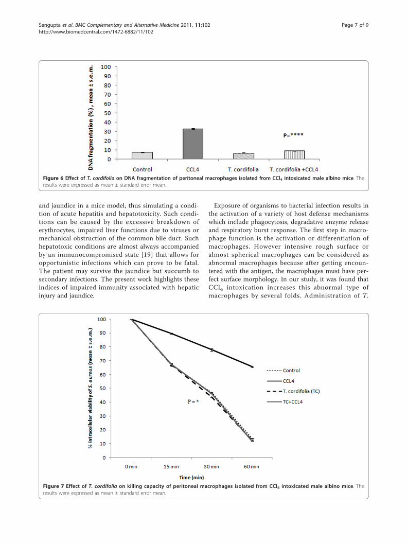
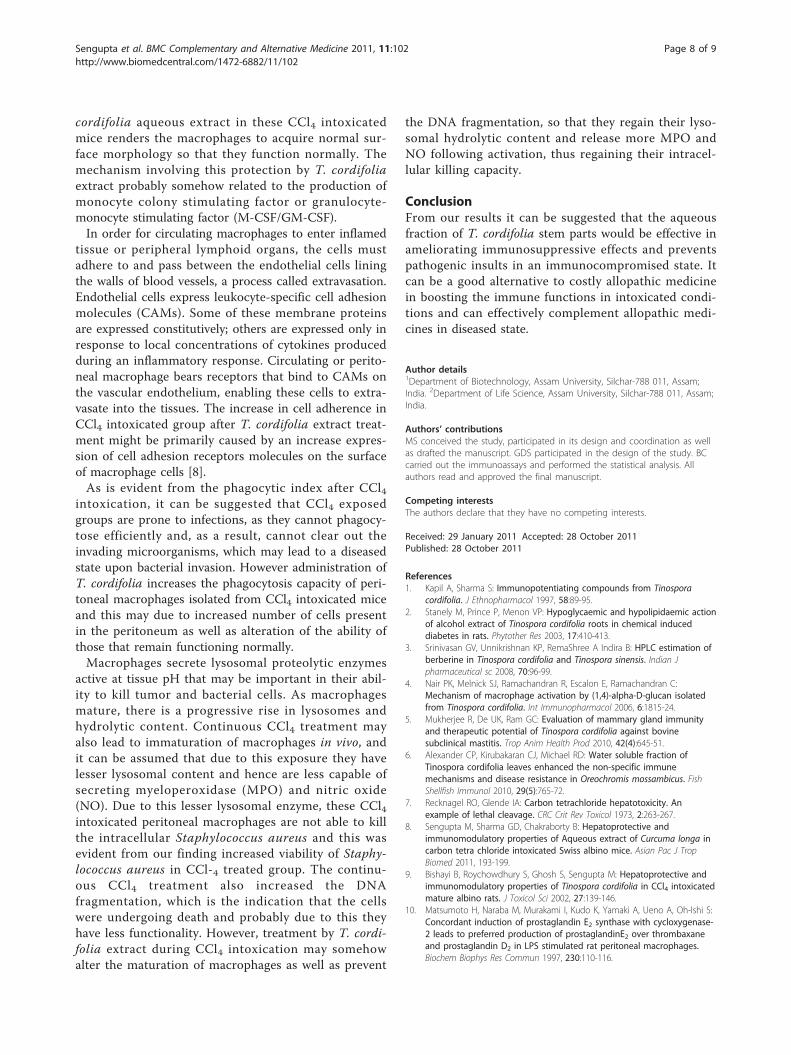
Effect of Tinospora cordifolia on induced DNAfragmentation of peritoneal macrophages by CCl4In our present study it was found that CCl4 induced theDNA fragmentation of peritoneal macrophages by five folds.So, the current work determines whether T. cordifoliaextract has any ameliorating effect on CCl4 induced DNAfragmentation in peritoneal macrophages. The result showedthat T. cordifolia prevented the DNA fragmentation of CCl4intoxicated peritoneal macrophages (Figure 6; P = ****).
Effect of Tinospora cordifolia on intracellular killingcapacity of peritoneal macrophages isolated from CCl4intoxicated male albino miceWhether T. cordifolia administration in CCl4 intoxicatedmice has any effect on the killing capacity of peritoneal
macrophages was a pertinent question. Our results sug-gested that CCl4 intoxication in control mice reducedthe intra cellular killing capacity by seven folds but afteradministration of T. cordifolia aqueous extract in CCl4intoxicated mice restored the killing capacity of perito-neal macrophages (Figure 7; P = *).No acute toxicity was identified on oral administration
of the aqueous extract of Tinospora cordifolia at a doseof 150 mg/kg body weight.
DiscussionThis study was undertaken to determine the effect of T.cordifolia on non specific immune functional activitiesof peritoneal macrophages in CCl4 intoxicated Swissalbino mice. CCl4 administration induces hepatic injury
Figure 4 Effect of T. cordifolia on myeloperoxidase release of peritoneal macrophages isolated from CCl4 intoxicated male albinomice. The results were expressed as mean ± standard error mean.
Figure 5 Effect of T. cordifolia on nitric oxide release of peritoneal macrophages isolated from CCl4 intoxicated male albino mice. Theresults were expressed as mean ± standard error mean.
Sengupta et al. BMC Complementary and Alternative Medicine 2011, 11:102http://www.biomedcentral.com/1472-6882/11/102
Page 6 of 9
and jaundice in a mice model, thus simulating a condi-tion of acute hepatitis and hepatotoxicity. Such condi-tions can be caused by the excessive breakdown oferythrocytes, impaired liver functions due to viruses ormechanical obstruction of the common bile duct. Suchhepatotoxic conditions are almost always accompaniedby an immunocompromised state [19] that allows foropportunistic infections which can prove to be fatal.The patient may survive the jaundice but succumb tosecondary infections. The present work highlights theseindices of impaired immunity associated with hepaticinjury and jaundice.
Exposure of organisms to bacterial infection results inthe activation of a variety of host defense mechanismswhich include phagocytosis, degradative enzyme releaseand respiratory burst response. The first step in macro-phage function is the activation or differentiation ofmacrophages. However intensive rough surface oralmost spherical macrophages can be considered asabnormal macrophages because after getting encoun-tered with the antigen, the macrophages must have per-fect surface morphology. In our study, it was found thatCCl4 intoxication increases this abnormal type ofmacrophages by several folds. Administration of T.
Figure 6 Effect of T. cordifolia on DNA fragmentation of peritoneal macrophages isolated from CCl4 intoxicated male albino mice. Theresults were expressed as mean ± standard error mean.
Figure 7 Effect of T. cordifolia on killing capacity of peritoneal macrophages isolated from CCl4 intoxicated male albino mice. Theresults were expressed as mean ± standard error mean.
Sengupta et al. BMC Complementary and Alternative Medicine 2011, 11:102http://www.biomedcentral.com/1472-6882/11/102
Page 7 of 9
cordifolia aqueous extract in these CCl4 intoxicatedmice renders the macrophages to acquire normal sur-face morphology so that they function normally. Themechanism involving this protection by T. cordifoliaextract probably somehow related to the production ofmonocyte colony stimulating factor or granulocyte-monocyte stimulating factor (M-CSF/GM-CSF).In order for circulating macrophages to enter inflamed
tissue or peripheral lymphoid organs, the cells mustadhere to and pass between the endothelial cells liningthe walls of blood vessels, a process called extravasation.Endothelial cells express leukocyte-specific cell adhesionmolecules (CAMs). Some of these membrane proteinsare expressed constitutively; others are expressed only inresponse to local concentrations of cytokines producedduring an inflammatory response. Circulating or perito-neal macrophage bears receptors that bind to CAMs onthe vascular endothelium, enabling these cells to extra-vasate into the tissues. The increase in cell adherence inCCl4 intoxicated group after T. cordifolia extract treat-ment might be primarily caused by an increase expres-sion of cell adhesion receptors molecules on the surfaceof macrophage cells [8].As is evident from the phagocytic index after CCl4
intoxication, it can be suggested that CCl4 exposedgroups are prone to infections, as they cannot phagocy-tose efficiently and, as a result, cannot clear out theinvading microorganisms, which may lead to a diseasedstate upon bacterial invasion. However administration ofT. cordifolia increases the phagocytosis capacity of peri-toneal macrophages isolated from CCl4 intoxicated miceand this may due to increased number of cells presentin the peritoneum as well as alteration of the ability ofthose that remain functioning normally.Macrophages secrete lysosomal proteolytic enzymes
active at tissue pH that may be important in their abil-ity to kill tumor and bacterial cells. As macrophagesmature, there is a progressive rise in lysosomes andhydrolytic content. Continuous CCl4 treatment mayalso lead to immaturation of macrophages in vivo, andit can be assumed that due to this exposure they havelesser lysosomal content and hence are less capable ofsecreting myeloperoxidase (MPO) and nitric oxide(NO). Due to this lesser lysosomal enzyme, these CCl4intoxicated peritoneal macrophages are not able to killthe intracellular Staphylococcus aureus and this wasevident from our finding increased viability of Staphy-lococcus aureus in CCl-4 treated group. The continu-ous CCl4 treatment also increased the DNAfragmentation, which is the indication that the cellswere undergoing death and probably due to this theyhave less functionality. However, treatment by T. cordi-folia extract during CCl4 intoxication may somehowalter the maturation of macrophages as well as prevent
the DNA fragmentation, so that they regain their lyso-somal hydrolytic content and release more MPO andNO following activation, thus regaining their intracel-lular killing capacity.
ConclusionFrom our results it can be suggested that the aqueousfraction of T. cordifolia stem parts would be effective inameliorating immunosuppressive effects and preventspathogenic insults in an immunocompromised state. Itcan be a good alternative to costly allopathic medicinein boosting the immune functions in intoxicated condi-tions and can effectively complement allopathic medi-cines in diseased state.
Author details1Department of Biotechnology, Assam University, Silchar-788 011, Assam;India. 2Department of Life Science, Assam University, Silchar-788 011, Assam;India.
Authors’ contributionsMS conceived the study, participated in its design and coordination as wellas drafted the manuscript. GDS participated in the design of the study. BCcarried out the immunoassays and performed the statistical analysis. Allauthors read and approved the final manuscript.
Competing interestsThe authors declare that they have no competing interests.
Received: 29 January 2011 Accepted: 28 October 2011Published: 28 October 2011
References1. Kapil A, Sharma S: Immunopotentiating compounds from Tinospora
cordifolia. J Ethnopharmacol 1997, 58:89-95.2. Stanely M, Prince P, Menon VP: Hypoglycaemic and hypolipidaemic action
of alcohol extract of Tinospora cordifolia roots in chemical induceddiabetes in rats. Phytother Res 2003, 17:410-413.
3. Srinivasan GV, Unnikrishnan KP, RemaShree A Indira B: HPLC estimation ofberberine in Tinospora cordifolia and Tinospora sinensis. Indian Jpharmaceutical sc 2008, 70:96-99.
4. Nair PK, Melnick SJ, Ramachandran R, Escalon E, Ramachandran C:Mechanism of macrophage activation by (1,4)-alpha-D-glucan isolatedfrom Tinospora cordifolia. Int Immunopharmacol 2006, 6:1815-24.
5. Mukherjee R, De UK, Ram GC: Evaluation of mammary gland immunityand therapeutic potential of Tinospora cordifolia against bovinesubclinical mastitis. Trop Anim Health Prod 2010, 42(4):645-51.
6. Alexander CP, Kirubakaran CJ, Michael RD: Water soluble fraction ofTinospora cordifolia leaves enhanced the non-specific immunemechanisms and disease resistance in Oreochromis mossambicus. FishShellfish Immunol 2010, 29(5):765-72.
7. Recknagel RO, Glende IA: Carbon tetrachloride hepatotoxicity. Anexample of lethal cleavage. CRC Crit Rev Toxicol 1973, 2:263-267.
8. Sengupta M, Sharma GD, Chakraborty B: Hepatoprotective andimmunomodulatory properties of Aqueous extract of Curcuma longa incarbon tetra chloride intoxicated Swiss albino mice. Asian Pac J TropBiomed 2011, 193-199.
9. Bishayi B, Roychowdhury S, Ghosh S, Sengupta M: Hepatoprotective andimmunomodulatory properties of Tinospora cordifolia in CCl4 intoxicatedmature albino rats. J Toxicol Sci 2002, 27:139-146.
10. Matsumoto H, Naraba M, Murakami I, Kudo K, Yamaki A, Ueno A, Oh-Ishi S:Concordant induction of prostaglandin E2 synthase with cycloxygenase-2 leads to preferred production of prostaglandinE2 over thrombaxaneand prostaglandin D2 in LPS stimulated rat peritoneal macrophages.Biochem Biophys Res Commun 1997, 230:110-116.
Sengupta et al. BMC Complementary and Alternative Medicine 2011, 11:102http://www.biomedcentral.com/1472-6882/11/102
Page 8 of 9
11. Qu J, Condliffe MA, Lawson M, Pelvin RJ, Chlivers E: Lack of effect ofrecombinant platelet derived growth factor on human neuttrophilfunction. J Immunol 1997, 159:2952-2959.
12. Lin TH, Mondol KL, Bolen JB, Haskell , Juliano RL: Integrin mediatedtyrosine phosphorylation and message induction in monocytic cells. JBiol Chem 1995, 270:16189-16202.
13. Czuprynski CJ, Henson PM, Campbell PA: Killing of Listeria monocytogenesby inflammatory neutrophils and mononuclear phagocytes fromimmune and non immune mice. J Leuk Biol 1984, 35:193-208.
14. Bos AR, Weaver R, Roos D: Characterization and qualification of theperoxidase in human neutrophils. Biochem Biophys Acta 1990,525:4133-41.
15. Sasaki M, Gonzales ZM, Huang H, Herring WJ, Ahn S, Ginty DD, Dawson VL,Dawson TM: Dynamic regulation of neuronal nitric oxide synthasetranscription by calcium influx through a CREB family transcriptionfactor-dependant mechanism. Proc Nalt Acad Sc 2000, 97:8617-8622, USA.
16. Perandones CE, Illera VA, Peckham D, Stunz LL, Ashman RF: Regulation ofApoptosis In Vitro in Mature Murine Spleen T Cells. J Immuno 1993,151:3521-3529.
17. Kurita-Ochiai T, Ochiai K, Fukushima K: Butyric-acid-induced Apoptosis inMurine Thymocytes and Splenic T- and B-cells Occurs in the Absence ofp53. J Dent Res 2000, 79:1948-54.
18. Leigh PCJ, Van FR, Zwet TL: In vitro determination of phagocytosis andintracellular killing by polymorphonuclear neutrophils and mononuclearphagocytes. In Handbook of Experimental Immunology.. 4 edition. Edited by:Weir DM editors. Blackwell scientific publications; 1986:46.1-46.19.
19. Charalambos GA, Philip AB, Julia AW, Diego V: The importance of immunedysfunction in determining outcome in acute liver failure. J Hepatol 2008,49:845-861.
Pre-publication historyThe pre-publication history for this paper can be accessed here:http://www.biomedcentral.com/1472-6882/11/102/prepub
doi:10.1186/1472-6882-11-102Cite this article as: Sengupta et al.: Effect of aqueous extract ofTinospora cordifolia on functions of peritoneal macrophages isolatedfrom CCl4 intoxicated male albino mice. BMC Complementary andAlternative Medicine 2011 11:102.
Submit your next manuscript to BioMed Centraland take full advantage of:
• Convenient online submission
• Thorough peer review
• No space constraints or color figure charges
• Immediate publication on acceptance
• Inclusion in PubMed, CAS, Scopus and Google Scholar
• Research which is freely available for redistribution
Submit your manuscript at www.biomedcentral.com/submit
Sengupta et al. BMC Complementary and Alternative Medicine 2011, 11:102http://www.biomedcentral.com/1472-6882/11/102
Page 9 of 9