Embed Size (px)
Citation preview
Abstracts / Osteoarthritis and Cartilage 20 (2012) S54–S296S148
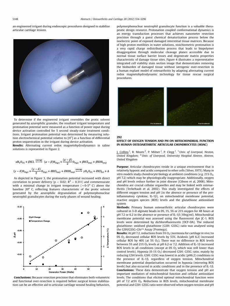
an engineered irrigant during endoscopic procedures designed to stabilizearticular cartilage lesions.
ĂTo determine if the engineered irrigant resembles the protic solventgenerated by azurophilic granules, the resultant irrigant temperature andprotonation potential were measured as a function of power input duringdevice activation controlled for 5 second steady-state treatment condi-tions. Irrigant protonation potential was determined by measuring solu-tion electrochemical potential relative to [H+] as a function of differentialproton sequestration in the irrigant during device activation.Results: Alternating current redox magnetohydrodynamics in salinesolutions is represented in Figure 2.
ĂAs depicted in Figure 3, the protonation potential increased with directcorrelation to power delivery (p < 0.02; R2 ¼ 0.311) and commensuratewith a minimal change in irrigant temperature (w0-5o C) above thebaseline 20o C, reflecting features characteristic of the protic solventgenerated by the azurophilic degranulation of polymorphonuclearneutrophil granulocytes during the early phases of wound healing.
ĂConclusions: Because resection precision that eliminates both volumetricand functional over-resection is required before surgical lesion stabiliza-tion can be an effective aid to articular cartilage wound healing behaviors,
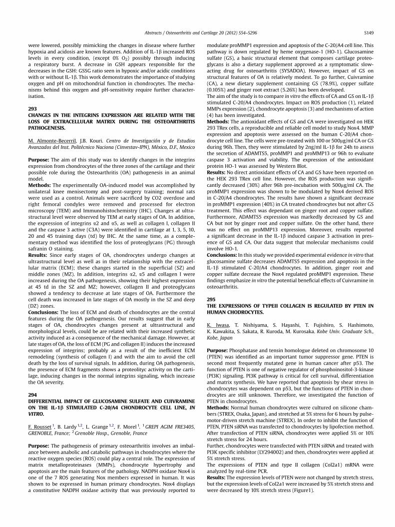
polymorphonuclear neutrophil granulocyte function is a valuable thera-peutic design resource. Protonation coupled conformational dynamics isan energy transduction processes that achieves nanometer resectionprecision through a guest chemical denaturization process below theisoelectric point of exposed damaged interstitial tissue matrices. Becauseof high proton motilities in water solutions, stoichiometric protonation isa very rapid charge redistribution process that leads to biopolymerdisaggregation through molecular cleavage planes accessible due tonormal tissue surface barrier losses and degenerate matrix propertiescharacteristic of damage tissue sites. Figure 4 illustrates a representativeintegrated cell viability stain section image that demonstrates removingthe bioburden of damaged tissue without iatrogenic over-resection ina human explant model of osteoarthritis by adapting alternating currentredox magnetohydrodynamic technology for tissue rescue surgicalprocedures.
Ă
292EFFECT OF OXYGEN TENSION AND PH ON MITOCHONDRIAL FUNCTIONIN HUMAN OSTEOARTHRITIC ARTICULAR CHONDROCYTES (HOAC)
J. Collins 1, R. Moots 2, P. Milner 1, P. Clegg 1. 1Univ. of Liverpool, Neston,United Kingdom; 2Univ. of Liverpool, University Hospital Aintree, Aintree,United Kingdom
Purpose: Articular chondrocytes reside in a unique environment that isrelatively hypoxic and acidic compared to other cells (Silver,1975). Many invitromodels study chondrocyte biology at ambient conditions (e.g. 21% O2,pH 7.2) which may be physiologically inappropriate. Additionally, oxygenand pH levels reduce further in joint disease (Gibson et al, 2008). Mito-chondria are crucial cellular organelles and may be linked with osteoar-thritis (Terkeltaub et al, 2002). This study investigated the effects ofdifferent oxygen tension and pH (in the absence or presence of the pro-inflammatory cytokine, IL-1b), on mitochondrial membrane potential,reactive oxygen species (ROS) levels and the glutathione antioxidantsystemMethods: Primary human osteoarthritic articular chondrocytes werecultured in 3-D alginate beads in 0%, 1%, 5% or 21% oxygen for 48 hours atpH 7.2 or 6.2 in the absence or presence of IL-1b (10ng/ml). Mitochondrialmembrane potential was assessed using the fluorescent dye JC-1. ROSlevels were determined by dichlorofluorescein (DCF-DA). The reducedglutathione: oxidised glutathione (GSH: GSSG) ratio was analysed usingthe GSH/GSSG-Glo� Assay (Promega).Results: At pH 7.2, reductions from 5% O2 (normoxia for cartilage in vivo) to0% O2 decreased cellular ROS levels by 53%. Acidosis (pH 6.2) increasedcellular ROS by 40% (at 5% O2). There was no difference in ROS levelsbetween 5% and 21% O2 levels at pH 6.2 or 7.2. Addition of IL-1b increasedROS levels in all conditions (except at 0% O2 which was still lower thancontrol levels). Hypoxia (0-1% O2) decreased GSH: GSSG ratio, mainly byreducing GSH levels. GSH: GSSG was lowest in acidic (pH6.2) conditions inthe presence of IL-1b, regardless of oxygen tension. Mitochondrialmembrane potential depolarisation occurred in hypoxia (mirroring ROSlevels) but also occurred in acidic conditions and in the presence of IL-1b.Conclusions: These data demonstrate that oxygen tension and pH areimportant mediators of mitochondrial function and cellular antioxidantlevels. The conditions that elicited optimal mitochondrial function werepH of 7.2 at5% O2. Reductions in ROS levels, mitochondrial membranepotential and GSH: GSSG ratiowere observedwhen oxygen tension and pH
Abstracts / Osteoarthritis and Cartilage 20 (2012) S54–S296 S149
were lowered, possibly mimicking the changes in disease where furtherhypoxia and acidosis are known features. Addition of IL-1b increased ROSlevels in every condition, (except 0% O2) possibly through inducinga respiratory burst. A decrease in GSH appears responsible for thedecreases in the GSH: GSSG ratio seen in hypoxic and/or acidic conditionswith or without IL-1b. This work demonstrates the importance of studyingoxygen and pH on mitochondrial function in chondrocytes. The mecha-nisms behind this oxygen and pH-sensitivity require further character-isation.
293CHANGES IN THE INTEGRINS EXPRESSION ARE RELATED WITH THELOSS OF EXTRACELLULAR MATRIX DURING THE OSTEOARTHRITISPATHOGENESIS.
M. Almonte-Becerril, J.B. Kouri. Centro de Investigación y de EstudiosAvanzados del Inst. Politécnico Naciona (Cinvestav-IPN), México, D.F., Mexico
Purpose: The aim of this study was to identify changes in the integrinsexpression from chondrocytes of the three zones of the cartilage and theirpossible role during the Osteoarthritis (OA) pathogenesis in an animalmodel.Methods: The experimentally OA-induced model was accomplished byunilateral knee menisectomy and post-surgery training; normal ratswere used as a control. Animals were sacrificed by CO2 overdose andright femoral condyles were removed and processed for electronmicroscopy (TEM) and Immunohistochemistry (IHC). Changes at ultra-structural level were observed by TEM at early stages of OA. In addition,the expression of integrins a2 and a5, as well as collagen I, collagen IIand the caspase 3 active (C3A) were identified in cartilage at 1, 3, 5, 10,20 and 45 training days (td) by IHC. At the same time, as a comple-mentary method was identified the loss of proteoglycans (PG) throughsafranin O staining.Results: Since early stages of OA, chondrocytes undergo changes atultrastructural level as well as in their relationship with the extracel-lular matrix (ECM); these changes started in the superficial (SZ) andmiddle zones (MZ). In addition, integrins a2, a5 and collagen I wereincreased during the OA pathogenesis, showing their highest expressionat 45 td in the SZ and MZ; however, collagen II and proteoglycansshowed a tendency to decrease at late stages of OA. Furthermore thecell death was increased in late stages of OA mostly in the SZ and deep(DZ) zones.Conclusions: The loss of ECM and death of chondrocytes are the centralfeatures during the OA pathogenesis. Our results suggest that in earlystages of OA, chondrocytes changes present at ultrastructural andmorphological levels, could be are related with their increased syntheticactivity induced as a consequence of the mechanical damage. However, atlate stages of OA, the loss of ECM (PG and collagen II) induces the increasedexpression of integrins; probably as a result of the inefficient ECMremodeling (synthesis of collagen I) and with the aim to avoid the celldeath by the loss of survival signals. In addition, during OA pathogenesis,the presence of ECM fragments shows a proteolityc activity on the carti-lage, inducing changes in the normal integrins signaling, which increasethe OA severity.
294DIFFERENTIAL IMPACT OF GLUCOSAMINE SULFATE AND CUIVRAMINEON THE IL-1b STIMULATED C-20/A4 CHONDROCYTE CELL LINE, INVITRO.
F. Rousset 1, B. Lardy 1,2, L. Grange 1,2, F. Morel 1. 1GREPI AGIM FRE3405,GRENOBLE, France; 2Grenoble Hosp., Grenoble, France
Purpose: The pathogenesis of primary osteoarthritis involves an imbal-ance between anabolic and catabolic pathways in chondrocytes where thereactive oxygen species (ROS) could play a central role. The expression ofmatrix metalloproteinases (MMPs), chondrocyte hypertrophy andapoptosis are the main features of the pathology. NADPH oxidase Nox4 isone of the 7 ROS generating Nox members expressed in human. It wasshown to be expressed in human primary chondrocytes. Nox4 displaysa constitutive NADPH oxidase activity that was previously reported to
modulate proMMP1 expression and apoptosis of the C-20/A4 cell line. Thispathway is down regulated by heme oxygenase-1 (HO-1). Glucosaminesulfate (GS), a basic structural element that composes cartilage proteo-glycans is also a dietary supplement approved as a symptomatic slow-acting drug for osteoarthritis (SYSADOA). However, impact of GS onstructural features of OA is relatively modest. To go further, Cuivramine(CA), a new dietary supplement containing GS (78.9%), copper sulfate(0.105%) and ginger root extract (5.26%) has been developed.The aim of the study is to compare in vitro the effects of CA and GS on IL-1bstimulated C-20/A4 chondrocytes. Impact on ROS production (1), relatedMMPs expression (2), chondrocyte apoptosis (3) andmechanisms of action(4) has been investigated.Methods: The antioxidant effects of GS and CA were investigated on HEK293 TRex cells, a reproducible and reliable cell model to study Nox4. MMPexpression and apoptosis were assessed on the human C-20/A4 chon-drocyte cell line. The cells were pre-treated with 100 or 500mg/ml CA or GSduring 96h. Then, they were stimulated by 2ng/ml IL-1b for 24h to assessthe secretion of ADAMTS5, proMMP1 and proMMP13 or 96h to evaluatecaspase 3 activation and viability. The expression of the antioxidantprotein HO-1 was assessed by Western Blot.Results: No direct antioxidant effects of CA and GS have been reported onthe HEK 293 TRex cell line. However, the ROS production was signifi-cantly decreased (30%) after 96h pre-incubation with 500mg/ml CA. TheproMMP1 expression was shown to be modulated by Nox4 derived ROSin C-20/A4 chondrocytes. The results have shown a significant decreasein proMMP1 expression (40%) in CA treated chondrocytes but not after GStreatment. This effect was dependant on ginger root and copper sulfate.Furthermore, ADAMTS5 expression was markedly decreased by GS andCA but not by ginger root and copper sulfate. On the other hand, therewas no effect on proMMP13 expression. Moreover, results reporteda significant decrease in the IL-1b induced caspase 3 activation in pres-ence of GS and CA. Our data suggest that molecular mechanisms couldinvolve HO-1.Conclusions: In this study we provided experimental evidence in vitro thatglucosamine sulfate decreases ADAMTS5 expression and apoptosis in theIL-1b stimulated C-20/A4 chondrocytes. In addition, ginger root andcopper sulfate decrease the Nox4 regulated proMMP1 expression. Thesefindings emphasize in vitro the potential beneficial effects of Cuivramine inosteoarthritis.
295THE EXPRESSIONS OF TYPEII COLLAGEN IS REGULATED BY PTEN INHUMAN CHODROCYTES.
K. Iwasa, T. Nishiyama, S. Hayashi, T. Fujishiro, S. Hashimoto,K. Kawakita, S. Sakata, R. Kuroda, M. Kurosaka. Kobe Univ. Graduate Sch.,Kobe, Japan
Purpose: Phosphatase and tensin homologue deleted on chromosome 10(PTEN) was identified as an important tumor suppressor gene. PTEN issecond most frequently mutated gene in human cancer after p53. Thefunction of PTEN is one of negative regulator of phosphoinositol-3-kinase(PI3K) signaling. PI3K pathway is critical for cell survival, differentiationand matrix synthesis. We have reported that apoptosis by shear stress inchondrocytes was dependent on p53, but the functions of PTEN in chon-drocytes are still unknown. Therefore, we investigated the function ofPTEN in chondrocytes.Methods: Normal human chondrocytes were cultured on silicone cham-bers (STREX, Osaka, Japan), and stretched at 5% stress for 6 hours by pulse-motor-driven stretch machine (STREX). In order to inhibit the function ofPTEN, PTEN siRNAwas transfected to chondrocytes by lipofection method.After transfection of PTEN siRNA, chondrocytes were applied 5% or 10%stretch stress for 24 hours.Further, chondrocytes were transfected with PTEN siRNA and treated withPI3K specific inhibitor (LY294002) and then, chondrocytes were applied at5% stretch stress.The expressions of PTEN and type II collagen (Col2a1) mRNA wereanalyzed by real-time PCR.Results: The expression levels of PTEN were not changed by stretch stress,but the expression levels of Col2a1 were increased by 5% stretch stress andwere decreased by 10% stretch stress (Figure1).