Embed Size (px)
Citation preview
ARTICLE
Effects of amantadine on the dynamics of membrane-boundinfluenza A M2 transmembrane peptide studied by NMRrelaxation
Sarah D. Cady Æ Mei Hong
Received: 8 May 2009 / Accepted: 26 June 2009 / Published online: 25 July 2009
� Springer Science+Business Media B.V. 2009
Abstract The molecular motions of membrane proteins in
liquid-crystalline lipid bilayers lie at the interface between
motions in isotropic liquids and in solids. Specifically,
membrane proteins can undergo whole-body uniaxial dif-
fusion on the microsecond time scale. In this work, we
investigate the 1H rotating-frame spin-lattice relaxation
(T1q) caused by the uniaxial diffusion of the influenza A M2
transmembrane peptide (M2TMP), which forms a tetra-
meric proton channel in lipid bilayers. This uniaxial diffu-
sion was proved before by 2H, 15N and 13C NMR lineshapes
of M2TMP in DLPC bilayers. When bound to an inhibitor,
amantadine, the protein exhibits significantly narrower
linewidths at physiological temperature. We now investi-
gate the origin of this line narrowing through temperature-
dependent 1H T1q relaxation times in the absence and
presence of amantadine. Analysis of the temperature
dependence indicates that amantadine decreases the corre-
lation time of motion from 2.8 ± 0.9 ls for the apo peptide
to 0.89 ± 0.41 ls for the bound peptide at 313 K. Thus the
line narrowing of the bound peptide is due to better
avoidance of the NMR time scale and suppression of
intermediate time scale broadening. The faster diffusion of
the bound peptide is due to the higher attempt rate of
motion, suggesting that amantadine creates better-packed
and more cohesive helical bundles. Analysis of the tem-
perature dependence of ln T�11q
� �indicates that the activa-
tion energy of motion increased from 14.0 ± 4.0 kJ/mol for
the apo peptide to 23.3 ± 6.2 kJ/mol for the bound peptide.
This higher activation energy indicates that excess aman-
tadine outside the protein channel in the lipid bilayer
increases the membrane viscosity. Thus, the protein-bound
amantadine speeds up the diffusion of the helical bundles
while the excess amantadine in the bilayer increases the
membrane viscosity.
Introduction
Nuclear magnetic resonance has long been used as a tool
for measuring molecular dynamics over a broad range of
time scales, from the fast picosecond–nanosecond regime
(Mandel et al. 1996), to the slow microsecond–millisecond
regime (Palmer et al. 2001; Schaefer et al. 1977; Shaw
et al. 2000), and to the ultra-slow supra-second regime
(deAzevedo et al. 2000; Schmidt et al. 1988). Some of the
most interesting applications are to biomolecules, where
molecular dynamics has a particularly strong connection to
function (Ishima and Torchia 2000; Kay 1998; Palmer
et al. 1996). In solution NMR studies of biomolecular
dynamics, 15N relaxation NMR has been the method of
choice and the Lipari–Szabo model-free formalism (Clore
et al. 1990; Lipari and Szabo 1982) has provided a simple
theoretical framework to separate the effects of internal
anisotropic motions from whole-body isotropic motion and
to extract the amplitudes and rates of internal motion.
In the solid state, the lack of isotropic molecular tum-
bling considerably simplifies studies of internal motions by
NMR. Many solid-state NMR techniques directly probe the
amplitudes of internal motions, most notably 2H quadru-
polar NMR (Jelinski et al. 1980), which has exquisite
angular resolution but no chemical resolution, and 1H–X
dipolar coupling techniques under magic-angle spinning
Electronic supplementary material The online version of thisarticle (doi:10.1007/s10858-009-9352-9) contains supplementarymaterial, which is available to authorized users.
S. D. Cady � M. Hong (&)
Department of Chemistry, Iowa State University, Ames,
IA 50011, USA
e-mail: [email protected]
123
J Biomol NMR (2009) 45:185–196
DOI 10.1007/s10858-009-9352-9
(MAS), which have chemical site resolution (Hong et al.
2002; Munowitz et al. 1982; Schaefer et al. 1983). Nuclear
spin relaxation times (T1, T2 and T1q) have also been used
to determine the correlation times and activation energies
of motional processes such as methyl three-site jumps and
aromatic ring flips. Combined, these amplitude and rate
measurements have provided detailed information on the
dynamics of structural proteins such as collagen and silk
(Jelinski et al. 1980; Yang et al. 2000), enzymes (Williams
and McDermott 1995), and lipid membranes (Blume et al.
1982; Smith and Oldfield 1984). Molecular motions of
synthetic polymers (Hagemeyer et al. 1989; Schaefer et al.
1990; Schaefer et al. 1984) and small molecules (Rothwell
and Waugh 1981) have also been investigated extensively
using solid-state NMR.
The dynamic environment of liquid-crystalline lipid
bilayers lies at the interface between isotropic fluids and
rigid solids and hence presents unique challenges to
understanding membrane protein dynamics. While mem-
brane protein sidechain motions have been studied by
NMR for decades (Huster et al. 2001; Kinsey et al. 1981;
Lee et al. 1993; Opella 1986), whole-body uniaxial rota-
tional diffusion of membrane proteins has been less
examined by NMR. As the symmetry axis of the lipid
bilayer, the bilayer normal is the axis around which
phospholipids undergo nanosecond rotational diffusion
(Bloom et al. 1991; Gennis 1989). Membrane proteins can
also undergo such rotational diffusion, because the same
principle that underlies the phospholipid motion, which is
Brownian diffusion in a two-dimensional fluid (Saffman
and Delbruck 1975), also applies to membrane proteins. A
number of examples of this uniaxial diffusion have now
been reported for membrane peptides and proteins (Hong
2007; Hong and Doherty 2006; Lewis et al. 1985; Mac-
donald and Seelig 1988; Pauls et al. 1985; Prosser et al.
1992; Tian et al. 1998; Yamaguchi et al. 2001). Their NMR
fingerprints include powder lineshapes with reduced
anisotropy and an asymmetry parameter (g) of 0, vanishing
intensity at the isotropic chemical shift of non-spinning
cross polarization (CP) spectra, and narrow lines in mac-
roscopically aligned samples whose alignment axis devi-
ates from the static magnetic field (Aisenbrey and
Bechinger 2004; Glaser et al. 2004; Park et al. 2006).
The influenza A M2 protein forms a proton channel in
the virus envelope that is important for the virus life cycle
(Pinto et al. 1992; Pinto and Lamb 2007). Acidification of
the virus interior uncoats the viral RNA and releases it into
the host cell. Amantadine binds the M2 proton channel and
prevents its opening, thus inhibiting viral replication (Hay
et al. 1985; Wang et al. 1993). The protein forms a tetra-
meric helical bundle in the membranes of both whole cells
(Sakaguchi et al. 1997) and synthetic lipids (Luo and Hong
2006). It undergoes uniaxial diffusion at a rate of *105/s in
DLPC bilayers based on 2H NMR spectra and the 2D
Brownian diffusion theory (Cady et al. 2007; Kovacs and
Cross 1997). The motional axis is the bilayer normal,
which is also the helical bundle axis. Since the TM helices
have tilt angles of *38� in DLPC bilayers (Cady and Hong
2008), the rotational diffusion has large amplitudes.
Combined with the fact that the motional rate is not orders
of magnitude different from the 1H–13C and 1H–15N
dipolar couplings, the motion strongly impacts the NMR
spectra: the ambient-temperature 1H-decoupled 13C and15N spectra of the protein in lipid bilayers are severely
exchange-broadened under both MAS and static conditions
(Cady et al. 2007; Li et al. 2007). Interestingly, upon
amantadine binding, the resonances in both MAS and static
solid-state NMR spectra of the protein sharpen consider-
ably. This line narrowing was also observed in solution
NMR spectra of M2 (18–60) bound to DHPC micelles
when an excess of the analogous rimantadine was added
(Schnell and Chou 2008).
Previously we have compared the 1H-decoupled 13C T2
relaxation times of the apo and bound M2TMP in DLPC
bilayers to understand the amantadine-induced line nar-
rowing. We found that the bound peptide has 30–150%
longer 13C T2 than the apo state at 303 K (Cady and Hong
2008; Cady et al. 2009), indicating that the line narrowing
has a significant contribution from dynamic changes of the
protein. However, the nature of this amantadine-induced
relaxation time increase has not been elucidated.
In this work, we have measured and analyzed temper-
ature-dependent 1H T1q relaxation times of the apo and
amantadine-bound M2TMP to better understand its
motional properties. We quantify the rates and activation
energies of the M2TMP microsecond motion in the
absence and presence of amantadine. We find that aman-
tadine increases the motional rates approximately threefold
at 313 K by increasing the attempt rates, thus alleviating
intermediate time scale broadening and narrowing the
spectral lines. Further, excess amantadine in the bilayer
increases the activation energy of the motion, whose
physical origin will be discussed.
Materials and methods
Peptides and lipids
FMOC-protected uniformly 13C, 15N-labeled amino acids
were either prepared in-house (Carpino and Han 1972) or
purchased from Sigma–Aldrich and Cambridge Isotope
Laboratories. The M2 transmembrane domain (residues
22–46) of the Influenza A Udorn strain (Ito et al. 1991) was
synthesized by PrimmBiotech (Cambridge, MA) and
purified to [95% purity. The amino acid sequence is
186 J Biomol NMR (2009) 45:185–196
123
SSDPL VVAASII GILHLIL WILDRL. Three labeled
peptides were used in this work, each with three to four
uniformly 13C, 15N-labeled residues. They are LAGI (L26,
A29, G34 and I35), VAIL (V27, A30, I33 and L38), and
VSL (V28, S31 and L36).
Membrane sample preparation
M2TMP was reconstituted into 1,2-dilauroyl-sn-glycero-3-
phosphatidylcholine (DLPC) bilayers by detergent dialysis
(Luo and Hong 2006). The lipid vesicle solution was pre-
pared by suspending dry DLPC powder in 1 ml phosphate
buffer (10 mM Na2HPO4/NaH2PO4, 1 mM EDTA,
0.1 mM NaN3) at pH 7.5, vortexing and freeze–thawing six
times to create uniform vesicles (Traikia et al. 2000).
M2TMP was dissolved in octyl-b-D-glucopyranoside (OG)
in 2 ml phosphate buffer, then mixed with an equal volume
of DLPC vesicles, and dialyzed against the phosphate
buffer at 4�C for 3 days. The final peptide/lipid molar ratio
was 1:15. The dialyzed peptide-DLPC solution was cen-
trifuged at 150,000g to give a pellet containing *50 wt%
water. For the amantadine-bound samples, 10 mM aman-
tadine hydrochloride was added to the phosphate buffer.
After pelleting, the amount of amantadine remaining in the
supernatant was quantified by 1H solution NMR, and the
bound fraction indicates a peptide:amantadine molar ratio
of *1:8 (Cady et al. 2009). All membrane-bound M2
samples were thus studied at pH 7.5, corresponding to the
closed state of the channel.
Solid-state NMR spectroscopy
NMR experiments were carried out on a Bruker AVANCE-
600 (14.1 Tesla) spectrometer (Karlsruhe, Germany) using
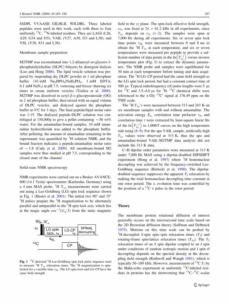
a 4 mm MAS probe. 1H T1q measurements were carried
out using a Lee-Goldburg (LG) spin lock sequence shown
in Fig. 1 (Huster et al. 2001). The initial two 90� and 35�1H pulses prepare the 1H magnetization to be alternately
parallel and antiparallel to the 1H spin lock axis, which lies
at the magic angle cos-1(1/H3) from the static magnetic
field in the yz plane. The spin-lock effective field strength,
xe, was fixed at 2p 9 61.2 kHz in all experiments, since
T1q depends on xe (1–2). The samples were spun at
7,000 Hz during all experiments. Six or seven spin lock
time points sSL were measured between 0 and 8 ms to
obtain the 1H T1q at each temperature, and six or seven
temperatures were measured per peptide to provide a suf-
ficient number of data points in the ln T�11q
� �versus inverse
temperature plot (Fig. 5) to extract the dynamic parame-
ters. The NMR probe and samples were equilibrated for
30 min at each temperature before tuning and data acqui-
sition. The 1H LG–CP period had the same field strength as
the LG spin lock period, but had a constant contact time of
300 ls. Typical radiofrequency (rf) pulse lengths were 5 ls
for 13C and 3.5–4.0 ls for 1H. 13C chemical shifts were
referenced to the a-Gly 13C0 signal at 176.49 ppm on the
TMS scale.
The 1H T1q’s were measured between 313 and 243 K on
six membrane samples with and without amantadine. The
activation energy Ea, correlation time prefactor s0, and
correlation time s were extracted by least-square linear fits
of the ln T�11q
� �to 1,000/T curves on the high temperature
side using (8–9). For the apo VAIL sample, artificially high
T1q values were observed at 313 K, thus the apo and
amantadine-bound VAIL-M2TMP data analysis did not
include the 313 K data.
C–H dipolar order parameters were measured at 313 K
under 7,000 Hz MAS using a dipolar-doubled DIPSHIFT
experiment (Hong et al. 1997) where 1H homonuclear
decoupling was achieved by the frequency-switched Lee-
Goldburg sequence (Bielecki et al. 1990). The dipolar-
doubled sequence suppresses the apparent T2 relaxation by
making the total homonuclear decoupling time constant at
one rotor period. The t1 evolution time was controlled by
the position of a 13C p pulse in the rotor period.
Theory
The membrane protein rotational diffusion of interest
generally occurs on the microsecond time scale based on
the 2D Brownian diffusion theory (Saffman and Delbruck
1975). Motions on this time scale can be probed by1H-decoupled S-spin spin–spin relaxation times (T2) and
rotating-frame spin-lattice relaxation times (T1q). The T2
relaxation times of an S spin dipolar coupled to an I spin
under conditions of random isotropic motion and I spin rf
decoupling depends on the spectral density at the decou-
pling field strength (Rothwell and Waugh 1981), which is
typically 50–100 kHz. However, measurement of 13C T2 by
the Hahn-echo experiment in uniformly 13C-labeled resi-
dues in proteins has the shortcoming that 13C–13C scalar
Fig. 1 13C-detected 1H Lee-Goldburg spin lock pulse sequence used
to measure 1H T1q relaxation times. The 1H magnetization is spin-
locked for a variable time sSL. The LG spin-lock and LG–CP have the
same field strength
J Biomol NMR (2009) 45:185–196 187
123
coupling contributes to the time-dependent intensity decay,
unless a selective 13C p pulse is applied to remove the
scalar interaction. Given the small chemical shift disper-
sion among the aliphatic carbons in proteins, very soft ppulses, which necessitate multiple experiments with shifted13C offsets, would be required to obtain homonuclear and
heteronuclear decoupled 13C T2’s of all sites. A simpler
alternative, then, for probing microsecond time scale
motion is to measure the T1q, since it is primarily sensitive
to spectral densities at the frequency of the spin-lock field
(Schaefer et al. 1977), which is also 50–100 kHz.
To obtain site-specific relaxation times, we measure the1H T1q through the directly bonded 13C sites by transferring
the 1H magnetization to 13C, and by using spin-diffusion-
free Lee-Goldburg spin lock instead of transverse spin lock
(van Rossum et al. 2000). The effective spin lock field is
thus tilted at the magic angle, cos-1(1/H3), from the static
magnetic field. The 1H magnetization, prepared along the
direction of the spin-lock field (Fig. 1), can only undergo
spin-lattice relaxation in the rotating frame.
The 1H T1q relaxation in 13C-labeled molecules is driven
by fluctuating 1H–1H and 1H–13C dipolar couplings due to
random molecular motions. The orientation-dependent
relaxation rate depends on spectral densities, J(x), at the
spin lock field xe, 2xe, Larmor frequencies xH, xC, and
the sum and difference of xH and xC (Huster et al. 2001;
Mehring 1983). Since the Larmor frequencies are three
to four orders of magnitude larger than xe (xH = 2p 9
600 MHz, xC = 2p 9 150 MHz, and xe = 2p 9 61.2
kHz in our experiments), for motions in the tens to hun-
dreds of kilohertz regime, one can safely ignore the spec-
tral density terms at the Larmor frequencies. Taking into
account MAS at a frequency xR, the powder averaged
T1q for the rotating-frame relevant part is (Fares et al.
2005):
T�11q
D E¼DM2ðS; dCH; dHH; bÞ � ½Jð2xe þ 2xRÞ
þ Jð2xe � 2xRÞ þ Jð2xe þ xRÞþ Jð2xe � xRÞ� ð1Þ
Here DM2 is the product of the dynamic portion of the
dipolar second moment and orientational terms that trans-
form spin interaction tensors from their molecule-fixed
principal axis frames to the rotor frame (Fares et al. 2005).
The dynamic portion of the dipolar second moment is
approximately (1 - S2) times the rigid-limit dipolar second
moment, where the order parameter S represents the ori-
entation-invariant part of the interaction. In solid-state
NMR, the order parameter can be independently measured
by various dipolar chemical-shift correlation experiments
(Hong et al. 2002; Huster et al. 2001; Schmidt-Rohr et al.
1992). The dependence of DM2 on the C–H (dCH) and H–H
(dHH) dipolar couplings in (1) reflects the fact that the
13C-detected 1H T1q relaxation is driven by C–H and H–H
dipolar couplings. b is the angle between B0 and the spin-
lock field and is cos-1(1/H3) in our experiments.
For motions with a single correlation time s, the spectral
density is given by (Lipari and Szabo 1982):
JðxiÞ ¼s
1þ x2i s
2; ð2Þ
Explicitly writing the spectral density terms in (1), we
obtain
T�11q
D E¼DM2 S; dCH; dHH;bð Þ
�"
s
1þ 2xe þ 2xRð Þ2s2þ s
1þ 2xe � 2xRð Þ2s2
þ s
1þ 2xe þxRð Þ2s2þ s
1þ 2xe �xRð Þ2s2
#
ð3Þ
The dependence of T1q on correlation time s can be
considered in three regimes. In the long correlation time or
strong collision limit where 2xes � 1, Eq. 3 is
approximated as:
T�11q
D E¼DM2 S;dCH;dHH;bð Þ
�"
1
2xeþ2xRð Þ2þ 1
2xe�2xRð Þ2:
þ 1
2xeþxRð Þ2þ 1
2xe�xRð Þ2
#�1s; 2xes� 1;
ð4Þ
In the short correlation time or weak collision regime,
(3) is simplified to:
T�11q
D E¼ 4DM2 S; dCH; dHH; bð Þ � s; 2xes� 1 ð5Þ
In the intermediate motional regime where 2xes = 1,
for xe � xR, the relaxation rate is the fastest,
corresponding to a T1q minimum:
T�11q
D E¼ 2DM2 S; dCH; dHH; bð Þ � s; 2xes ¼ 1 ð6Þ
For an activated motional process, the correlation time is
given by the Arrhenius law:
s ¼ s0 eEa=RT ; ð7Þ
where Ea is the activation energy and s0 is the prefactor
describing the attempt rate of motion. The larger the
attempt rate, the smaller the s0, and the shorter the
correlation time s. Substituting s into (5), we find that Ea
can be extracted from a plot of ln T�11q
� �versus 1,000/T in
the short s limit:
188 J Biomol NMR (2009) 45:185–196
123
ln T�11q
D E¼ lnð4DM2s0Þ þ
Ea
1000 R� 1000
T; 2xes� 1
ð8Þ
Equation 8 indicates that the natural logarithm of
relaxation rates is linear with 1,000/T with a slope of Ea/
1,000 R in the short correlation time regime. Moreover, the
pre-exponential factor s0 can be extracted from the intercept
of the linear fit. For this purpose, we use the second moment
expression from Mehring (1983), which takes into account
both C–H and H–H dipolar relaxation as well as the LG spin
lock factor (Huster et al. 2001):
4DM2s0 ¼ ð1� S2Þ 3
10d2
HH þ1
15d2
CH
� �s0; 2xes� 1
ð9Þ
The motional correlation time is fully determined once
the activation energy Ea and the pre-exponential factor s0
are obtained from the slope and the intercept, respectively.
In principle, an alternative method to determine the pre-
exponential factor s0 is to exploit the T1q minimum tem-
perature, where 2xes = 1. However, as we show below,
the T1q minima observed for membrane-bound M2TMP
result from the lipid phase transition, and are thus not the
true minima of a single motional process. On the other
hand, the lipid-induced T1q minima should not affect the
high-temperature slopes or intercepts, thus the activation
energy Ea and s0 can still be extracted reliably from these
features.
The T1q in the long correlation time limit can in prin-
ciple also be used to extract Ea and s. Equations 4 and 5
indicate that the slope of the ln T�11q
� �plot with 1/T on the
low temperature side has the same magnitude but the
opposite sign from that of the high temperature side. But
this scenario is true only if a single motional process with
the same Ea persists throughout the temperature range.
Thus, the lipid phase transition makes this assumption
invalid. Since our purpose is to understand the physiolog-
ical temperature dynamics of M2TMP, below we will
analyze only the high temperature regime of the T1q data to
extract M2TMP motional parameters.
Results
Site-specific 1H T1q relaxation times were measured on six
membrane samples with three sets of labeled residues
(LAGI, VAIL, and VSL) without and with amantadine.
The eleven labeled residues are distributed from position
26 to 38 in the transmembrane domain, with I32 and H37
being the only residues not measured in this range.
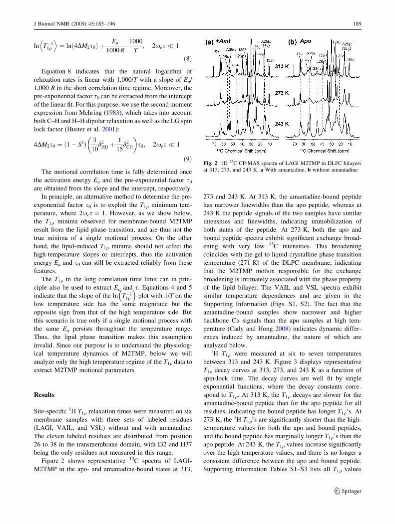
Figure 2 shows representative 13C spectra of LAGI-
M2TMP in the apo- and amantadine-bound states at 313,
273 and 243 K. At 313 K, the amantadine-bound peptide
has narrower linewidths than the apo peptide, whereas at
243 K the peptide signals of the two samples have similar
intensities and linewidths, indicating immobilization of
both states of the peptide. At 273 K, both the apo and
bound peptide spectra exhibit significant exchange broad-
ening with very low 13C intensities. This broadening
coincides with the gel to liquid-crystalline phase transition
temperature (271 K) of the DLPC membrane, indicating
that the M2TMP motion responsible for the exchange
broadening is intimately associated with the phase property
of the lipid bilayer. The VAIL and VSL spectra exhibit
similar temperature dependences and are given in the
Supporting Information (Figs. S1, S2). The fact that the
amantadine-bound samples show narrower and higher
backbone Ca signals than the apo samples at high tem-
perature (Cady and Hong 2008) indicates dynamic differ-
ences induced by amantadine, the nature of which are
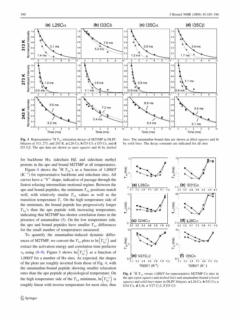
analyzed below.1H T1q were measured at six to seven temperatures
between 313 and 243 K. Figure 3 displays representative
T1q decay curves at 313, 273, and 243 K as a function of
spin-lock time. The decay curves are well fit by single
exponential functions, where the decay constants corre-
spond to T1q. At 313 K, the T1q decays are slower for the
amantadine-bound peptide than for the apo peptide for all
residues, indicating the bound peptide has longer T1q’s. At
273 K, the 1H T1q’s are significantly shorter than the high-
temperature values for both the apo and bound peptides,
and the bound peptide has marginally longer T1q’s than the
apo peptide. At 243 K, the T1q values increase significantly
over the high temperature values, and there is no longer a
consistent difference between the apo and bound peptide.
Supporting information Tables S1–S3 lists all T1q values
Fig. 2 1D 13C CP-MAS spectra of LAGI M2TMP in DLPC bilayers
at 313, 273, and 243 K. a With amantadine, b without amantadine
J Biomol NMR (2009) 45:185–196 189
123
for backbone Ha, sidechain Hb, and sidechain methyl
protons in the apo and bound M2TMP at all temperatures.
Figure 4 shows the 1H T1q’s as a function of 1,000/T
(K-1) for representative backbone and sidechain sites. All
curves have a ‘‘V’’ shape, indicative of passage through the
fastest relaxing intermediate motional regime. Between the
apo and bound peptides, the minimum T1q positions match
well, with relatively similar T1q values as well as the
transition temperature Tc. On the high temperature side of
the minimum, the bound peptide has progressively longer
T1q’s than the apo peptide with increasing temperature,
indicating that M2TMP has shorter correlation times in the
presence of amantadine (5). On the low temperature side,
the apo and bound peptides have smaller T1q differences
for the small number of temperatures measured.
To quantify the amantadine-induced dynamic differ-
ences of M2TMP, we convert the T1q plots to ln T�11q
� �and
extract the activation energy and correlation time prefactor
s0 using (8–9). Figure 5 shows ln T�11q
� �as a function of
1,000/T for a number of Ha sites. As expected, the shapes
of the plots are roughly inverted from those of Fig. 4, with
the amantadine-bound peptide showing smaller relaxation
rates than the apo peptide at physiological temperature. On
the high temperature side of the T1q minimum, ln T�11q
� �is
roughly linear with inverse temperature for most sites, thus
Fig. 3 Representative 1H T1q relaxation decays of M2TMP in DLPC
bilayers at 313, 273, and 243 K. a L26 Ca, b I33 Cd, c I35 Ca, and dI35 Cb. The apo data are shown as open squares and fit by dashed
lines. The amantadine-bound data are shown as filled squares and fit
by solid lines. The decay constants are indicated for all sites
Fig. 4 1H T1q versus 1,000/T for representative M2TMP Ca sites in
the apo (open squares and dashed line) and amantadine-bound (closedsquares and solid line) states in DLPC bilayers. a L26 Ca, b S31 Ca, cG34 Ca, d L36, e V27 Cc2, f I35 Cd
190 J Biomol NMR (2009) 45:185–196
123
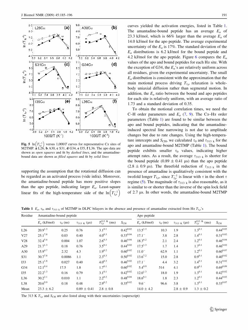
supporting the assumption that the rotational diffusion can
be regarded as an activated process (vide infra). Moreover,
the amantadine-bound peptide has more positive slopes
than the apo peptide, indicating larger Ea. Least-square
linear fits of the high-temperature side of the ln T�11q
� �
curves yielded the activation energies, listed in Table 1.
The amantadine-bound peptide has an average Ea of
23.3 kJ/mol, which is 66% larger than the average Ea of
14.0 kJ/mol for the apo peptide. The average experimental
uncertainty of the Ea is 17%. The standard deviation of the
Ea distributions is 6.2 kJ/mol for the bound peptide and
4.2 kJ/mol for the apo peptide. Figure 6 compares the Ea
values of the apo and bound peptides for each Ha site. With
the exception of G34, the Ea’s are relatively uniform across
all residues, given the experimental uncertainty. The small
Ea distribution is consistent with the approximation that the
main motional process driving T1q relaxation is whole-
body uniaxial diffusion rather than segmental motion. In
addition, the Ea ratio between the bound and apo peptides
for each site is relatively uniform, with an average ratio of
1.73 and a standard deviation of 0.35.
To obtain the motional correlation times, we need the
C–H order parameters and Ea (7, 9). The Ca–Ha order
parameters (Table 1) are found to be similar between the
apo and bound peptides, indicating that the amantadine-
induced spectral line narrowing is not due to amplitude
changes but due to rate changes. Using the high-tempera-
ture intercepts and SCH, we calculated s0 and s313 k for the
apo and amantadine-bound M2TMP (Table 1). The bound
peptide exhibits smaller s0 values, indicating higher
attempt rates. As a result, the average s313 k is shorter for
the bound peptide (0.89 ± 0.41 ls) than the apo peptide
(2.8 ± 0.9 ls). The threefold reduction of s313 k in the
presence of amantadine is qualitatively consistent with the
twofold longer T1q, since T�11q is linear with s in the short s
regime (5). The magnitude of s313 k is also reasonable, as it
is similar to or shorter than the inverse of the spin lock field
of 2.7 ls. In other words, the amantadine-bound M2TMP
Fig. 5 ln T�11q
� �versus 1,000/T curves for representative Ca sites of
M2TMP. a L26, b A30, c S31, d G34, e I35, f L36. The apo data are
shown as open squares and fit by dashed lines, and the amantadine-
bound data are shown as filled squares and fit by solid lines
Table 1 Ea, s0, and s313 k of M2TMP in DLPC bilayers in the absence and presence of amantadine extracted from Ha T1q’s
Residue Amantadine-bound peptide Apo peptide
Ea (kJ/mol) s0 (ns) s313 K (ls) T1q313 K (ms) SCH Ea (kJ/mol) s0 (ns) s313 K (ls) T1q
313 K (ms) SCH
L26 20.91.3 0.25 0.76 3.10.1 0.420.02 13.51.3 10.3 1.9 1.30.1 0.440.02
V27 25.17.8 0.03 0.40 4.00.3 0.330.04 17.1- 3.8 2.8 1.40.1 0.310.02
V28 32.45.4 0.004 1.07 2.60.3 0.460.04 18.36.2 2.1 2.4 1.20.1 0.460.04
A29 21.71.3 0.18 0.76 3.50.1 0.440.02 17.51.3 1.7 1.4 1.70.1 0.460.02
A30 15.94.7 2.32 4.3 1.90.2 0.600.02 11.0- 62.9 1.1 1.20.1 0.600.07
S31 30.77.8 0.0086 1.1 2.30.3 0.500.02 13.67.2 15.0 2.8 0.90.1 0.400.02
I33 25.11.8 0.027 0.40 4.00.3 0.460.02 17.1- 4.4 3.2 1.40.1 0.310.02
G34 12.10.4 17.5 1.8 1.70.1 0.600.02 5.40.9 514 4.1 0.90.1 0.690.09
I35 22.21.3 0.16 0.79 3.10.1 0.420.02 12.01.3 18.0 1.9 1.30.1 0.420.02
L36 30.22.1 0.010 1.1 2.20.2 0.480.04 18.68.1 1.8 2.3 1.20.1 0.440.02
L38 20.69.9 0.18 0.48 2.90.2 0.350.02 9.6- 96.6 3.8 1.30.1 0.330.07
Mean 23.3 ± 6.2 0.89 ± 0.41 2.8 ± 0.8 14.0 ± 4.2 2.8 ± 0.9 1.3 ± 0.2
The 313 K T1q and SCH are also listed along with their uncertainties (superscript)
J Biomol NMR (2009) 45:185–196 191
123
has a shorter correlation time than the characteristic time
scale of the spin-lock field, thus alleviating exchange
broadening, while the apo peptide correlation time is closer
to the NMR time scale, thus giving rise to broader lines.
Decreasing the temperature to 273 K increased the s to
6.0 ls and 3.4 ls for the apo and bound peptides,
respectively.
It is also interesting to examine the sidechain 1H T1q
trends. Table S3 shows that the methyl proton T1q’s are
much longer than the backbone Ha T1q’s and are relatively
similar between the apo and bound peptides at high tem-
peratures. Figure 7 shows representative ln T�11q
� �plots of
two methyl groups and one methine Cb site. The methyl
protons have more similar high-temperature slopes
between the apo and bound peptides, with an apparent
activation energy difference of only 20% between the two
states. The similarities can be attributed to three-site jumps
of the methyl groups that are additional to the uniaxial
diffusion, which reduce the dynamic difference between
the apo and bound peptide. In comparison, the Cb sites
show more distinct activation energies between the apo and
bound states, consistent with the Ha T1q behavior.
Discussion
M2TMP uniaxial diffusion is the main motion driving
T1q relaxation
The motivation for this work is to quantify the motion of
M2TMP that is responsible for exchange broadening of its
NMR spectra in phosphocholine bilayers at physiological
temperatures, and to understand the origin of amantadine-
induced line narrowing.
The exchange broadening is due to microsecond motion
of the peptide that interferes with 13C–1H and 15N–1H
dipolar decoupling and cross polarization. We assign the
motion to whole-body uniaxial rotational diffusion of the
M2 helical bundle. The presence of this uniaxial diffusion
has been previously shown based on the lineshapes of 2H
quadrupolar spectra, 15N static powder spectra, and 15N–1H
and 13C–1H dipolar couplings (Cady et al. 2007). Two lines
of evidence support the assignment of the T1q relaxation
mechanism to this uniaxial diffusion. First, all eleven
measured residues exhibit rapid T1q relaxation at high
temperatures, and all T1q values are increased by amanta-
dine. Such across-the-board effects can only result from a
whole-body motion. Second, the two-dimensional Brown-
ian diffusion theory of Saffman and Delbruck predicts a
rate of 105/s for the uniaxial diffusion of the M2 helical
bundle, which agrees well with the time scale probed by
the T1q experiments.
The presence of whole-body motion does not exclude
additional internal motions. Sidechain motions are cer-
tainly present, although they do not interfere with the
extraction of the rates and activation energy of uniaxial
diffusion from the backbone Ha T1q data. The clearest
manifestation of sidechain motions is the fact that the
methyl protons have longer T1q’s than the backbone pro-
tons, and the methyl 1H T1q’s are similar between the apo
and bound peptides at high temperatures (Table S3). These
observations are not surprising, since the fast methyl
Fig. 6 Activation energy Ea (kJ/mol) extracted from the high-
temperature slopes of the Ha ln T�11q
� �versus 1,000/T curves. The
amantadine-bound M2TMP (filled bars) has larger activation energies
than the apo peptide (open bars)
Fig. 7 a–c ln T�11q
� �versus 1,000/T curves for several M2TMP
sidechains. a L26 methyl Cd1/ Cd2, b A30 Cb and I33 Cc2 methyl
groups. c L36 Cb. Apo data: open squares and dashed lines.
Amantadine-bound data: filled squares and solid lines. d Activation
energy (kJ/mol) of the methyl protons. The average Ea difference
between the apo and bound peptides is *20%
192 J Biomol NMR (2009) 45:185–196
123
rotation on the nanosecond time scale pre-averages the
dipolar second moment, and thus reduce the relaxation
rates (1). The local nature of the methyl rotation also makes
the motion less sensitive to amantadine binding. The full
T1q dependence includes spectral densities at the 1H and13C Larmor frequencies, which are more relevant time
scales for the methyl rotation. Thus, (1) does not apply
fully to methyl groups. Since the main purpose of this study
is to understand the motion that causes exchange broad-
ening of the NMR spectra, but sidechain methyl 13C signals
do not suffer from exchange broadening, we do not con-
sider the combined motion of three-site jumps and uniaxial
diffusion experienced by the methyl groups further.
Another manifestation of possible segmental motions is the
distribution of Ea. Specifically, the G34 Ea is two standard
deviations lower than the average, suggesting the local
motion at this residue (vide infra).
Correlation time of M2TMP diffusion and origin
of spectral line narrowing by amantadine
The threefold shorter correlation time of the amantadine-
bound M2TMP (Table 1) explains the amantadine-induced
line narrowing of the peptide spectra. The faster motional
rates better avoid the intermediate time scale condition,
thus alleviating line broadening. The faster diffusion rates
result from the one to two orders of magnitude reduction of
the prefactor s0, as obtained from the high-temperature
intercepts of the ln T�11q
� �curves. The intercept depends
both on s0 and the dipolar second moment (9), whose exact
magnitude differs somewhat between different theories,
depending on how many spin interactions are included and
whether the MAS or the static condition is operative (Fares
et al. 2005; Huster et al. 2001; Mehring 1983). However,
the ratio of the intercepts between the apo and bound
peptide depends only on s0. Thus, the relative size of s0
between the apo peptide (long s0) and the bound peptide
(short s0) is unambiguous.
The shorter s0 of the amantadine-bound M2TMP indi-
cates higher attempt rates of motion, which suggest that the
M2 helices form better-packed tetramers in the presence of
amantadine. Amantadine may interact with all four helices
of the tetramer, thus serving as a non-covalent linker that
brings the four helices together to form a more cohesive
tetrameric bundle. A more cohesive helical bundle can
diffuse faster, and would also have structurally and
dynamically more homogeneous individual helices. This
interpretation is consistent with the relative lack of corre-
lation time distribution for the bound peptide (vide infra),
as manifested by the sharpness of the T1q minima, and is
also consistent with disulfide cross linking data that indi-
cate increased tetramer association in the presence of
amantadine (Cristian et al. 2003). Thus, the reduced s0 of
the bound M2TMP suggests that amantadine is centrally
located in the pore of the channel, shared by all four
helices, consistent with the binding site seen in a recent
crystal structure (Stouffer et al. 2008). In comparison, a
solution NMR study (Schnell and Chou 2008) found four
rimantadine molecules at the lipid-facing surface of each
channel. It is difficult to imagine how this surface binding
motif would homogenize the peptide conformation and
speed up its motion.
A question that is beyond the scope of the current study
is the correlation time distribution of apo M2TMP, which is
manifested by the broad T1q minima of the apo peptide.
The s distribution suggests that without amantadine, the
M2 helical bundles are floppier, with more internal degrees
of freedom, and may exhibit dynamic heterogeneities
between different tetramers. Such dynamic heterogeneity
may be functionally relevant, as it may allow the apo
peptide to adopt appropriate conformations to achieve its
many functions, including channel activation, gating, and
inhibition.
The activation energy of M2TMP uniaxial diffusion
is related to membrane viscosity
A basic assumption in our dynamic analysis is that the
rotational diffusion of a membrane protein in lipid bilayers
can be considered an activated process that follows an
Arrhenius law (7). The linearity of the observed ln T�11q
� �
with respect to 1,000/T at high temperatures (Fig. 5) vali-
dates this assumption, but it is of interest to consider the
physical basis for the activated diffusion. Membrane pro-
tein diffusion in lipid bilayers requires the availability of
free volume in the vicinity of the protein, which is achieved
by discrete hopping of the lipid molecules. Thus, activated
lateral and rotational diffusion of membrane proteins has
its molecular origin in the free volume theory of liquids
(Cohen and Turbill 1959; Galla et al. 1979). Considered in
this light, the activation energy essentially reflects the
macroscopic viscosity of the membrane. Higher activation
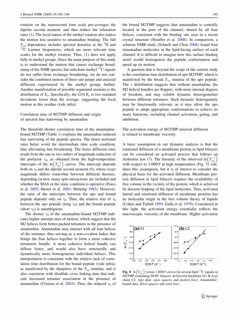
Fig. 8 ln T�11q
� �versus 1,000/T curves for several lipid 13C signals in
M2TMP-containing DLPC bilayers. a Glycerol backbone G3. b Acyl
chain C2. Apo data: open squares and dashed lines; Amantadine-
bound data: filled squares and solid lines
J Biomol NMR (2009) 45:185–196 193
123
energies indicate higher viscosities. Thus, the higher Ea of
the amantadine-bound peptide indicates that excess
amantadine increases the membrane viscosity. If this is
true, then the lipid 1H T1q values should be affected by the
excess amantadine outside the channel in the bilayer, in the
same manner as the protein.
Examination of several resolved lipid 13C signals con-
firms that there is indeed an amantadine-induced increase
of the lipid Ea. Figure 8 shows the ln T�11q
� �curves of the
lipid glycerol G3 and the acyl chain C2 signals. The
excess-amantadine-containing membrane clearly has larger
slopes than the amantadine-free membrane, similar to the
peptide T1q behavior. The amantadine-bound samples in
our experiments contain eightfold molar excess of aman-
tadine to the peptide, or 32-fold excess amantadine to the
channel. The excess amantadine partitions into the bilayer
(Subczynski et al. 1998; Wang et al. 2004). Paramagnetic
relaxation enhancement NMR data indicate that amanta-
dine is located at the interfacial region of the DMPC
bilayer with the amine group pointing to the surface (Li
et al. 2008). This depth and orientation are consistent with
the amphipathic nature of amantadine, with its hydrophobic
adamantane cage close the hydrocarbon core and its polar
ammonium moiety pointing to the polar region of the
bilayer.
Our recently measured lipid 13C T2 relaxation times at
313 K in peptide-free DLPC bilayers showed no discern-
ible difference between amantadine-containing and aman-
tadine-free bilayers (Cady et al. 2009), in contrast to the
current lipid 1H T1q data. This discrepancy is likely due to
the presence of the peptide in the current measurements but
the lack of the peptide in the previous T2 experiments.
Membrane viscosity is a function of molecular crowd-
ing: peptide-containing bilayers have much smaller free
volumes for amantadine than pure lipid bilayers, thus
amantadine may cause detectable fluidity changes only in
M2-containing bilayers.
The viscosity origin of the activation energy of M2TMP
diffusion also explains the asymmetric slope of the
ln T�11q
� �curves on the two sides of the T1q minimum. Both
the apo and bound peptides have steeper slopes or larger
apparent activation energies on the low temperature side.
Since this T1q minimum is associated with phase transition
of the DLPC bilayer, the slope increase at low temperature
is caused by the significant viscosity increase of the gel-
phase membrane compared to the liquid-crystalline phase.
Thus, the M2TMP rotational diffusion is not a single
motional process above and below the lipid phase transi-
tion temperature, in contrast to most other motions that
have been characterized by NMR (Douglass and Jones
1966; Rothwell and Waugh 1981).
The viscosity origin of the activation energy may also
partly explain the Ea distribution among the residues. Since
a perfectly rigid-body motion should have a single Ea, the
observed Ea distribution beyond the experimental uncer-
tainty must reflect additional motions, which may result
from varying membrane viscosity across the bilayer
thickness. The abnormally low Ea of G34 may then be
partly due to the location of G34 at the center of the TM
helix and thus at the center of the bilayer, which has the
lowest viscosity due to large-amplitude motions of the lipid
chain ends. While the exact nature of the local G34 motion
is unknown, the conclusion of increased conformational
flexibility is consistent with a large number of NMR
parameters for this site: its 15N anisotropic chemical shifts,
N–H dipolar coupling (Hu et al. 2007), and 15N isotropic
chemical shifts were all found to change with drug binding
(Cady et al. 2009; Wang et al. 2009) and membrane
composition (Luo et al. 2009).
The lipid-mediated influences of excess amantadine on
the diffusion dynamics of M2TMP do not change the
previous conclusion that the protein-bound amantadine
causes site-specific conformational changes through direct
amantadine-protein interactions. The structural changes are
manifested as chemical shift perturbations that are highly
site specific, with residues S31, G34, and V28 exhibiting
large chemical shift perturbations while other residues
showing little changes (Cady and Hong 2008; Cady et al.
2009).
Conclusion
The temperature-dependent 1H T1q relaxation times shown
here indicate that the uniaxial diffusion of the M2TMP in
liquid-crystalline DLPC bilayers causes efficient 1H T1q
relaxation, and the diffusion rates are increased by aman-
tadine through an increase in the attempt frequencies. The
average motional correlation time at 313 K is 0.89 ls for
the bound peptide and 2.8 ls for the apo peptide. The faster
diffusion of the bound peptide suggests that amantadine
induces more homogeneous and better-packed M2TMP
tetramers by coordinating with all four helices. Thus, the
well documented amantadine-induced NMR line narrowing
is due to suppression of the intermediate time scale
broadening.
From the linear relation of ln T�11q
� �with 1/T at high
temperature, we extracted the activation energy of the M2
uniaxial diffusion. The average Ea is 23.3 kJ/mol for the
bound peptide and 14.0 kJ/mol for the apo peptide. The
higher Ea of the bound peptide is attributed to larger
membrane viscosity by excess amantadine, which is con-
firmed by the lipid T1q data.
Similar exchange broadening of the apo protein and line
narrowing of the bound protein have also been reported for
solution NMR spectra of M2 (18–60) in DHPC micelles
194 J Biomol NMR (2009) 45:185–196
123
(Schnell and Chou 2008). There, the exchange broadening
of the apo protein spectra was alleviated by the addition of
40 mM rimantadine. At 53-fold molar excess to the protein
(0.75 mM), the excess rimantadine is expected to signifi-
cantly increase the viscosity of the micelle interior, which
may affect the dynamics of the micelle-bound rimantadine
in a way that facilitates the observation of its NOEs with
the protein.
The present study illustrates the rich information that
can be obtained from temperature-dependent relaxation
studies of membrane proteins. Compared to most relaxa-
tion NMR studies so far, which were targeted to either
small molecules with a single motion (Rothwell and
Waugh 1981) or macromolecules with a well-defined local
motion (Batchelder et al. 1982), the present study examines
the rates and energetics of the global motion of a mem-
brane protein (Reuther et al. 2006). It gives a glimpse into
the energetic driving force behind membrane protein uni-
axial diffusion, which is not easily obtained by other bio-
physical techniques (Gennis 1989).
References
Aisenbrey C, Bechinger B (2004) Investigations of polypeptide
rotational diffusion in aligned membranes by 2H and 15N solid-
state NMR spectroscopy. J Am Chem Soc 126:16676–16683
Batchelder LS, Sullivan CE, Jelinski LW, Torchia DA (1982)
Characterization of leucine side-chain reorientation in colla-
gen-fibrils by solid-state 2H NMR. Proc Natl Acad Sci USA
79:386–389
Bielecki A, Kolbert AC, de Groot HJM, Griffin RG, Levitt MH
(1990) Frequency-switched Lee-Goldburg sequences in solids.
Adv Magn Reson 14:111–124
Bloom M, Evans E, Mouritsen OG (1991) Physical properties of the
fluid lipid-bilayer component of cell membranes: a perspective.
Q Rev Biophys 24:293–397
Blume A, Rice DM, Wittebort RJ, Griffin RG (1982) Molecular
dynamics and conformation in the gel and liquid-crystalline
phases of phosphatidylethanolamine bilayers. Biochemistry
21:6220–6230
Cady SD, Hong M (2008) Amantadine-induced conformational and
dynamical changes of the influenza M2 transmembrane proton
channel. Proc Natl Acad Sci USA 105:1483–1488
Cady SD, Goodman C, Tatko CD, DeGrado WF, Hong M (2007)
Determining the orientation of uniaxially rotating membrane
proteins using unoriented samples: a 2H, 13C, AND 15N solid-
state NMR investigation of the dynamics and orientation of a
transmembrane helical bundle. J Am Chem Soc 129:5719–5729
Cady SD, Mishanina TV, Hong M (2009) Structure of amantadine-
bound M2 transmembrane peptide of influenza A in lipid
bilayers from magic-angle-spinning solid-state NMR: the role of
Ser31 in amantadine binding. J Mol Biol 385:1127–1141
Carpino LA, Han GY (1972) The 9-fluorenylmethoxycarbonyl amino-
protecting group. J Org Chem 37:3404–3409
Clore GM, Szabo A, Bax A, Kay LE, Driscoll PC, Gronenborn AM
(1990) Deviations from the simple 2-parameter model-free
approach to the interpretation of N-15 nuclear magnetic relax-
ation of proteins. J Am Chem Soc 112:4989–4991
Cohen MH, Turbill D (1959) Molecular transport in liquids and
glasses. J Chem Phys 31:1164–1169
Cristian L, Lear JD, DeGrado WF (2003) Use of thiol-disulfide
equilibria to measure the energetics of assembly of transmem-
brane helices in phospholipid bilayers. Proc Natl Acad Sci USA
100:14772–14777
deAzevedo ER, Hu WG, Bonagamba TJ, Schmidt-Rohr K (2000)
Principles of centerband-only detection of exchange in solid-state
nuclear magnetic resonance, and extension to four-time center-
band-only detection of exchange. J Chem Phys 112:8988–9001
Douglass DC, Jones GP (1966) Nuclear magnetic relaxation of n-
alkanes in rotating frame. J Chem Phys 45:956–963
Fares C, Qian J, Davis JH (2005) Magic angle spinning and static
oriented sample NMR studies of the relaxation in the rotating
frame of membrane peptides. J Chem Phys 122:194908
Galla HJ, Hartmann W, Theilen U, Sackmann E (1979) On two-
dimensional passive random walk in lipid bilayers and fluid
pathways in biomembranes. J Membr Biol 48:215–236
Gennis RB (1989) Biomembranes: molecular structure and function.
Springer, New York
Glaser RW, Sachse C, Durr UH, Wadhwani P, Ulrich AS (2004)
Orientation of the antimicrobial peptide PGLa in lipid mem-
branes determined from 19F-NMR dipolar couplings of 4-CF3-
phenylglycine labels. J Magn Reson 168:153–163
Hagemeyer A, Schmidt-Rohr K, Spiess HW (1989) 2D NMR
experiments for studying molecular order and dynamics in static
and rotating solids. Adv Magn Reson 13:85–130
Hay AJ, Wolstenholme AJ, Skehel JJ, Smith MH (1985) The
molecular basis of the specific anti-influenza action of amanta-
dine. EMBO J 4:3021–3024
Hong M (2007) Structure, topology, and dynamics of membrane
peptides and proteins from solid-state NMR spectroscopy. J Phys
Chem B 111:10340–10351
Hong M, Doherty T (2006) Orientation determination of membrane-
disruptive proteins using powder samples and rotational diffusion:
a simple solid-state NMR approach. Chem Phys Lett 432:296–300
Hong M, Gross JD, Rienstra CM, Griffin RG, Kumashiro KK,
Schmidt-Rohr K (1997) Coupling amplification in 2D MAS
NMR and its application to torsion angle determination in
peptides. J Magn Reson 129:85–92
Hong M, Yao XL, Jakes K, Huster D (2002) Investigation of
molecular motions by Lee-Goldburg cross-polarization NMR
spectroscopy. J Phys Chem B 106:7355–7364
Hu J, Asbury T, Achuthan S, Li C, Bertram R, Quine JR, Fu R, Cross
TA (2007) Backbone structure of the amantadine-blocked trans-
membrane domain M2 proton channel from influenza A virus.
Biophys J 92:4335–4343
Huster D, Xiao LS, Hong M (2001) Solid-state NMR investigation of
the dynamics of the soluble and membrane-bound colicin Ia
channel-forming domain. Biochemistry 40:7662–7674
Ishima R, Torchia DA (2000) Protein dynamics from NMR. Nat
Struct Biol 7:740–743
Ito T, Gorman OT, Kawaoka Y, Bean WJ, Webster RG (1991)
Evolutionary analysis of the influenza A virus M gene with
comparison of the M1 and M2 proteins. J Virol 65:5491–5498
Jelinski LW, Sullivan CE, Torchia DA (1980) 2H NMR study of
molecular motion in collagen fibrils. Nature 284:531–534
Kay LE (1998) Protein dynamics from NMR. Nat Struct Biol
(suppl):513–517
Kinsey RA, Kintanar A, Tsai MD, Smith RL, Janes N, Oldfield E
(1981) First observation of amino acid side chain dynamics in
membrane proteins using high field deuterium nuclear magnetic
resonance spectroscopy. J Biol Chem 256:4146–4149
Kovacs FA, Cross TA (1997) Transmembrane four-helix bundle of
influenza A M2 protein channel: structural implications from
helix tilt and orientation. Biophys J 73:2511–2517
J Biomol NMR (2009) 45:185–196 195
123
Lee K-C, Hu W, Cross TA (1993) 2H NMR determination of the
global correlation time of the gramicidin channel in a lipid
bilayer. Biophys J 65:1162–1167
Lewis BA, Harbison GS, Herzfeld J, Griffin RG (1985) NMR
structural analysis of a membrane protein: bacteriorhodopsin
peptide backbone orientation and motion. Biochemistry 24:
4671–4679
Li C, Qin H, Gao FP, Cross TA (2007) Solid-state NMR character-
ization of conformational plasticity within the transmembrane
domain of the influenza A M2 proton channel. Biochim Biophys
Acta 1768:3162–3170
Li C, Yi M, Hu J, Zhou HX, Cross TA (2008) Solid-state NMR and
MD simulations of the antiviral drug amantadine solubilized in
DMPC bilayers. Biophys J 94:1295–1302
Lipari G, Szabo A (1982) Model-free approach to the interpretation of
nuclear magnetic-resonance relaxation in macromolecules. 1.
Theory and range of validity. J Am Chem Soc 104:4546–4559
Luo W, Hong M (2006) Determination of the oligomeric number and
intermolecular distances of membrane protein assemblies by
anisotropic (1)H-driven spin diffusion NMR spectroscopy. J Am
Chem Soc 128:7242–7251
Luo W, Cady SD, Hong M (2009) Immobilization of the influenza A
M2 transmembrane peptide in virus-envelope mimetic lipid
membranes: a solid-state NMR investigation. Biochemistry
48:6361–6368
Macdonald PM, Seelig J (1988) Dynamic properties of gramicidin A
in phospholipid membranes. Biochemistry 27:2357–2364
Mandel AM, Akke M, Palmer AG (1996) Dynamics of ribonuclease
H: temperature dependence of motions on multiple time scales.
Biochemistry 35:16009–16023
Mehring M (1983) Principles of high resolution NMR in solids.
Springer, Berlin
Munowitz M, Aue WP, Griffin RG (1982) Two-dimensional separa-
tion of dipolar and scaled isotropic chemical shift interactions in
magic angle NMR spectra. J Chem Phys 77:1686–1689
Opella SJ (1986) Protein dynamics by solid state nuclear magnetic
resonance. Methods Enzymol 131:327–361
Palmer AG, Williams J, McDermott A (1996) Nuclear magnetic
resonance studies of biopolymer dynamics. J Phys Chem
100:13293–13310
Palmer AG, Kroenke CD, Loria JP (2001) Nuclear magnetic resonance
methods for quantifying microsecond-to-millisecond motions in
biological macromolecules. Methods Enzymol 339:204–238
Park SH, Mrse AA, Nevzorov AA, De Angelis AA, Opella SJ (2006)
Rotational diffusion of membrane proteins in aligned phospho-
lipid bilayers by solid-state NMR spectroscopy. J Magn Reson
178:162–165
Pauls KP, MacKay AL, Soderman O, Bloom M, Tanjea AK, Hodges
RS (1985) Dynamic properties of the backbone of an integral
membrane polypeptide measured by 2H-NMR. Eur Biophys J
12:1–11
Pinto LH, Lamb RA (2007) Controlling influenza virus replication by
inhibiting its proton flow. Mol Biosyst 3:18–23
Pinto LH, Holsinger LJ, Lamb RA (1992) Influenza virus M2 protein
has ion channel activity. Cell 69:517–528
Prosser RS, Davis JH, Mayer C, Weisz K, Kothe G (1992) Deuterium
NMR relaxation studies of peptide-lipid interactions. Biochem-
istry 31:9355–9363
Reuther G, Tan KT, Vogel A, Nowak C, Arnold K, Kuhlmann J,
Waldmann H, Huster D (2006) The lipidated membrane anchor
of full length N-Ras protein shows an extensive dynamics as
revealed by solid-state NMR spectroscopy. J Am Chem Soc
128:13840–13846
Rothwell WP, Waugh JS (1981) Transverse relaxation of dipolar
coupled spin systems under Rf-irradiation—detecting motions in
solids. J Chem Phys 74:2721–2732
Saffman PG, Delbruck M (1975) Brownian motion in biological
membranes. Proc Natl Acad Sci USA 72:3111–3113
Sakaguchi T, Tu Q, Pinto LH, Lamb RA (1997) The active oligomeric
state of the minimalistic influenza virus M2 ion channel is a
tetramer. Proc Natl Acad Sci USA 94:5000–5005
Schaefer J, Stejskal EO, Buchdahl R (1977) Magic-angle C-13 NMR
analysis of motion in solid glassy polymers. Macromolecules
10:384–405
Schaefer J, Mckay RA, Stejskal EO (1983) Dipolar rotational spin-
echo 13C NMR of polymers. J Magn Reson 52:123–129
Schaefer J, Stejskal EO, Mckay RA (1984) Phenylalanine ring
dynamics by solid-state 13C NMR. J Magn Reson 57:85–92
Schaefer D, Spiess HW, Suter UW, Fleming WW (1990) 2D solid-
state NMR studies of ultraslow chain motion: glass transition in
aPP versus helical jumps in iPP. Macromolecules 23:3431–3439
Schmidt C, Blumich B, Spiess HW (1988) Deuteron two-dimensional
exchange NMR in solids. J Magn Reson 79:269–290
Schmidt-Rohr K, Clauss J, Spiess HW (1992) Correlation of structure,
mobility, and morphological information in heterogeneous
polymer materials by two-dimensional wideline-separation
NMR spectroscopy. Macromolecules 25:3273–3277
Schnell JR, Chou JJ (2008) Structure and mechanism of the M2
proton channel of influenza A virus. Nature 451:591–595
Shaw WJ, Long JR, Campbell AA, Stayton PS, Drobny GP (2000) A
solid state NMR study of dynamics in a hydrated salivary peptide
adsorbed to hydroxyapatite. J Am Chem Soc 122:7118–7119
Smith RL, Oldfield E (1984) Dynamic structure of membranes by
deuterium NMR. Science 225:280–288
Stouffer AL, Acharya R, Salom D, Levine AS, Di Costanzo L, Soto
CS, Tereshko V, Nanda V, Stayrook S, DeGrado WF (2008)
Structural basis for the function and inhibition of an influenza
virus proton channel. Nature 451:596–599
Subczynski WK, Wojas J, Pezeshk V, Pezeshk A (1998) Partitioning
and localization of spin-labeled amantadine in lipid bilayers: an
EPR study. J Pharm Sci 87:1249–1254
Tian F, Song Z, Cross TA (1998) Orientational constraints derived
from hydrated powder samples by two-dimensional PISEMA. J
Magn Reson 135:227–231
Traikia M, Warschawski DE, Recouvreur M, Cartaud J, Devaux PF
(2000) Formation of unilamellar vesicles by repetitive freeze-
thaw cycles: characterization by electron microscopy and P-31-
nuclear magnetic resonance. Eur Biophys J 29:184–195
van Rossum BJ, de Groot CP, Ladizhansky V, Vega S, de Groot HJM
(2000) A method for measuring heteronuclear (H-1-C-13)
distances in high speed MAS NMR. J Am Chem Soc 122:
3465–3472
Wang C, Takeuchi K, Pinto LH, Lamb RA (1993) Ion channel
activity of influenza A virus M2 protein: characterization of the
amantadine block. J Virol 67:5585–5594
Wang J, Schnell JR, Chou JJ (2004) Amantadine partition and
localization in phospholipid membrane: a solution NMR study.
Biochem Biophys Res Commun 324:212–217
Wang J, Cady SD, Balannik V, Pinto LH, DeGrado WF, Hong M
(2009) Discovery of spiro-piperidine inhibitors and their mod-
ulation of the dynamics of the M2 proton channel from influenza
A virus. J Am Chem Soc 131:8066–8076
Williams JC, McDermott AE (1995) Dynamics of the flexible loop of
triosephosphate isomerase: the loop motion is not ligand gated.
Biochemistry 34:8309–8319
Yamaguchi S, Huster D, Waring A, Lehrer RI, Tack BF, Kearney W,
Hong M (2001) Orientation and dynamics of an antimicrobial
peptide in the lipid bilayer by solid-state NMR. Biophys J
81:2203–2214
Yang Z, Liivak O, Seidel A, LaVerde G, Zax DB, Jelinski LW (2000)
Supercontraction and backbone dynamics in spider silk: 13C and
2H NMR studies. J Am Chem Soc 122:9019–9025
196 J Biomol NMR (2009) 45:185–196
123


![Membrane-bound ATP Fuels the Na/K Pump · Membrane-bound ATP Fuels the Na/K Pump Studies on Membrane-bound Glycolytic Enzymes on Inside-Out Vesicles ... (0.125% [wt/vol] Ponceau-S,](https://img.pdfslide.net/doc/110x75/61493cb1080bfa6260147b2a/membrane-bound-atp-fuels-the-nak-pump-membrane-bound-atp-fuels-the-nak-pump-studies.jpg)