Embed Size (px)
Citation preview

Progress In Electromagnetics Research C, Vol. 11, 121–136, 2009
EFFECTS OF MICROWAVE ON WATER AND ITSINFLUENCE ON DRUG DISSOLUTION
T.-W. Wong, A. Iskhandar, M. Kamal, S.-J. JumiN.-H.Kamarudin,N.-Z. Mohamad Zin, and N.-H. Mohd Salleh
Non-destructive Biomedical and Pharmaceutical Research CentreParticle Design Research GroupFaculty of PharmacyUniversiti Teknologi MARA MalaysiaPuncak Alam, Selangor 42300, Malaysia
Abstract—Use of water with different molecular mobilities couldaffect drug dissolution of a dosage form and such profile of water mightbe modifiable using microwave. This study investigated the effectsof microwave on water and its influences on dissolution of free drugsand drugs in calcium-crosslinked alginate beads using sulphanilamideand sulphamerazine as hydrophilic and hydrophobic model drugsrespectively. The water was treated by microwave at 300 W or withoutpre-treatment. The drug dissolution, pH and molecule mobilityprofiles of untreated and microwave-treated water were examined.Microwave-treated water had higher pH and water molecule mobility.The latter was characterized by higher conductivity, lower molecularinteraction and crystallinity profiles. The dissolution of hydrophilicand hydrophobic free or encapsulated drugs was enhanced usingmicrowave-treated water due to its higher molecular mobility. Theuntreated water of the same pH as microwave-treated water did notenhance drug dissolution. The drug dissolution from beads wasincreased by higher water uptake leading to matrix erosion and poreformation using microwave-treated water and was not promoted by theformation of non-crosslinked hydrated alginic acid matrix in untreatedwater of lower pH. Microwave treatment of water increased watermolecule mobility and can promote drug dissolution.
Corresponding author: T.-W. Wong ([email protected]).
122 Wong et al.
1. INTRODUCTION
Microwave is an electromagnetic wave with wavelengths longer thanthose of terahertz waves, but shorter than radiowaves [1]. It hasfrequencies between 300MHz and 300GHz. Microwave is not a formof heat, but a form of energy which manifests as heat through itsinteraction with materials. The transmission of microwave to an objectresults in vibration of molecules by induced or permanent dipoles. Theintensity of vibration is dependent on the size, shape and polarizabilityof the molecules, as well as, the extent of intermolecular bonding ofthe object. Practically, the amount of energy absorbed by an object,P , is defined as:
P = 2πfE2E0Er tan δ (1)
where f = frequency of microwave, E = electric field, E0 = dielectricconstant of free space, Er = dielectric constant of object and tan δ =loss tangent.
Microwave has been utilized to design controlled-release alginate,alginate-chitosan, pectinate-chitosan and poly(methyl vinyl ether-co-maleic acid) beads [2–5]. The drug release characteristics ofthese beads were dependent on the propensity of polymer-polymerand drug-polymer interaction brought about by microwave. Themicrowave has also been investigated as the alternative mode tocrosslink gelatin matrix which is available as microspheres suspendedin a polar acetone [6]. It is found that only a short span of 10min is required for effective crosslinking of gelatin microspheres bymicrowave unlike when thermal denaturation method is used [6–8].The bioavailability of poorly water-soluble drugs is limited by theirdissolution in gastrointestinal tract [9]. Kerc et al. (1998), Bergeseet al. (2003) and Moneghini et al. (2008) have explored the usefulnessof microwave as the tool to prepare fast-release solid dispersion [10–12].They reported that the drug release propensity of microwave-treatedsample is greatly higher than those of pure drug, samples which areuntreated by microwave or treated by vacuum at 100◦C, or obtainedby solvent deposition method. A review of microwave application indesign of drug delivery system, namely agglomerates, beads, tablets,microparticles, nanoparticles and solid dispersion, has been reportedlately by Wong (2008) [4].
The process of drug dissolution proceeds through migrationof drug molecules into the cavity of liquid dissolution mediumand subsequent formation of bonding between drug and watermolecules [13]. The lattice theory postulates that a liquid mediumhas crystalline or quasi-crystalline structures [14]. A proportion ofvolume occupied by the liquid is empty in liquid lattice network which
Progress In Electromagnetics Research C, Vol. 11, 2009 123
constitutes free volume of liquid. The dissolution and diffusion of drugmolecules are accompanied by solute molecules moving from one emptycavity to another within a liquid lattice. Technically, a higher fractionof free cavity may be formed in bulk water following the transmission ofmicrowave to water which results in vibration of water molecules, localsuperheating and lost of crystalline structure of water network [15]. Itis hypothesized that the propensity of drug dissolution can be enhancedthrough employing microwave-treated liquid as dissolution medium.As dissolution profile of a drug can dictate its bioavailability, thepresent study sets to investigate the effects of microwave on aqueousdissolution medium and its influence on dissolution profiles of freedrugs of varying degrees of hydrophobicity as well as encapsulateddrugs in alginate beads.
2. MATERIALS AND METHODS
2.1. Materials
Sulphanilamide and sulphamerazine (Sigma, USA) were employedas model hydrophilic and hydrophobic drugs with the respectivemolecular weights of 172.2 and 264.3 g/mole. They were used asreceived without further purification. Sodium alginate (ManugelrDMB, ISP, USA: mannuronic acid (M)/guluronic acid (G) ratio =0.59) was used as matrix polymer for drug encapsulation in beadswith calcium chloride dihydrate (Merck, Germany) as crosslinkingagent. Deionized water was used as model dissolution medium. Itwas obtained by means of filtration and ion exchange processes (Elga,Veolia Water Systems, UK). Other chemical employed was hydrochloricacid (Merck, Germany) for digestion of beads in atomic absorptionspectrophotometric assay.
2.2. Preparation of Alginate Beads
An aqueous dispersion containing 2 %w/w of sodium alginate and1 %w/w of drug was introduced dropwise into an aqueous solutioncontaining 6 %w/w of calcium chloride dihydrate by extrusion througha 1.6 mm diameter orifice at a flow rate of 60 droplets/min aidedby peristaltic pump (Watson-Marlow Bredel Pumps, UK). The bulkof the calcium chloride solution was subjected to magnetic stirringthroughout the preparation process and the stirring was continuedfor an additional period of 15 min after the last addition of thesodium alginate-drug dispersion. The formed alginate beads wereremoved from the calcium chloride solution by filtration and washedwith deionized water. Blank beads were prepared in the same manner
124 Wong et al.
for all formulations, except that no drug was incorporated. All beadswere oven-dried at 40 ± 0.5◦C for 3 days and subsequently equilibratedto a constant weight by storing in a desiccator at 25 ± 1◦C.
2.3. Microwave Treatment of Dissolution Medium
An appropriate volume of deionized water was filtered under thenegative pressure. An amount of 520 g filtrate was subjected tomicrowave irradiation at 300W for 10 min at 2450 ± 50MHz (EM-GA, Sanyo, Japan). The microwave-treated water was cooled to ambienttemperature and its weight was adjusted to 500 g prior to subsequentexperiments on drug dissolution and calcium release.
2.4. Drug Dissolution
The dissolution profiles of free drug and drug encapsulated in alginatebeads were determined using untreated water and water treated bymicrowave at 300W as dissolution media. An accurately weighedamount of drug or beads was placed in 500 g of dissolution medium andagitated at 50 strokes/min using a shaker bath (Memmert GmbH+Co.KG, Germany) at 37 ± 0.2◦C. Aliquots were withdrawn at varioustime intervals and assayed spectrophotometrically for sulphanilamideand sulphamerazine at 260 and 259.9 nm respectively (Cary 50Conc, Varian Australia Pty Ltd, Australia). The percentage ofdrug dissolution was calculated with respect to drug load added todissolution medium or drug content of beads. The drug content wasexpressed as the percentage of drug encapsulated in a unit weight ofbeads. The drug content was determined by subjecting the samesample of beads from the drug dissolution study for an additional15 h of magnetic stirring followed by ultrasonication for at least 3consecutive periods of 10 min before assaying for drug. Blank beadswere taken as control sample. At least triplicates were carried out foreach batch of sample and the results averaged. The kinetics of drugreleased from beads was investigated by fitting the drug release datainto Korsmeyer-Peppas dissolution model as previously described [2].The drug release rate constant (k) and release exponent (n) indicativeof drug release mechanism were computed.
2.5. Calcium Release
The release profiles of calcium ions from alginate beads were examinedusing the similar protocol as drug dissolution study except that thealiquots were assayed using atomic absorption spectrophotometer (Z-2000, Hitachi Hi-Technologies Corporation, Japan). The percentage of
Progress In Electromagnetics Research C, Vol. 11, 2009 125
calcium release was calculated with respect to total calcium contentof beads. The total calcium content was determined by subjectingalginate beads to heating in 1 : 1 ratio of 37% hydrochloric acid anddeionised water mixture. The calcium content was expressed as thepercentage of calcium in a unit weight of beads At least triplicateswere carried out for each batch of sample and the results averaged.
2.6. Drug Aqueous Solubility
A saturated drug solution was obtained by stirring an excessiveamount of drug in a 50 ml volumetric flask filled with water. Thedrug solution was agitated at 50 strokes/min using a shaker bath(Memmert GmbH+Co. KG, Germany) at 37 ± 0.2◦C for 24 hours.Aliquots were withdrawn and assayed spectrophotometrically forsulphanilamide and sulphamerazine at 260 and 259.9 nm respectively(Cary 50 Conc, Varian Australia Pty Ltd, Australia). The calculateddrug concentration represented the aqueous solubility of drug in thespecified volume of water. At least triplicates were carried out for eachbatch of sample and the results averaged.
2.7. Bead Size and Shape
The size and shape of beads were determined using a digimatic verniercaliper system (Mitutoyo, Japan). The length and breadth weremeasured from each bead and its size calculated from the average ofthese two dimensions. The shape of bead was represented by aspectratio which is the quotient of its length to breadth. An aspect ratioof value unity represents a perfect sphere while higher values representgreater elongation. For each formulation, 20 beads were randomlyselected for measurement and the results averaged.
2.8. Scanning Electron Microscopy (SEM)
The surface structure of beads was examined using SEM technique(FEI Quanta 200F, Holland). The beads were fixed with a carbontape onto studs, and the prepared studs were viewed directly undera scanning electron microscope at a magnification level of 1000 ×.Representative sections were photographed.
2.9. Bead Swelling, Erosion and Water Uptake
The analysis of bead swelling, erosion and water uptake capacity wasconducted by immersing an accurately weighed bead with known size in10ml of untreated water or water treated by microwave at 300 W. The
126 Wong et al.
bead was subjected to agitation at 50 strokes/min using a shaker bath(Memmert GmbH+Co. KG, Germany) at 37 ± 0.2◦C. At specifiedintervals, the weight and size of wet beads were characterized afterremoving its surface moisture through running the bead gently over adry petri dish till no sign of moisture left on the immediate dish surfacecontacted by bead. The bead was then oven-dried at 40 ± 0.5◦C for 3days and subsequently equilibrated to a constant weight by storing ina desiccator at 25 ± 1◦C.
The swelling (SI), erosion (EI) and water uptake (WUI) indicesof bead were defined as:
SI = (St − Si)/Si · 100% (2)
where Si = initial dry bead diameter and St = wet bead diameter attime, t.
EI = Wi −Wt(d)/Wi · 100% (3)
where Wi = initial dry bead weight and Wt(d) = dry weight of beadcollected at t.
WUI = Wt −Wt(d)/Wt(d) · 100% (4)
where Wt = wet weight of beads at t.In computation of bead erosion and water uptake indices, the
weight measurement of beads was corrected for embedded drug andcalcium content at t. The response of beads towards dissolutionmedium was merely evaluated for their changes in polymeric domain.Ten replicates were conducted and the results averaged.
2.10. Dissolution Medium Temperature
The temperature of dissolution medium was determined usingan infrared thermometer (Thermo-Hunter, Optex, Japan) withmeasurement conducted in a non-contact mode. The temperatureof the untreated water was examined at the ambient temperature.The temperature of the microwave-treated water was examinedimmediately after the treatment by microwave. At least triplicateswere carried out for each batch of sample and the results averaged.
2.11. Dissolution Medium pH
The pH of dissolution medium was determined by means of a pH meter(Mettler Toledo 320, China) at 25 ± 1◦C. At least triplicates werecarried out for each batch of sample and the results averaged.
Progress In Electromagnetics Research C, Vol. 11, 2009 127
2.12. Dissolution Medium Conductivity
The conductivity of dissolution medium was determined by means of aconductivity meter (WTW Series conductivity 720, Inolab, Germany)at 25 ± 1◦C. At least triplicates were carried out for each batch ofsample and the results averaged.
2.13. Dissolution Medium Crystallinity
The crystallinity state of dissolution medium was evaluated using X-ray diffractometer (Ultima IV, Rigaku Corporation, Japan) with Cu-Kα radiation generated at 40 kV and 40 mA. The X-ray diffraction wasoperated at a scanning speed of 3◦/min, ranging from 3◦ to 70◦ (2θ).At least triplicates were carried out for each batch of sample and theresults averaged.
2.14. Fourier Transform Infrared (FTIR) Spectroscopy
An appropriately weighed amount of dry potassium bromide (KBrFTIR grade, Aldrich, Germany) was ground into a fine powder usingagate mortar and pestle before compressing it into a disc. Each disc wasadded with 30 mg of dissolution medium and scanned at a resolutionof 4 cm−1 over a wavenumber region of 400 to 4000 cm−1 using a FTIRspectrometer (Spectrum RX1 FTIR system, Perkin Elmer, USA) at25 ± 1◦C. The characteristic peaks of IR transmission spectra wererecorded. At least triplicates were carried out for each batch of sampleand the results averaged.
3. RESULTS AND DISCUSSION
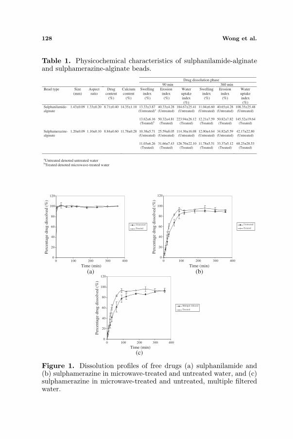
Sulphanilamide and sulphamerazine were hydrophilic and hydrophobicdrugs respectively with the former exhibited 13.6 folds of solubilityin an aqueous solution (sulphanilamide solubility in untreated water= 464.4 ± 12.5mg/100 ml; sulphamerazine solubility in untreatedwater = 34.2 ± 1.2mg/100 ml). Encapsulation of sulphanilamide andsulphamerazine in alginate beads via crosslinking process using calciumchloride solution brought about the formation of matrices with sizesof 1.43 ± 0.09 and 1.20 ± 0.09mm, aspect ratios of 1.33 ± 0.20 and1.10 ± 0.10, drug contents of 8.71 ± 0.40 and 8.84 ± 0.60%, as wellas, calcium content of 14.35 ± 1.10 and 11.78 ± 0.28% respectively(Table 1).
128 Wong et al.
Table 1. Physicochemical characteristics of sulphanilamide-alginateand sulphamerazine-alginate beads.
Drug dissolution phase
90 min 360 min
Bead type Size
(mm)
Aspect
ratio
Drug
content
(%)
Calcium
content
(%)
Swelling
index
(%)
Erosion
index
(%)
Water
uptake
index
(%)
Swelling
index
(%)
Erosion
index
(%)
Water
uptake
index
(%)Sulphanilamide-
alginate
1.43±0.09 1.33±0.20 8.71±0.40 14.35±1.10 13.33±3.87
(Untreated)a
13.62±6.16
(Treated)b
40.33±4.28
(Untreated)
50.32±4.81
(Treated)
184.67±25.41
(Untreated)
223.94±26.12
(Treated)
11.04±6.60
(Untreated)
12.21±7.59
(Treated)
40.65±4.28
(Untreated)
50.82±7.82
(Treated)
108.35±25.48
(Untreated)
145.52±19.64
(Treated)
Sulphamerazine-
alginate
1.20±0.09 1.10±0.10 8.84±0.60 11.78±0.28 10.38±5.71
(Untreated)
11.03±6.26
(Treated)
25.59±8.05
(Untreated)
31.66±7.43
(Treated)
114.30±16.08
(Untreated)
126.70±22.10
(Treated)
12.00±4.64
(Untreated)
11.78±5.51
(Treated)
34.82±5.59
(Untreated)
33.37±5.12
(Treated)
42.17±22.80
(Untreated)
68.23±28.53
(Treated)
aUntreated denoted untreated water.
bTreated denoted microwave-treated water.
0
20
40
60
80
100
120
0 100 200 300 400
Untreated
Treated
0
20
40
60
80
100
120
0 100 200 300 400
Untreated
Treated
0
20
40
60
80
100
120
0 100 200 300 400
Multiple filtered
Treated
Time (min)
Time (min) Time (min)
Per
cen
tag
e d
rug
dis
solv
ed (
%)
Per
cen
tag
e d
rug
dis
solv
ed (
%)
Per
cen
tag
e d
rug
dis
solv
ed (
%)
(a)
(c)
(b)
Figure 1. Dissolution profiles of free drugs (a) sulphanilamide and(b) sulphamerazine in microwave-treated and untreated water, and (c)sulphamerazine in microwave-treated and untreated, multiple filteredwater.
Progress In Electromagnetics Research C, Vol. 11, 2009 129
3.1. Drug Dissolution
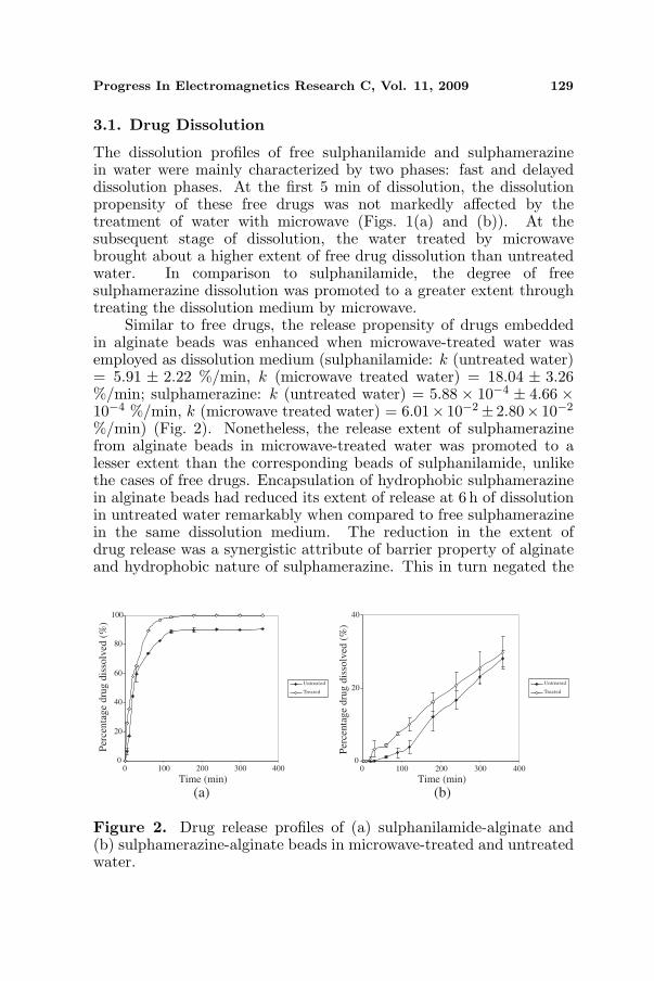
The dissolution profiles of free sulphanilamide and sulphamerazinein water were mainly characterized by two phases: fast and delayeddissolution phases. At the first 5 min of dissolution, the dissolutionpropensity of these free drugs was not markedly affected by thetreatment of water with microwave (Figs. 1(a) and (b)). At thesubsequent stage of dissolution, the water treated by microwavebrought about a higher extent of free drug dissolution than untreatedwater. In comparison to sulphanilamide, the degree of freesulphamerazine dissolution was promoted to a greater extent throughtreating the dissolution medium by microwave.
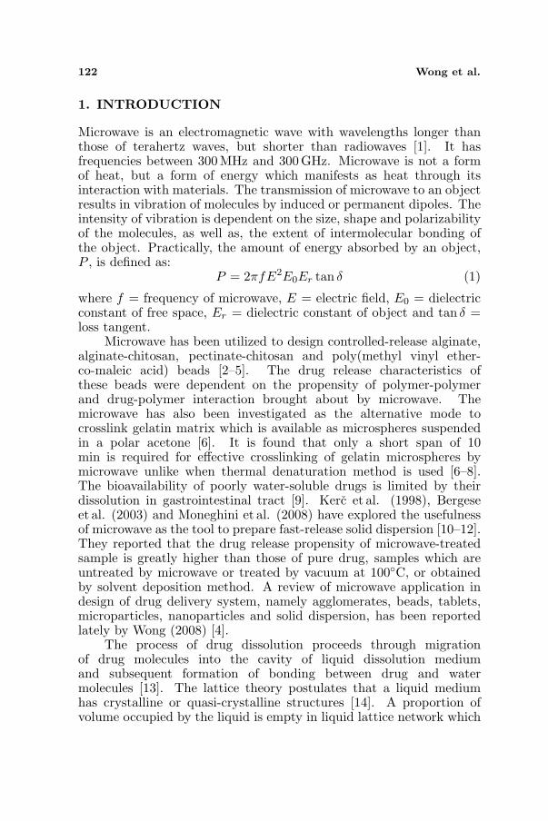
Similar to free drugs, the release propensity of drugs embeddedin alginate beads was enhanced when microwave-treated water wasemployed as dissolution medium (sulphanilamide: k (untreated water)= 5.91 ± 2.22 %/min, k (microwave treated water) = 18.04 ± 3.26%/min; sulphamerazine: k (untreated water) = 5.88 × 10⁻⁴ ± 4.66 ×10⁻⁴ %/min, k (microwave treated water) = 6.01× 10⁻²± 2.80× 10⁻²
%/min) (Fig. 2). Nonetheless, the release extent of sulphamerazinefrom alginate beads in microwave-treated water was promoted to alesser extent than the corresponding beads of sulphanilamide, unlikethe cases of free drugs. Encapsulation of hydrophobic sulphamerazinein alginate beads had reduced its extent of release at 6 h of dissolutionin untreated water remarkably when compared to free sulphamerazinein the same dissolution medium. The reduction in the extent ofdrug release was a synergistic attribute of barrier property of alginateand hydrophobic nature of sulphamerazine. This in turn negated the
0
20
40
60
80
100
0 100 200 300 400
Untreated
Treated
0
20
40
0 100 200 300 400
Untreated
Treated
Per
cen
tag
e d
rug
dis
solv
ed (
%)
Per
cen
tag
e d
rug
dis
solv
ed (
%)
Time (min)Time (min)
(a) (b)
Figure 2. Drug release profiles of (a) sulphanilamide-alginate and(b) sulphamerazine-alginate beads in microwave-treated and untreatedwater.

130 Wong et al.
sensitivity of encapsulated sulphamerazine to drug release modulationvia treating the dissolution medium by microwave. With referenceto drug release exponent values, the release of sulphanilamide andsulphamerazine from beads was governed largely by drug diffusionand polymer relaxation respectively (sulphanilamide: n = 0.42± 0.12;sulphamerazine: n = 1.50± 0.49).
3.2. Physicochemical Properties of Water
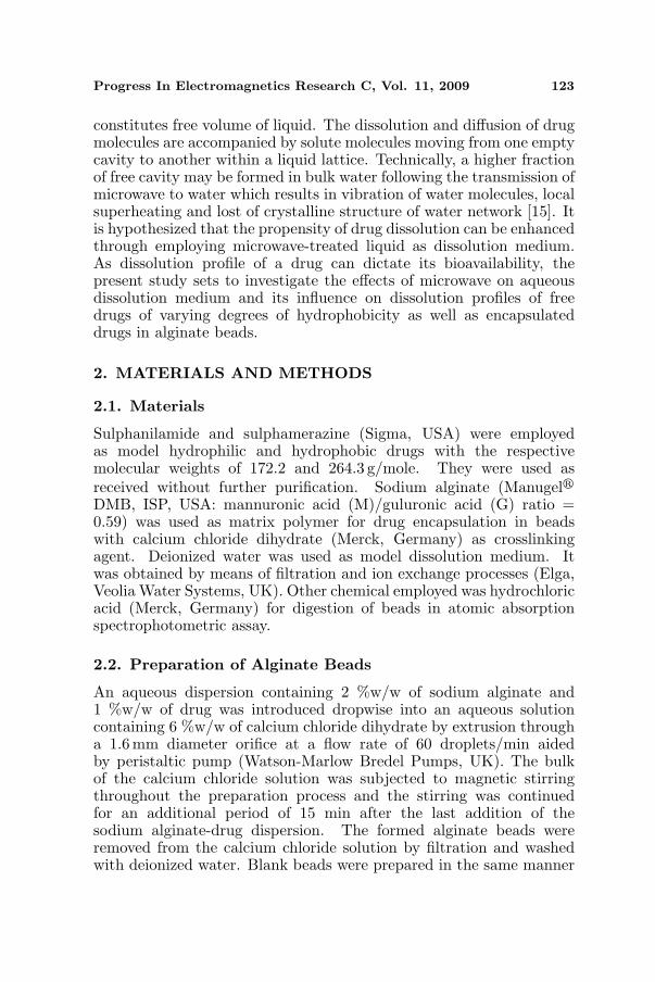
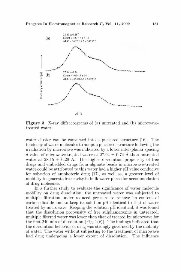
The treatment of water by microwave brought about changes in pH andmobility of water molecules (Table 2). These changes were possiblyinduced by the heating effect of microwave as the untreated water hada temperature of 22.0 ± 0◦C whereas the water treated by microwavehad a temperature of 85.2 ± 0.7◦C. The pH of untreated waterwas lower than microwave-treated water (Student’s-t-test, p < 0.05).This was likely due to the removal of carbon dioxide from waterby heat in the latter. The conductivity of microwave-treated waterwas higher than untreated water (Student’s-t-test, p < 0.05). Itwas envisaged that the mobility of water molecules was promotedby microwave through reducing their clustering propensity in bulkphase. This was further supported by the FTIR study of which alower transmission intensity ratio ascribing O-H peak at wavenumberof 3445.5 ± 2.7 cm−1 to 1635.0 ± 0.5 cm−1 was attained by watertreated with microwave (Student’s-t-test, p < 0.05). In addition, X-ray diffractometry analysis indicated that microwave-treated water hada lower degree of crystallinity than untreated water (Fig. 3). Thecrystallinity count of microwave-treated water was significantly lowerthan untreated water at 2θ = 27.94◦ (Student’s-t-test, p < 0.05).The area under curve of crystallinity count-2θ plot of microwave-treated water was similarly lower than untreated water (Student’s-t-test, p < 0.05). Upon heating of water by microwave, the water clusterwill adopt a more mobile and less ordered structure, and an expanded
Table 2. Physicochemical characteristics of microwave-treated anduntreated water.
Water type Temperature
(°C)
Conductivity
(uS/cm)a
pHa
FTIR transmission intensity
ratioa
FTIR transmission band
wavenumber
(cm-1
)
Untreated 22.0 ± 0.00 0.90 ± 0.00 4.81 ± 0.04 0.3163 ± 0.0717 3448.0 ± 5.2/1634.8 ± 0.8
Microwave-treated 85.19 ± 0.68 0.97 ± 0.06 4.91 ± 0.02 0.1963 ± 0.0407 3442.6 ± 6.3/1635.5 ± 0.4
aExperiments conducted at 25 ± 1°C.
Progress In Electromagnetics Research C, Vol. 11, 2009 131
2 ( )
Inte
nsi
ty c
ou
nt
(cp
s)(a)
(b)
28.15 ± 0.28
Count = 4297.7 ± 81.3
AUC = 3632018.3 ± 38755.3
27.94 ± 0.74
Count = 4094.5 ± 64.1
AUC = 3364465.5 ± 56493.5
°
°
°θ
Figure 3. X-ray diffractograms of (a) untreated and (b) microwave-treated water.
water cluster can be converted into a puckered structure [16]. Thetendency of water molecules to adopt a puckered structure following theirradiation by microwave was indicated by a lower inter-planar spacingd value of microwave-treated water at 27.94 ± 0.74 A than untreatedwater at 28.15 ± 0.28 A. The higher dissolution propensity of freedrugs and embedded drugs from alginate beads in microwave-treatedwater could be attributed to this water had a higher pH value conducivefor solvation of amphoteric drug [17], as well as, a greater level ofmobility to generate free cavity in bulk water phase for accommodationof drug molecules.
In a further study to evaluate the significance of water moleculemobility on drug dissolution, the untreated water was subjected tomultiple filtration under reduced pressure to remove its content ofcarbon dioxide and to keep its solution pH identical to that of watertreated by microwave. Keeping the solution pH identical, it was foundthat the dissolution propensity of free sulphamerazine in untreated,multiple filtered water was lower than that of treated by microwave forthe first 240 min of dissolution (Fig. 1(c)). The findings indicated thatthe dissolution behavior of drug was strongly governed by the mobilityof water. The water without subjecting to the treatment of microwavehad drug undergoing a lower extent of dissolution. The influence

132 Wong et al.
of microwave irradiation on drug dissolution was less dependent onchanges in the pH of liquid medium than mobility of water molecules.The similar mode of mechanism of microwave was identified whenmicrowave-treated water was used to examine the activity of Ca2+-dependent K+ channels of cultured kidney cells [18]. The microwave-treated water was deemed to exhibit “solution memory” [18]. It couldprobably explain that the dissolution propensity of drug was higher inmicrowave-treated water than untreated water even if this water wasre-equilibrated from 85.2 ± 0.7◦C to 37 ± 0.2◦C prior test and thedrug dissolution test was conducted over a period of several hours.
3.3. Bead Swelling, Erosion and Water Uptake
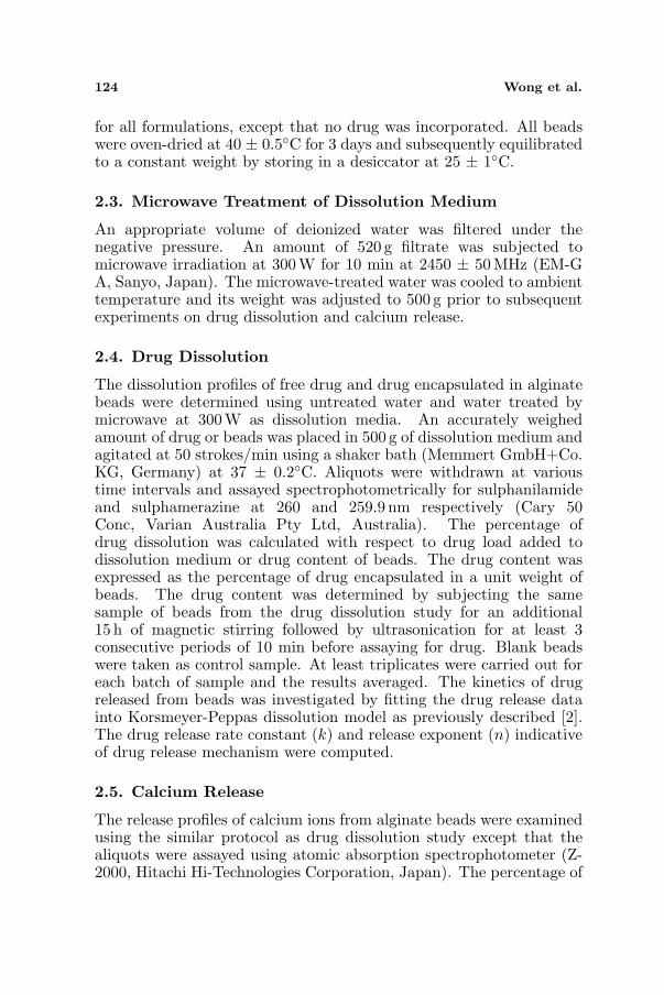
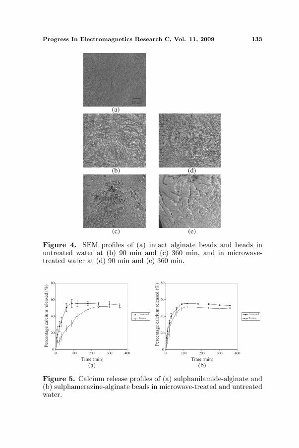
With reference to sulphanilamide-alginate and sulphamerazine-alginate beads, the drug dissolution process was accompanied byminimal differences in swelling profile but an increase in water uptakeand erosion extent of matrices when microwave-treated water wasemployed as dissolution medium (Table 1). The microwave-treatedwater molecules were more mobile and exhibited higher penetrationintensity from exterior to core of beads. The containment of a higherfraction of water in beads was deemed to generate a higher level ofhydrostatic pressure thereby promoting erosion and drug dissolutionof matrix. The erosion characteristics of beads differed with the useof microwave-treated water. Surface morphology analysis of intactand eroded beads indicated that matrix undergoing drug dissolutionin microwave-treated water tended to form deep pores, whereas beadsin untreated water had inter-connecting polymeric fibrils filled inthe forming pores (Fig. 4). The higher erosion extent of beads inmicrowave-treated water was associated with deep pore formation.This reduced the barrier for drug dissolution and promoted a higherpropensity of drug release from beads.
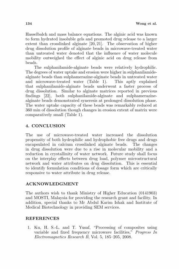
The increase in water uptake, erosion and drug dissolution ofbeads in microwave-treated water was not ascribed to loss of calciumions as the crosslinker of matrix. Fig. 5 shows that calcium ions ofbeads were lost to a greater degree in untreated water than microwave-treated water. The loss of calcium ions from beads could havepromoted by H+/Ca2+ exchange to a greater extent in the more acidicuntreated water than the more mobile microwave-treated water. ThepKa values of β-D-mannuronic acid and α-L-guluronic acid of alginatewere 3.38 and 3.65 respectively [19]. Using guluronic acid-rich alginatewith M/G ratio of 0.59 as matrix polymer, the formed beads wereexpected to have an excess of 5.64 percent alginic acid in the moreacidic untreated water (pH = 4.81 ± 0.04) than microwave-treatedwater (pH = 4.91 ± 0.02) from an estimation made by Henderson-
Progress In Electromagnetics Research C, Vol. 11, 2009 133
10 µm
(a)
(e)(c)
(d)(b)
Figure 4. SEM profiles of (a) intact alginate beads and beads inuntreated water at (b) 90 min and (c) 360 min, and in microwave-treated water at (d) 90 min and (e) 360 min.
0
20
40
60
80
0 100 200 300 400
Untreated
Treated
0
20
40
60
80
0 100 200 300 400
Untreated
Treated
Per
cen
tag
e ca
lciu
m r
elea
sed
(%
)
Time (min)Time (min)
(a) (b)
Per
centa
ge
calc
ium
rel
ease
d (
%)
Figure 5. Calcium release profiles of (a) sulphanilamide-alginate and(b) sulphamerazine-alginate beads in microwave-treated and untreatedwater.
134 Wong et al.
Hasselbalch and mass balance equations. The alginic acid was knownto form hydrated insoluble gels and promoted drug release to a largerextent than crosslinked alginate [20, 21]. The observation of higherdrug dissolution profile of alginate beads in microwave-treated waterthan untreated water denoted that the influence of water moleculemobility outweighed the effect of alginic acid on drug release frombeads.
The sulphanilamide-alginate beads were relatively hydrophilic.The degrees of water uptake and erosion were higher in sulphanilamide-alginate beads than sulphamerazine-alginate beads in untreated waterand microwave-treated water (Table 1). This aptly explainedthat sulphanilamide-alginate beads underwent a faster process ofdrug dissolution. Similar to alginate matrices reported in previousfindings [22], both sulphanilamide-alginate and sulphamerazine-alginate beads demonstrated syneresis at prolonged dissolution phase.The water uptake capacity of these beads was remarkably reduced at360 min of dissolution though changes in erosion extent of matrix werecomparatively small (Table 1).
4. CONCLUSION
The use of microwave-treated water increased the dissolutionpropensity of both hydrophilic and hydrophobic free drugs and drugsencapsulated in calcium crosslinked alginate beads. The changesin drug dissolution were due to a rise in molecular mobility and areduction in crystallinity of water network. Future study shall focuson the interplay effects between drug load, polymer microstructuralnetwork and water attributes on drug dissolution. This is essentialto identify formulation conditions of dosage form which are criticallyresponsive to water attribute in drug release.
ACKNOWLEDGMENT
The authors wish to thank Ministry of Higher Education (0141903)and MOSTI, Malaysia for providing the research grant and facility. Inaddition, special thanks to Mr Abdul Karim Ishak and Institute ofMedical Biotechnology in providing SEM services.
REFERENCES
1. Ku, H. S.-L. and T. Yusaf, “Processing of composites usingvariable and fixed frequency microwave facilities,” Progress InElectromagnetics Research B, Vol. 5, 185–205, 2008.
Progress In Electromagnetics Research C, Vol. 11, 2009 135
2. Wong, T.-W., A.-W. Selasiah, and Y. Anthony, “Effects ofmicrowave on drug release property of poly (methyl vinyl ether-co-maleic acid) matrix,” Drug Dev. Ind. Pharm., Vol. 33, 737–746,2007.
3. Wong, T.-W., A.-W. Selasiah, and Y. Anthony, “Drug releaseresponses of zinc ion crosslinked poly(methyl vinyl ether-co-maleicacid) matrix towards microwave,” Int. J. Pharm., Vol. 357, 154–163, 2008.
4. Wong, T.-W., “Use of microwave in processing of drug deliverysystems,” Curr. Drug Deliv., Vol. 5, No. 2, 77–84, 2008.
5. Wong, T.-W. and N. Sumiran, “Drug release property of chitosan-pectinate beads and its changes under the influence of microwave,”Eur. J. Pharm. Biopharm., Vol. 69, 176–188, 2008.
6. Vandelli, M.-A., M. Romagnoli, A. Monti, M. Gozzi, P. Guerra,F. Rivasi, and F. Forni, “Microwave-treated gelatin microspheresas drug delivery system,” J. Controlled Release, Vol. 96, 67–84,2004.
7. Yannas, I.-V. and A.-V. Tobolsky, “Crosslinking of gelatine bydehydration,” Nature, Vol. 215, 509–510, 1967.
8. Welz, M.-M. and C.-M. Ofiner III, “Examination of self-crosslinked gelatin as a hydrogel for controlled-release,” J. Pharm.Sci., Vol. 81, 85–90, 1992.
9. Lee, C.-C., C.-L.-C. Ong, P.-W.-S. Heng, L.-W. Chan, and T.-W. Wong, “Interactive mixture as rapid drug delivery system,”Drug Dev. Ind. Pharm., Vol. 34, 206–214, 2008.
10. Kerc, J., S. Srcic, and B. Kofler, “Alternative solvent-freepreparation methods for felodipine surface solid dispersions,”Drug Dev. Ind. Pharm., Vol. 24, No. 4, 359–363, 1998.
11. Bergese, P., I. Colombo, D. Gervasoni, and L.-E. Depero,“Microwave generated nanocomposites for making insoluble drugssoluble,” Mater. Sci. Eng. C, Vol. 23, 791–795, 2003.
12. Moneghini, M., B. Bellich, P. Baxa, and F. Princivalle,“Microwave generated solid dispersions containing ibuprofen,” Int.J. Pharm., Vol. 361, 125–130, 2008.
13. Aulton, M., “Dissolution and solubility,” Pharmaceutics: TheScience of Dosage Form Design, 2nd edition, M. E. Aulton (ed.),15–32, Churchill Livingstone, London, 2002.
14. Aulton, M., “Properties of solutions,” Pharmaceutics: TheScience of Dosage Form Design, 2nd edition, M. E. Aulton (ed.),33–40, Churchill Livingstone, London, 2002.
15. Pan, X., H. Liu, Z. An, J. Wang, and G. Niu, “Microwave

136 Wong et al.
enhanced dehydration and solvent washing purification ofpenicillin G sulfoxide,” Int. J. Pharm., Vol. 220, 33–41, 2001.
16. Chaplin, M.-F., “A proposal for the structuring of water,”Biophys. Chem., Vol. 83, 211–221, 1999.
17. Wong, T.-W., H.-Y. Lee, L.-W. Chan, and P.-W.-S. Heng,“Release characteristics of pectinate microspheres prepared by anemulsification technique,” J. Microencapsulation, Vol. 19, No. 4,511–522, 2002.
18. Fesenko, E.-E., V.-I. Geletyuk, V.-N. Kazachenko, and N.-K. Chemeris, “Preliminary microwave irradiation of watersolutions changes their channel-modifying activity,” FEBSLetters, Vol. 366, 49–52, 1995.
19. Haug, A., “Composition and properties of alginates,” Thesis,Norweigian Institute of Technology, Trondheim, 1964.
20. Tu, J., S. Bolla, J. Barr, J. Miedema, X. Li, and B. Jasti,“Alginate microparticles prepared by spray-coagulation method:Preparation, drug loading and release characterization,” Int. J.Pharm., Vol. 303, 171–181, 2005.
21. Pongjanyakul, T. and S. Puttipipatkhachorn, “Modulating drugrelease and matrix erosion of alginate matrix capsules bymicroenvironmental interaction with calcium ion,” Eur. J. Pharm.Biopharm., Vol. 67, 187–195, 2007.
22. Velings, N.-M. and M.-M. Mestdagh, “Physico-chemical proper-ties of alginate gel beads,” Polym. Gels Networks, Vol. 3, 311–330,1995.