Embed Size (px)
Citation preview
Effects of Vasoconstriction on Dermal Pharmacokinetics and Local Tissue Distribution of Compounds
PARMINDER SINGH' AND MICHAEL s. ROBERTSSX
Received March 4, 1993, from the 'Department of Pharmacy, The University of Queensland, Queensland, Australia 4072, and the $Department of Medicine, The University of Queensland, Princess Alexandra Hospital, Brisbane, Queensland, Australia 4 102. Accepted for publication August 19, 1993@.
Abstract 0 The effects of the local vasoconstrictor phenylephrine on thedermal absorption kinetics and local tissue distribution of compounds were investigated in rats. Phenylephrine (0.0025 %-0. 1 %)and tracer quantities of salicylic acid, lidocaine, and water were applied in an aqueous solution to the exposed rat dermis. The disappearance of salicylic acid from the solution into the rat dermis and its appearance in blood, local underlying tissues, and similar tissues on the contralateral side was quantified. The clearance of salicylic acid into the dermis decreased and the concentrations of salicylic acid in underlying tissues increased with an increase in phenylephrine concentration (up to 0.0 1 % ). The concentrations of salicylicacid in plasma and contralateral tissues decreased with increasing phenylephrine concentrations. At higher phenylephrine concentrations, no significant increase in local tissue concentrations of salicylic acid was observed. The effects of phenylephrine on local tissue levels of lidocaine and tritiated water paralleled those found for salicylic acid. The concentration-depth profiles for solutes in underlying tissues with variable blood flows were described by a compartment-in-series pharmacokinetic model in which each tissue's blood flows to and from a central compartment were incorporated. The values predicted under varying degrees of vaso- constriction were found to compare well with the experimentally determined concentrations of salicylic acid, lidocaine, and water in tissues below a dermal application site, in the presence of phenylephrine. Phenylephrine can significantly increase quantities of solutes delivered to local tissues after dermal application, the observed effects being due to the vasoconstrictive properties of phenylephrine. Blood flow changes in skin can have profound effects on dermal pharmacokinetics and relative processes of local and systemic solute distribution.
Dermal blood supply plays an important role in the clearance of topically applied compounds.',2 Certain lipophilic solutes such as lidocaine can bypass the dermal microcirculation to accu- mulate in underlying tissues.3~~ Direct penetration for a number of other compounds following topical application has been reviewed.5 Ionizable solutes such as salicylic acid penetrate into the deeper underlying tissues only to a limited depth and are predominantly removed by the dermal blood supply.3
Epinephrine and phenylephrine are often used to decrease systemic absorption of local anesthetics from subcutaneous and other injection sites6v7 by decreasing the local blood flow. The peak systemic concentrations of timolol after ocular adminis- tration in rabbits have also been shown to decrease significantly in the presence of phenylephrine and epinephrine.*s9 The actions of vasoconstrictors are physiologically mediated by the adrenergic receptors of the sympathetic nervous system.6 The adrenergic receptors consist of the a-receptors, which mediate peripheral vasoconstriction; the (3-1 receptors, which modulate increases in cardiac rate and contractility; and the p-2 receptors, causing peripheral vasodilation in the vascular beds of skeletal muscle.6 Phenylephrine, a synthetic vasoconstrictive agent, differs from epinephrine in the absence of a hydroxyl group at position 4 of the aromatic nucleus. As a consequence, phenylephrine is essentially a pure a-adrenergic agonist devoid of the systemic side effects normally associated with p-agonists. It has no direct
@Abstract published in Advance ACS Abstracts, January 15, 1994.
Elimination ke'
Clfpwdm
muscle . - ,i Cdm. Vdm
Flgure 1-A @ [ pharmacokinetic model for local tissue penetration of dermally
applied solutes. Refer to the text for the meaning of various terms.
cardiac effects and cardiac irregularities are rare even when high doses are given intravenously.6 Subcutaneous epinephrine produces sinus tachycardia in the rat, while phenylephrine has no such effect."J In addition, central nervous system stimulation is minimal with phenylephrine as compared to epinephrine.6 Phenylephrine is considered to be a preferred local vasocon- strictive agent under conditions of severe tachycardia from untreated hyperthyroidism, for patients receiving inhalation agents which sensitize the heart to epinephrine and for peridural analgesia in labor and delivery by cesarian section."
Lidocaine and salicylic acid are commonly used in topical formulations as local anesthetic and analgesic, respectively. Although their local effects have previously been demonstrated,
0 1994. American Chemical Society and American Pharmaceutical Association
0022-3549/94/ 1200- 783$04.50/0 Journal of Pharmaceutical Sciences / 783 Vol. 83, No. 6, June 1994
it would be clinically important to suggest ways to further localize these compounds to tissues below the application site and at the same time minimize their entry into the systemic circulation. Better therapy will thus be possible with reduced systemic absorption and consequently fewer side effects of compounds administered topically mainly for their local action. The object of the present study was to quantitate the effects of phenyl- ephrine in terms of dermal absorption kinetics and local tissue distribution of salicylic acid, lidocaine, and tritiated water after dermal application. A model previously introduced to describe dermal and underlying tissue pharmacokinetics of solutes12-14 was tested to predict local tissue levels of compounds under varying degree of vasoconstriction. The predicted values were than compared with experimentally obtained concentrations of salicylic acid, lidocaine, and tritiated water in tissues below a dermal site of application, in the presence of phenylephrine.
Theoretical Section
We have previously suggested a physiologically based phar- macokinetic model to describe dermal and underlying tissue pharmacokinetics of compounds12-14 after topical application. Briefly, each tissue can be described as compartments-in-series, each joined in parallel to a central plasma compartment (Figure 1). After dermal application solute diffuses in the ith tissue compartment from the tissue compartment overlying it (i + 1) and into the tissue compartment underlying it (i - 1). The distribution into the plasma compartment is defined by the blood supply to individual tissues. The differential mass balance equations can then be written for each compartment to describe inflow, outflow, and disappearance of the drug. The rate of change of concentration of solute in the dermal compartment is given by eq 112-14
where Co = the concentration in donor solution in the dermal absorption cell at zero time, v d = the apparent volume of distribution of total solute in the dermis (=VudUd), Vud = the apparent volume of distribution of unbound solute in the dermis, C1B-d = the clearance between the cell solution and the dermis (mL/h), k,+ = the rate constant of transfer between the cell solution and the dermis (h-l), Cld-,, = the clearance between the dermis and the subcutaneous tissue (mL/h), Qd = the blood flow to the dermis (mL/h/g), Cb = the concentration of solute in the blood (fraction of the initial concentration), Cd = the concentration of solute in the dermis (fraction of the initial concentration), C,, = the concentration of solute in the subcu- taneous tissue (fraction of the initial concentration), fud = the fraction unbound in the dermis, fub = the fraction unbound in the blood, fu,, = the fraction unbound in the subcutaneous tissue. Equation 1 reduces to eq 2 when fud = fuse, fublfud = RMd, where
RMd is the dermis-plasma partition coefficient and noting that the concentration in the dermis is expressed per gram of tissue and is estimated by dividing the observed concentration cd by the weight of the tissue assuming a tissue density of unity.13J4 The rate of change of concentration in all deeper underlying tissue compartments with time for the model in Figure 1 can be described by eq 312-14
0.6 6
- 0
0.0 0.00 0 . 0 2 0 . 0 4 0 . 0 6 0 . 0 8 0.10
Phenylephrine Concentration (%)
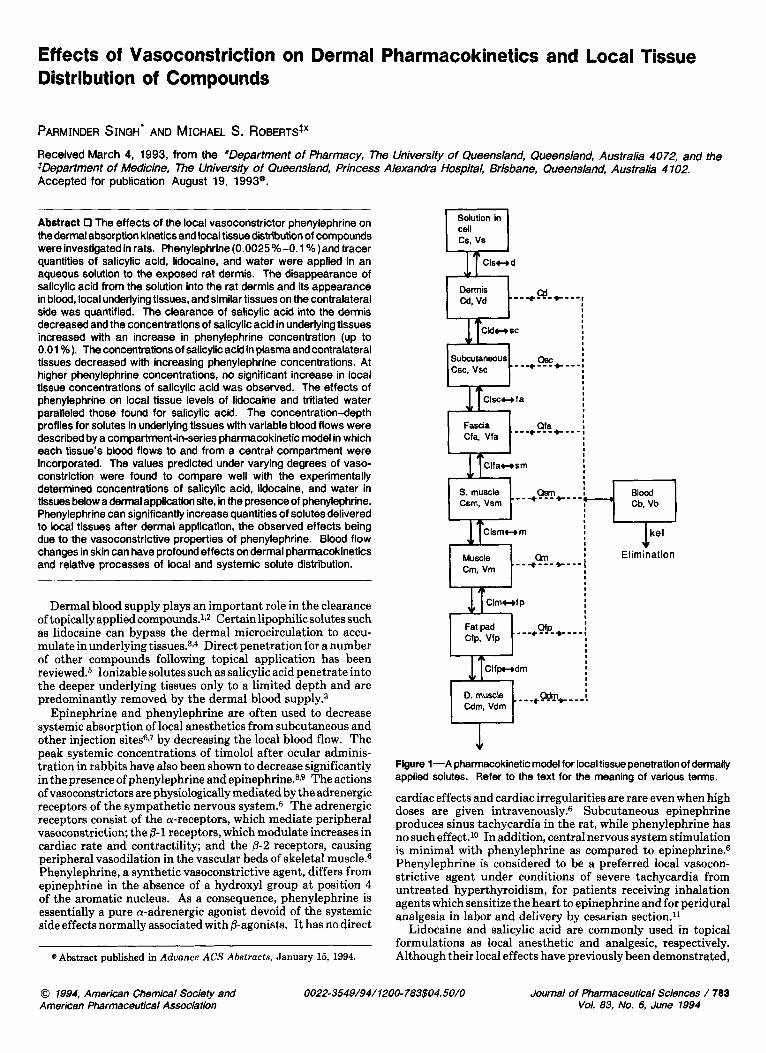
Figure P-Effect of phenylephrine concentration on salicylic acid clearance after application to rat dermis.
Table 1-Effect of Phenylephrlne on Dermal Clearance of TrHlated Water and Lldocalne (Mean f SD)
Treatment Water (mL/h) Lidocaine (mL/h)
Control 1.10 f 0.24 0.51 f 0.15 Phenylephrine 0.53 f 0.05 0.37 f 0.01 1:20 000 1:5000 0.04 f 0.02 0.34 f 0.02
Sacrificed 0.36 f 0.02 0.33 f 0.06
where ClT,i+l-i = the clearance between the i + 1 tissue and the ith tissue (mL/h), c1T,i-i-1= the clearance between the ith tissue and the i - 1 tissue (mL/h), CT,~ = the concentration in the ith tissue (fraction of the initial concentration), cT,i+l = the concentration in the i + 1 tissue (fraction of the initial concentration), CT,i-1 = the concentration in the i - 1 tissue (fraction of the initial concentration), QT,~ = the blood flow to the ith tissue (mL/h/g), RMT,~ = the ith tissue-plasma partition coefficient and fuT is the unbound fraction in the tissues.13
In a previous report it was also shown that dermal clearance of tritiated water can serve as an index for measuring skin blood flow.12 On the basis of this premise, the blood flow to the dermis was estimated by monitoring the clearance of tritiated water in the presence of phenylephrine. Simulations in the above model were performed by assuming normal blood flow, blood flow corresponding to two phenylephrine concentrations (1:20 000 and 1:5000), and no blood flow in tissues. The concentrations in the underlying tissues after dermal application of either tritiated water, lidocaine, or salicylic acid were then predicted and compared with those obtained experimentally without phenylephrine, with phenylephrine (1:20 000 and 1:5000), and in the sacrificed animal.
Experimental Section
Chemicals and Instruments-[14ClSalicylic acid (specific activity 56 mCi/mmol, purity >98.0%) was a gift from Hamilton Labs Pty Ltd, [l4C1lidocaine hydrochloride (specific activity 48 mCi/mmol, purity > 97.0%) and tritiated water (1 mCi/g) were purchased from New England Nuclear. Salicylic acid and phenylephrine hydrochloride were from Sigma Chemical Co., and lidocaine hydrochloride was a gift from Astra Pharmaceuticals Pty. Ltd. Zimmer's electrodermatome (Model 901) was used from removing rat epidermis. Tissue solubilizer (NCS) and liquid scintillation cocktails (OCS, organic counting scintillant, andBCS, biodegradable counting scintillant) were used for tissue and aqueous samples (respectively) were purchased from Amersham International. All other reagents used were of analytical grade. A liquid scintillation
704 / Journal of Pharmaceutical Sciences Vol. 83, No. 6, June 1994
A-Dermls T
0.0 0 . 0 0 0.02 0.04 0 .06 0 .08 0 .10
PE conc. (%)
C-Fascia 0.06
0.05 C 0 - ; 0.04
0.03 u C
$ 0.02
0-01- 0.00 0 . 0 0 0 .02 0.04 0 . 0 6 0 .08 0 .10
PE conc. (%)
c 0 .- L
L. - m
c 0)
I E-Muscle
0.Ooo 1 I
0 . 0 0 0 . 0 2 0.04 0 . 0 6 0 .08 0 . 1 0
PE conc. (%)
0.10 r B-Subcutaneous
0.00 omma 0.00 0 . 0 2 0.04 0 . 0 6 0 . 0 8 0.10
PE conc. (%)
Om5 1 D-Supetflclal muscle
e 0 - L.
t C a u C 0 0
L
0.OW 0 . 0 0 0 .02 0 .04 0 . 0 6 0 .08 0 .10
PE conc. (%)
F-Fat pad OernOf
1 0.001 5
rn 0.#)10
0.ooo5
0.0000 0 . 0 0 0 . 0 2 0.04 0 . 0 6 0 .08 0 .10
PE conc. (%)
0.010 H-Plasma 0.003
G-Deep muscle 0.008
0 C -
.1 0 L 0.006-
ID - L. c a u C 0) - c 0.004
0 0
- - c) 0.002
e
g 0.001 0 0 - 0 . m
I I I 1 0 . m - 0.OOO I I I
0 . 0 0 0 . 0 2 0 .04 0 .06 0 .08 0 .10 0 . 0 0 0 . 0 2 0.04 0 .06 0 .08 0 . 1 0 PE conc. (%) PE conc. (%)
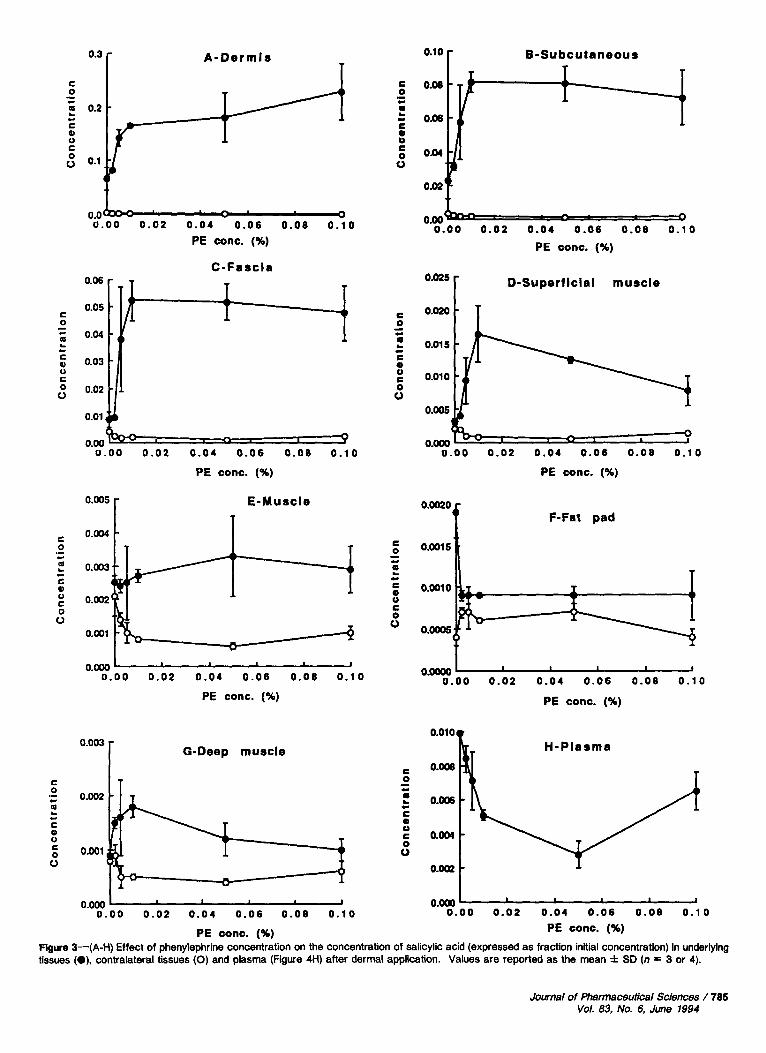
FIgure 3-(A-H) Effect of phenylephrine concentration on the concentration of salicylic acid (expressed as fraction initial concentration) in underlying tissues (.), contralateral tissues (0) and plasma (Figure 4H) after dermal application. Values are reported as the mean f SD (n = 3 or 4).
Journal of Pharmaceutical Sciences / 785 Val. 83, No. 6, June 1994
counter (Tri-carb 4000 series, United Technologies Packard) was used to determine the radioactivity in the samples.
Animals-Male Wistar rats (30+350 g) were used in the studies. The animals were housed under standard laboratory conditions at 20.0
0.5 "C and 5575% relative humidity and supplied with normal pellet diet and water adlibitium. All experiments had previously been approved by the Animal Experimentation Committees of the University of Queensland and the Princess Alexandra Hospital. In Vivo Dermal P e n e t r a t i o n a n d Local T i s s u e U p t a k e
Studies-The rats were lightly anesthetized by pentobarbitone (35 mg/ kg), and their body temperature was maintained at 37 "C by placing them on a heating pad. The hair from a4 cm2 dorsum area was removed by electric clippers and the epidermis removed by means of an electrodermatome set a t a thickness of 80 ~ m . 3 A dermal absorption cell was adhered to the exposed rat dermis and warmed to 37 O C by means of an external heating device.3J5
A solution of solute (3 mL) spiked with corresponding labeled substance and containing an appropriate concentration of phenylephrine, previously warmed to 37 "C, was introduced into the dermal absorption cell and the solution stirred by a glass stirrer driven by an external motor. Samples (10 pL) were removed at time intervals of 5,10,15,30, 45,60,75,90, and 120 min and placed in preweighed scintillation vials. The total volume sampled over a 2-h period was less than 1% of the original volume and considered negligible. The glass cell was removed from the rat skin at the end of 2-h dermal perfusion study and the application area wiped dry with blotting paper. A blood sample was then taken from the tail vein, and the animals were sacrificed with an overdose of anesthetic ether. Immediately thereafter the tissues below the treated site i.e. skin, subcutaneous tissue, fascia, muscle lining .or superficial muscle, muscle, fat pad, and deep muscle were removed by dissection and placed in preweighed scintillation vials.3 Similarly, the tissues below the contralateral side were also removed. Tissue and plasma samples were stored at -20 OC prior to analysis.
In Vivo Dermal Penetration and Local Tissue Uptake Studies (Sacrificed Animals)-The rats were initially anesthetized by intra- peritoneal injection of pentobarbitone (35 mg/kg) and, after removal of the epidermis as described above, were sacrificed by an overdose of ether. Dermal perfusion and tissue uptake studies were then conducted in postmortem rats.
Blood Flow Measurements-The blood flows to different tissues estimated in our earlier study were used.12 The dermal clearance of tritiated water has been shown to closely reflect the blood flow to the dermis.12 The phenylephrine-induced reduction in blood flow to the dermis was determined by monitoring the dermal clearance of tritiated water in presence of phenylephrine.
Sample Treatment-Aqueous samples removed from the glass cells in in vivo dermal perfusion studies were directly mixed with 5 mL of BCS and counted on liquid scintillation counter. The tissue samples were solubilized with 50 fiL of water and 1 mL of NCS at 50 "C for 6-8 h. After cooling of the digested samples to room temperature, 0.03% glacial acetic acid was added to each tissue sample followed by 10 mL of OCS. The plasma samples were solubilized with tissue solubilizer (5 parts for 1 part of plasma) at room temperature and treated with glacial acetic acid before adding OCS. Each sample was then counted on the liquid scintillation counter for 10 min3
Analysis of Data-Zero-time samples from the cell were used to represent the initial solution concentration, and the 14C activity in the tissues and plasma was converted to the fraction of the initial solution concentration (concentration fraction). Clearance (Cl) of the solute into the dermis was estimated from the plot of the percent solute remaining in the dermal cell vs time using eq 3
C1= kV (3)
where k is the disappearance rate constant and V is the volume (mL) of the solution applied to the d e r m i ~ . ~ J ~
The MINIM computer program16 was used to numerically integrate eqs 2 and 3 to estimate the concentrations in the dermis, subcutaneous tissue, fascia, and superficial muscle, assuming experimentally obtained blood flows, unbound fractions in the blood/tissues, and apparent tissue- tissue clearance^'^-^^ as constants. In order to estimate the muscle concentrations, eq 5 was numerically integrated for subcutaneous tissue, fascia, superficial muscle and muscle using a sixth order polynomial input function representation of the predicted solute concentrations in the dermis-time profile. Simulations in the model were performed by varying the blood-flow term in eqs 2 and 3 and assumingapparent tissue- tissue clearances as constants. The predicted estimates for solute
I r
1 ' C 0
U
LL
.- L
E .01 '
.001 - 0
~
2 4 6 8 1 0 1 2 Tissue depth
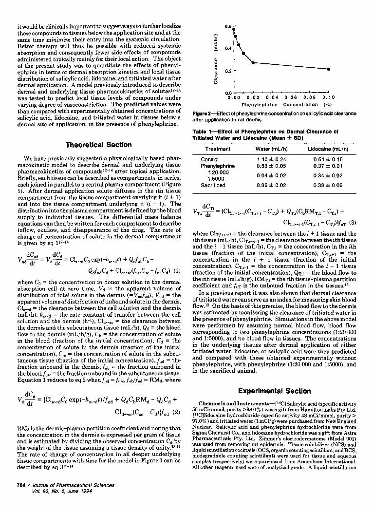
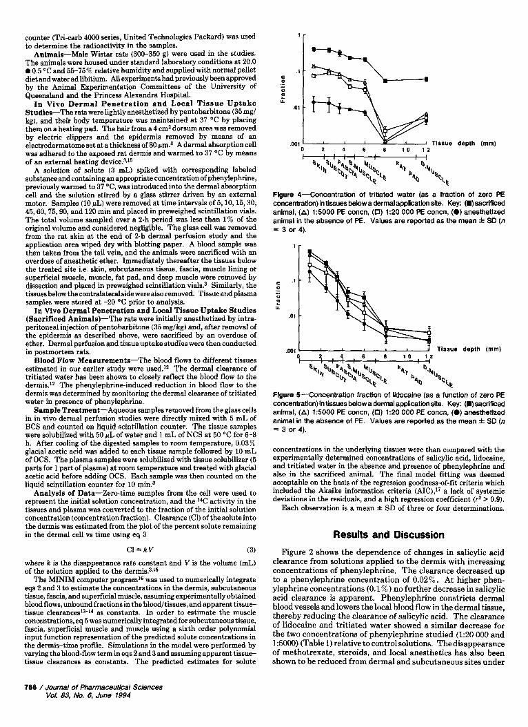
Figure 4-Concentration of tritiated water (as a fraction of zero PE concentration) in tissues below a dermal application site. Key: (W) sacrificed animal, (A) 15000 PE concn, (0) 1:20 000 PE concn, (0) anesthetized animal in the absence of PE. Values are reported as the mean f SD (n = 3 or 4).
.001 I Tissue depth (mm)
Flgure 5-Concentration fraction of lidocaine (as a function of zero PE concentration) in tissues below a dermal application site. Key: (.) sacrificed animal, (A) 1:5000 PE concn, (0) 1:20 000 PE concn. (0) anesthetized animal in the absence of PE. Values are reported as the mean f SD (n = 3 or 4).
concentrations in the underlying tissues were than compared with the experimentally determined concentrations of salicylic acid, lidocaine, and tritiated water in the absence and presence of phenylephrine and also in the sacrificed animal. The final model fitting was deemed acceptable on the basis of the regression goodness-of-fit criteria which included the Akaike information criteria (AIC),17 a lack of systemic deviations in the residuals, and a high regression coefficient (r2 > 0.9).
Each observation is a mean f SD of three or four determinations.
Results and Discussion Figure 2 shows the dependence of changes in salicylic acid
clearance from solutions applied to the dermis with increasing concentrations of phenylephrine. The clearance decreased up to a phenylephrine concentration of 0.02 % . At higher phen- ylephrine concentrations (0.1 % ) no further decrease in salicylic acid clearance is apparent. Phenylephrine constricts dermal blood vessels and lowers the local blood flow in the dermal tissue, thereby reducing the clearance of salicylic acid. The clearance of lidocaine and tritiated water showed a similar decrease for the two concentrations of phenylephrine studied (1:20 000 and 1:5000) (Table 1) relative to control solutions. The disappearance of methotrexate, steroids, and local anesthetics has also been shown to be reduced from dermal and subcutaneous sites under
706 / Journal of Pharmaceutical Sciences Vot. 83, No. 6, June 1994
150
Dermis
& 0 .- L
m 100 U
50
0 0 .0025 0 . 0 0 5 0 .01
8o
T
0 .02 0 . 1
140
105
0 - c
70
35
0
Subcutaneous 1 0 0 . 0 0 2 5 0 .005 0 . 0 1
PE conc. (%)
Fascia -
20
' 0 0 .0025 0 . 0 0 5 0.01
PE conc. ('A)
r Superficial muscle
T
h I2 0 . 1
20 0
6
- c
a
10
0 0 .0025 0.005 0.01 0 . 0 2 0 .1 0.02 0 . 1
PE conc. (%) PE conc. (%)
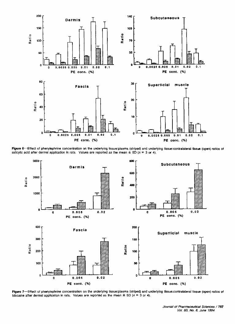
Figure 6-Effect of phenylephrine concentration on the underlying tissue:plasma (striped) and underlying tissue:contralateral tissue (open) ratios of salicylic acid after dermal application in rats. Values are reported as the mean f SD (n = 3 or 4).
0
m .- c
a
T Dermis 3000 i 2oo:L 1000
U 0 0 . 0 0 5 0 . 0 2
PE conc. (%)
Fascia
T
0
6 R
- ).
"[ Subcutaneous T 600
400
200
n 0 0 . 0 0 5 0 . 0 2
PE conc. (Oh)
150
Superf icial muscle
T 0 0 - .-
c L
g 200 a 100 a
100 50
n n 0 0 . 0 0 5 0 . 0 2
PE conc. (%)
" 0 0 . 0 0 5 0 . 0 2
PE conc. (%)
Figure 7-Effect of phenylephrine concentration on the underlying tissue:plasma (striped) and underlying tissue:contralateral tissue (open) ratios of lidocaine after dermal application in rats. Values are reported as the mean i SD (n = 3 or 4).
Journal of Pharmaceutical Sciences / 787 Vol. 83, No. 6, June 1994
Subcutaneous
40 Dermls
0
B - * a
30
20
10
n
0 .- c
20
n " 0 0 . 0 0 5
PE conc. (%) 0 . 0 2 "
0 0 .005 0 . 0 2
PE conc. (%)
3o 1 Superficial muscle Fascla
20 L I 0 -
c
a 10
n I
0 0 . 0 0 5 0 . 0 2
PE conc. (%)
" 0 0 . 0 0 5 0 . 0 2
PE conc. (%)
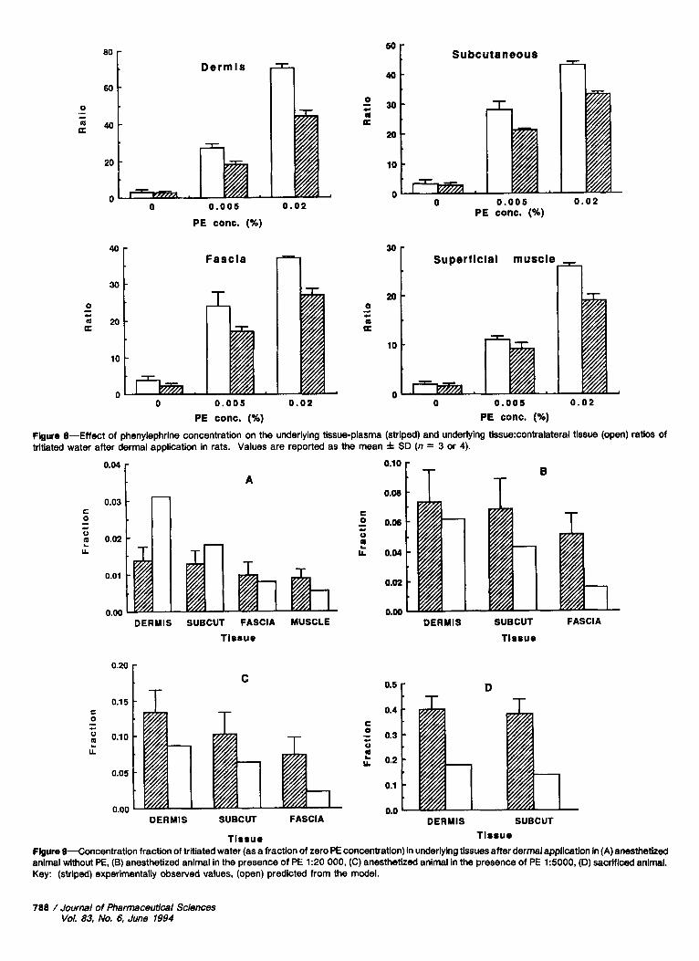
Figure 8-Effect of phenylephrine concentration on the underlying tissue-plasma (striped) and underlying tissue:contralateral tissue (open) ratios of tritiated water after dermal application in rats. Values are reported as the mean f SD (n = 3 or 4).
B
O'" 0.08 1 T T A
0.03 C 0
0
L
.- c
2 0.02
0.01
C 0 0.06
P ).
V
LL 0.04
0.02
0.00
T h DERMIS SUBCUT FASCIA
Tissue DERMIS SUBCUT FASCIA MUSCLE
Tissue
C
T D OS I T 0.4
C 0
V 3 0.3
; 0.2
0.1
0.0
c 0 .- c
0.10 2 L
0.05
0.00 DERMIS SUBCUT FASCIA
Tlssue
-.- DERMIS SUBCUT
Tissue Figure 9-Concentration fraction of tritiated water (as a fraction of zero PE concentration) in underlying tissues after dermal application in (A) anesthetized animal without PE. (B) anesthetized animal in the presence of PE 1:20 000, (C) anesthetized animal in the presence of PE 15000, (D) sacrificed animal. Key: (striped) experimentally observed values, (open) predicted from the model.
788 /Journal of Pharmaceutical Sciences Vol. 83, No. 6, June 1994
r
A
r
DERMIS SUBCUT FASCIA S.MUSCLE MUSCLE
Tissue
C 0
0
Y
- c
!!
0 8
0.6 ET 0 c c
0
0.4 0
L
- - + c
z !! u.
0.2
0.0
C
DERMIS SUBCUT FASCIA S.MUSCLE MUSCLE
T i s s u e
0.3
*3
0.1
0.0
B
T
DERMIS SUBCUT FASCIA S.MU5CLE MUSCLE
Tissue
D
DERMIS SUBCUT FASCIA S.MUSCLE MUSCLE
Tissue
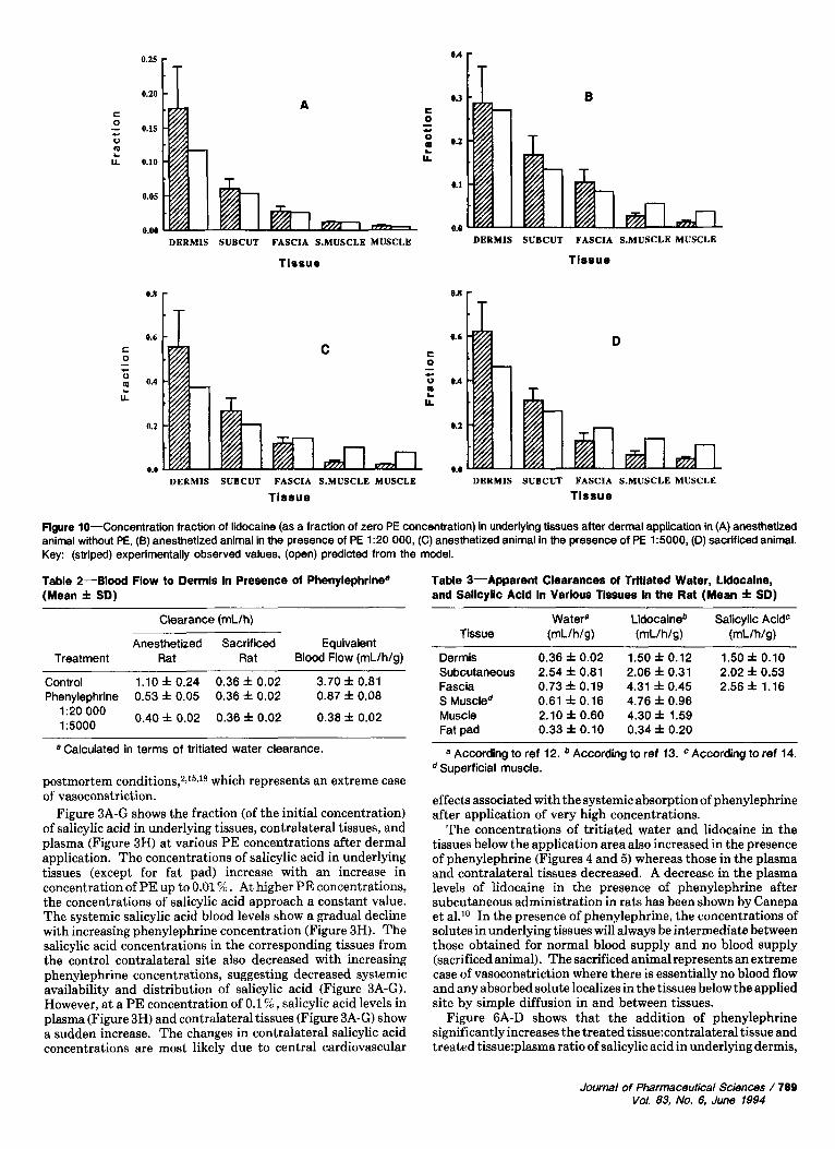
Figure 10-Concentration fraction of lidocaine (as a fraction of zero PE concentration) in underlying tissues after dermal application in (A) anesthetized animal without PE, (6) anesthetized animal in the presence of PE 1:20 000, (C) anesthetized animal in the presence of PE 1:5000, (D) sacrificed animal. Key: (striped) experimentally observed values, (open) predicted from the model.
Table 2-Blood Flow to Dermis In Presence of Phenylephrlnea (Mean f SD)
Clearance (mL/h) ~
Anesthetized Sacrificed Equivalent Treatment Rat Rat Blood Flow (mL/h/g)
Control 1.10 f 0.24 0.36 f 0.02 3.70 f 0.81 Phenylephrine 0.53 f 0.05 0.36 f 0.02 0.87 f 0.08
1:20 Oo0 0.40 f 0.02 0.36 f 0.02 0.38 f 0.02 15000
a Calculated in terms of tritiated water clearance.
postmortem conditions72J5J8 which represents an extreme case of vasoconstriction.
Figure 3A-G shows the fraction (of the initial concentration) of salicylic acid in underlying tissues, contralateral tissues, and plasma (Figure 3H) at various PE concentrations after dermal application. The concentrations of salicylic acid in underlying tissues (except for fat pad) increase with an increase in concentration of PE up to 0.01 5%. At higher PE concentrations, the concentrations of salicylic acid approach a constant value. The systemic salicylic acid blood levels show a gradual decline with increasing phenylephrine concentration (Figure 3H). The salicylic acid concentrations in the corresponding tissues from the control contralateral site also decreased with increasing phenylephrine concentrations, suggesting decreased systemic availability and distribution of salicylic acid (Figure 3A-G). However, at a PE concentration of 0.1 % , salicylic acid levels in plasma (Figure 3H) and contralateral tissues (Figure 3A-G) show a sudden increase. The changes in contralateral salicylic acid concentrations are most likely due to central cardiovascular
Table 3-Apparent Clearances of Trltlated Water, Lldocalne, and Sallcyllc Acid In Various Tissues In the Rat (Mean f SD)
Watera Lidocaine* Salicylic AcidC Tissue (rnL/ h/g ) (rnL/h/g) (rnL/h/g)
~
Dermis 0.36 f 0.02 1.50 f 0 . 1 2 1.50 f 0.10 Subcutaneous 2.54 f 0.81 2.06 f 0.31 2.02 f 0.53 Fascia 0.73 f 0.19 4.31 f 0.45 2.56 f 1.16 S Muscled 0.61 f 0.16 4.76 f 0.96 Muscle 2.10 f 0.60 4.30 f 1.59 Fat pad 0.33 f 0.10 0.34 f 0.20
a According to ref 12. According to ref 13. According to ref 14. Superficial muscle.
effects associated with the systemic absorption of phenylephrine after application of very high concentrations.
The concentrations of tritiated water and lidocaine in the tissues below the application area also increased in the presence of phenylephrine (Figures 4 and 5) whereas those in the plasma and contralateral tissues decreased. A decrease in the plasma levels of lidocaine in the presence of phenylephrine after subcutaneous administration in rats has been shown by Canepa et a1.10 In the presence of phenylephrine, the concentrations of solutes in underlying tissues will always be intermediate between those obtained for normal blood supply and no blood supply (sacrificed animal). The sacrificed animal represents an extreme case of vasoconstriction where there is essentially no blood flow and any absorbed solute localizes in the tissues below the applied site by simple diffusion in and between tissues.
Figure 6A-D shows that the addition of phenylephrine significantly increases the treated tissue:contralateral tissue and treated tissue:plasma ratio of salicylic acid in underlying dermis,
Journal of Pharmaceutical Sciences / 789 Vol. 83, No. 6. June 1994
O.l0 0.20
0.15
0.10
0.05
0.00
A
-
-
-
-
-
T 0.00 1 T C 0 0.06 .- c 0
LL 0.04
0.02
0.00 _.__
DERMIS SUBCUT FASCIA
T i s s u e
0.25 r 0.20 I T C
C 0
0
.- 0.15
m LL 0.10 L
0.05
DERMIS SUBCUT FASCIA
Tissue
c 0
V
U
- c.
t
DERMIS SUBCUT FASCIA
Tissue
O.’ F T D 0.4
c - 0 0.3 c 0
t 0.2
0.1
0.0 DERMIS SUBCUT FASCIA
Tissue
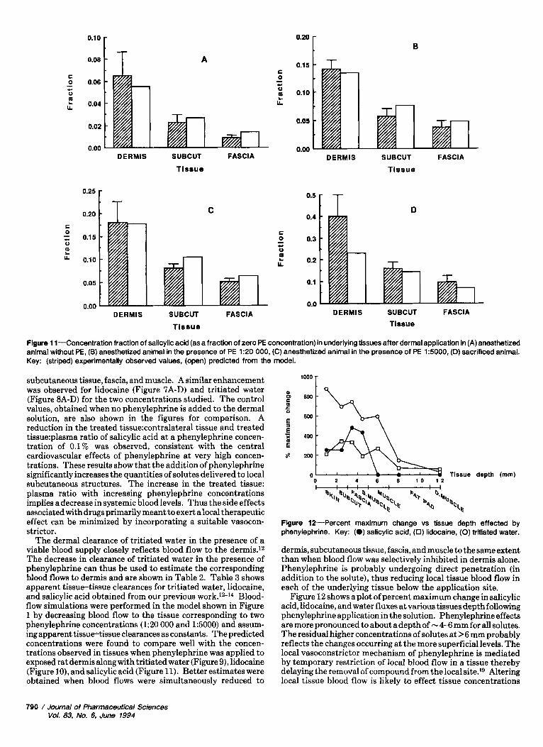
Flgure 1 1-Concentration fraction of salicylic acid (as a fraction of zero PE concentration) in underlying tissues after dermal application in (A) anesthetized animal without PE, (B) anesthetized animal in the presence of PE 1:20 000, (C) anesthetized animal in the presence of PE 1:5000. (D) sacrificed animal. Key: (striped) experimentally observed values, (open) predicted from the model.
subcutaneous tissue, fascia, and muscle. A similar enhancement was observed for lidocaine (Figure 7A-D) and tritiated water (Figure 8A-D) for the two concentrations studied. The control values, obtained when no phenylephrine is added to the dermal solution, are also shown in the figures for comparison. A reduction in the treated tissue:contralateral tissue and treated tissue:plasma ratio of salicylic acid at a phenylephrine concen- tration of 0.1% was observed, consistent with the central cardiovascular effects of phenylephrine at very high concen- trations. These results show that the addition of phenylephrine significantly increases the quantities of solutes delivered to local subcutaneous structures. The increase in the treated tissue: plasma ratio with increasing phenylephrine concentrations implies a decrease in systemic blood levels. Thus the side effects associated with drugs primarily meant to exert a local therapeutic effect can be minimized by incorporating a suitable vasocon- strictor.
The dermal clearance of tritiated water in the presence of a viable blood supply closely reflects blood flow to the dermis.12 The decrease in clearance of tritiated water in the presence of phenylephrine can thus be used to estimate the corresponding blood flows to dermis and are shown in Table 2. Table 3 shows apparent tissue-tissue clearances for tritiated water, lidocaine, and salicylic acid obtained from our previous work.12-14 Blood- flow simulations were performed in the model shown in Figure 1 by decreasing blood flow to the tissue corresponding to two phenylephrine concentrations (1:20 000 and 1:5000) and assum- ing apparent tissue-tissue clearances as constants. The predicted concentrations were found to compare well with the concen- trations observed in tissues when phenylephrine was applied to exposed rat dermis along with tritiated water (Figure 9), lidocaine (Figure lo), and salicylic acid (Figure 11). Better estimates were obtained when blood flows were simultaneously reduced to
Tissue depth (rnrn) 0 2 4 6 8 1 0 1 2
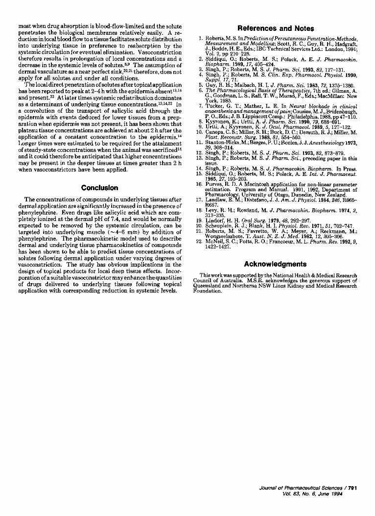
Flgure 12-Percent maximum change vs tissue depth effected by phenylephrine. Key: (0) salicylic acid, (0) lidocaine, (0) trltiated water.
dermis, subcutaneous tissue, fascia, and muscle to the same extent than when blood flow was selectively inhibited in dermis alone. Phenylephrine is probably undergoing direct penetration (in addition to the solute), thus reducing local tissue blood flow in each of the underlying tissue below the application site.
Figure 12 shows a plot of percent maximum change in salicylic acid, lidocaine, and water fluxes at various tissues depth following phenylephrine application in the solution. Phenylephrine effects are more pronounced to about a depth of - 4-6 mm for all solutes. The residual higher concentrations of solutes at >6 mm probably reflects the changes occurring at the more superficial levels. The local vasoconstrictor mechanism of phenylephrine is mediated by temporary restriction of local blood flow in a tissue thereby delaying the removal of compound from the local site.19 Altering local tissue blood flow is likely to effect tissue concentrations
790 / Journal of Pharmaceutical Sciences Vol. 83, No. 6, June 1994
most when drug absorption is blood-flow-limited and the solute penetrates the biological membranes relatively easily. A re- duction in local blood flow to a tissue facilitates solute distribution into underlying tissue in preference to reabsorption by the systemic circulation for eventual elimination. Vasoconstriction therefore results in prolongation of local concentrations and a decrease in the systemic levels of s01utes.~~~ The assumption of dermal vasculature as a near perfect sink,20.21 therefore, does not apply for all solutes and under all conditions.
The local direct penetration of solutes after topical application has been reported to peak at 2-4 h with the epidermis absent13J4 and present.22 At later times systemic redistribution dominates as a determinant of underlying tissue concentrat ion~.~3J~~~~ In a convolution of the transport of salicylic acid through the epidermis with events deduced for lower tissues from a prep- aration when epidermis was not present, it has been shown that plateau tissue concentrations are achieved at about 2 h after the application of a constant concentration to the epidermis.14 Longer times were estimated to be required for the attainment of steady-state concentrations when the animal was sacrificed14 and it could therefore be anticipated that higher concentrations may be present in the deeper tissues at times greater than 2 h when vasoconstrictors have been applied.
The concentrations of compounds in underlying tissues after dermal application are significantly increased in the presence of phenylephrine. Even drugs like salicylic acid which are com- pletely ionized at the dermal pH of 7.4, and would be normally expected to be removed by the systemic circulation, can be targeted into underlying muscle (-4-6 mm) by addition of phenylephrine. The pharmacokinetic model used to describe dermal and underlying tissue pharmacokinetics of compounds has been shown to be able to predict tissue concentrations of solutes following dermal application under varying degrees of vasoconstriction. The study has obvious implications in the design of topical products for local deep tissue effects. Incor- poration of a suitable vasoconstrictor may enhance the quantities of drugs delivered to underlying tissues following topical application with corresponding reduction in systemic levels.
References and Notes 1. Roberta, M. S. InPrediction of PercutaneousPenetration-Methods,
Measurement and Modelling; Scott, R. C., Guy, R. H., Hadgraft, J., Bodde, H. E., Eds.; IBC Technical Services Ltd.: London, 1991; Vol. 2, pp 210-228.
2. Siddiqui, 0.; Roberts, M. S.; Polack, A. E. J. Pharmacokin. Biopharm. 1989,17,405-424.
3. Singh, P.; Roberta, M. S. J. Pharm. Sci. 1993,82, 127-131. 4. Singh, P.; Roberts, M. S. Clin. Exp. Pharmacol. Physiol. 1990,
Suppl. 17, 71. 5. Guy, R. H.; Maibach, H. I. J. Pharm. Sci. 1983, 72,1375-1380. 6. The Pharmacological Basis of Therapeutics, 7th ed.; Gilman, A.
G., Goodman, L. S., Rall, T. W., Murad, F., Eds.; MacMillan: New York, 1985.
7. Tucker, G. T.; Mather, L. E. In Neural blockade in clinical anaesthesia and management ofpain; Cousins, M. J., Bridenbaugh, P. O., Eds.; J. B. Lippincott Comp.: Philadelphia, 1988, pp 47-110.
8. Kyyronen, K.; Urtti, A. J. Pharm. Sci. 1990, 79, 688-691. 9. Urtti, A.; Kyyronen, K. J. Ocul. Pharmacol. 1989,5, 127-132.
10. Canepa, C. S.; Miller, S. H.; Buck, D. C.; Demuth, R. J.; Miller, M.
11. Stanton-Hicks,M.;Berges,P. U.; Bonica, J. J. Anesthesiology 1973,
12. Singh, P.; Roberts, M. S. J. Pharm. Sci. 1993,82, 873-879. 13. Singh, P.; Roberts, M. S. J. Pharm. Sci., preceding paper in this
14. Singh, P.; Roberta, M. S. J. Pharmacokin. Biopharm. In Press. 15. Siddiqui, 0.; Roberts, M. S.; Polack, A. E. Znt. J. Pharmaceut.
16. Purves, R. D. A Macintosh application for non-linear parameter estimation. Program and Manual. 1991, 1992, Department of Pharmacology, University of Otago, Dunedin, New Zealand.
17. Landlaw, E. M.; Distefano, J. J. Am. J. Physiol. 1984,246, R665- R667.
18. Levy, R. H.; Rowland, M. J. Pharmacokin. Biopharm. 1974, 2,
19. Lindorf, H. H. Oral Surg. 1979,48, 292-297. 20. Scheuplein, R. J.; Blank, H. I. Physiol. Rev. 1971, 51, 702-747. 21. Roberts, M. S.; Favretto, W. A.; Meyer, A.; Reckmann, M.;
Wongseelashote, T. Aust. N. 2. J. Med. 1982, 12, 305-306. 22. McNeil, S. C.; Potts, R. 0.; Francoeur, M. L. Pharm. Res. 1992,9,
Plast. Reconstr. Surg. 1988,81, 554-560.
39, 308-314.
issue.
1985,27,193-203.
313-335.
1422-1427.
Acknowledgments This work was supported by the National Health & Medical Research
Council of Australia. M.S.R. acknowledges the generous support of Queensland and Northerna NSW Lions Kidney and Medical Research Foundation.
Journal of Pharmaceutical Sciences / 79 1 Vol. 83, No. 6, June 1994