Embed Size (px)
Citation preview
Chinese Medicine, 2014, 5, 223-230 Published Online December 2014 in SciRes. http://www.scirp.org/journal/cm http://dx.doi.org/10.4236/cm.2014.54027
How to cite this paper: Gao, Z.L., Zhang, C., Jin, L.W. and Yao, W. (2014) Efficacy of Sea Buckthorn Therapy in Patients with Nonalcoholic Fatty Liver Disease. Chinese Medicine, 5, 223-230. http://dx.doi.org/10.4236/cm.2014.54027
Efficacy of Sea Buckthorn Therapy in Patients with Nonalcoholic Fatty Liver Disease Zeli Gao1*, Cheng Zhang2, Liwen Jin2, Wei Yao2 1Department of Gastroenterology, People’s Hospital of Pudong New Area, Shanghai, China 2Department of Gastroenterology, Zhoupu Hospital, Pudong New Area, Shanghai, China Email: *[email protected] Received 22 September 2014; revised 30 October 2014; accepted 10 November 2014
Copyright © 2014 by authors and Scientific Research Publishing Inc. This work is licensed under the Creative Commons Attribution International License (CC BY). http://creativecommons.org/licenses/by/4.0/
Abstract Aim: To evaluate the clinical efficacy of the traditional Chinese medicine sea buckthorn (SBT) in the treatment of patients with nonalcoholic fatty liver disease (NAFLD). Method: 94 patients with NAFLD were randomly divided into two groups: 48 cases of patients received oral sea buckthorn 1.5 g (3 times a day) for three months as the treated group, and 46 cases received only the vehicle for three months as the control group. Serum lipids, transaminase and serum liver fibrosis indices were assessed at baseline and after SBT treatment. All patients underwent liver CT and Fibroscan examination at baseline and after treatment. Results: SBT treatment resulted in a significant de-crease in the serum levels of alanine aminotransferase (ALT), LDL-C, hyaluronic acid and collagen type IV. The liver stiffness measurement (LSM) of the treated patients was significantly lower than that in the control or baseline. The CT liver/spleen ratio of the treated patients was also signifi-cantly increased. Conclusion: The results of our study demonstrated the beneficial effects of SBT on serum lipids, transaminase, and liver/spleen ratio and liver stiffness in patients with NAFLD, which may be further developed as a promising therapy for the treatment of NAFLD.
Keywords Sea Buckthorn (Hippophae rhamnoides L), Nonalcoholic Fatty Liver Disease, Blood Lipid, Traditional Chinese Medicine
1. Introduction Nonalcoholic fatty liver disease (NAFLD) is a one of the most common causes of chronic liver injury in the
*Corresponding author.
Z. L. Gao et al.
224
world [1] [2]. Although NAFLD is usually benign in early stages in most cases; about 40% - 50% NAFLD cases progress to fibrosis, and 15% - 20% cases progress to cirrhosis or even end-stage liver disease. In NAFLD cirr-hosis, 30% to 40% of patients will experience a liver-related death [3]. So, it is important to find ways or medi-cines to treat this disease.
Multiple factors have been reported to interact with the development of NAFLD. It has been shown that insu-lin resistance (IR), oxidative stress, genetic and immune factors, obesity, dyslipidemia, diabetes, and hyperten-sion, are key risk factors for the development and progression of NAFLD [4]. It has been proved that insulin sensitizers, weight loss medications, lipid lowering medications (Statins), antioxidants all have some therapeutic effects in treatment of NAFLD; however, there is no definitive therapeutic strategy for effective treatment of NAFLD [5].
SBT (Hippophae rhamnoides L) is a thorny nitrogen-fixing deciduous shrub, native to Asia and Europe. The pharmacological effects of sea buckthorn were recorded in some medicinal classics. Recently, sea buckthorn was reported to have diverse pharmacological effects [6]. It was found that, in a high-fat diet fed mouse model, administration of flavonoids isolated from seed residues of Hippophae rhamnoides L (FSH) markedly reduced the levels of both serum total cholesterol and low-density lipoprotein-cholesterol, and significantly lowered total cholesterol and triglyceride concentrations in liver. Furthermore, the FSH treatment not only significantly sup-pressed the rise in serum glucose, but also improved the impaired glucose tolerance. The sea buckthorn leaf ex-tract was found to have significant anti-inflammatory activity in adjuvant induced arthritis (AIA) rat model and in lipopolysaccharide-induced inflammatory response [7] [8]. Crude extracts containing carotenoids from the berries of various cultivars of SBT have been shown to have antioxidant capacity [9]. The SBT leaves and seed oil also have hepatoprotective activity evaluated using CCl4-induced hepatic damage in animals [10] [11]. In consideration of these demonstrated effects of SBT, we presume that SBT may have a therapeutic effect in pa-tients with NAFLD. Therefore, the aim of this study was to investigate the effects of SBT in the treatment of pa-tients with NAFLD.
2. Patients and Methods This randomized, prospective, clinical trial was carried out at the outpatient department of Gastroenterology of Shanghai Pudong New District ZhouPu Hospital and Shanghai Baoshan Central Hospital. The recruitment of patients started in March 2008 and was concluded in March 2011. Of the patients diagnosed with NAFLD by ultrasonographic evidence and elevated aminotransferase activity, 94 cases were consistent with the inclusion and exclusion criteria as follows.
2.1. Inclusion Criteria 2.1.1. Diagnostic Criteria of NAFLD NAFLD was diagnosed according to the criteria in Guidelines for Management of Nonalcoholic Fatty Liver Disease set by Workshop on Fatty Liver and Alcoholic Liver Disease specified by the Chinese Society of Hepa-tology .
Inclusion criteria were as follows: 1) patients with no history of alcohol consuming or an alcohol-drinking history of less than 140 g (ethanol) weekly for man and less than 70 g for women; 2) patients with no histories of viral hepatitis, drug-induced liver disease, total parenteral nutrition, and hepatolenticular degeneration or au-toimmune liver disease that could cause fatty liver; 3) patients whose histological changes in liver biopsy met the pathological diagnostic criteria of fatty liver disease; 4) patients whose liver imaging met the imaging diag-nostic criteria of diffuse fatty liver, which could not be explained by other reasons; and 5) patients whose serum ALT and/or AST or GGT continued to rise in recent six months or longer.
2.1.2. Exclusion Criteria Patients were excluded if they were in agreement with the following criteria: 1) patients whose fatty liver was due to chronic cardiac insufficiency or malnutrition; 2) patients whose fatty liver was associated with viral hepa-titis, hepaticdecompensation, positive testing for hepatitis B virus or hepatitis C virus, any other suspected caus-es of liver disease by history; 3) patients who had received treatment for fatty liver by other Chinese or modern medicines; 4) patients with serious primary diseases, alcohol or drug abuse; 5) patients with encyesis; 6) patients with genetic diseases; and 7) patients who did not cooperate with the treatment or could not guarantee comple-
Z. L. Gao et al.
225
tion of the treatment.
2.2. Grouping and Treatment Method The patients enrolled (n = 94) were randomly divided into two groups: the study group (n = 48, 25 male and 23 female) and the control group (n = 46, 27 male and 19 female). The age of the patients ranged from 18 to 67 years. The patients of the two groups were comparable in sex, age, duration of disease and severity of the pa-thological condition.
The study group was given SBT capsules (Hebei Shengxing Seabuckthorn Pharmaceuticals, Shijiazhuang City, China) at a dose of 1.5 g in capsules 3 times/day for 90 consecutive days. The control group was given starch 1.5 g in capsules 3 times a day (manufactured by the same company). Both groups were advised to follow a diet tailored on the individual requirement, a regimen of daily physical exercise of walking for 30 - 60 min/day for 3 months during the treatment. Patients with diabetes or hypertension were given the corresponding treat-ment at the same time.
Changes in symptoms and signs, blood pressure (BP), physical fitness index and adverse drug reactions were assessed during the 3-month treatment period.
Diabetes was considered present if patients took medications for diabetes (metformin or sulfamides) or if the fasting serum glucose level was 7 mmol/L or above in untreated patients. Arterial hypertension was considered present if patients took antihypertensive drugs.
2.3. Evaluation and Monitoring 2.3.1. Laboratory Tests Laboratory tests were performed at the baseline and at the end of treatment. Fasting blood was collected from peripheral vein at observational points, using the automatic multi-parameter analyzer (Siemens, Germany). The following liver function associated laboratory parameters including serum aspartate aminotransferase (AST) and ALT levels, gamma glutamyl transferase (r-GT), blood glucose, serum lipid indices including triglycerides, (TG), total cholesterol (TC), high density lipoprotein cholesterol (HDL-C), low density lipoprotein cholesterol, (LDL-C), were evaluated. Liver fibrosis indices, including: laminin (LN), hyaluronic acid (HA, procollagen III), PC III and collagen Type IV (C IV), were measured by radioimmunoassay (RIA).
2.3.2. B-Ultrasound Examination and Quantitative Standard Liver ultrasounds measurement were performed at baseline and post-treatment, by a single, blinded, radiologist in the two hospitals in order to avoid personal errors. The ultrasonographic diagnosis of fatty liver was con-firmed in the presence of the following four findings: 1) a diffuse hyperechoic echotexture (bright liver); 2) in-creased liver echotexture compared with the kidneys; 3) vascular blurring; and 4) deep attenuation.
2.3.3. Transient Elastography Liver stiffness measurement (LSM) was performed with a Fibroscan (EchoSens, Paris, France), based on elas-tometry or one dimensional transient elastography. Details of the technique and the examination procedure have been described in previous reports [12] [13].
2.3.4. CT Examination and Quantitative Standard The patients with NAFLD of both groups underwent liver CT scan. The degree of NAFLD was determined by the liver/spleen ratio: 1) mild fatty liver: liver density decreased, the liver CT value was less than the spleen CT value, and the liver/spleen ratio was smaller than 1.0; 2) moderate fatty liver: the liver/spleen ratio was smaller than 0.7, and the intrahepatic vascular display was unclear; and 3) severe fatty liver: liver density decreased sig-nificantly and was even below zero, the liver/spleen ratio was ≤0.5, and the intrahepatic vascular display was clear.
2.4. Statistical Analysis The obtained data were analyzed by SAS 6.12 software. The clinical and laboratory findings within groups were compared by analysis of variance (ANOVA). A confidence interval of 95% was used and a two-tailed, values of
Z. L. Gao et al.
226
p < 0.05, p < 0.01 and p < 0.001 were considered statistically significant. For non-parametric data were com-pared using chi-square test.
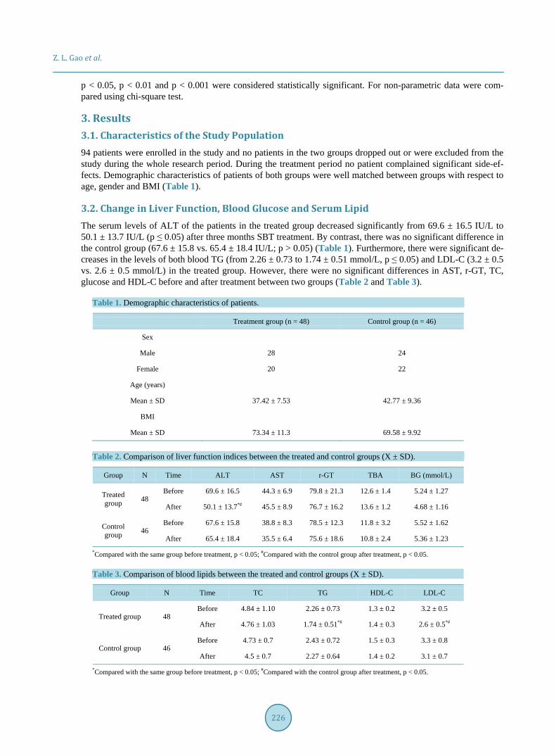
3. Results 3.1. Characteristics of the Study Population 94 patients were enrolled in the study and no patients in the two groups dropped out or were excluded from the study during the whole research period. During the treatment period no patient complained significant side-ef- fects. Demographic characteristics of patients of both groups were well matched between groups with respect to age, gender and BMI (Table 1).
3.2. Change in Liver Function, Blood Glucose and Serum Lipid The serum levels of ALT of the patients in the treated group decreased significantly from 69.6 ± 16.5 IU/L to 50.1 ± 13.7 IU/L (p ≤ 0.05) after three months SBT treatment. By contrast, there was no significant difference in the control group (67.6 ± 15.8 vs. 65.4 ± 18.4 IU/L; p > 0.05) (Table 1). Furthermore, there were significant de-creases in the levels of both blood TG (from 2.26 ± 0.73 to 1.74 ± 0.51 mmol/L, p ≤ 0.05) and LDL-C (3.2 ± 0.5 vs. 2.6 ± 0.5 mmol/L) in the treated group. However, there were no significant differences in AST, r-GT, TC, glucose and HDL-C before and after treatment between two groups (Table 2 and Table 3).
Table 1. Demographic characteristics of patients.
Treatment group (n = 48) Control group (n = 46)
Sex
Male 28 24
Female 20 22
Age (years)
Mean ± SD 37.42 ± 7.53 42.77 ± 9.36
BMI
Mean ± SD 73.34 ± 11.3 69.58 ± 9.92
Table 2. Comparison of liver function indices between the treated and control groups (X ± SD).
Group N Time ALT AST r-GT TBA BG (mmol/L)
Treated group 48
Before 69.6 ± 16.5 44.3 ± 6.9 79.8 ± 21.3 12.6 ± 1.4 5.24 ± 1.27
After 50.1 ± 13.7*# 45.5 ± 8.9 76.7 ± 16.2 13.6 ± 1.2 4.68 ± 1.16
Control group 46
Before 67.6 ± 15.8 38.8 ± 8.3 78.5 ± 12.3 11.8 ± 3.2 5.52 ± 1.62
After 65.4 ± 18.4 35.5 ± 6.4 75.6 ± 18.6 10.8 ± 2.4 5.36 ± 1.23
*Compared with the same group before treatment, p < 0.05; #Compared with the control group after treatment, p < 0.05.
Table 3. Comparison of blood lipids between the treated and control groups (X ± SD).
Group N Time TC TG HDL-C LDL-C
Treated group 48 Before 4.84 ± 1.10 2.26 ± 0.73 1.3 ± 0.2 3.2 ± 0.5
After 4.76 ± 1.03 1.74 ± 0.51*# 1.4 ± 0.3 2.6 ± 0.5*#
Control group 46 Before 4.73 ± 0.7 2.43 ± 0.72 1.5 ± 0.3 3.3 ± 0.8
After 4.5 ± 0.7 2.27 ± 0.64 1.4 ± 0.2 3.1 ± 0.7
*Compared with the same group before treatment, p < 0.05; #Compared with the control group after treatment, p < 0.05.
Z. L. Gao et al.
227
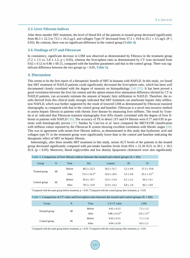
3.3. Liver Fibrosis Indices After three months SBT treatment, the level of blood HA of the patients in treated group decreased significantly from 86.3 ± 22.3 to 73.5 ± 16.3 μg/L and collagen Type IV decreased from 37.3 ± 19.8 to 25.1 ± 3.5 ng/L (P ≤ 0.05). By contrast, there was no significant difference in the control group (Table 4).
3.4. Findings of CT and Fibroscan In consistency, significant decrease in LSM was observed as demonstrated by Fibrosca in the treatment group (7.2 ± 1.5 vs. 5.8 ± 1.2; p ≤ 0.05), whereas the liver/spleen ratio as demonstrated by CT scan increased from 0.62 ± 0.12 to 0.86 ± 60.15, compared with the baseline parameters and that in the control group. There was sig-nificant difference between the two groups (p < 0.05, Table 5).
4. Discussion This seems to be the first report of a therapeutic benefit of SBT in humans with NAFLD. In this study, we found that SBT treatment of NAFLD patients could significantly decreased the liver/spleen ratio, which has been well documented closely correlated with the degree of steatosis on histopathology [14] [15]. It has been proved a good correlation between the liver fat content and the spleen-minus-liver attenuation difference elicited by CT in NAFLD patients, can accurately estimate the amount of hepatic fatty infiltration in NAFLD. Therefore, the re-sults derived from this clinical study strongly indicated that SBT treatment can ameliorate hepatic fatty infiltra-tion NAFLD, which was further supported by the result of lowered LSM as demonstrated by Fibroscan transient elastography as compared with that in the control group and baseline. Fibroscan is a novel non-invasive method to assess hepatic fibrosis in patients with chronic liver disease by measuring liver stiffness. The result by Yone-da et al. indicated that Fibroscan transient elastography liver KPa closely correlated with the degree of liver fi-brosis in patients with NAFLD [16]. The accuracy of TE to detect ≥F3 and F4 fibrosis were 0.77 and 0.95 in pa-tients with histologically proven [17]. Studies by Cast’era et al. have compared the METAVIR classification with stiffness values reported by the Fibroscan R system showing excellent correlation with fibrotic stages [18]. This was in agreement with serum liver fibrosis indices, as demonstrated in this study that hyaluronic acid and collagen type IV in the treatment group were significantly lower than in the control and baseline indicating the therapeutic effect of SBT on hepatic fibrosis.
Interestingly, after three months SBT treatment in this study, serum ALT levels of the patients in the treated group decreased significantly compared with pre-intake baseline levels from 69.6 ± 21.56 IU/L to 50.1 ± 18.5 IU/L (p < 0.05). Moreover, blood triglycerides and low density lipoprotein cholesterol were also significantly
Table 4. Comparison of liver fibrosis indices between the treated and control groups (X ± SD).
Group N Time HA Lamnin III IV
Treated group 48 Before 86.3 ± 22.3 56.5 ± 31.7 5.5 ± 0.8 37.3 ± 19.8
After 73.5 ± 16.3*# 52.6 ± 18.5 5.5 ± 0.6 25.1 ± 3.5*#
Control group 46 Before 85.4 ± 19.7 55.5 ± 12.4 6.1 ± 2.2 34.5 ± 9.1
After 78.3 ± 15.8 51.9 ± 14.2 6.8 ± 1.8 36.1 ± 8.8
*Compared with the same group before treatment, p < 0.05; #Compared with the control group after treatment, p < 0.05.
Table 5. Comparison of CT value and liver/spleen ratio between the treated and control groups (X ± SD).
Group N Time L/S CT value LSM
Ttreated group 48 Before 0.62 ± 0.12 7.2 ± 1.5
After 0.86 ± 0.15*# 5.8 ± 1.2*#
Control group 46 Before 0.62 ± 0.11 7.1 ± 1.4
After 0.64 ± 0.10 6.9 ± 1.1
*Compared with the same group before treatment, p < 0.05; #Compared with the control group after treatment, p < 0.05.
Z. L. Gao et al.
228
decreased after 3 month SBT treatment. This result derived from human study was consistent with the that re- ported by Wang et al. [19], in that a total SBT flavone could significantly reduce serum cholesterol and TG le-vels, and increase HDL-C level in mice with experimental hyperlipidemia. These results indicated that SBT may play important roles in hepatoprotection and improvement of lipid metabolism in NAFLD.
Until now, the exact pathogenesis of NAFLD remains unclear. The pathogenesis of NAFLD and NASH has classically been considered as a “two hit” theory: i) increased fat flux to the liver resulting in hepatic triglyceride accumulation, which predisposes a patient to the second hit; ii) increased oxidative stress, lipoperoxidation, and increased reactive oxygen species resulting in hepatocyte death and scar formation [20]. The major methods for the treatment of NAFLD at present include optimizing diet and increasing physical activity for the sake of cur-ing obesity, diabetes and hyperlipidemia. The main aim of these treatments is to improve insulin resistance through using lipid-lowering and liver-protecting drugs such as fibrates and insulin sensitizers to maintain lipid metabolism, energy metabolism and antioxidant balance. However, these agents may also induce some adverse effects such as hepatic and muscular injuries, thus limiting their wider clinical applications [21] [22].
The present study was attempted to find effective drugs from the pool of traditional Chinese medicines that have no or significant less toxic effects, and known therapeutic effects in improving lipid metabolism and pro-moting NAFLD reversal.
SBT, also called Hippophae rhamnoides L, is an edibble plant and used as a traditional Chinese medicine, Tibetan medicine and Mongolian medicine. Various pharmacological activities of SBT, such as cytoprotective, anti-stress, immunomodulatory, radioprotective, anti-atherogenic, anti-tumor, anti-microbial and tissue regene-ration, have been reported [7]. Moreover, it was also reported that SBT has antihypertensive effects at least in part through improving insulin sensitivity and blocking angiotensin signal pathway indicating its potential use in the management of hyperinsulinemia in non-diabetic state with cardiovascular diseases [23]. A recent study re-ported that SBT can significantly increase the activities of superoxide dismutase (SOD), glutathione peroxidase (GSH-Px), and GSH content in liver, and significantly reduced the elevated levels of alanine aminotransferase (ALT), aspartate aminotransferase (AST), alkaline phosphatase (ALP), triglyceride (TG), and cholesterol in carbon tetrachloride (CCl4)-induced hepatic damage in ICR mice [11]. In agreement, other studies also demon-strated a protective effect of SBT against chronic hypoxic injury using primary rat hepatocytes through main-taining better levels/activities of GSH, GPx, and SOD [24].
Lipid accumulation in the liver is the major hallmark of NAFLD. In this study, we found that SBT has an ef-fect of ameliorating lipid metabolism in NAFLD patients, although the mechanism is yet unknown. Alterations in immune response have been implicated in the pathogenesis of NAFLD. It has been proved that abnormalities in lipid and lipoprotein metabolism accompanied by chronic inflammation are considered to be the central pathway for the development of several obesity-related co-morbidities such as NAFLD and cardio-vascular dis-ease (CVD) [25] [26]. NAFLD is associated with increased levels of the proinflammatory T helper 1-associated cytokines TNF-α and interleukin-12 (IL-12) [20]. Adipocytokines including TNF-α, IL-6, leptin and adiponectin, are important inflammatory proteins generated by visceral adipose. They all flow directly to the liver via the portal vein and have been implicated in the pathogenesis of NAFLD and NASH [27]. It was reported that intra-peritoneal injection of SBT flavone could significantly increase the white blood cell and bone marrow nucleated cell counts, stimulate the production of interleukin-6 (IL-6) and tumor necrosis factor-alpha (TNF-α) in human blood mononuclear cells (PBMCs) [28] [29]. SBT leaf extract also has a significant anti-inflammatory activity in arthritis (AIA) rat mode
5. Conclusions Our study demonstrated the beneficial effects of SBT treatment on serum lipids, transaminase; liver/spleen ratio and liver stiffness in patients with NAFLD; we thought that the SBT mediated effects of amelioration of lipid metabolism, manipulation of adipocytokine. SBT is especially suitable for complicating with hepatic dysfunc-tion, obese patients with non-alcoholic fatty liver disease. SBT can improve insulin resistance, which has signif-icantly reduced effect on TG accumulation in the liver, inhibit lipid peroxidation in liver cells, and improve liver microcirculation and liver fibrosis.
SBT provides a new medication for the treatment of NAFLD with traditional Chinese medicine. Clinical practice needs to be further implemented.
The limitation of this study is that the diagnosis of NAFLD was made mainly based on ultrasound, lacking
Z. L. Gao et al.
229
pathological evidence. Pathological type and histological characteristic of the subjects enrolled in this study are quite unknown. Although ultrasound and CT studies demonstrated the significant improvement of the histologi-cal features of liver in NASH patients after SBT treatment, the supporting evidence of the liver pathology is de-sired. The second limitation of this study is that we cannot exclude the synergetic effect of other factors, such as the limited diet and physical exercise et al. Furthermore, in some patient with diabetes, hypoglycemic agents were used. It has been reported that diformin in itself can be used for the treatment of NAFLD. Furthermore, the observation period is too short for observation of the long-term effects of SBT treatment on NAFLD. Nonethe-less, more extensive studies are needed for identification and isolation of the effective components or com-pound(s) in SBT and for elucidation of the underlying mechanism of SBT in the treatment of fatty liver. In con-clusion, the promising results derived from this study will open up a new horizon for substantial treatment of fatty liver.
Acknowledgements The authors are grateful to Professor Zhang Yusheng from the Department of Radiology for providing CT ex-amination; Professor Tang Mingrong from the Department of Clinical Laboratory for the laboratory test; and Professor Ma Yuan from the Department of B-ultrasound for providing support for the present study.
Funding Supported by the Laboratory of Liver and Kidney Disease (Shanghai University of Traditional Chinese Medi-cine) under the Ministry of Education of China (No. GS090501).
Authors Contribution Gao Zeli designed the research strategy, performed the literature search and drafted the paper, analyzed the data and drafted the paper. Zhang Cheng, Jin Liwen and Yao Wei collected the clinical materials.
References [1] Park, H., Shima, T., Yamaguchi, K., et al. (2011) Efficacy of Long-Term Ezetimibe Therapy in Patients with Nonal-
coholic Fatty Liver Disease. Journal of Gastroenterology, 46, 101-107. http://dx.doi.org/10.1007/s00535-010-0291-8 [2] Smith, B.W. and Adams, L.A. (2011) Non-Alcoholic Fatty Liver Disease. Critical Reviews in Clinical Laboratory
Sciences, 48, 97-113. http://dx.doi.org/10.3109/10408363.2011.596521 [3] Fon Tacer, K. and Rozman, D. (2011) Nonalcoholic Fatty Liver Disease: Focus on Lipoprotein and Lipid Deregulation.
Journal of Lipids, 2011, Article ID: 783976. http://dx.doi.org/10.1155/2011/783976 [4] Targher, G., Bertolini, L. and Poli, F. (2005) Nonalcoholic Fatty Liver Disease and Risk of Future Cardiovascular
Events among Type 2 Diabetic Patients. Diabetes, 54, 3541-3546. http://dx.doi.org/10.2337/diabetes.54.12.3541 [5] Chuthan Sourianarayanane, A., Pagadala, M.R. and Kirwan, J.P. (2013) Management of Non-Alcoholic Fatty Liver
Disease. Minerva Gastroenterologica e Dietologica, 59, 69-87. [6] Kruczek, M., Swiderski, A., Mech-Nowak, A., et al. (2012) Antioxidant Capacity of Crude Extracts Containing Caro-
tenoids from the Berries of Various Cultivars of Sea Buckthorn (Hippophae rhamnoides L). Acta Biochimica Polonica, 59, 135-137.
[7] Ganju, L., Padwad, Y., Singh, R., Karan, D., Chanda, S., Chopra, M.K., Bhatnagar, P., Kashyap, R. and Sawhney, R.C. (2005) Anti-Inflammatory Activity of Seabuckthorn (Hippophae rhamnoides) Leaves. International Immunopharma-cology, 5, 1675-1684. http://dx.doi.org/10.1016/j.intimp.2005.03.017
[8] Padwad, Y., Ganju, L., Jain, M., Chanda, S., Karan, D., Kumar, R., Banerjee, P.K. and Sawhney, R.C. (2006) Effect of Leaf Extract of Sea Buckthorn on Lipopolysaccharide Induced Inflammatory Response in Murine Macrophages. In-ternational Immunopharmacology, 6, 46-52. http://dx.doi.org/10.1016/j.intimp.2005.07.015
[9] Wang, J., Zhang, W., Zhu, D., et al. (2011) Hypolipidaemic and Hypoglycaemic Effects of Total Flavonoids from Seed Residues of Hippophae rhamnoides L in Mice Fed a High-Fat Diet. Journal of the Science of Food and Agriculture, 91, 1446-1451. http://dx.doi.org/10.1002/jsfa.4331
[10] Geetha, S., Jayamurthy, P., Pal, K., Pandey, S. and Sawhney, R.C. (2008) Hepatoprotective Activity of Sea Buckthorn (Hippophae rhamnoides L) against Carbon Tetrachloride Induced Hepatic Damage in Rats. Journal of Science of Food and Agriculture, 88, 1592-1597. http://dx.doi.org/10.1002/jsfa.3255
[11] Hsu, Y.W., Tsai, C., Chen, W.K. and Lu, F.J. (2009) Protective Effects of Seabuckthorn (Hippophae rhamnoides L)
Z. L. Gao et al.
230
Seed Oil against Carbon Tetrachloride-Induced Hepatotoxicity in Mice. Food and Chemical Toxicology, 47, 2281- 2288. http://dx.doi.org/10.1016/j.fct.2009.06.015
[12] Sandrin, L., Tanter, M., Gennisson, J.L., Catheline, S. and Fink, M. (2002) Shear Elasticity Probe for Soft Tissues with 1-D Transient Elastography. IEEE Transactions on Ultrasonics, Ferroelectrics, and Frequency Control, 49, 436-446. http://dx.doi.org/10.1109/58.996561
[13] Sandrin, L., Fourquet, B., Hasquenoph, J.M., Yon, S., Fournier, C., Mal, F., et al. (2003) Transient Elastography: A New Noninvasive Method for Assessment of Hepatic Fibrosis. Ultrasound in Medicine and Biology, 29, 1705-1713. http://dx.doi.org/10.1016/j.ultrasmedbio.2003.07.001
[14] Oliva, M.R., Mortele, K.J., Segatto, E., Glickman, J.N., Erturk, S.M., Ros, P.R. and Silverman, S.G. (2006) Computed Tomography Features of Nonalcoholic Steatohepatitis with Histopathologic Correlation. Journal of Computer Assisted Tomography, 30, 37-43. http://dx.doi.org/10.1097/01.rct.0000193818.31749.84
[15] Tobari, M., Hashimoto, E., Yatsuji, S., Torii, N. and Shiratori, K. (2009) Imaging of Nonalcoholic Steatohepatitis: Advantages and Pitfalls of Ultrasonography and Computed Tomography. Internal Medicine, 48, 739-746. http://dx.doi.org/10.2169/internalmedicine.48.1869
[16] Castera, L., Forns, X. and Alberti, A. (2008) Non-Invasive Evaluation of Liver Fibrosis Using Transient Elastography. Journal of Hepatology, 48, 835-847. http://dx.doi.org/10.1016/j.jhep.2008.02.008
[17] Mahadeva, S., Mahfudz, A.S., Vijayanathan, A., Goh, K.L., Kulenthran, A. and Cheah, P.L. (2013) Performance of Transient Elastography (TE) and Factors Associated with Discordance in Nonalcoholic Fatty Liver Disease. Journal of Digestive Diseases, 14, 604-610. http://dx.doi.org/10.1111/1751-2980.12088
[18] Yoneda, M., Yoneda, M., Mawatari, H., Fujita, K., Endo, H., Iida, H., et al. (2008) Noninvasive Assessment of Liver Fibrosis by Measurement of Stiffness in Patients with Nonalcoholic Fatty Liver Disease (NAFLD). Digestive and Liver Disease, 40, 371-378. http://dx.doi.org/10.1016/j.dld.2007.10.019
[19] Wang, J., Zhang, W., Zhu, D., Zhu, X.L., Pang, X.F. and Qu, W.J. (2011) Hypolipidaemic and Hypoglycaemic Effects of Total Flavonoids from Seed Residues of Hippophae rhamnoides L in Mice Fed a High-Fat Diet. Journal of the Science of Food and Agriculture, 91, 1446-1451. http://dx.doi.org/10.1002/jsfa.4331
[20] Smith, B.W. and Adams, L.A. (2011) Non-Alcoholic Fatty Liver Disease. Critical Reviews in Clinical Laboratory Sciences, 48, 97-113. http://dx.doi.org/10.3109/10408363.2011.596521
[21] Rallidis, L.S., Drakoulis, C.K. and Parasi, A.S. (2004) Pravastatin in Patients with Nonalcoholic Steatohepatitis: Re-sults of a Pilot Study. Atherosclerosis, 174, 193-196. http://dx.doi.org/10.1016/j.atherosclerosis.2004.01.008
[22] Lewis, J.H. (2012) Clinical Perspective: Statins and the Liver—Harmful or Helpful? Digestive Diseases and Sciences, 57, 1754-1763. http://dx.doi.org/10.1007/s10620-012-2207-3
[23] Tulsawani, R., Gupta, R. and Misra, K. (2013) Efficacy of Aqueous Extract of Hippophae rhamnoides and Its Bio-Ac- tive Flavonoids against Hypoxia-Induced Cell Death. Indian Journal of Pharmacology, 45, 258-263. http://dx.doi.org/10.4103/0253-7613.111943
[24] Pang, X., Zhao, J., Zhang, W., Zhuang, X.Y., Wang, J.S., Xu, R.Q., Xu, Z.L. and Qu, W.J. (2008) Antihypertensive Effect of Total Flavones Extracted from Seed Residues of Hippophae rhamnoides L in Sucrose-Fed Rats. Journal of Ethnopharmacology, 117, 325-331. http://dx.doi.org/10.1016/j.jep.2008.02.002
[25] Targher, G., Bertolini, L., Padovani, R., Rodella, S., Zoppini, G., Pichiri, I., et al. (2010) Prevalence of Non-Alcoholic Fatty Liver Disease and Its Association with Cardiovascular Disease in Patients with Type 1 Diabetes. Journal of He-patology, 53, 713-718. http://dx.doi.org/10.1016/j.jhep.2010.04.030
[26] Loria, P., Lonardo, A., Bellentani, S., Day, C.P., Marchesini, G. and Carulli, N. (2007) Non-Alcoholic Fatty Liver Disease (NAFLD) and Cardiovascular Disease: An Open Question. Nutrition, Metabolism and Cardiovascular Dis-eases, 17, 684-698. http://dx.doi.org/10.1016/j.numecd.2007.01.007
[27] Kremer, M., Thomas, E., Milton, R.J., Perry, A.W., van Rooijen, N., Wheeler, M.D., et al. (2010) Kupffer Cell and In-terleukin-12-Dependent Loss of Natural Killer T Cells in Hepatosteatosis. Hepatology, 51, 130-141. http://dx.doi.org/10.1002/hep.23292
[28] Ganju, L., Padwad, Y., Singh, R., Karan, D., Chanda, S., Chopra, M.K., et al. (2005) Anti-Inflammatory Activity of Seabuckthorn (Hippophae rhamnoides L) Leaves. International Immunopharmacology, 5, 1675-1684. http://dx.doi.org/10.1016/j.intimp.2005.03.017
[29] Mishra, K.P., Chanda, S., Karan, D., Ganju, L. and Sawhney, R.C. (2008) Effect of Seabuckthorn (Hippophae rham-noides) Flavone on Immune System: An in Vitro Approach. Phytotherapy Research, 22, 1490-1495. http://dx.doi.org/10.1002/ptr.2518