Embed Size (px)
Citation preview
EHS 201 GENERAL MICROBIOLOGY Course Team Dr. Chinedu E. Ihejirika & Dr. Onyenonachi C. Ihejirika (Course Developer/Writers) -Federal University of Technology, Owerri
Prof. Folakemi Omojasola (Course Editor) -University of Ilorin Dr. Oluremi Saliu (Course Coordinator)-NOUN Prof. Shehu Usman Adamu (Programme Coordinator)- NOUN
NATIONAL OPEN UNIVERSITY OF NIGERIA
COURSE GUIDE

EHS 201 COURSE GUIDE
ii
© 2021 by NOUN Press National Open University of Nigeria Headquarters University Village Plot 91, Cadastral Zone NnamdiAzikiwe Expressway Jabi, Abuja Lagos Office 14/16 Ahmadu Bello Way Victoria Island, Lagos e-mail: [email protected] URL: www.nou.edu.ng All rights reserved. No part of this book may be reproduced, in any form or by any means, without permission in writing from the publisher. Printed 2021 ISBN: 978-978-058-326-2

EHS 201 COURSE GUIDE
iii
CONTENTS PAGE Introduction ………………………………………………….. iv What You Will Learn in this Course ………………………… iv Course Aim …………………………………………………... v Course Objectives ……………………………………………. v Working through this Course ………………………………… v Course Materials ……………………………………………… vi Study Units …………………………………………………… vi Presentation Schedule ………………………………………... vii Assessment …………………………………………………… vii Tutor Marked Assignments (TMAS) ………………………… viii Final Examination and Grading ……………………………… viii Course Marking Scheme ……………………………………... viii Course Overview ……………………………………………... ix How to get the most out of this Course ………………………. ix Facilitators/Tutors and Tutorials …………………………… x Summary …………………………………………………….. xi
EHS 201 COURSE GUIDE
iv
INTRODUCTION This course, EHS201: General Microbiology, is a first semester course. It is a two credit unit course available to all student of undergraduate students in Environmental Health Science. General Microbiology is a foundational course for students studying Environmental Health Science is made easier to understand and to accept as it explored the basis of cell biology, explaining microorganisms as the smallest unit of life that can only be seen with the aid of a microscope. In the act of understanding environmental health sciences, microbiology takes a major position as microorganisms exist in all types of environment, such as the soil, water and the atmosphere including other living things like plants and animals, man not left out. In this course, the student shall be exposed to the understanding of the history and scope of microbiology, basic characteristics of microorganisms, basic cellular compositions and structures. Microorganisms as major causes of diseases can best be controlled by understanding their cultural characteristics, growth and growth its prevention and control to avoid the spread of diseases. The importance of microbiology to food processes, food production, agriculture and waste treatment will also aid the students in understanding exploring the values of microorganisms in the environment especially in developing country like Nigeria. WHAT YOU ARE TO LEARN IN THIS COURSE The course content consist of a unit of the course guide which tells you briefly what the course is about, what course materials you need and how to work with such materials. It also gives you some guideline for the time you are expected to spend on each unit in order to complete it successfully. It guides you concerning your tutor-marked assignment which will be placed in the assignment file. Regular tutorial classes related to the course will be conducted and it is advisable for you to attend these sessions. It is expected that the course will prepare you for challenges you are likely to meet in the field of Environmental Health Science.
EHS 201 COURSE GUIDE
v
COURSE AIMS The course aim is to provide you with an understanding of General Microbiology. It is intended to let you appreciate the proportion occupied by Microbiology in the management of environmental health in a developing country like Nigeria. COURSE OBJECTIVE To achieve the aim set out, the course has a set of objectives. Each unit under a module has specified objectives which are stated at the beginning of the unit. You are advised to read the objectives before you study the unit because you may need to make reference to them during your study to check on your own progress. It is also good that you endeavour to check the unit objectives after completion of each unit to decipher level of accomplishment. After going through the course, you should be able to: i. Understand the concept, history and scope of microbiology, ii. identify the general characteristics of microorganisms,
prokaryotic and eukaryotic structures iii. Understand microbial nutrition, growth, reproduction and control
measures iv. knowledge of the prevention and control of microbial diseases
through their pathogenicity, study of antimicrobial chemotherapy and clinical microbiology
v. Inculcate the role of microorganisms in food processing, environmental management, agriculture and the industry.
WORKING THROUGH THIS COURSE To complete this course you are expected to read each study unit, read the textbooks and other materials which may be provided by the National Open University of Nigeria. Each unit contains self-assessment exercises. In the course you would be required to submit assignment for assessment. At the end of the course there is final examination. The course should take about 15 weeks to complete. Listed below are the components of the course, what you have to do and how to allocate your time to each unit, in order to complete the course successfully and timely. The course demands that you should spend good time to read and my advice for you is that you should endeavour to attend tutorial session
EHS 201 COURSE GUIDE
vi
where you will have the opportunity of comparing knowledge with colleagues. COURSE MATERIALS The main components of the course are: 1. The course guide 2. Study unit 3. References/further readings 4. Assignments 5. Presentation schedule STUDY UNITS The course units in this course are as follow: Module 1 Introduction to Microbiology Unit 1 History and Scope of Microbiology Unit 2 General Characteristics of Microorganisms Unit 3 Prokaryotic and Eukaryotic Microorganisms Module 2 Microbial Nutrition, Growth, Reproduction and Control Unit 1 Microbial Nutrition Unit 2 Cell Reproduction and Microbial Growth Unit 3 Control of Microorganisms Module 3 Microbial Metabolism Unit 1 Introduction to Microbial Metabolism Unit 2 Catabolism Unit 3 Anabolism Module 4 Prevention and Control of Microbial Diseases Unit 1 Pathogenicity of Microorganisms Unit 2 Antimicrobial Chemotherapy Unit 3 Clinical Microbiology Module 5 Microbes in The Environment, Agriculture and Industry Unit 1 Microbiologyin Food Processing Unit 2 Environmental Microbiology Unit 3 Microbiology in Agriculture and the Industry In Module 1 (Introduction to Microbiology), Unit 1 focuses on the history and scope of microbiology. Unit 2 deals with the General Characteristics of Microorganisms. Unit 3 is about the prokaryotic and
EHS 201 COURSE GUIDE
vii
eukaryotic microorganisms. In Module 2 (Microbial Nutrition, Growth, Reproduction and Control), Unit 1 deals with the microbial nutrition. Unit 2 has to do with Cell Reproduction and Microbial Growth. Control of Microorganisms is treated in Unit 3. In Module 3 (Microbial Metabolism), Units 1dwelt on the Introduction to Microbial Metabolism. Unit 2 dealt with the Catabolism, while Unit 3 discussed on Anabolism.In Module 4 (Prevention and Control of Microbial Diseases), Unit 1 focuses on the Pathogenicity of Microorganisms. Unit 2 deals with the Antimicrobial Chemotherapy. Unit 3 is about the Clinical Microbiology.In Module 5 (Microbes in the Environment, Agriculture, and Industry), Unit 1 deals with the Microbiology in Food and Industry. Unit 2 has to do with Environmental Microbiology. Microbiology in Agriculture is treated in Unit 3. Each unit consists of one or two weeks work and include an introduction, objectives, main content, reading materials, exercises, conclusion, summary, Tutor marked Assignments (TMAs), references and other resources. The various units direct you to work on exercises related to the require reading. In general, the exercises test you on the materials you have just covered or require you to apply it in a way that will assist you to evaluate your own progress and to reinforce your understanding of the material. Alongside the TMAs, these exercises will help you achieve the stated learning objectives of the individual units and course as a whole. PRESENTATION SCHEDULE Your course materials have important dates for the early and timely completion and submission of your TMAs and attending tutorials. You are expected to submit all your assignments by the stipulated time and date and guard against falling behind in your work. ASSESSMENT There are three parts to the course assessment and these include self-assessment exercises, Tutor marked Assessments and the written examination or end of course examination. It is advisable that you do all the exercises. In tackling the assignments, you are expected to use the information, knowledge and techniques gathered during the course. The assignments must be submitted to your facilitator for formal assessment in line with the deadlines stated in the presentation schedule.The work you submit to your tutor for assessment will count for 30% of your total course work. At the end of the course you will need to sit for a final end of course examination of about three hours duration. This examination will count for 70% of your total course mark.
EHS 201 COURSE GUIDE
viii
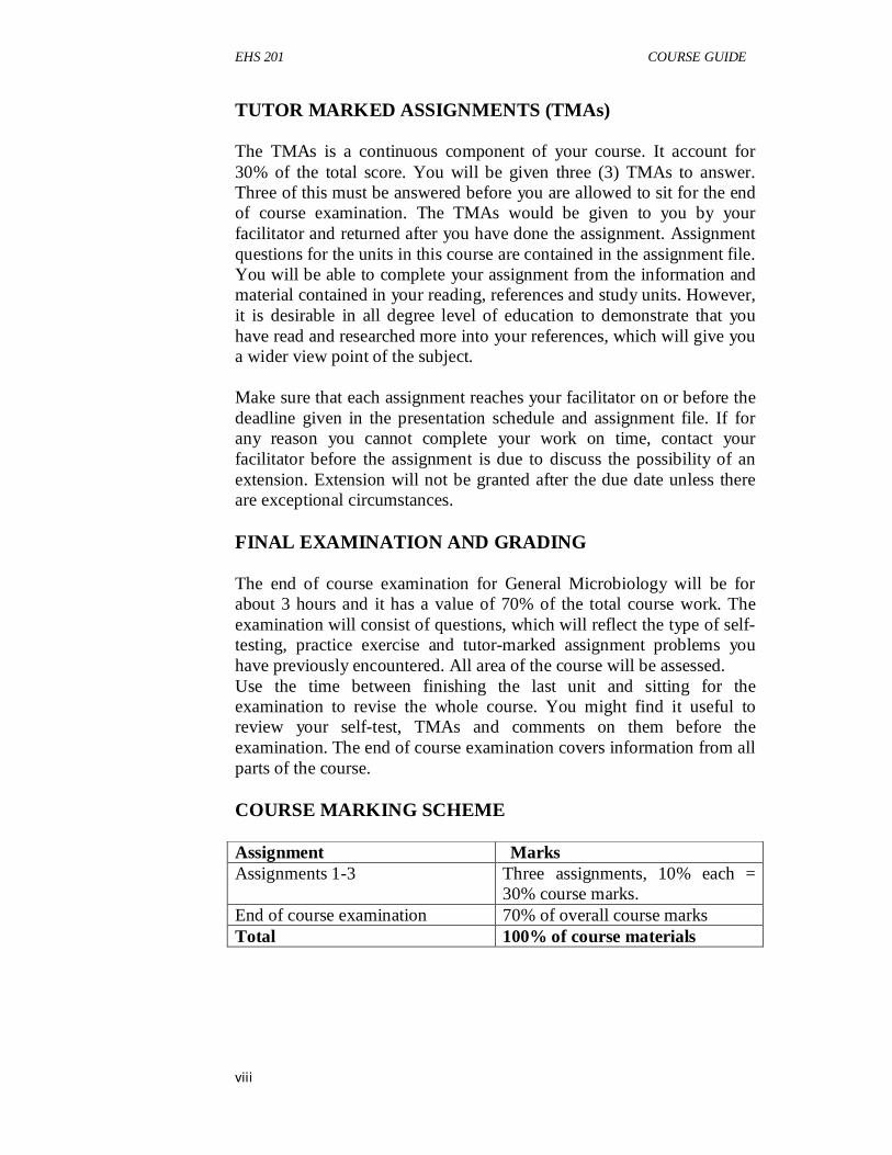
TUTOR MARKED ASSIGNMENTS (TMAs) The TMAs is a continuous component of your course. It account for 30% of the total score. You will be given three (3) TMAs to answer. Three of this must be answered before you are allowed to sit for the end of course examination. The TMAs would be given to you by your facilitator and returned after you have done the assignment. Assignment questions for the units in this course are contained in the assignment file. You will be able to complete your assignment from the information and material contained in your reading, references and study units. However, it is desirable in all degree level of education to demonstrate that you have read and researched more into your references, which will give you a wider view point of the subject. Make sure that each assignment reaches your facilitator on or before the deadline given in the presentation schedule and assignment file. If for any reason you cannot complete your work on time, contact your facilitator before the assignment is due to discuss the possibility of an extension. Extension will not be granted after the due date unless there are exceptional circumstances. FINAL EXAMINATION AND GRADING The end of course examination for General Microbiology will be for about 3 hours and it has a value of 70% of the total course work. The examination will consist of questions, which will reflect the type of self-testing, practice exercise and tutor-marked assignment problems you have previously encountered. All area of the course will be assessed. Use the time between finishing the last unit and sitting for the examination to revise the whole course. You might find it useful to review your self-test, TMAs and comments on them before the examination. The end of course examination covers information from all parts of the course. COURSE MARKING SCHEME Assignment Marks
Assignments 1-3
Three assignments, 10% each = 30% course marks.
End of course examination 70% of overall course marks Total 100% of course materials
EHS 201 COURSE GUIDE
ix
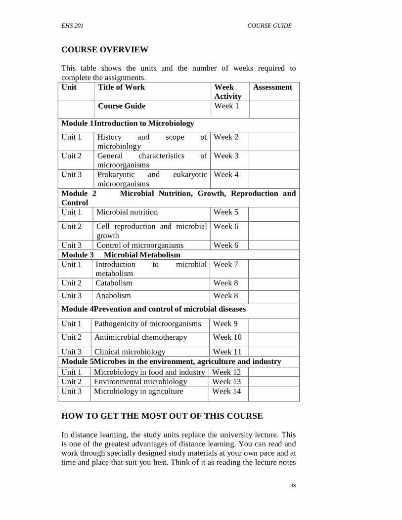
COURSE OVERVIEW This table shows the units and the number of weeks required to complete the assignments. Unit Title of Work Week
Activity Assessment
Course Guide Week 1
Module 1Introduction to Microbiology
Unit 1 History and scope of microbiology
Week 2
Unit 2 General characteristics of microorganisms
Week 3
Unit 3 Prokaryotic and eukaryotic microorganisms
Week 4
Module 2 Microbial Nutrition, Growth, Reproduction and Control Unit 1 Microbial nutrition Week 5
Unit 2 Cell reproduction and microbial growth
Week 6
Unit 3 Control of microorganisms Week 6 Module 3 Microbial Metabolism Unit 1 Introduction to microbial
metabolism Week 7
Unit 2 Catabolism Week 8
Unit 3 Anabolism Week 8
Module 4Prevention and control of microbial diseases
Unit 1 Pathogenicity of microorganisms Week 9
Unit 2 Antimicrobial chemotherapy Week 10
Unit 3 Clinical microbiology Week 11 Module 5Microbes in the environment, agriculture and industry Unit 1 Microbiology in food and industry Week 12 Unit 2 Environmental microbiology Week 13 Unit 3 Microbiology in agriculture Week 14
HOW TO GET THE MOST OUT OF THIS COURSE In distance learning, the study units replace the university lecture. This is one of the greatest advantages of distance learning. You can read and work through specially designed study materials at your own pace and at time and place that suit you best. Think of it as reading the lecture notes

EHS 201 COURSE GUIDE
x
instead of listening to a lecturer. In the same way that a lecturer might set you some reading task, the study units tell you when to read your other material. Just as a lecturer might give you an in-class exercise, your study units provide exercise for you to do at appropriate points. The following are practical strategies for working through the course: i. read the course guide thoroughly organize a study schedule ii. stick to your own created study schedule iii. read the introduction and objectives very well assemble your
study materials iv. work through the unit v. keep in mind that you will learn a lot by doing all your
assignment carefully vi. review the stated objectives vii. do not proceed to the next unit until you are sure you have
understood the previous unit viii. keep to your schedules of studying and assignments ix. review the course and prepare yourself for the final examination. FACILITATORS/TUTORS AND TUTORIALS There are 15 hours of tutorials provided in support of this course. You will be notified of the dates, times and location of the tutorials as well as the name and the phone number of your facilitator, as soon as you are allocated a tutorial group. Your facilitator will mark and comment on your assignments, keep a close watch on your progress and any difficulties you might face and provide assistance to you during the course. You are expected to mail your Tutor marked Assignment to your facilitator before the schedule date (at least two working days are required). They will be marked by your tutor and returned to you as soon as possible. Do not delay to contact your facilitator by telephone or e-mail if you need assistance. The following might be circumstances in which you would find assistance necessary, hence you would have to contact your facilitator if: You do not understand any part of the study or the assigned readings. You have difficulty with self-tests. You have a question or problem with an assignment or with the grading of an assignment. You should endeavour to attend the tutorials. This is the only chance to have face to face contact with your course facilitator and to ask

EHS 201 COURSE GUIDE
xi
question which are answered instantly. You can raise any problem encountered in the course of your study. To gain more benefit from course tutorials prepare a question list before attending them. You will learn a lot from participating actively in discussions. SUMMARY General microbiology provides the student with adequate training and exposure to cell biology, identification, characterization, culturing, prevention and control of microbial growth and diseases. Not all microorganisms are pathogenic and many are involved in food processing, soil fertilization and agriculture and environmental management and remediation. Upon completing this course, you will be equipped with the knowledge of managing microbial diseases agents and application and control of microbial pathogens for general environmental health. Presently, bioremediation and water treatment processes have continuously employed microorganisms and the knowledge of General Microbiology will be ultimately beneficial. In addition, you will be able to answer questions on the subject such as: What is the meaning of microorganisms? What do you understand by microbiology? What are the methods of characterizing and identifying microorganisms? What are the importance of microorganisms in food processing, agriculture and environmental management? How do you control microbial growth and diseases? What are chemotherapeutic agents? What are the physical methods of preventing microbial growth? What are prokaryotic and eukaryotic microorganisms? The above list is just a few of the question expected and is by no means exhaustive. To gain most from this course you are advised to consult relevant books to widen your knowledge on the topic. I wish you success in the course. It is my hope you will find it both illuminating and useful.
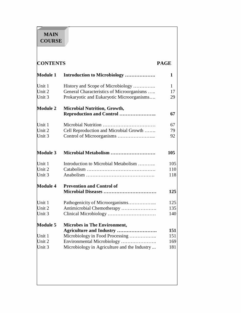
CONTENTS PAGE Module 1 Introduction to Microbiology ………………. 1 Unit 1 History and Scope of Microbiology ………….. 1 Unit 2 General Characteristics of Microorganisms ….. 17 Unit 3 Prokaryotic and Eukaryotic Microorganisms…. 29 Module 2 Microbial Nutrition, Growth,
Reproduction and Control ………………….. 67
Unit 1 Microbial Nutrition …………………………… 67 Unit 2 Cell Reproduction and Microbial Growth ……. 79 Unit 3 Control of Microorganisms …………………... 92 Module 3 Microbial Metabolism ………………………. 105 Unit 1 Introduction to Microbial Metabolism ……….. 105 Unit 2 Catabolism ……………………………………. 110 Unit 3 Anabolism ……………………………………. 118 Module 4 Prevention and Control of
Microbial Diseases …………………………… 125 Unit 1 Pathogenicity of Microorganisms……………... 125 Unit 2 Antimicrobial Chemotherapy …………………. 135 Unit 3 Clinical Microbiology ………………………… 140 Module 5 Microbes in The Environment,
Agriculture and Industry ……………………. 151 Unit 1 Microbiology in Food Processing …………….. 151 Unit 2 Environmental Microbiology …………………. 169 Unit 3 Microbiology in Agriculture and the Industry ... 181
MAIN COURSE
EHS 201 MODULE 1
1
MODULE 1 INTRODUCTION TO MICROBIOLOGY Unit 1 History and Scope of Microbiology Unit 2 General Characteristics of Microorganisms Unit 3 Prokaryotic and Eukaryotic Microorganisms
UNIT 1 HISTORY AND SCOPE OF MICROBIOLOGY CONTENTS 1.0 Introduction 2.0 Objectives 3.0 Main Content
3.1 Definition of microbiology 3.2 Types of microscopes 3.3 Discovery of microorganisms 3.4 Early observation of microbial growth 3.5 History of microbial sterilization 3.6 History of industrial microbiology 3.7 Discovery of microbes as causative agents of diseases 3.8 Development of pure culture techniques 3.9 Discovery of microbes as biogeochemical agents 3.10 Microbial growth beyond the nineteenth century 3.11 Scope and importance of microbiology
4.0 Conclusion 5.0 Summary 6.0 Tutor-Marked Assignments 7.0 References/Further Reading 1.0 INTRODUCTION The importance of the study of General Microbiology as a major prerequisite in Environmental Health Science can never be over emphasized. The history of the discovery of microorganisms, applications of microscopes and other techniques have created opportunities for improved approaches in the study of microbiology, microbial diversity, its isolation and characterization. Beyond the early efforts of many scientist like Anton van Leeuwenhoek (1632-1723), Louis Pasteur (1822-1895) and others, recent developments in microbiology and environmental health studies have be developed. This Unit therefore shall expose the student to the history of the ancient microbiology, the origin of the present efforts in microbiology and to close the gap in knowledge and enhance greater exploits in microbiology.
EHS 201 GENERAL MICROBIOLOGY
2
2.0 OBJECTIVES By the end of this unit, you will be able to: • define microbiology • state the types of microscope • discuss the discovery of microorganisms • narrate early observation of microbial growth • narrate the history of microbial sterilization • discuss the history of industrial microbiology • state the discovery of microbes as causative agents of diseases. 3.0 MAIN CONTENT
The main content of this Unit shall include the definition and concept of microbiology, types of microscope, discovery of microorganisms, importance of microorganisms and the projection of microbiology beyond the 21st Century. 3.1 Definition of Microbiology Microbiology is the study of organisms, called microorganisms that are too small to be perceived clearly by the unaided human eye. Special techniques are required to isolate and grow them. An object that has a diameter of less than 0.1 mm, cannot seen with the naked eye at all and very little detail can be perceived in an object with a diameter of 1mm. Therefore, organisms with a diameter of 1 mm or less are microorganisms and fall into the broad domain of microbiology. Microorganisms have a wide taxonomic distribution; they include some metazoan animals, protozoa, many algae and fungi, bacteria, and viruses. The existence of this microbial world was unknown until the invention of microscopes, optical instruments that serve to magnify objects so small that they cannot be clearly seen by the unaided human eye. Microscopes, invented at the beginning of the seventeenth century, opened the biological realm of the very small to systematic scientific exploration. 3.2 Types of Microscopes Early microscopes were of two kinds. The first were simple microscopes with a single lens of very short focal length, consequently incapable of a high magnification; such instruments did not differ in optical principle from ordinary magnifying glasses able to increase an image several fold, which had been known since antiquity. The second were compound microscopes with a double lens system consisting of an ocular and
EHS 201 MODULE 1
3
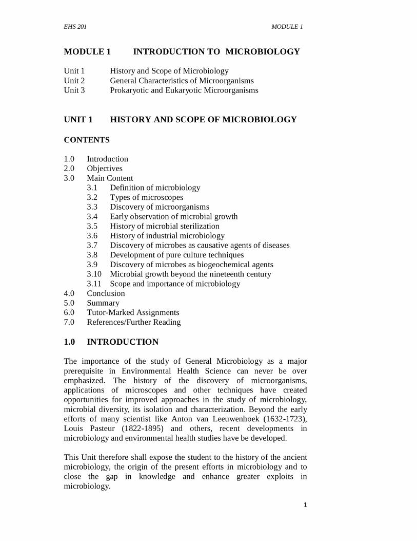
objective. The compound microscope, with its greater intrinsic power of magnification, eventually displaced completely the simple instrument; all our contemporary microscopes are of the compound type. However, nearly all the great original microscopic discoveries were made with simple microscopes. 3.3 Discovery of Microorganisms The discoverer of the microbial world was a Dutch merchant, Anton van Leeuwenhoek.
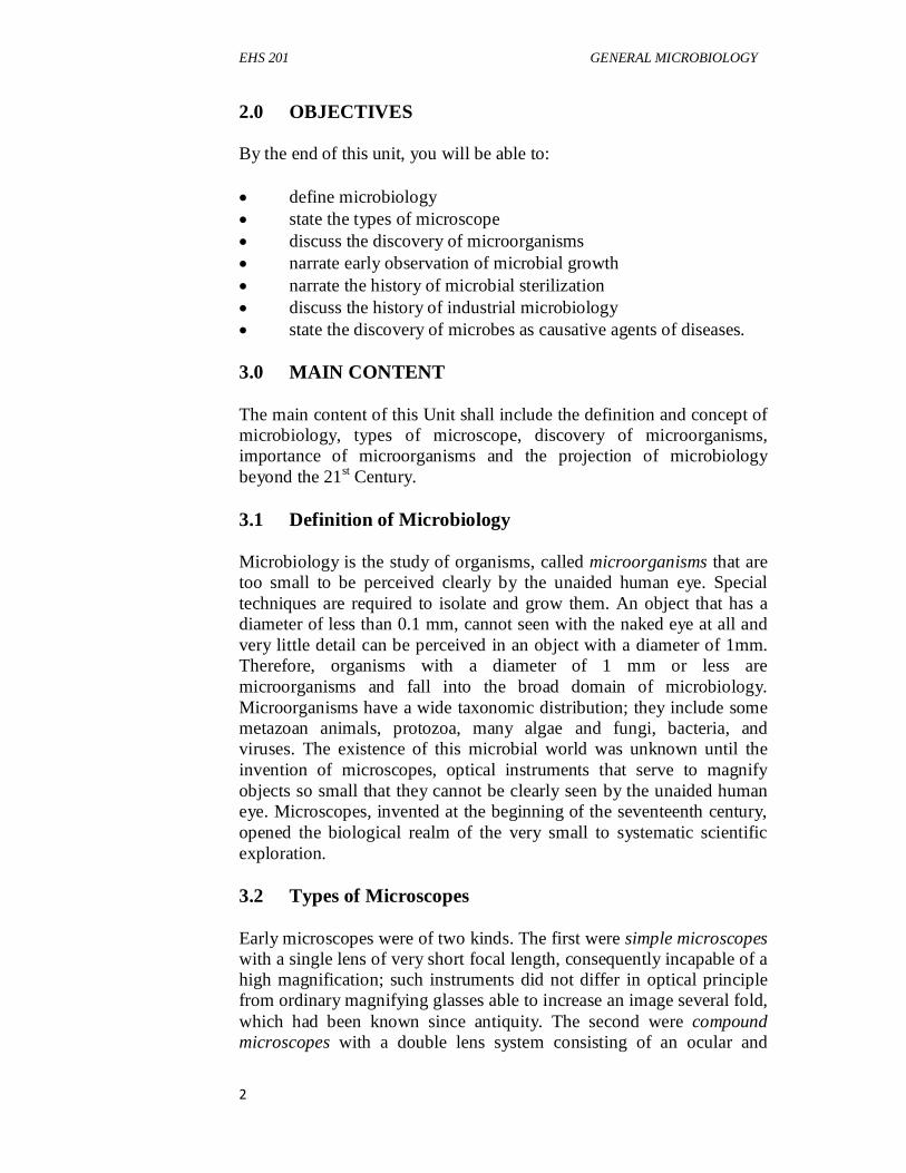
Fig. 3.1: Anton van Leeuwenhoek (1632-1723). In this portrait, he is holding one of his microscopes. Courtesy of the Rijksmuseum, Amsterdam). Source: (Stanier et al., 1987). Leeuwenhoek's microscopes (Figure 3.2) bore little resemblance to the instruments with which we are familiar. The almost spherical lens (a) was mounted between two small metal plates. The specimen was placed on the point of a blunt pin (b) attached to the back plate and was brought into focus by manipulating two screws (c) and (d), which varied the position of the pin relative to the lens. During this operation the observer held the instrument with its other face very close to his eye and squinted through the lens. No change of magnification was possible, the magnifying power of each microscope being an intrinsic property of its lens. Despite the simplicity of their construction, Leeuwenhoek's microscopes were able to give clear images at magnifications that ranged, depending on the focal length of the lens, from about 50 to nearly 300 diameters. The highest magnification that he could obtain was consequently somewhat less than one-third of the highest magnification that is obtainable with a modern compound light microscope. Leeuwenhoek constructed hundreds of such instruments, a few of which survive today.
EHS 201 GENERAL MICROBIOLOGY
4
Fig. 3.2: A drawing to show the construction of one of Leeuwenhoek's microscopes: (a) lens, (b) mounting pin, (c) and (d) focusing screws. After C. E. Dobell, Antony van Leeuwenhoek and HisLittle Animals (New York: Russell and Russell, Inc., 1932). Source: (Stanier et al., 1987). 3.4 Early Observation of Microbial Growth By 1860 some scientists had begun to realize that there is a causal relationship between the development of microorganisms in organic infusions and the chemical changes that take place in these infusions; microorganisms are the agents that bring about the chemical changes. The great pioneer in these studies was Louis Pasteur (Fig. 3.3). However, the acceptance of this concept was conditional on the demonstration that spontaneous generation does not occur. Stung by the continued claims of adherents to the doctrine of spontaneous generation, Pasteur finally turned his attention to this problem. His work on the subject was published in 1861 as a Memoir on the Organized Bodies Which Exist in the Atmosphere. Pasteur first demonstrated that air does contain microscopically observable "organized bodies." He aspirated large quantities of air through a tube that contained a plug of guncotton to serve as a filter. The guncotton was then removed and dissolved in a mixture of alcohol and ether, and the sediment was examined microscopically. In addition to inorganic matter, it contained considerable numbers of small round or oval bodies, indistinguishable from microorganisms. Pasteur next confirmed the fact that heated air can be supplied to a boiled infusion without giving rise to microbial development. Having established this
EHS 201 MODULE 1
5
point, he went on to show that in a closed system the addition of a piece of germ-laden guncotton to a sterile infusion invariably provoked microbial growth.
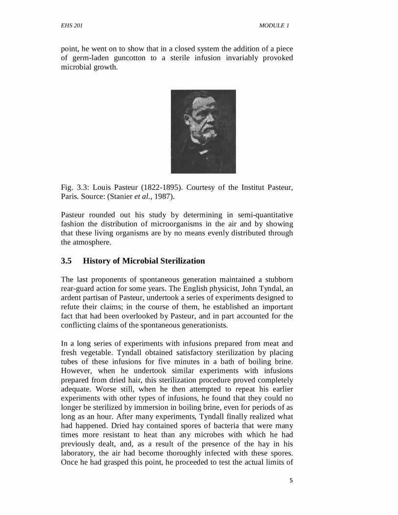
Fig. 3.3: Louis Pasteur (1822-1895). Courtesy of the Institut Pasteur, Paris. Source: (Stanier et al., 1987). Pasteur rounded out his study by determining in semi-quantitative fashion the distribution of microorganisms in the air and by showing that these living organisms are by no means evenly distributed through the atmosphere. 3.5 History of Microbial Sterilization The last proponents of spontaneous generation maintained a stubborn rear-guard action for some years. The English physicist, John Tyndal, an ardent partisan of Pasteur, undertook a series of experiments designed to refute their claims; in the course of them, he established an important fact that had been overlooked by Pasteur, and in part accounted for the conflicting claims of the spontaneous generationists. In a long series of experiments with infusions prepared from meat and fresh vegetable. Tyndall obtained satisfactory sterilization by placing tubes of these infusions for five minutes in a bath of boiling brine. However, when he undertook similar experiments with infusions prepared from dried hair, this sterilization procedure proved completely adequate. Worse still, when he then attempted to repeat his earlier experiments with other types of infusions, he found that they could no longer be sterilized by immersion in boiling brine, even for periods of as long as an hour. After many experiments, Tyndall finally realized what had happened. Dried hay contained spores of bacteria that were many times more resistant to heat than any microbes with which he had previously dealt, and, as a result of the presence of the hay in his laboratory, the air had become thoroughly infected with these spores. Once he had grasped this point, he proceeded to test the actual limits of
EHS 201 GENERAL MICROBIOLOGY
6
heat resistance of the spores of hay bacteria and found that boiling infusions for even as long as 5 hours would not render them sterile with certainty. From these results he concluded that bacteria have phases, one relatively thermolabile (destroyed by boiling for five minutes) and one thermoresistant to an almost incredible extent. Tyndall then proceeded to develop a method of sterilization by discontinuous heating, later called tyndallization, which could be used to kill all bacteria in infusions. Since growing bacteria are easily killed by brief boiling, all that is necessary is to allow the infusion to stand for a certain period to permit germination of the spores with a consequent loss of their heat resistance. A very brief period of boiling can then be used, and repeated, if need be several times at intervals to catch any spores later in germination. Tyndall found that discontinuous boiling for 1 minute on five successive occasions would make an infusion sterile whereas a single continuous boiling for one hour "could not. Recognition of the tremendous heat resistance of bacterial spores was essential to the development of adequate procedures for sterilization. 3.6 History of Industrial Microbiology During the long controversy over spontaneous generation, a correlation between the growth of microorganisms in organic infusions and the onset of chemical changes in the infusion itself was frequently observed. These chemical changes were designated as "fermentation" and "putrefaction." Putrefaction, a process of decomposition that results in the formation of ill-smelling products, occurs characteristically in meat and is a consequence of the breakdown of proteins, the principal organic constituents in such natural materials. Fermentation, a process that results in the formation of alcohols or organic acids, occurs characteristically in plant materials as a consequence of the breakdown of carbohydrates, the predominant organic compounds in plant tissues. In 1837 three men, C. Cagniard-Latour, Th. Schwann, and F. Kiitzing, independently proposed that the yeast that appears during alcoholic fermentation is a microscopic plant and that the conversion of sugars to ethyl alcohol and carbon dioxide characteristic of the alcoholic fermentation is a physiological function of the yeast cell. This theory was bitterly attacked by such leading chemists of the time as J. J. Berzelius, J. Liebig, and F. Wohler, who held the view that fermentation and putrefaction are purely chemical processes.
EHS 201 MODULE 1
7
3.7 Discovery of Microbes as Causative Agents of Diseases During his studies on fermentation, Pasteur, ever conscious of the practical applications of his scientific work, devoted considerable attention to the spoilage of beer and wine, which he showed to be caused by the growth of undesirable microorganisms. Pasteur used a peculiar and significant term to describe these microbially induced spoilage processes; he called them "diseases" of beer and wine. In fact, he was already considering the possibility that microorganisms may act as agents of infectious disease in higher organisms. Some evidence in support of this hypothesis already existed. It had been shown in 1813 that specific fungi can cause diseases of wheat and rye, and in 1845 M. J. Berkeley had proved that the great Potato Blight of Ireland, a natural disaster that deeply influenced Irish history, was caused by a fungus. The first recognition that fungi may be specifically associated with a disease of animals came in 1836 through the work of A. Bassi in Italy on a fungal disease of silkworms. A few years later J. L. Sch6nlein showed that certain skin diseases of man are caused by fungal infections. Despite these indications, very few medical scientists were willing to entertain the notion that the major infectious diseases of man could be caused by microorganisms, and fewer still believed that organisms as small and apparently simple as the bacteria could act as agents of disease. Early discovery of microbes as agents of diseases include; 1. Surgical Antisepsis: The introduction of anesthesia about 1840
made possible a very rapid development of surgical methods. Speed was no longer a primary consideration, and the surgeon was able to undertake operations of a length and complexity that would have been unthinkable previously. However, with the elaboration of surgical technique, a problem that had always existed became more and more serious: surgical sepsis, or the infections that followed surgical intervention and often resulted in the death of the patient. Pasteur's studies on the problem of spontaneous generation had shown the presence of microorganisms in the air and at the same time indicated various ways in which their access to and development in organic infusions could be prevented. A young British surgeon, Joseph Lister, who was deeply impressed by Pasteur's work, reasoned that surgical sepsis might well result from microbial infection of the tissues exposed during operation.
2. Discovery of Anthrax: The discovery that bacteria can act as
specific agents of infectious disease in animals was made through the study of anthrax, a serious infection of domestic animals that
EHS 201 GENERAL MICROBIOLOGY
8
is transmissible to humans. In the terminal stages of a generalized anthrax infection, the rod-shaped bacteria responsible for the disease occur in enormous numbers in the bloodstream. These objects were first observed as early as 1850, and their presence in the blood of infected animals was reported by a series of investigators during the following 15 years. Particularly careful and detailed studies were carried out between 1863 and 1868 by C. J. Davaine, who showed that the rods are invariably present in diseased animals but are undetectable in healthy ones and that the disease can be transmitted to healthy animals by inoculation with blood containing these rod-shaped elements.
This series of experiments fulfilled the criteria which had been laid down 36 years before by J. Henle as logically necessary to establish the causal relationship between a specific microorganism and a specific disease. In generalized form, these criteria are: (1) The microorganism must be present in every case of the disease; (2) The microorganism must be isolated from the diseased host and
grown in pure culture; (3) The specific disease must be reproduced when a pure culture of
the microorganism is inoculated into a healthy susceptible host; and
(4) The microorganism must be recoverable once again from the experimentally infected host. Since Koch was the first to apply these criteria experimentally, they are now generally known as Koch's postulates.
Koch carried out another series of experiments that demonstrated the biological specificity of disease agents. He showed that another spore-forming bacterium, the hay bacillus, does not cause anthrax upon injection, and he also differentiated bacteria that cause other infections from the anthrax organism. From these studies he concluded that only one kind of bacillus is able to cause this specific disease process, while other bacteria either do not produce disease following inoculation, or give rise to other kinds of disease. In the meantime, Pasteur had found a collaborator, J. Joubert, with a knowledge of medical problems. Unaware of Robert Koch's work, Pasteur and Joubert undertook the study of anthrax. They did not add anything new to the conclusions reached by Koch, but they confirmed his work and provided additional demonstrations that the bacillus, and not some other agent, was the specific cause of the disease.
EHS 201 MODULE 1
9
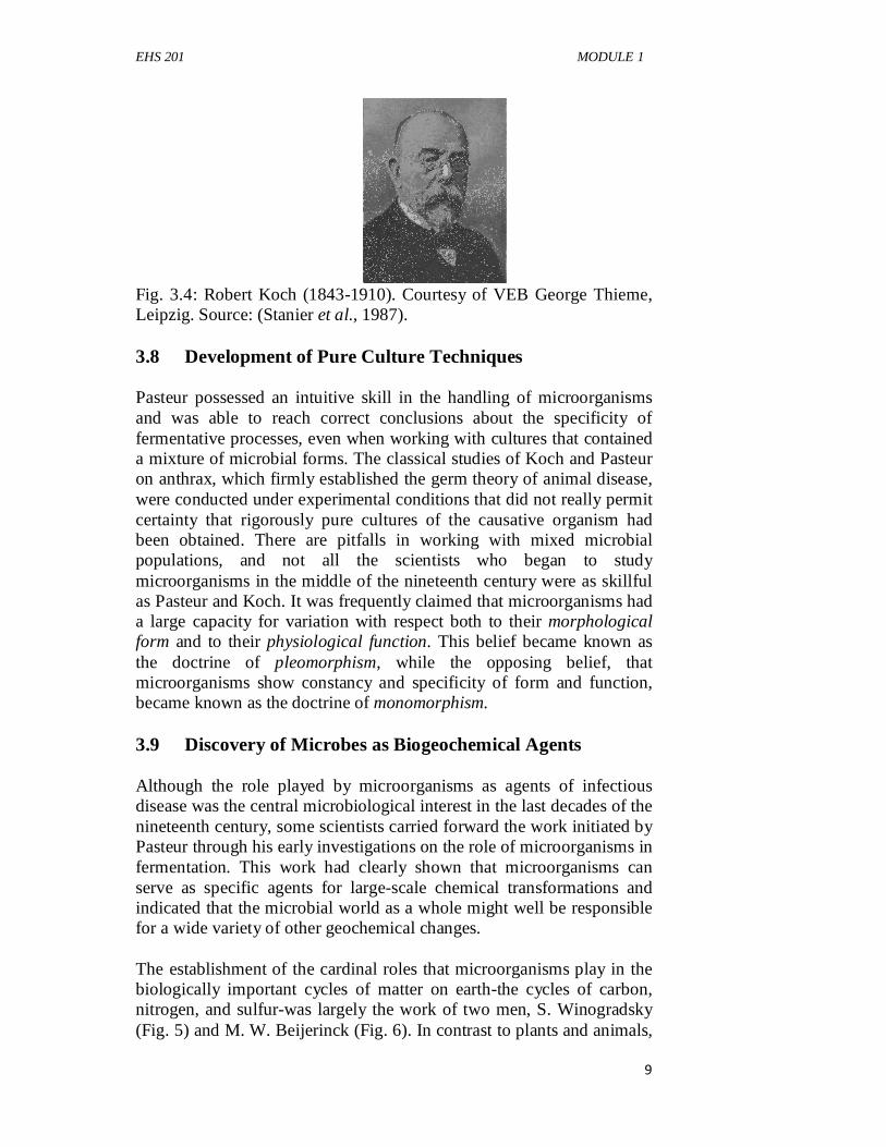
Fig. 3.4: Robert Koch (1843-1910). Courtesy of VEB George Thieme, Leipzig. Source: (Stanier et al., 1987). 3.8 Development of Pure Culture Techniques Pasteur possessed an intuitive skill in the handling of microorganisms and was able to reach correct conclusions about the specificity of fermentative processes, even when working with cultures that contained a mixture of microbial forms. The classical studies of Koch and Pasteur on anthrax, which firmly established the germ theory of animal disease, were conducted under experimental conditions that did not really permit certainty that rigorously pure cultures of the causative organism had been obtained. There are pitfalls in working with mixed microbial populations, and not all the scientists who began to study microorganisms in the middle of the nineteenth century were as skillful as Pasteur and Koch. It was frequently claimed that microorganisms had a large capacity for variation with respect both to their morphological form and to their physiological function. This belief became known as the doctrine of pleomorphism, while the opposing belief, that microorganisms show constancy and specificity of form and function, became known as the doctrine of monomorphism. 3.9 Discovery of Microbes as Biogeochemical Agents Although the role played by microorganisms as agents of infectious disease was the central microbiological interest in the last decades of the nineteenth century, some scientists carried forward the work initiated by Pasteur through his early investigations on the role of microorganisms in fermentation. This work had clearly shown that microorganisms can serve as specific agents for large-scale chemical transformations and indicated that the microbial world as a whole might well be responsible for a wide variety of other geochemical changes. The establishment of the cardinal roles that microorganisms play in the biologically important cycles of matter on earth-the cycles of carbon, nitrogen, and sulfur-was largely the work of two men, S. Winogradsky (Fig. 5) and M. W. Beijerinck (Fig. 6). In contrast to plants and animals,
EHS 201 GENERAL MICROBIOLOGY
10
microorganisms show an extraordinarily wide range of physiological diversity. Many groups are specialized for carrying out chemical transformations that cannot be performed at all by plants and animals, and thus play vital parts in the turnover of matter on earth.
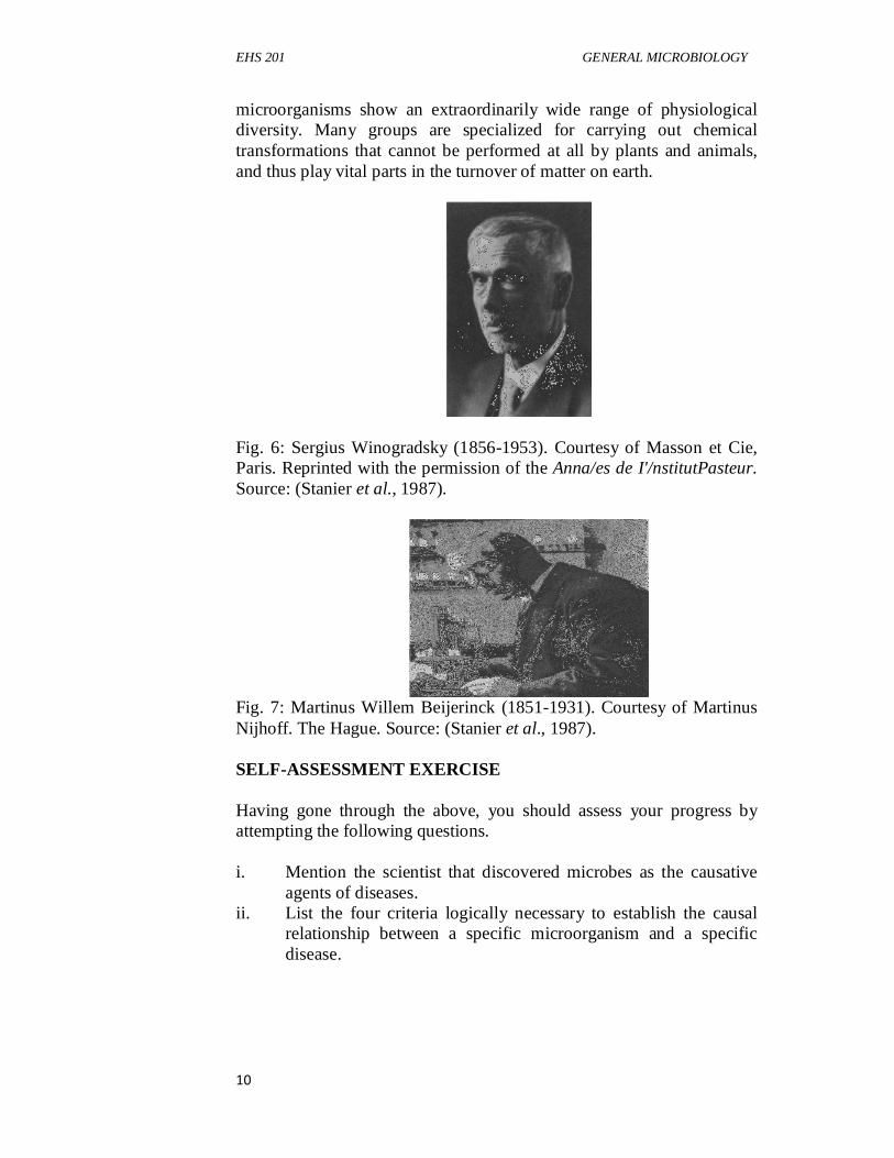
Fig. 6: Sergius Winogradsky (1856-1953). Courtesy of Masson et Cie, Paris. Reprinted with the permission of the Anna/es de I'/nstitutPasteur. Source: (Stanier et al., 1987).
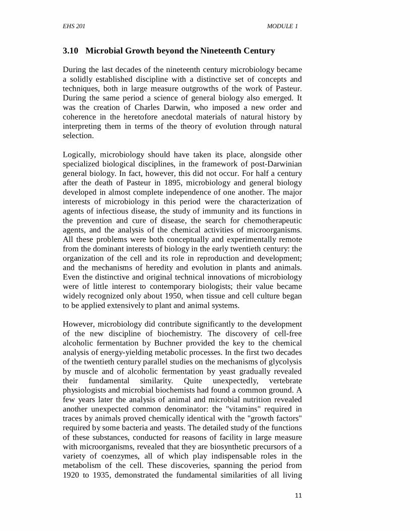
Fig. 7: Martinus Willem Beijerinck (1851-1931). Courtesy of Martinus Nijhoff. The Hague. Source: (Stanier et al., 1987). SELF-ASSESSMENT EXERCISE Having gone through the above, you should assess your progress by attempting the following questions. i. Mention the scientist that discovered microbes as the causative
agents of diseases. ii. List the four criteria logically necessary to establish the causal
relationship between a specific microorganism and a specific disease.
EHS 201 MODULE 1
11
3.10 Microbial Growth beyond the Nineteenth Century During the last decades of the nineteenth century microbiology became a solidly established discipline with a distinctive set of concepts and techniques, both in large measure outgrowths of the work of Pasteur. During the same period a science of general biology also emerged. It was the creation of Charles Darwin, who imposed a new order and coherence in the heretofore anecdotal materials of natural history by interpreting them in terms of the theory of evolution through natural selection. Logically, microbiology should have taken its place, alongside other specialized biological disciplines, in the framework of post-Darwinian general biology. In fact, however, this did not occur. For half a century after the death of Pasteur in 1895, microbiology and general biology developed in almost complete independence of one another. The major interests of microbiology in this period were the characterization of agents of infectious disease, the study of immunity and its functions in the prevention and cure of disease, the search for chemotherapeutic agents, and the analysis of the chemical activities of microorganisms. All these problems were both conceptually and experimentally remote from the dominant interests of biology in the early twentieth century: the organization of the cell and its role in reproduction and development; and the mechanisms of heredity and evolution in plants and animals. Even the distinctive and original technical innovations of microbiology were of little interest to contemporary biologists; their value became widely recognized only about 1950, when tissue and cell culture began to be applied extensively to plant and animal systems. However, microbiology did contribute significantly to the development of the new discipline of biochemistry. The discovery of cell-free alcoholic fermentation by Buchner provided the key to the chemical analysis of energy-yielding metabolic processes. In the first two decades of the twentieth century parallel studies on the mechanisms of glycolysis by muscle and of alcoholic fermentation by yeast gradually revealed their fundamental similarity. Quite unexpectedly, vertebrate physiologists and microbial biochemists had found a common ground. A few years later the analysis of animal and microbial nutrition revealed another unexpected common denominator: the "vitamins" required in traces by animals proved chemically identical with the "growth factors" required by some bacteria and yeasts. The detailed study of the functions of these substances, conducted for reasons of facility in large measure with microorganisms, revealed that they are biosynthetic precursors of a variety of coenzymes, all of which play indispensable roles in the metabolism of the cell. These discoveries, spanning the period from 1920 to 1935, demonstrated the fundamental similarities of all living
EHS 201 GENERAL MICROBIOLOGY
12
systems at the metabolic level-a doctrine proclaimed by biochemists and microbiologists under the slogan "the unity of biochemistry." The second great advance of biology in the early twentieth century-the creation of the discipline of genetics, formed through the convergence of cytology and Mendelian analysis-had no immediate impact on microbiology. Indeed, it long seemed doubtful whether the mechanisms of inheritance operative in plants and animals likewise functioned in bacteria. The first important contact between genetics and microbiology occurred in 1941, when Beadle and Tatum succeeded in isolating a series of biochemical mutants from the fungus Neurospora. This opened the way to the analysis of the consequences of mutation in biochemical terms, and Neurospora joined the fruit fly and the maize plant as a material of choice for genetic research. In 1943 an analysis by Delbriick and Luria of mutation in bacteria provided the technical and conceptual basis for genetic work on these microorganisms. Soon afterward several mechanisms of genetic transfer were shown to exist in bacteria, all significantly different from the mechanism of sexual recombination in plants and animals. In 1944 the work of Avery, McLeod and McCarty on the process of bacterial genetic transfer known as transformation revealed that it is mediated by free deoxyribonucleic acid (DNA). The chemical nature of the hereditary material was thus discovered. The confluence of microbiology, genetics, and biochemistry between 1940 and 1945 brought to an end the long isolation of microbiology from the main currents of biological thought. It also set the stage for the second major revolution in, biology, to which microbiologists made many contributions of fundamental importance: the advent of molecular biology. 3.11 Scope and Importance of Microbiology As the scientist-writer Steven Jay Gould (1941–2002) emphasized, we live in the age of bacteria. They were the first living organisms on our planet, likely created the atmosphere that allowed the evolution of oxygen-consuming life-forms, and now live virtually everywhere life is possible. Furthermore, the biosphere depends on their activities, and they influence human society in countless ways. Because microorganisms play such diverse roles, modern microbiology is a large discipline with many different specialties; it has a great impact on fields such as medicine, agricultural and food sciences, ecology, genetics, biochemistry, and molecular biology. One indication of the importance

EHS 201 MODULE 1
13
of microbiology is the Nobel Prize given for work in physiology or medicine. About one-third of these prizes have been awarded to scientists working on microbiological problems. Microbiology has both basic and applied aspects. The basic aspects are concerned with the biology of microorganisms themselves. The applied aspects are concerned with practical problems such as disease, water and wastewater treatment, food spoilage and food production, and industrial uses of microbes. It is important to note that the basic and applied aspects of microbiology are intertwined. Basic research is often conducted in applied fields, and applications often arise out of basic research. A discussion of some of the major fields of microbiology and the occupations within them follows. Although pathogenic microbes are the minority, they garner considerable interest. Thus, one of the most active and important fields in microbiology is medical microbiology, which deals with diseases of humans and animals. Medical microbiologists identify the agents causing infectious diseases and plan measures for their control and elimination. Frequently they are involved in tracking down new, unidentified pathogens such as the agent that causes variant Creutzfeldt-Jakob disease (the human version of “mad cow disease”), Hantavirus, West Nile virus, and the virus responsible for SARS. These microbiologists also study the ways in which microorganisms cause disease. As noted earlier, major epidemics have regularly affected human history. The 1918 influenza pandemic is of particular note; it killed more than 20 million people in about one year. Public health microbiology is concerned with the control and spread of such communicable diseases. Public health microbiologists and epidemiologists monitor the amount of disease in populations. Based on their observations, they can detect outbreaks and developing epidemics, and implement appropriate control measures in response. They also conduct surveillance for new diseases as well as bioterrorism events. Those public health microbiologists working for local governments monitor community food establishments and water supplies in an attempt to keep them safe and free from infectious disease agents. Immunology is concerned with how the immune system protects the body from pathogens and the response of infectious agents. It is one of the fastest growing areas in science. Much of the growth began with the discovery of HIV, which specifically targets cells of the immune system. Immunology also deals with health problems such as the nature and treatment of allergies and autoimmune diseases such as rheumatoid arthritis.
EHS 201 GENERAL MICROBIOLOGY
14
Agricultural microbiology is concerned with the impact of microorganisms on agriculture. Microbes such as nitrogen-fixing bacteria play critical roles in the nitrogen cycle and affect soil fertility. Other microbes live in the digestive tracts of ruminants such as cattle and break down the plant materials these animals ingest. There are also plant and animal pathogens that can have significant economic impacts if not controlled. Agricultural microbiologists work on methods to increase soil fertility and crop yields, study rumen microorganisms in order to increase meat and milk production, and try to combat plant and animal diseases. Currently many agricultural microbiologists are studying the use of bacterial and viral insect pathogens as substitutes for chemical pesticides. Microbial ecology is concerned with the relationships between microorganisms and the components of their living and nonliving habitats. Microbial ecologists study the global and local contributions of microorganisms to the carbon, nitrogen, and sulfur cycles, including the role of microbes in both the production and removal of greenhouse gases such as carbon dioxide and methane. The study of pollution effects on microorganisms also is important because of the impact these organisms have on the environment. Microbial ecologists are employing microorganisms in bioremediation to reduce pollution. The study of the microbes normally associated with the human body has become a new frontier in microbial ecology. Numerous foods are made using microorganisms. On the other hand, some microbes cause food spoilage or are pathogens spread through food. An excellent example of the latter is Escherichia coli O157:H7, which in 2006 caused a widespread outbreak of disease when it contaminated a major source of spinach in the United States. Scientists working in food and dairy microbiology continue to explore the use of microbes in food production. They also work to prevent microbial spoilage of food and the transmission of food-borne diseases. There is also considerable research on the use of microorganisms themselves as a nutrient source for livestock and humans. In 1929 Alexander Fleming discovered that the fungus Penicillium produced what he called penicillin, the first antibiotic that could successfully control bacterial infections. Although it took World War II for scientists to learn how to mass-produce it, scientists soon found other microorganisms capable of producing additional antibiotics as well as compounds such as citric acid, vitamin B12, and monosodium glutamate (MSG). Today, industrial microbiologists use microorganisms to make products such as antibiotics, vaccines, steroids, alcohols and other solvents, vitamins, amino acids, and enzymes. Industrial microbiologists
EHS 201 MODULE 1
15
identify microbes of use to industry. They also utilize techniques to improve production by microbes and devise systems for culturing them and isolating the products they make. Microbes are metabolically diverse and can employ a wide variety of energy sources, including organic matter, inorganic molecules (e.g., H2 and NH3), and sunlight. Microbiologists working in microbial physiology and biochemistry study many aspects of the biology of microorganisms, including their metabolic capabilities. They may also study the synthesis of antibiotics and toxins, the ways in which microorganisms survive harsh environmental conditions, and the effects of chemical and physical agents on microbial growth and survival. Microbial genetics and molecular biology focus on the nature of genetic information and how it regulates the development and function of cells and organisms. The bacteria E. coli and Bacillussubtilis, the yeast Saccharomyces cerevisiae (baker’s yeast), and bacterial viruses such as T4 and lambda continue to be important model organisms used to understand biological phenomena. Microbial geneticists also play a significant role in applied microbiology because they develop techniques that are useful in agricultural microbiology, industrial microbiology, food and dairy microbiology, and medicine. Because of the practical importance of microbes and their use as model organisms, the future of microbiology is bright. However, it is important to remember that future advances in microbiology will build on the foundations laid by earlier scientists. 4.0 CONCLUSION General microbiology is made more understandable by explaining the history, definition and concept microbiology, the ancient discoveries in the field of microbiology and the scope and importance of microbiology. 5.0 SUMMARY In this unit, the student has learnt the meaning of microbiology, history of the discovery of many aspects of microbiology, the scope and importance of microbiology. Beyond ancient microbial technology, the student will be challenged with the need for improved scientific approaches to microbiology to meet millennium demands.
EHS 201 GENERAL MICROBIOLOGY
16
6.0 TUTOR-MARKED ASSIGNMENTS 1 (a) Define microbiology (b) Who discovered the microbial world? 2 (a) Discuss briefly microbial growth beyond the 21st century.
(b) List five important applications of microbiology. 7.0 REFERENCES/FURTHER READING Brock, T. D. (1961).Milestones in Microbiology. Englewood Cliffs,
N.J.: Prentice Hall, Inc. Bulloch, W. (1979).The History of Bacteriology. New York: Oxford
University Press, 1960; Republished: New York: Dover Publications, Inc.
Stanier, R.Y., Ingraham, J.L., Wheelis, M.L. & Painter, P.R. (1987).
General Microbiology, Fifth Edition. Macmillan Press LTD Houndmills, Basingstoke, Hampshire RG21 6XS and London pp.2-15.
Willey, J.M., Sherwood, L.M. & Woolverton, C.J. (2009). Prescott’s
principles of ‘microbiology—(1st ed). McGraw-Hill. pp. 1-12.
EHS 201 MODULE 1
17
UNIT 2 GENERAL CHARACTERISTICS OF MICROORGANISMS CONTENTS 1.0 Introduction 2.0 Objectives 3.0 Main Content
3.1 Microbial Taxonomy 3.2 Characteristics of Microorganisms
3.2.1 Viruses 3.2.2 Bacteria 3.2.3 Fungi
3.3 Rapid Methods of Identification 3.4 Morphological Characteristics 3.5 Biochemical Characteristics
4.0 Conclusion 5.0 Summary 6.0 Tutor-Marked Assignments 7.0 References/Further Reading 1.0 INTRODUCTION Many microorganisms can be identified by particular growth patterns and biochemical characteristics. These characteristics vary, depending on whether the clinical microbiologist is dealing with viruses, fungi (yeasts, molds), parasites (protozoa, helminths), common gram-positive or gram-negative bacteria, rickettsias, chlamydiae, or mycoplasmas. These processes in microbial identification and characterization are veritable tools in the study of microbiology which enhances other related studies especially in environmental health science and diseases control. It will further help to classify and characterize many organisms of food processing, agricultural, environmental management and industrial importance. 2.0 OBJECTIVES By the end of this unit, you will be able to: • discuss microbial taxonomy, • characterise different types of microorganisms • explain Rapid Methods of Identification of Microorganisms • characterise microorganisms based on morphology and
biochemical reactions.

EHS 201 GENERAL MICROBIOLOGY
18
3.0 MAIN CONTENT The main content of this unit shall include understanding of microbial taxonomy, characterization of different types of microorganisms, rapid methods of microbial identification, morphological and biochemical characterization. 3.1 Microbial Taxonomy Taxonomy is a system of orderly classification of living organisms into categories called taxons and it aims to classify living organisms by differentiating them and establishing relationships between groups of organisms. Taxonomists are those who study the classification of organisms, these persons can highly argumentative, different opinions on classification of organisms exist by Taxonomists, when you put two of them together and you may get three opinions classification of organism. Taxonomy is based on the Linnaean binomial system.The original rationale behind this system is not used now, Carolus Linnaeus (he was Swedish, but he Latinized his name) lived in a time centuries ago when it was not appreciated that evolution occurred. He classified organisms mostly according to similar appearance, but this can be misleading, in the absence of evolutionary theory fish and whales are grouped together for instance, because they look alike. The formal binomial naming method created by Linnaeus is still used, but the modern classification rationale is based on evolutionary relatedness. All living cellular things (biological entities other than viruses and prions) have species and a genus designation, and organisms are placed into groupings that reflect their evolutionary relationships. The basic taxonomic group in microbial taxonomy is the species. Taxonomists working with higher organisms define their species differently than microbiologists. Prokaryotic species are characterized by differences in their phenotype and genotype. Phenotype is the collection of visible characteristics and the behavior of a microorganism. Genotype is the genetic make- up of a microorganism The binomial (scientific) nomenclature assigns each microbe 2 names; Genus (noun), first letter always capitalized and species (adjective) lowercase. Both are written italicized or underlined. For example, Staphylococcus aureus (S. aureus), Escherichia coli (E. coli). These look like really fussy, picky rules, but this is essential, serious misunderstandings can occur if this convention is not followed. It is totally unacceptable to write “Escherichia coli” this was (not underlined or italicized). “Escherichia Coli” written in this form is also is incorrect, reason being that the first coli capitalized. It is also wrong to write thus; escherichia coli, the term is italicized but the first letter of the generic
EHS 201 MODULE 1
19
name must begin with a capital letter. It is acceptable and a usual practice to just use the first letter of the genus of a species Within each Kingdom each organism is nested into a hierarchical classification of taxons in the order - Kingdom, Phylum-Division, Class, Order, Family, Genus, Species. The order of this list is important (which accounts for use of the term hierarchical), each taxon holds progressively more numbers of taxonomically different organisms, as one moves up the list from species level, thus a genus contains a number of species, a family contains a number of genera and thus contains more species than a single genus in that classification since each family contains a number of genera each with their own species. 3.2 Characteristics of Microorganisms 3.2.1 Viruses Viruses are identified by isolation in conventional cell (tissue) culture, by immunodiagnosis (fluorescent antibody, enzyme immunoassay, radioimmunoassay, latex agglutination, and immunoperoxidase), and by molecular detection methods such as nucleic acid probes and PCR amplification assays. Several types of systems are available for virus cultivation: cell cultures, embryonated hen’s eggs, and experimental animals; these are discussed shortly. Cell cultures are divided into three general classes: 1. Primary cultures: These consist of cells derived directly from
tissues such as monkey kidney and mink lung cells that have undergone one or two passages (subcultures) since harvesting.
2. Semicontinuous cell cultures or low-passage cell lines: These are obtained from subcultures of a primary culture and usually consist of diploid fibroblasts that undergo a finite number of divisions.
3. Continuous or immortalized cell cultures, such as HEp-2 cells: These are derived from transformed cells that are generally epithelial in origin. These cultures grow rapidly, are heteroploid (having a chromosome number that is not a simple multiple of the haploid number), and can be subcultured indefinitely.
Each type of cell culture favors the growth of a different array of viruses, just as bacterial culture media have differing selective and restrictive properties for growth of bacteria. Viral replication in cell cultures is detected in two ways:

EHS 201 GENERAL MICROBIOLOGY
20
(1) By observing the presence or absence of cytopathic effects (CPEs) and
(2) By hemadsorption. A cytopathic effect is an observable morphological change that occurs in cells because of viral replication.
Examples include ballooning, binding together, clustering, or even death of the culture cells. During the incubation period of a cell culture, red blood cells can be added. Several viruses alter the plasma membrane of infected culture cells so that red blood cells adhere firmly to them. This phenomenon is called hemadsorption. 3.2.2 Bacteria Isolation and growth of bacteria are required before many diagnostic tests can be used to confirm the identification of the pathogen. The presence of bacterial growth usually can be recognized by the development of colonies on solid media or turbidity in liquid media. The time for visible growth to occur is an important variable in the clinical laboratory. For example, most pathogenic bacteria require only a few hours to produce visible growth, whereas it may take weeks for colonies of mycobacteria or mycoplasmas to become evident. The clinical microbiologist as well as the clinician should be aware of reasonable reporting times for various cultures. The initial identity of a bacterial organism may be suggested by; (1) The source of the culture specimen; (2) Its microscopic appearance and Gram reaction; (3) Its pattern of growth on selective, differential, or metabolism-
determining media; and (4) Its hemolytic, metabolic, and fermentative properties on the
various media. For example, methylene blue is often used to inhibit the growth of Gram-positive bacteria, whereas phenylethyl alcohol is often used to inhibit Gram-negative bacteria. Sheep blood–supplemented agars can be used to determine hemolytic capabilities. After the microscopic and growth characteristics of a pure culture of bacteria are examined, specific biochemical tests can be performed. Classic dichotomous keys are coupled with the biochemical tests for the identification of bacteria from specimens. Generally, fewer than 20 tests are required to identify clinical bacterial isolates to the species level.
EHS 201 MODULE 1
21
Certain bacteria require special considerations. For instance, the rickettsias, chlamydiae, and mycoplasmas differ from other bacterial pathogens in a variety of ways. Rickettsias can be diagnosed by immunoassays or by isolation of the microorganism. Because isolation is both hazardous and expensive, immunological methods are preferred. Isolation of rickettsias and diagnosis of rickettsial diseases are generally confined to reference and specialized research laboratories. Chlamydiae can be demonstrated in tissues and cell scrapings with Giemsa staining, which detects the characteristic intracellular inclusion bodies. Immunofluorescent staining of tissues and cells with monoclonal antibody reagents is a more sensitive and specific means of diagnosis. The most sensitive methods for demonstrating chlamydiae in clinical specimens involve nucleic acid sequencing and PCR based methods. The most routinely used techniques for identification of the mycoplasmas are immunological (hemagglutinin), complement-fixing antigen-antibody reactions using the patient’s sera and PCR. These microorganisms are slow growing; therefore positive results from isolation procedures are rarely available before 30 days—a long delay with an approach that offers little advantage over standard techniques. DNA probes are also used for the detection of Mycoplasma pneumoniae in clinical specimens. 3.2.3 Fungi Fungal cultures remain the standard for the recovery of fungi from patient specimens; however, the time needed to culture fungi varies anywhere from a few days to several weeks, depending on the organism. For this reason, fungal cultures demonstrating no growth should be maintained for a minimum of 30 days before they are discarded as a negative result. Cultures should be evaluated for rate and appearance of growth on at least one selective and one nonselective agar medium, with careful examination of colonial morphology, color, and dimorphism. Typically, the isolation of fungi is accomplished by concurrent culture of the specimen on media that is respectively supplemented and unsupplemented with antibiotics and cycloheximide. Antibiotics inhibit bacteria that may be in the specimen and cycloheximide inhibits saprophytic (living on decaying matter) molds. However, a number of media formulations are routinely used to culture specific fungi. Fungal serology (e.g., complement fixation and immunodiffusion) is designed to detect serum antibody but is limited to a few fungi. The cryptococcal latex antigen test is routinely used for the direct detection of Cryptococcus neoformans in serum and cerebrospinal fluid. In the clinical laboratory, nonautomated and automated methods for rapid identification (minutes to hours) are used to detect most yeasts. Any
EHS 201 GENERAL MICROBIOLOGY
22
biochemical methods used to detect fungi should always be accompanied by morphological studies examining for pseudohyphae, yeast cell structure, chlamydospores, and so on. SELF-ASSESSMENT EXERCISE Having gone through the above, you should assess your progress by attempting the following questions. i. Mention three types of microorganisms. ii. List the four initial identities that may suggest a bacterial
organism. 3.3 Rapid Methods of Identification Clinical microbiology has benefited greatly from technological advances in equipment, computer software and databases, molecular biology, and immunochemistry. With new technology, it has been possible to shift from the multistep methods previously discussed to unitary procedures and systems that incorporate standardization, speed, reproducibility, miniaturization, mechanization, and automation. These rapid identification methods can be divided into three categories: (1) Manual biochemical “kit” systems, (2) Mechanized/automated systems, and (3) Immunologic systems. One example of a “kit approach” biochemical system for the identification of members of the family Enterobacteriaceaeand other Gram-negative bacteria is the API 20E system. It consists of a plastic strip with 20 microtubes containing dehydrated biochemical substrates that can detect certain biochemical characteristics. The biochemical substrates in the20 microtubes are inoculated with a pure culture of bacteriaevenly suspended in sterile physiological saline. After 5 to 12hours of incubation, the 20 test results are converted to a seven ornine-digit profile. This profile number can be used witha computer or a book called the API Profile Index to identifythe bacterium.
3.4 Morphological Characteristics i. The Isolation of Pure Cultures by Plating Methods
Due to the microscopic nature (the small size) of microorganisms, the amount of information that can be obtained about their properties from the examination of individuals is limited; for the most part, the microbiologist studies populations, containing millions or billions of individuals. Such populations are obtained by growing microorganisms, under more or less well-defined conditions, as cultures. A culture that contains only
EHS 201 MODULE 1
23
one kind of microorganism is known as a pure or axenic culture. A culture that contains more than one kind of microorganism is known as a mixed culture; if it contains only two kinds of microorganisms, deliberately maintained in association with one another, it is known as a o-membered culture.
Pure cultures of microorganisms that form discrete colonies on solid media (e.g., yeasts, most bacteria, many fungi and unicellular algae) may be most simply obtained by one of the modifications of the plating method. Several plating methods can be used to determine the number of viable microbes in a sample. These are referred to as viable counting methods (plate counts) because they count only those cells that are able to reproduce when cultured. Two commonly procedures used are the spread-plate and the pour-plate techniques. This method involves the separation and immobilization of individual organisms on or in a nutrient medium solidified with agar or some other appropriate gelling agent. Each viable organism gives rise, through growth, to a colony from which transfers can be readily made.
Microorganisms do not require much space for development; hence an artificial environment can be created within the confines of a test tube, a flask, or a Petri dish, the three kinds of containers most commonly used to cultivate microorganisms. The culture containers must be rendered initially sterile (free of any living microorganism) and, after the introduction of the desired type of microorganism, it must be protected from subsequent external contamination. The primary source of external contamination is the atmosphere, which always contains floating microorganisms.
Plating techniques are simple, sensitive, and widely used for viable counts of bacteria and other microorganisms in samples of food, water, and soil. Several problems, however, can lead to inaccurate counts. Low counts will result if clumps of cells are not broken up and the microorganisms well dispersed
a. Streak Plate Method
The streaked plate is in general the most useful plating method. A sterilized bent wire is dipped into a suitable diluted suspension of organisms and is then used to make a series of parallel, non-overlapping streaks on the surface of an already solidified agar plate. The inoculum is progressively diluted with each successive streak, so that even if the initial streaks yield confluent growth, well isolated colonies develop along the lines of later streaks. The inoculum (the microbial material used to seed or inoculate a culture vessel) is commonly introduced on a metal wire or loop,
EHS 201 GENERAL MICROBIOLOGY
24
which is rapidly sterilized just before its use by heating in a flame Transfers of liquid cultures can also be made by pipette. For this purpose, the mouth end of the pipette may be plugged with cotton wool, and the pipette is sterilized in a paper wrapping or in a glass or metal container, which keeps both inner and outer surfaces free of contamination until the time of use.
b. Pour Plate Method
Alternatively, isolations can be made with poured plates, in this method, fixed amount of inoculum (generally 1 ml) from a broth/sample is placed in the center of sterile Petri dish using a sterile pipette. Molten cooled agar (approx. 15mL) is then poured into the Petri dish containing the inoculum and mixed well. After the solidification of the agar, the plate is inverted and incubated at 37°C for 24-48 hours. Microorganisms will grow both on the surface and within the medium. Colonies that grow within the medium generally are small in size and may be confluent; the few that grow on the agar surface are of the same size and appearance as those on a streak plate. Each (both large and small) colony is carefully counted (using magnifying colony counter if needed). Each colony represents a “colony forming unit” (CFU). The isolation of anaerobic bacteria by plating methods poses special problems. Provided that the desired organisms are not rapidly killed by exposure to oxygen, plates may be prepared in the usual manner and then incubated in closed containers, from which the oxygen is removed either by chemical absorption or evacuation. For more oxygen-sensitive anaerobes, a modification of the pour plate method, known as the dilution shake culture, is preferred. A tube of melted and cooledagar medium is inoculated and mixed, and approximately one-tenth of its contents is transferred to a second tube, which is then mixed and used to inoculate a third tube in a similar fashion. After 6 to 10 successive dilutions have been prepared, the tubes are rapidly cooled and sealed, by pouring a layer of sterile petroleum jelly and paraffin on the surface, thus preventing access of air to the agar column. In shake culture the colonies develop deep in the agar column and are thus not easily accessible for transfer. To make a transfer, the petroleum jelly-paraffin seal is removed with a sterile needle, and the agar column is extruded from the tube into a sterile petri dish by gently blowing a stream of gas through a capillary pipette inserted between the tube wall and the agar. The column is sectioned into discs with a sterile knife to permit examination and transfer of colonies.
EHS 201 MODULE 1
25
ii. Selective Media A selective medium is prepared by the addition of specific substances to a culture medium that will permit growth of one group of bacteria while inhibiting growth of some other groups. These are examples: Salmonella-Shigella agar (SS) is used to isolate Salmonella and Shigella species. Its bile salt mixture inhibits many groups of coliforms. Both Salmonella and Shigella species produce colorless colonies because they are unable to ferment lactose. Lactose-fermenting bacteria will produce pink colonies. Mannitol salt agar (MS) is used for the isolation of staphylococci. The selectivity is obtained by the high (7.5%) salt concentration that inhibits growth of many groups of bacteria. The mannitol in this medium helps in differentiating the pathogenic from the nonpathogenic staphylococci, as the former ferment mannitol to form acid while the latter do not. Thus this medium is also differential. Bismuth sulfite agar (BS) is used for the isolation of Salmonella enterica serovar Typhi, especially from stool and food specimens. S. enterica serovar Typhi reduces the sulfite to sulfide, resulting in black colonies with a metallic sheen. The addition of blood, serum, or extracts to tryptic soy agar or broth will support the growth of many fastidious bacteria. These media are used primarily to isolate bacteria from cerebrospinal fluid, pleural fluid, sputum, and wound abscesses.
iii. Differential Media
The incorporation of certain chemicals into a medium may result in diagnostically useful growth or visible change in the medium after incubation. These are examples:
1. Eosin methylene blue agar (EMB) differentiates between lactose fermenters and nonlactose fermenters. EMB contains lactose, salts, and two dyes—eosin and methylene blue. E. coli, which is a lactose fermenter, will produce a dark colony or one that has a metallic sheen. S. enterica serovar Typhi, a nonlactose fermenter, will appear colorless.
2. MacConkey agar is used for the selection and recovery of Enterobacteriaceae and related gram-negative rods. The bile salts and crystal violet in this medium inhibit the growth of gram-positive bacteria and some fastidious gram-negative bacteria. Because lactose is the sole carbohydrate, lactose-fermenting
EHS 201 GENERAL MICROBIOLOGY
26
bacteria produce colonies that are various shades of red, whereas nonlactose fermenters produce colorless colonies.
3. Hektoen enteric agar is used to increase the yield of Salmonella and Shigella species relative to other microbiota. The high bile salt concentration inhibits the growth of gram-positive bacteria and retards the growth of many coliform strains.
4. Blood agar: addition of citrated blood to tryptic soy agar makes possible variable hemolysis, which permits differentiation of some species of bacteria. Three hemolytic patterns can be observed on blood agar.
1. α-hemolysis—greenish to brownish halo around the colony (e.g., Streptococcus gordonii, Streptococcus pneumoniae).
2. β-hemolysis—complete lysis of blood cells resulting in a clearing effect around growth of the colony (e.g., Staphylococcus aureus and Streptococcus pyogenes).
3. Nonhemolytic—no change in medium (e.g., Staphylococcus epidermidis and Staphylococcus saprophyticus).
3.4 Biochemical Characteristics Some media are used to test bacteria for particular metabolic activities, products, or requirements. These are examples: a. Urea broth: It is used to detect the enzyme urease. Some enteric
bacteria are able to break down urea, using urease, into ammonia and CO2.
b. Triple sugar iron (TSI) agar: It contains lactose, sucrose, and glucose plus ferrous ammonium sulfate and sodium thiosulfate. TSI is used for the identification of enteric organisms based on their ability to attack glucose, lactose, or sucrose, and to liberate sulfides from ammonium sulfate or sodium thiosulfate.
c. Citrate agar: It contains sodium citrate, which serves as the sole source of carbon, and ammonium phosphate, the sole source of nitrogen. Citrate agar is used to differentiate enteric bacteria on the basis of citrate utilization.
d. Lysine iron agar (LIA): It is used to differentiate bacteria that can either deaminate or decarboxylate the amino acid lysine. LIA contains lysine, which permits enzyme detection, and ferric ammonium citrate for the detection of H2S production.
e. Sulfide, indole, motility (SIM) medium: It is used for three different tests. One can observe the production of sulfides, formation of indole (a metabolic product from tryptophan utilization), and motility. This medium is generally used for the differentiation of enteric organisms.
EHS 201 MODULE 1
27
4.0 CONCLUSION General microbiology is better appreciated by understanding microbiological taxonomy, assimilating the different typology of microorganisms, identification of microorganisms, demonstrating the morphological and biochemical characterization of microorganisms, and by applying the Rapid Method of Identification of microorganisms. 5.0 SUMMARY The student is expected to have learnt the taxonomy of microorganisms, typology of microorganisms, identification of microorganisms, morphological and biochemical characteristics of microorganisms and the application of Rapid Method of Identification of microorganisms. 6.0 TUTOR-MARKED ASSIGNMENT 1. Explain the pour plate and streak plate methods of isolation of
pure cultures of microorganisms. 2. How do you determine the initial identity of a bacterium? 3. What are the following: selective medium and differential
medium? 7.0 REFERENCES/FURTHER READING Baron, E.J., Pererson, L.R., & Finegold, S.M. (1994). Bailey and Scott's
Diagnostic.Microbiology. (9th ed.). CV Mosby, St. Louis. Koneman, E.W., Allen, S.D., Schreckenberg, P.C.,& Winn, W.C.
(1992). Atlas and Textbook of Diagnostic Microbiology. (4th ed. ).JB Lippincott, Philadelphia.
Kunin, C.M. (1987). Detection, Prevention and Management of Urinary
Tract Infections. (4th ed).. Lea & Febiger, Philadelphia. Murray, P.R., Baron, E.J., Pfaller, M.A., Tenover, P.C.,& Yolken, R.H.
(1995). Manual of Clinical Microbiology. (6th ed.). American Society for Microbiology, Washington, DC.
Pennington, J.E. (1994). Respiratory Infections: Diagnosis and
Management. (3rd ed.). Raven Press, New York. Willey, J.M., Sherwood, L.M. & Woolverton, C.J. (2009). Prescott’s
Principles of ‘Microbiology. (1st ed). McGraw-Hill. pp. 1-12.

EHS 201 GENERAL MICROBIOLOGY
28
Woods G. L. & Washington, J. A. (1995). The Clinician and the Microbiology Laboratory. Mandell, G.L., Bennett, J.E., &Dolin R (Eds): Principles and Practice of Infectious Diseases.( 4th ed.). Churchill Livingstone, New York.
EHS 201 MODULE 1
29
UNIT 3 PROKARYOTIC AND EUKARYOTIC MICROORGANISMS CONTENTS 1.0 Introduction 2.0 Objectives 3.0 Main Content 3.1 Structure of Prokaryotic Cell 3.1.1 Shape, Arrangement and Size of Prokaryotic Cell 3.1.1.1 Prokaryotic cell organization 3.1.1.2 Bacterial Cell Walls 3.1.2 Capsules and Slime Layers 3.1.3 Pili and Fimbriae 3.1.4 Flagella 3.2 Bacterial Motility and Chemotaxis 3.2.1 Flagellar Movement 3.2.2 Spirochete Motility 3.2.3 Twitching and Gliding Motility 3.2.4 Chemotaxis 3.3 Bacterial Endospores 3.4 Comparison of Prokaryotic and Eukaryotic Cells 3.5 Eucaryotic Microorganisms 3.5.1 Eucaryotic Cell Structure and Function 3.5.1.1. Eucaryotic Membrane 3.5.1.2 Eukaryotic Cytoplasm 3.5.1.3 Endoplasmic Reticulum 3.5.1.4 Golgi Apparatus 3.5.1.5 Lysosomes 3.6 Biosynthetic-Secretory Pathway 3.7 Endocytic Pathway 3.8 Organelles Involved in Genetic Control of the Cell 3.9 Organelles Involved in Energy Conservation 3.10 Overview of Fungal Structure and Function 3.10.1 Fungal Structure 3.10.2 Fungal Reproduction 3.11 Viruses and other Acellular Agents 3.11.1 Introduction to Viruses 3.11.2 Structure of Viruses 3.11.3 Types of Viral Infections 3.11.1.1 Infections of Procaryotic Cells 3.11.1.2 Infection of Eucaryotic Cells 4.0 Conclusion 5.0 Summary 6.0 Tutor-Marked Assignment 7.0 References/Further Reading
EHS 201 GENERAL MICROBIOLOGY
30
1.0 INTRODUCTION In the study of General Microbiology, the structure and functions of the cellular components define the characteristics of the particular microorganism and confer on it the classification and diversity. The divisions of microorganisms into two major groups of prokaryotes and eukaryote have served as fundamental in understanding the diversity and ecological distribution and functions of each microorganism. The structure equips the microorganism with varying abilities that enable the diversity in an ecosystem. These structures also confer on the microorganisms’ infectivity, virulence, resistance to physical and chemotherapeutic agents of control and survival under unfavorable conditions. 2.0 OBJECTIVES By the end of this unit, you will be able to: • explain the structures of eukaryotic and prokaryotic cells • discuss the functions of individual organelles and structures of
the microorganisms.
3.0 MAIN CONTENT The main content shall discuss structures of prokaryotic and eukaryotic cells. It shall explain the functions of cellular organelles. It shall also discuss bacterial motility and chemotaxis, and bacterial endospores. 3.1 Structure of Bacteria Cell About 1950 the development of the electron microscope and of associated preparative techniques for biological materials made it possible to examine the structure of cells with a degree of resolution many times greater than that previously possible by the use of the light microscope. Within a few years many hitherto unperceived features of cellular fine structure were revealed. This led to the recognition of a profoundly important dichotomy among the various groups of organisms with respect to the internal architecture of the cell: two radically different kinds of cells exist in the contemporary living world. The more complex eukaryotic cell is the unit of structure in plants, metazoan animals, protozoa, fungi, and all save one of the groups that had traditionally been assigned to the algae. Despite the extraordinary diversity of the eucaryotic cell as a result of its evolutionary specialization in these groups, as well as the modifications that it can undergo during the differentiation of plants and animals, its
EHS 201 MODULE 1
31
basic architecture always has many common denominators. The less complex prokaryotic cell is the unit of structure in two microbial groups: the eubacteria (including the cyanobacteria, formerly known as the "blue-green algae") and the rchaebacteria, a heterogeneous group of microorganisms with procaryotic structure but with a cell chemistry that is strikingly different from that of the eubacteria. Indeed, the differences between the eubacteria and the archaebacteria are so profound that most microbiologists now believe that this distinction reflects an evolutionary separation as fundamental as that which divides the eukaryotes from either of the two groups of bacteria. These newly recognized lines of demarcation run through Haeckel's proposed kingdom of protists. Protozoa, fungi, and algae (with the exception of the "blue-green algae") are eukaryotes, which share with plants and animals a common cell structure (eukaryotic) and many details of cell chemistry and function. The eubacteria include most bacterial groups (including the cyanobacteria). The archaebacteria include only three known groups indistinguishable from the eubacteria on structural grounds but profoundly different chemically. It is likely that a number of additional groups of archaebacteria will be recognized as details of the cell biology of poorly studied groups of bacteria accumulate. We can thus distinguish on the basis of cell structure and function three major groups of cellular organisms the eukaryotes, the eubacteria, and the archaebacteria. The eukaryotes can be subdivided into three further groups: the plants, the animals, and the protists (a term that we shall restrict to the eukaryotic microorganisms). 3.1.1 Shape, Arrangement and Size of Bacteria Cell One might expect that small, relatively simple organisms like prokaryotes would be uniform in shape and size. This is not the case, as the microbial world offers almost endless variety in terms of morphology. However, the two most common shapes are cocci and rods (fig. 3.1). Cocci (S., coccus) are roughly spherical cells. They can exist singly or can be associated in characteristic arrangements that are frequently useful in their identification. Diplococci (S., diplococcus) arise when cocci divide and remain together to form pairs. Long chains of cocci result when cells adhere after repeated divisions in one plane; this pattern is seen in the genera Streptococcus, Enterococcus, and Lactococcus (figure 3.1 a). Staphylococcus divides in random planes to generate irregular, grapelike clumps (figure 3.1 b). Divisions in two or three planes can produce symmetrical clusters of cocci. Members of the genus Micrococcus often divide in two planes to form square groups of four cells called tetrads. In the genus Sarcina, cocci divide in three planes, producing cubical packets of eight cells.
EHS 201 GENERAL MICROBIOLOGY
32
Bacillus megaterium is an example of a bacterium with a rod shape (figure 3.1 c). Rods, sometimes called bacilli (s., bacillus) differ considerably in their length-to-width ratio, the coccobacillibeing so short and wide that they resemble cocci. The shape ofthe rod’s end often varies between species and may be flat, rounded, cigar-shaped, or bifurcated. Although many rods occursingly, some remain together after division to form pairs or chains (e.g., Bacillus megaterium is found in long chains). Although procaryotes are most often simple spheres or rods, other cell shapes and arrangements are observed. Vibrios most closely resemble rods, as they are comma-shaped (figure3.2 a). Spirilla are rigid, spiral-shaped procaryotes that usually have tufts of flagella at one or both ends of the cell (figure 3.2 b). Spirochetes are flexible, spiral-shaped bacteria that have a unique, internal flagellar arrangement (figure 3.2 c). Actinomycetes typically form long fi laments called hyphae that may branch to produce a network called a mycelium (figure 3.2 d). In this sense, they are similar to filamentous fungi, a group of eucaryotic microbes. The oval- to pear-shaped Hyphomicrobium (figure 3.2 e) produces a bud at the end of a long hypha. A few procaryotes actually are flat. For example, square archaea living in salt ponds are shaped like flat, squareto-rectangular boxes about 2 µm by 2 to 4 µm and only 0.25 µm thick (figure 3.2 f). The myxobacteria are of particular note. These bacteria sometimes aggregate to form complex structures called fruiting bodies. Finally, some procaryotes are variable in shape and lack a single, characteristic form. These are called pleomorphic. Spirochaetes; Class Alphapro teobacteria: The Caulobacteraceae andHyphomicrobiaceae; Class Deltaproteobacteria: Order Mxyococ cales. Procaryotes vary in size as much as in shape (figure 3.3). Escherichia coli is a rod of about average size, 1.1 to 1.5 µm wide by 2.0 to 6.0 µm long. Near the small end of the size continuum are members of the genus Mycoplasma, an interesting group of bacteria that lack cell walls. For many years, it was thought that they were the smallest procaryotes at about 0.3 µm in diameter, approximately the size of the poxviruses. However, even smaller procaryotes have been discovered. Nanobacteria and nanoarchaea range from around 0.2 µm to less than 0.05 µm in diameter. Their discovery was quite surprising because theoretical calculations predicted that the smallest cells would be about 0.14 to 0.2 µm in diameter. At the other end of the continuum are bacteria such as the spirochetes, which can reach 500 µm in length, and the photosynthetic bacterium Oscillatoria, which is about 7 µm in diameter (the same diameter as a red blood cell). The huge bacterium Epulopiscium fishelsoni lives in the intestine of the brown surgeonfi sh, Acanthurus nigrofuscus.E. fishelsoni grows as large as 600 by 80 µm, a little smaller than a printed hyphen. An even larger bacterium,
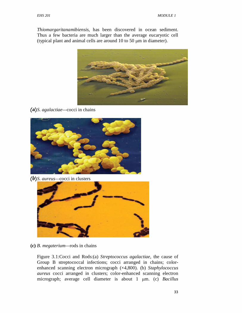
EHS 201 MODULE 1
33
Thiomargaritanamibiensis, has been discovered in ocean sediment. Thus a few bacteria are much larger than the average eucaryotic cell (typical plant and animal cells are around 10 to 50 µm in diameter).
(a) S. agalactiae—cocci in chains
(b) S. aureus—cocci in clusters
(c) B. megaterium—rods in chains Figure 3.1:Cocci and Rods:(a) Streptococcus agalactiae, the cause of Group B streptococcal infections; cocci arranged in chains; color-enhanced scanning electron micrograph (×4,800). (b) Staphylococcus aureus cocci arranged in clusters; color-enhanced scanning electron micrograph; average cell diameter is about 1 µm. (c) Bacillus
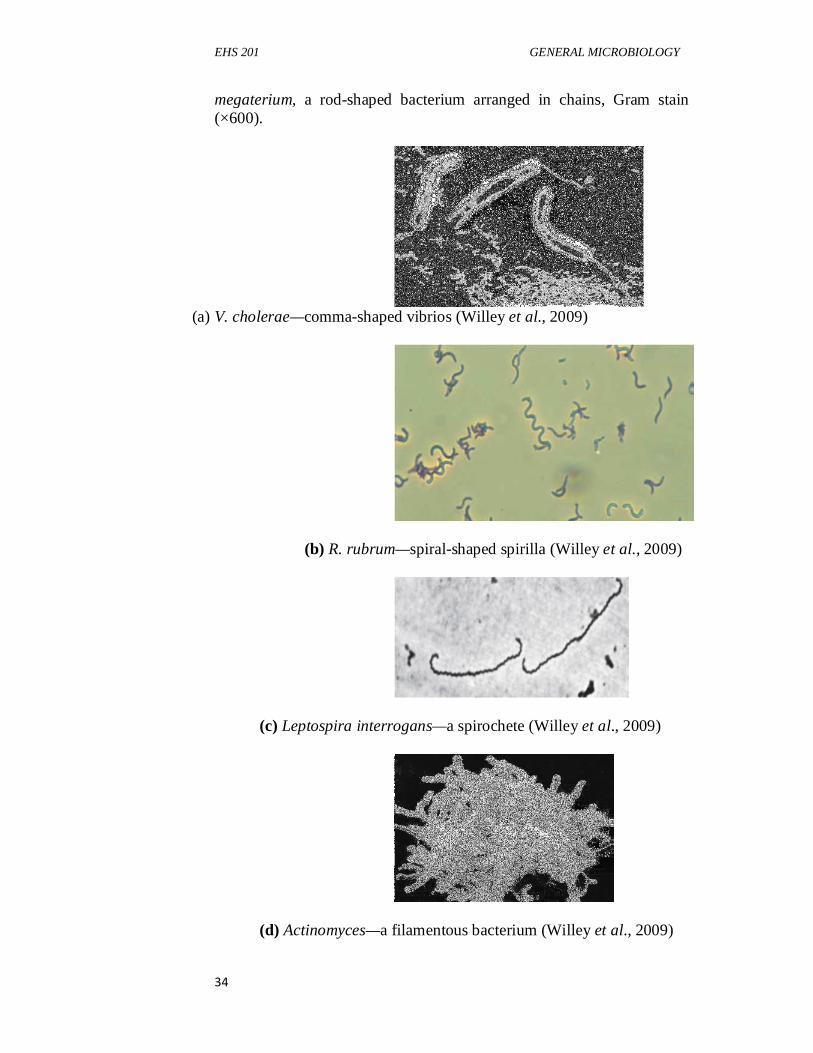
EHS 201 GENERAL MICROBIOLOGY
34
megaterium, a rod-shaped bacterium arranged in chains, Gram stain (×600).
(a) V. cholerae—comma-shaped vibrios (Willey et al., 2009)
(b) R. rubrum—spiral-shaped spirilla (Willey et al., 2009)
(c) Leptospira interrogans—a spirochete (Willey et al., 2009)
(d) Actinomyces—a filamentous bacterium (Willey et al., 2009)
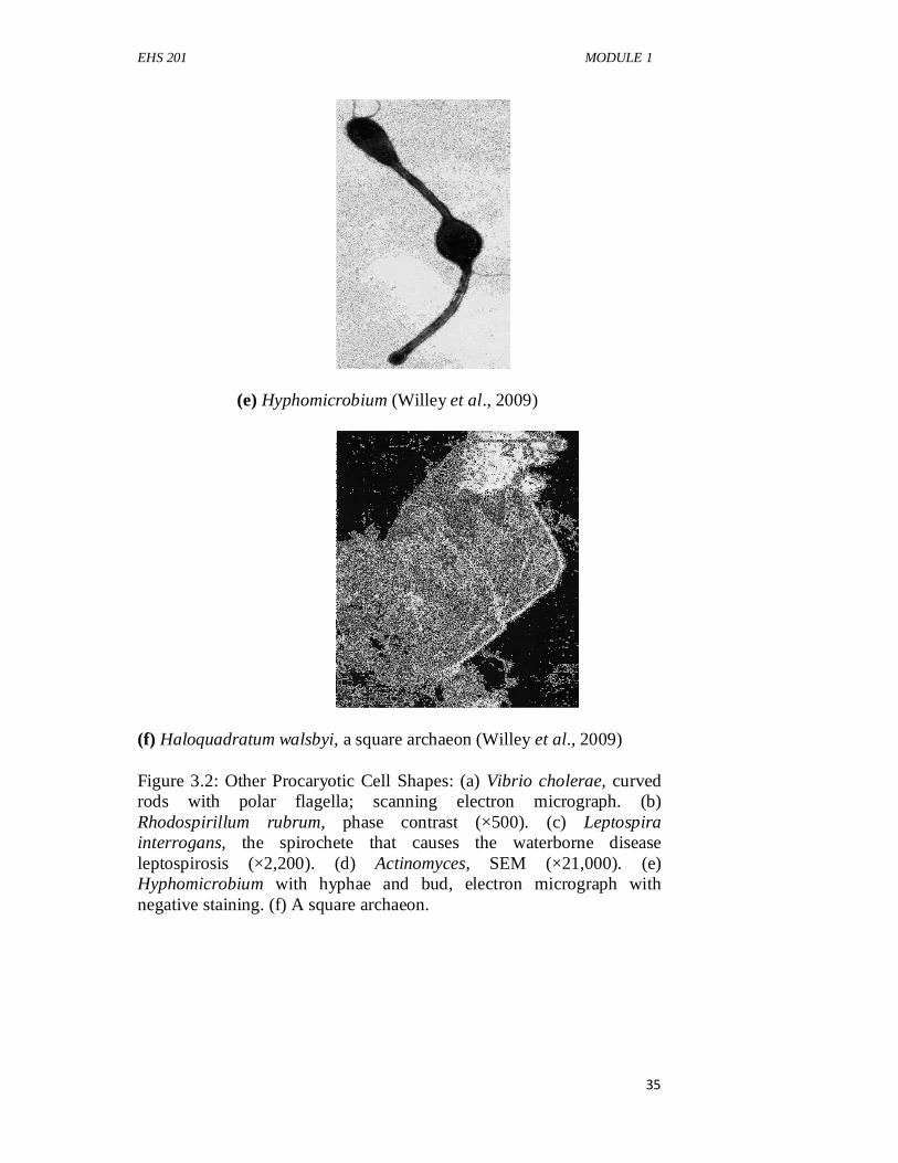
EHS 201 MODULE 1
35
(e) Hyphomicrobium (Willey et al., 2009)
(f) Haloquadratum walsbyi, a square archaeon (Willey et al., 2009) Figure 3.2: Other Procaryotic Cell Shapes: (a) Vibrio cholerae, curved rods with polar flagella; scanning electron micrograph. (b) Rhodospirillum rubrum, phase contrast (×500). (c) Leptospira interrogans, the spirochete that causes the waterborne disease leptospirosis (×2,200). (d) Actinomyces, SEM (×21,000). (e) Hyphomicrobium with hyphae and bud, electron micrograph with negative staining. (f) A square archaeon.
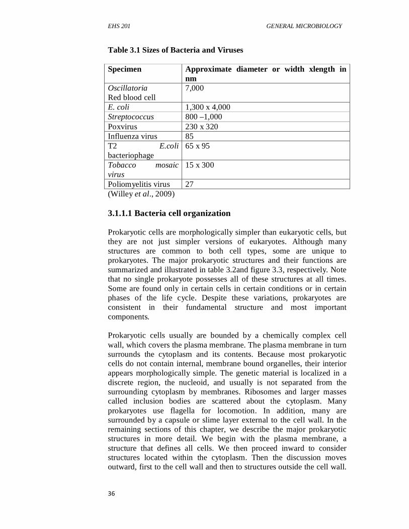
EHS 201 GENERAL MICROBIOLOGY
36
Table 3.1 Sizes of Bacteria and Viruses Specimen Approximate diameter or width xlength in
nm Oscillatoria Red blood cell
7,000
E. coli 1,300 x 4,000 Streptococcus 800 –1,000 Poxvirus 230 x 320 Influenza virus 85 T2 E.coli bacteriophage
65 x 95
Tobacco mosaic virus
15 x 300
Poliomyelitis virus 27 (Willey et al., 2009) 3.1.1.1 Bacteria cell organization Prokaryotic cells are morphologically simpler than eukaryotic cells, but they are not just simpler versions of eukaryotes. Although many structures are common to both cell types, some are unique to prokaryotes. The major prokaryotic structures and their functions are summarized and illustrated in table 3.2and figure 3.3, respectively. Note that no single prokaryote possesses all of these structures at all times. Some are found only in certain cells in certain conditions or in certain phases of the life cycle. Despite these variations, prokaryotes are consistent in their fundamental structure and most important components. Prokaryotic cells usually are bounded by a chemically complex cell wall, which covers the plasma membrane. The plasma membrane in turn surrounds the cytoplasm and its contents. Because most prokaryotic cells do not contain internal, membrane bound organelles, their interior appears morphologically simple. The genetic material is localized in a discrete region, the nucleoid, and usually is not separated from the surrounding cytoplasm by membranes. Ribosomes and larger masses called inclusion bodies are scattered about the cytoplasm. Many prokaryotes use flagella for locomotion. In addition, many are surrounded by a capsule or slime layer external to the cell wall. In the remaining sections of this chapter, we describe the major prokaryotic structures in more detail. We begin with the plasma membrane, a structure that defines all cells. We then proceed inward to consider structures located within the cytoplasm. Then the discussion moves outward, first to the cell wall and then to structures outside the cell wall.
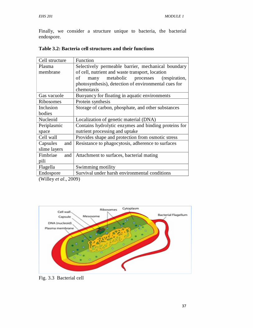
EHS 201 MODULE 1
37
Finally, we consider a structure unique to bacteria, the bacterial endospore. Table 3.2: Bacteria cell structures and their functions Cell structure Function Plasma membrane
Selectively permeable barrier, mechanical boundary of cell, nutrient and waste transport, location of many metabolic processes (respiration, photosynthesis), detection of environmental cues for chemotaxis
Gas vacuole Buoyancy for floating in aquatic environments Ribosomes Protein synthesis Inclusion bodies
Storage of carbon, phosphate, and other substances
Nucleoid Localization of genetic material (DNA) Periplasmic space
Contains hydrolytic enzymes and binding proteins for nutrient processing and uptake
Cell wall Provides shape and protection from osmotic stress Capsules and slime layers
Resistance to phagocytosis, adherence to surfaces
Fimbriae and pili
Attachment to surfaces, bacterial mating
Flagella Swimming motility Endospore Survival under harsh environmental conditions (Willey et al., 2009)
Fig. 3.3 Bacterial cell
EHS 201 GENERAL MICROBIOLOGY
38
3.1.1.1 Bacteria Cell Membrane Membranes are an absolute requirement for all living organisms. Cells must interact in a selective fashion with their environment, acquire nutrients, and eliminate waste. They also have to maintain their interior in a constant, highly organized state in the face of external changes. The plasma membrane encompasses the cytoplasm of both prokaryotic and eukaryotic cells. It is the chief point of contact with the cell’s environment and thus is responsible for much of its relationship with the outside world. The plasma membranes of prokaryotic cells are particularly important because they must fill an incredible variety of roles. In addition to retaining the cytoplasm, the plasma membrane also serves as a selectively permeable barrier: it allows particular ions and molecules to pass, either into or out of the cell, while preventing the movement of others. Thus the membrane prevents the loss of essential components through leakage while allowing the movement of other molecules. Because many substances cannot cross the plasma membrane without assistance, it must aid such movement when necessary. Transport systems are used for such tasks as nutrient uptake, waste excretion, and protein secretion. The prokaryotic plasma membrane also is the location of a variety of crucial metabolic processes: respiration, photosynthesis, and the synthesis of lipids and cell wall constituents. Finally, the membrane contains special receptor molecules that help prokaryotes detect and respond to chemicals in their surroundings. Clearly the plasma membrane is essential to the survival of microorganisms. All membranes have a common, basic design. However, prokaryotic membranes can differ dramatically in terms of the lipids they contain. Indeed, membrane chemistry can be used to identify particular prokaryotic species. To understand these chemical differences and the many functions of the plasma membrane, it is necessary to become familiar with membrane structure. In this section, the fundamental design of all membranes is discussed. This is followed by a consideration of the significant differences between bacterial and archaeal membranes. i. Bacteria Cytoplasm
The cytoplasm is bounded by the plasma membrane and contains inclusion bodies, ribosomes, the nucleoid, and plasmids. It usually lacks membrane-delimited organelles and is largely water (about 70% of prokaryotic mass is water). Until recently, it was thought to lack a cytoskeleton. The plasma membrane and everything within is called the protoplast; thus the cytoplasm is a major part of the protoplast.
EHS 201 MODULE 1
39
For many years it was thought that prokaryotes lacked the high level of cytoplasmic organization present in eukaryotic cells because they lacked a cytoskeleton. Recently homologs of all three eukaryotic cytoskeletal elements (microfilaments, intermediate filaments, and microtubules) have been identified in bacteria, and two have been identified in archaea.
The cytoskeletal filaments of prokaryotes are structurally similar to their eukaryotic counterparts and carry out similar functions: they participate in cell division, localize proteins to certain sites in the cell, and determine cell shape.
ii. Inclusion Bodies
Inclusion bodies aregranules of organic or inorganic material that often are clearly visible in a light microscope, are present in the cytoplasm. These bodies usually are used for storage (e.g., of carbon compounds, inorganic substances, and energy) or to reduce osmotic pressure by tying up molecules in particulate form. Some inclusion bodies lie free in the cytoplasm—for example, polyphosphate granules, cyanophycin granules, and some glycogen granules. Other inclusion bodies are enclosed by a shell about 2 to 4 nm thick, which is single-layered and may consist of proteins or a membranous structure composed of proteins and phospholipids. Examples of enclosed inclusion bodies are poly-β-hydroxybutyrate granules, some glycogen and sulfur granules, carboxysomes, and gas vacuoles. The quantity of inclusion bodies used for storage varies with the nutritional status of the cell. For example, polyphosphate granules are depleted in freshwater habitats that are phosphate limited. A brief description of several important inclusion bodies follows.
Organic inclusion bodies usually contain either glycogen or poly-β-hydroxyalkanoates (e.g., poly-β-hydroxybutyrate). Glycogen is a polymer of glucose units composed of long chains formed by α-(1→4) glycosidic bonds and branching chains connected to them by α-(1→6) glycosidic bonds. Poly-�-hydroxybutyrate (PHB) contains β-hydroxybutyrate molecules joined by ester bonds between the carboxyl and hydroxyl groups of adjacent molecules. Usually only one of these polymers is found in a species, but some photosynthetic bacteria have both glycogen and PHB. PHB accumulates in distinct bodies, around 0.2 to 0.7 µm in diameter, that are readily stained with Sudan black for light microscopy and are seen as empty “holes” in the electron microscope. This is because the solvents used to prepare specimens for electron microscopy dissolve these hydrophobic inclusion bodies.
EHS 201 GENERAL MICROBIOLOGY
40
Glycogen is dispersed more evenly throughout the cytoplasm as small granules (about 20 to 100 nm in diameter) and often can be seen only with the electron microscope. If cells contain a large amount of glycogen, staining with an iodine solution will turn them reddish-brown. Glycogen and PHB inclusion bodiesare carbon storage reservoirs providing material for energy andbiosynthesis. Many bacteria also store carbon as lipiddroplets.
Cyanobacteria, a group of photosynthetic bacteria, have two distinctive organic inclusion bodies. Cyanophycin granulesare composed of large polypeptides containing approximately equal amounts of the amino acids arginine and aspartic acid. The granules often are large enough to be visible in the light microscope and store extra nitrogen for the bacteria. Carboxysomesare present in many cyanobacteria and other CO2 -fixing bacteria. They are polyhedral, about 100 nm in diameter, and contain the enzyme ribulose-1, 5-bisphosphate carboxylase (Rubisco). Rubisco is the critical enzyme for CO2 fixation, the process of converting CO2 into sugar. The enzyme assumes a paracrystalline arrangement in the carboxysome, which serves as a reserve of the enzyme. Carboxysomes also may be a site of CO2 fixation.
iii. Gas Vacuole
A most remarkable organic inclusion body is the gas vacuole,a structure that provides buoyancy to some aquatic prokaryotes.Gas vacuoles are present in many photosynthetic bacteria and afew other aquatic prokaryotes such as Halobacterium (a salt-lovingarchaeon) and Thiothrix (a filamentous bacterium). Gas vacuolesare aggregates of enormous numbers of small, hollow, cylindricalstructures called gas vesicles. Gas vesicle walls arecomposed entirely of a single small protein. These protein subunitsassemble to form a rigid, enclosed cylinder that is hollow andimpermeable to water but freely permeable to atmospheric gases.Prokaryotes with gas vacuoles can regulate their buoyancy to floatat the depth necessary for proper light intensity, oxygen concentration,and nutrient levels. They descend by simply collapsingvesicles and float upward when new ones are constructed.
Two major types of inorganic inclusion bodies are seen in prokaryotes: polyphosphate granules and sulfur granules. Many bacteria store phosphate as polyphosphate granules,also called volutin granules or metachromatic granules.Polyphosphate is a linear polymer of orthophosphates joined by ester bonds. Thus polyphosphate granules store the phosphate needed for synthesis
EHS 201 MODULE 1
41
of important cell constituents such as nucleic acids. In some cells they act as an energy reserve, and polyphosphate can serve as an energy source in some reactions. Polyphosphate granules are sometimes called metachromatic granules because they show the metachromatic effect; that is, they appear red or a different shade of blue when stained with the blue dyes methylene blue or toluidine blue. Sulfur granules are used by some prokaryotes to store sulfur temporarily. For example, photosynthetic bacteria can use hydrogen sulfide as a photosynthetic electron donor and accumulate the resulting sulfur either in the periplasmic space or in special cytoplasmic globules.
iv. Ribosomes
When examined with the electron microscope, the cytoplasm of prokaryotes is often packed with ribosomes,and others may be loosely attached to the plasma membrane. Ribosomes are very complex structures made of both protein and ribonucleic acid (RNA). They are the site of protein synthesis; cytoplasmic ribosomes synthesize proteins destined to remain within the cell, whereas plasma membrane-associated ribosomes make proteins for transport to the outside.
Prokaryotic ribosomes are smaller than the ribosomes of eukaryotic cells. Prokaryotic ribosomes are called 70S ribosomes (as opposed to 80S in eukaryotes), have dimensions of about 14 to 15 nm by 20 nm, a molecular weight of approximately 2.7 million, and are constructed of a 50S and a 30S subunit. TheS in 70S and similar values stands for Svedberg unit.This is the unit of the sedimentation coefficient, a measure of the sedimentationvelocity in a centrifuge; the faster a particle travels whencentrifuged, the greater its Svedberg value or sedimentation coefficient. The sedimentation coefficient is a function of a particle’smolecular weight, volume, and shape. Heavier and more compactparticles normally have larger Svedberg numbers andsediment faster.
v. Nucleoid
Probably the most striking difference between prokaryotes and eukaryotes is the way their genetic material is packaged. Eukaryotic cells have two or more chromosomes contained within a membrane-bound organelle, the nucleus. In contrast, prokaryotes lack a membrane-delimited nucleus. Instead, the prokaryotic chromosome is located in an irregularly shaped region called the nucleoid(other names are also used: the nuclear body, chromatin body, and nuclear region). Most prokaryotes contain a single circle of double-stranded deoxyribonucleic
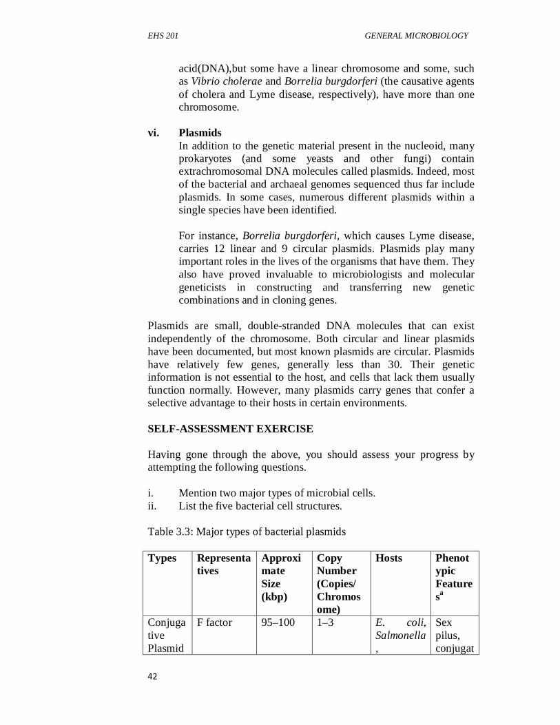
EHS 201 GENERAL MICROBIOLOGY
42
acid(DNA),but some have a linear chromosome and some, such as Vibrio cholerae and Borrelia burgdorferi (the causative agents of cholera and Lyme disease, respectively), have more than one chromosome.
vi. Plasmids
In addition to the genetic material present in the nucleoid, many prokaryotes (and some yeasts and other fungi) contain extrachromosomal DNA molecules called plasmids. Indeed, most of the bacterial and archaeal genomes sequenced thus far include plasmids. In some cases, numerous different plasmids within a single species have been identified. For instance, Borrelia burgdorferi, which causes Lyme disease, carries 12 linear and 9 circular plasmids. Plasmids play many important roles in the lives of the organisms that have them. They also have proved invaluable to microbiologists and molecular geneticists in constructing and transferring new genetic combinations and in cloning genes.
Plasmids are small, double-stranded DNA molecules that can exist independently of the chromosome. Both circular and linear plasmids have been documented, but most known plasmids are circular. Plasmids have relatively few genes, generally less than 30. Their genetic information is not essential to the host, and cells that lack them usually function normally. However, many plasmids carry genes that confer a selective advantage to their hosts in certain environments. SELF-ASSESSMENT EXERCISE Having gone through the above, you should assess your progress by attempting the following questions. i. Mention two major types of microbial cells. ii. List the five bacterial cell structures. Table 3.3: Major types of bacterial plasmids Types Representa
tives Approximate Size (kbp)
Copy Number (Copies/ Chromosome)
Hosts Phenotypic Featuresa
Conjugative Plasmid
F factor 95–100 1–3 E. coli, Salmonella,
Sex pilus, conjugat
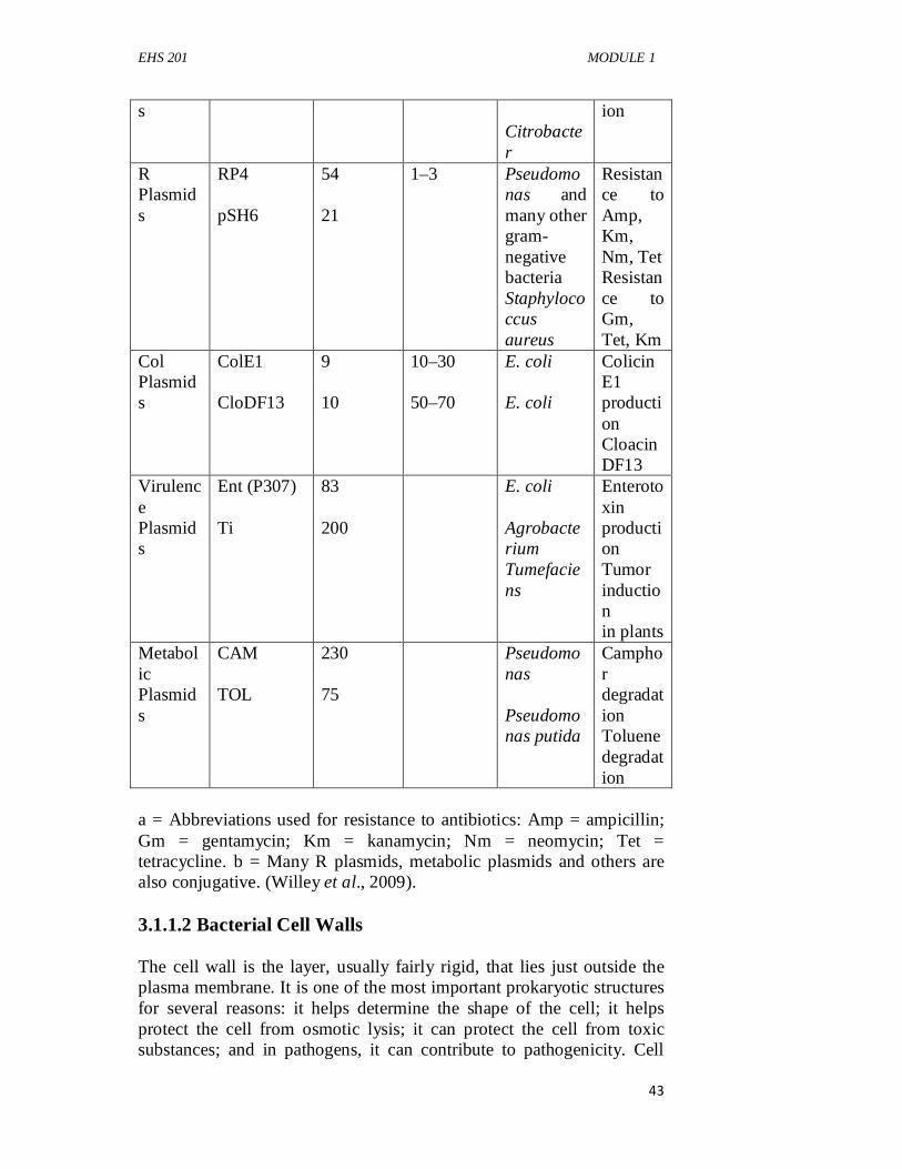
EHS 201 MODULE 1
43
s Citrobacter
ion
R Plasmids
RP4 pSH6
54 21
1–3
Pseudomonas and many other gram-negative bacteria Staphylococcus aureus
Resistance to Amp, Km, Nm, Tet Resistance to Gm, Tet, Km
Col Plasmids
ColE1 CloDF13
9 10
10–30 50–70
E. coli E. coli
Colicin E1 production Cloacin DF13
Virulence Plasmids
Ent (P307) Ti
83 200
E. coli Agrobacterium Tumefaciens
Enterotoxin production Tumor induction in plants
Metabolic Plasmids
CAM TOL
230 75
Pseudomonas Pseudomonas putida
Camphor degradation Toluene degradation
a = Abbreviations used for resistance to antibiotics: Amp = ampicillin; Gm = gentamycin; Km = kanamycin; Nm = neomycin; Tet = tetracycline. b = Many R plasmids, metabolic plasmids and others are also conjugative. (Willey et al., 2009). 3.1.1.2 Bacterial Cell Walls The cell wall is the layer, usually fairly rigid, that lies just outside the plasma membrane. It is one of the most important prokaryotic structures for several reasons: it helps determine the shape of the cell; it helps protect the cell from osmotic lysis; it can protect the cell from toxic substances; and in pathogens, it can contribute to pathogenicity. Cell
EHS 201 GENERAL MICROBIOLOGY
44
walls are so important that relatively few prokaryotes lack them. Those that do have other features that fulfill cell wall function. The bacterial cell wall also is the site of action of several antibiotics. Therefore, it is important to understand its structure. After Christian Gram developed the Gram stain in 1884, it soon became evident that most bacteria could be divided into two major groups based on their response to the Gram-stain procedure. Gram-positive bacteria stained purple, whereas Gram-negative bacteria were colored pink or red by the technique. The true structural difference between these two groups did not become clear until the advent of the transmission electron microscope. The Gram-positive cell wall consists of a single, 20 to 80 nm thick homogeneous layer of peptidoglycan(murein)lying outside the plasma membrane. In contrast, the Gram-negative cell wall is quite complex. It has a 2 to 7 nm peptidoglycan layer covered by a 7 to 8 nm thick outermembrane.Because of the thicker peptidoglycan layer, the walls of Gram-positive cells are more resistant to osmotic pressure than those of Gram-negative bacteria. Microbiologists often call all the structures from the plasma membrane outward the cell envelope. Therefore this includes the plasma membrane, cell wall, and structures such as capsules when present. One important feature of the cell envelope is a space that is frequently seen between the plasma membrane and the outer membrane in electron micrographs of Gram-negative bacteria. It also is sometimes observed between the plasma membrane and the wall in Gram-positive bacteria. This space is called the periplasmicspace.The substance that occupies the periplasmic space is the periplasm. The nature of the periplasmic space and periplasm differs in Gram-positive and Gram-negative bacteria. These differences are pointed out in the more detailed discussions of Gram-positive and Gram-negative cell walls that follow. i. Gram-Positive Cell Walls
Gram-positive bacteria normally have cell walls that are thick and composed primarily of peptidoglycan. Peptidoglycan in Gram-positive bacteria often contains a peptide interbridge. In addition, Gram-positive cell walls usually contain large amounts of teichoic acids,polymers of glycerol or ribitol joined by phosphate groups. Amino acids such as D -alanine or sugars such asglucose are attached to the glycerol and ribitol groups. The teichoicacids are covalently connected to the peptidoglycan itselfor to plasma membrane lipids; in the latter case, they are calledlipoteichoic acids. Teichoic acids appear to extend to the surfaceof the peptidoglycan. Because they are negatively charged, theyhelp give the Gram-positive cell wall its negative charge. Thefunctions of techoic acids are still unclear, but they may be
EHS 201 MODULE 1
45
importantin maintaining the structure of the wall. Teichoic acids arenot present in Gram-negative bacteria.
ii. Gram-Negative Cell Walls
Even a brief inspection of gram-negative cell walls are much more complex than gram-positive walls. The thin peptidoglycan layer next to the plasma membrane and bounded on either side by the periplasmic space usually constitutes only 5 to 10% of the wall weight. In E. coli, it is about 2 nm thick and contains only one or two sheets of peptidoglycan.
The periplasmic space of gram-negative bacteria is also strikingly different from that of gram-positive bacteria. It ranges in width from 1 nm to as great as 71 nm. Some recent studies indicate that it may constitute about 20 to 40% of the total cell volume, and it is usually 30 to 70 nm wide.
The outer membrane lies outside the thin peptidoglycan layer and is linked to the cell in two ways. The first is by Braun’s lipoprotein, the most abundant protein in the outer membrane. This small lipoprotein is covalently joined to the underlying peptidoglycan and is embedded in the outer membrane by its hydrophobic end. The second linking mechanism involves the many adhesion sites joining the outer membrane and the plasma membrane. The two membranes appear to be in direct contact at these sites. In E. coli, 20 to 100 nm areas of contact between the two membranes can be seen. Adhesion sites may be regions of direct contact or possibly true membrane fusions.
Possibly the most unusual constituents of the outer membrane are its lipopolysaccharides (LPSs).These large, complex molecules contain both lipid and carbohydrate, and consist of three parts: (1) Lipid A, (2) The core polysaccharide, and (3) The O side chain. LPS has many important functions. Because the core polysaccharide usually contains charged sugars and phosphate, LPS contributes to the negative charge on the bacterialsurface. LPS helps stabilize outer membrane structure because lipid A is a major constituent of the exterior leaflet of the outer membrane. LPS may contribute to bacterial attachment to surfaces and biofilm formation. A major function of LPS is that it helps create a permeability barrier. The geometry of LPS and interactions between neighboring LPS molecules are thought to restrict the entry of bile salts, antibiotics, and other toxic substances that might
EHS 201 GENERAL MICROBIOLOGY
46
kill or injure the bacterium. LPS also plays a role in protecting pathogenic gram-negative bacteria from host defenses. The O side chain of LPS is also called the O antigen because it elicits an immune response by an infected host. This response involves the production of antibodies that bind the strain-specific form of LPS that elicited the response. However, many gram-negative bacteria can rapidly change the antigenic nature of their O side chains, thus thwarting host defenses. Importantly, the lipid A portion of LPS is toxic; as a result, LPS can act as an endotoxin and cause some of the symptoms that arise in gram-negative bacterial infections. If LPS or lipid A enters the bloodstream, a form of septic shock develops, for which there is no direct treatment. Despite the role of LPS in creating a permeability barrier, the outer membrane is more permeable than the plasma membrane and permits the passage of small molecules such as glucose and other monosaccharides. This is due to the presence of porin proteins. Most porin proteins cluster together to form a trimer in the outer membrane. Each porin protein spans the outer membrane and is more or less tube-shaped; its narrow channel allows passage of molecules smaller than about 600 to 700 daltons. However, larger molecules such as vitamin B 12 also cross the outer membrane. Such large molecules do not pass through porins; instead, specific carriers transport them across the outer membrane. 3.1.2 Capsules and Slime Layers Some procaryotes have a layer of material lying outside the cell wall. This layer has different names depending on its characteristics. When the layer is well organized and not easily washed off, it is called a capsule. It is called a slime layerwhen itis a zone of diffuse, unorganized material that is removed easily.When the layer consists of a network of polysaccharides extending from the surface of the cell, it is referred to as theglycocalyx, a term that can encompass both capsules and slime layers because they usually are composed of polysaccharides. However, some slime layers and capsules are constructed of other materials. For example, Bacillus anthracis has a proteinaceous capsule composed of poly- D -glutamic acid. Capsules are clearly visible in the light microscope when negative stains or special capsule stains are employed; they also can be studied with the electron microscope. Although capsules are not required for growth and reproduction in laboratory cultures, they confer several advantages when procaryotes grow in their normal habitats. They help pathogenic bacteria resist phagocytosis by host phagocytes. Streptococcus pneumoniae provides a dramatic example. When it lacks a capsule, it is destroyed easily and does not cause disease. On the other hand, the capsulated variant quickly
EHS 201 MODULE 1
47
kills mice. Capsules contain a great deal of water and can protect against desiccation. They exclude viruses and most hydrophobic toxic materials such as detergents. The glycocalyx also aids in attachment to solid surfaces, including tissue surfaces in plant and animal hosts. Gliding bacteria often produce slime, which in some cases has been shown to facilitate motility. 3.1.3 Pili and Fimbriae Many procaryotes have short, fine, hair like appendages that are thinner than flagella. These are usually called fimbriae(s., fimbria) or pili (s., pilus). Although many people use the terms fimbriae and pili interchangeably, we distinguish between fimbriae and sex pili. A cell may be covered with up to 1,000 fimbriae, but they are only visible in an electron microscope due to their small size. They are slender tubes composed of helically arranged protein subunits and are about 3 to 10 nm in diameter and up to several micrometers long. Some types of fimbriae attach bacteria to solid surfaces such as rocks in streams and host tissues, and some are involved in motility. Many bacteria have about one to 10 sex pili(s., sex pilus) per cell. These hair like structures differ from fimbriae in the following ways. Pili often are larger than fimbriae (around 9 to 10 nm in diameter). They are genetically determined by conjugative plasmids and are required for conjugation. Some bacterial viruses attach specifically to receptors on sex pili at the start of their reproductive cycle. 3.1.4 Flagella Most motile procaryotes move by use of flagella(s., flagellum), threadlike locomotor appendages extending outward from the plasma membrane and cell wall. Bacterial flagella are slender, rigid structures, about 20 nm across and up to 20 µm long. Flagella are so thin they cannot be observed directly with a bright-field microscope but must be stained with special techniques designed to increase their thickness. The detailed structure of a flagellum can only be seen in the electron microscope. Bacterial species often differ distinctively in their patterns of flagella distribution, and these patterns are useful in identifying bacteria. Monotrichousbacteria (trichous means hair) have one flagellum; if it is located at an end, it is said to be a polar flagellum. Amphitrichousbacteria (amphi means on both sides) have a single flagellum at each pole. In contrast, lophotrichous bacteria (lopho means tuft) have a cluster of flagella at one or both ends. Flagella are spread
EHS 201 GENERAL MICROBIOLOGY
48
evenly over the whole surface of peritrichous(peri means around) bacteria. 3.2 Bacterial Motility and Chemotaxis Several structures outside the cell wall contribute to the motility of prokaryotes. Four major methods of movement have been observed in Bacteria: 1.The swimming movement conferred by flagella; 2. The corkscrew movement of spirochetes; 3. The twitching motility associated with fimbriae; and 4. Gliding motility. Bacteria have not evolved motility to move aimlessly. Rather, motility is used to move toward nutrients such as sugars and amino acids and away from many harmful substances and bacterial waste products. Bacteria also can respond to environmental cues such as temperature (thermotaxis), light (phototaxis), oxygen (aerotaxis), osmotic pressure (osmotaxis), and gravity. Movement toward chemical attractants and away from repellents is known as chemotaxis. 3.2.1 Flagellar Movement Prokaryotic flagella operate differently from eukaryotic flagella. Eukaryotic flagella flex and bend, resulting in a whiplash that moves the cell. The filament of a prokaryotic flagellum is in the shape of a rigid helix, and the cell moves when this helix rotates like a propeller on a boat. The flagellar motor can rotate veryrapidly. The E. coli motor rotates 270 revolutions per second (rps); Vibrio alginolyticus averages 1,100 rps. The direction of flagellar rotation determines the nature of bacterial movement. Monotrichous, polar flagella rotate counterclockwise (when viewed from outside the cell) during normal forward movement, whereas the cell itself rotates slowly clockwise. The rotating helical flagellar filament thrusts the cell forward with the flagellum trailing behind. Monotrichous bacteria stop and tumble randomly by reversing the direction of flagellar rotation. Peritrichously flagellated bacteria operate in a somewhat similar way. To move forward, the flagella rotate counterclockwise. As they do so, they bend at their hooks to form a rotating bundle that propels the cell forward. Clockwise rotation of the flagella disrupts the bundle and the cell tumbles. The motor that drives flagellar rotation is located at the base of the flagellum, where it is associated with the basal body. Torque generated
EHS 201 MODULE 1
49
by the motor is transmitted by the basal body to the hook and filament. The motor is composed of two components: 1. The rotor, and 2. The stator. The flagellum is a very effective swimming device. From the bacterium’s point of view, swimming is quite a difficult task because the surrounding water seems as viscous as molasses. The cell must bore through the water with its corkscrew-shaped flagella, and if flagellar activity ceases, it stops almost instantly. Despite such environmental resistance to movement, bacteria can swim from 20 to almost 90 µm/second. This is equivalent to traveling from 2 to over 100 cell lengths per second. In contrast, an exceptionally fast human might be able to run around 5 to 6 body lengths per second. 3.2.2 Spirochete Motility Although spirochetes have flagella, they work in a different manner. In many spirochetes, multiple flagella arise from each end of the cell and associate to form an axial fibril, which winds around the cell. The flagella do not extend outside the cell wall but rather remain in the periplasmic space and are covered by an outer sheath. The way in which axial fibrils propel the cell has not been fully established. They are thought to rotate like the external flagella of other bacteria, causing the corkscrew-shaped outer sheath to rotate and move the cell through the surrounding liquid, even very viscous liquids. Flagellar rotation may also flex or bend the cell and account for the creeping or crawling movement observed when spirochetes are in contact with a solid surface. 3.2.3 Twitching and Gliding Motility Twitching and gliding motility occur when cells are on a solid surface. Both types of motility can involve fimbriae, the production of slime, or both. Thus they are considered together. Several types of fimbriae have been identified on prokaryotic cells. Type IV fimbriae are present at one or both poles of some bacteria and are involved in twitching motility and in the gliding motility of some bacteria. Twitching motility is characterized by short, intermittent, jerky motions of up to several micrometers in length and is normally seen on very moist surfaces. It occurs only when cells are in contact with each other; isolated cells rarely move by this mechanism. Considerable
EHS 201 GENERAL MICROBIOLOGY
50
evidence exists that the fimbriae alternately extend and retract to move bacteria during twitching motility. Gliding motility is smooth and varies greatly in rate (from 2 to over 600 µm per minute) and in the nature of the motion. Although first observed over 100 years ago, the mechanism by which many bacteria glide remains a mystery. Some glide along in a direction parallel to the longitudinal axis of their cells. Others travel with a screw-like motion or even move in a direction perpendicular to the long axis of the cells. Still others rotate around their longitudinal axis while gliding. Such diversity in gliding movement correlates with the observation that more than one mechanism for gliding motility exists. Some types involve type IV fimbriae, some involve slime, and some involve mechanisms that have not yet been elucidated. 3.2.4 Chemotaxis The movement of cells toward chemical attractants or away from chemical repellents is called chemotaxis. Chemotaxis is readily observed in petri dish cultures. If bacteria are placed in the center of a dish of semisolid agar containing an attractant, the bacteria will exhaust the local supply of the nutrient and swim outward following the attractant gradient they have created. The result is an expanding ring of bacteria. When a disk of repellent is placed in a petri dish of semisolid agar and bacteria, the bacteria will swim away from the repellent, creating a clear zone around the disk. Attractants and repellents are detected by chemoreceptors, proteins that bind chemicals and transmit signals to other components of the chemosensing system. The chemosensing systems are very sensitive and allow the cell to respond to very low levels of attractants (about 10−8 M for some sugars). In gram-negative bacteria, the chemoreceptor proteins are located in the periplasmic space or in the plasma membrane. Some receptors also participate in the initial stages of sugar transport into the cell. Clearly, the bacterium must have some mechanism for sensing that it is getting closer to the attractant (or moving away from the repellent). The behavior of the bacterium is shaped by temporal changes in chemical concentration. The bacterium moves toward the attractant because it senses that the concentration of the attractant is increasing. Likewise, it moves away from a repellent because it senses that the concentration of the repellent is decreasing. The bacterium’s chemoreceptors play a critical role in this process.
EHS 201 MODULE 1
51
3.3 Bacterial Endospores Several genera of gram-positive bacteria, including Bacillus and Clostridium (rods), and Sporosarcina (cocci), can form a resistant, dormant structure called an endospore. Endospores develop within vegetative bacterial cells and are extraordinarily resistant to environmental stresses such as heat, ultraviolet radiation, gamma radiation, chemical disinfectants, and desiccation. In fact, some endospores have remained viable for around 100,000 years. Because of their resistance and the fact that several species of endospore-forming bacteria are dangerous pathogens, endospores are of great practical importance in food, industrial, and medical microbiology. This is because it is essential to be able to sterilize solutions and solid objects. Endospores often survive boiling for an hour or more; therefore autoclaves must be used to sterilize many materials. In the environment, endospores aid in survival when moisture or nutrients are scarce. Endospores are also of considerable theoretical interest. Because bacteria manufacture these intricate structures in a very organized fashion over a period of a few hours, spore formation is well suited for research on the construction of complex biological structures. This has made the endospore-forming Bacillus subtilis an important model organism. Endospores can be examined with both light and electron microscopes. Because endospores are impermeable to most stains, they often are seen as colorless areas in bacteria treated with methylene blue and other simple stains; special endospore stains are used to make them clearly visible. Endospore position in the mother cell (sporangium) frequently differs among species, making it of value in identification. Endospores may be centrally located, close to one end (subterminal), or terminal. Sometimes an endospore is so large that it swells the sporangium. Electron micrographs show that endospore structure is complex. The spore often is surrounded by a thin, delicate covering called the exosporium. A spore coat lies beneath the exosporium, is composed of several protein layers, and may be fairly thick. It is impermeable to many toxic molecules and is responsible for the spore’s resistance to chemicals. The coat also is thought to contain enzymes involved in germination. The cortex, which may occupy as much as half the spore volume, rests beneath the spore coat. It is made of apeptidoglycan that is less cross-linked than that in vegetative cells. The spore cell wall (or core wall) is inside the cortex and surrounds the protoplast or spore core. The core has normal cell structures such as ribosomes and a nucleoid but is metabolically inactive.
EHS 201 GENERAL MICROBIOLOGY
52
The transformation of dormant spores into active vegetative cells seems almost as complex a process as sporulation. It occurs in three stages: (1) Activation, (2) Germination, and (3) Outgrowth. Activation is a process that prepares spores for germination and usually results from treatments such as heating. This is followed by germination, the breaking of the spore’s dormant state. This process is characterized by spore swelling, rupture, or absorption of the spore coat, loss of resistance to heat and other stresses, loss of refractility, release of spore components, and increase in metabolic activity. Many normal metabolites or nutrients (e.g., amino acids and sugars) can trigger germination after activation. Germination is followed by the third stage, outgrowth. The spore protoplast makes new components, emerges from the remains of the spore coat, and develops again into an active bacterium. 3.4 Comparison of Prokaryotic and Eukaryotic Cells A comparison of the cells in showed there are many fundamental differences between eukaryotic and prokaryotic cells. Eukaryotic cells have a membrane-enclosed nucleus. In contrast, prokaryotic cells lack a true, membrane-delimited nucleus. Bacteria and Archaea are prokaryotes; all other organisms—fungi, protists, plants, and animals—are eukaryotic. Most prokaryotes are smaller than eukaryotic cells, often about the size of eukaryotic mitochondria and chloroplasts. The presence of the eukaryotic nucleus is the most obvious difference between these two cell types, but many other major distinctions exist. It is clear that prokaryotic cells are much simpler structurally. In particular, an extensive and diverse collection of membrane-delimited organelles is missing. Furthermore, prokaryotes are functionally simpler in several ways. They lack mitosis and meiosis, and have a simpler genetic organization. Many complex eukaryotic processes are absent in prokaryotes: endocytosis, intracellular digestion, directed cytoplasmic streaming, and ameboid movement, are just a few. Despite the many significant differences between these two basic cell forms, they are remarkably similar on the biochemical level, as we discuss in succeeding chapters. With a few exceptions, the genetic code is the same in both, as is the way in which the genetic information in DNA is expressed. The principles underlying metabolic processes and many important metabolic pathways are identical. Thus beneath the profound structural and functional differences between prokaryotes and
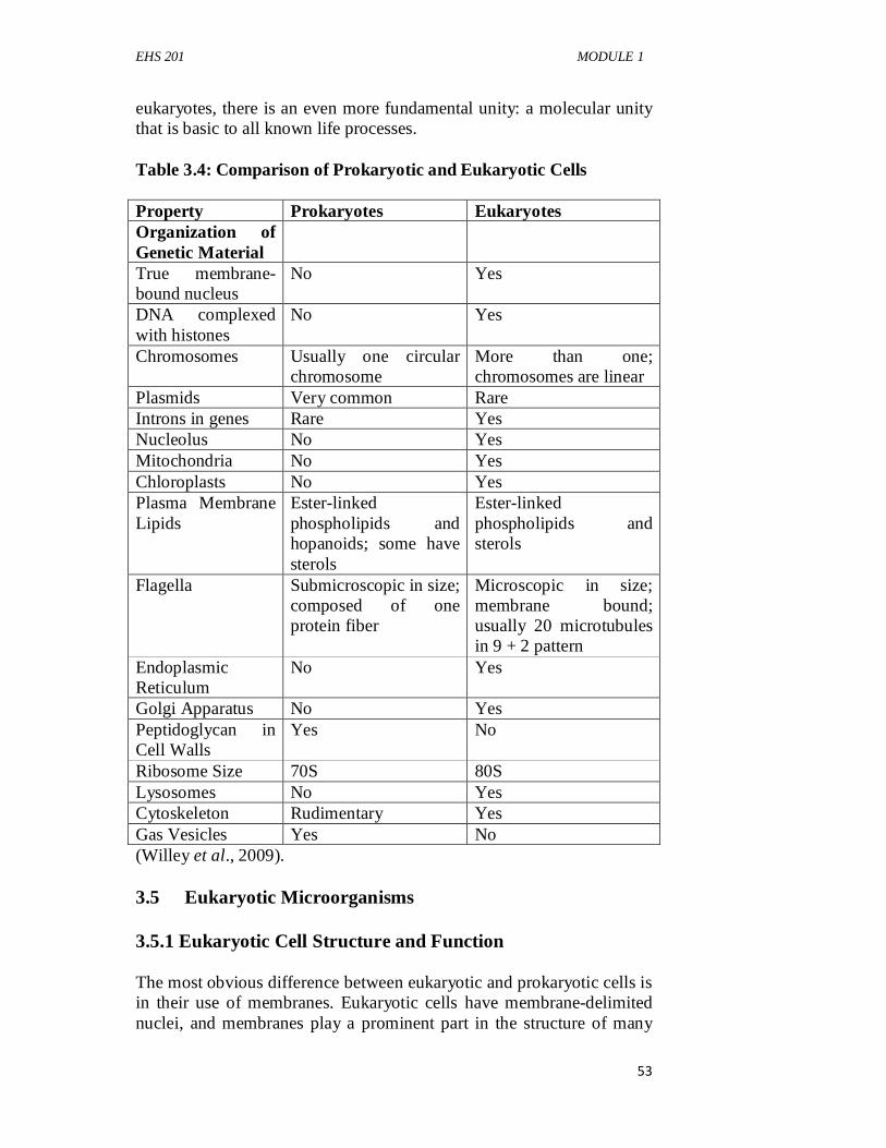
EHS 201 MODULE 1
53
eukaryotes, there is an even more fundamental unity: a molecular unity that is basic to all known life processes. Table 3.4: Comparison of Prokaryotic and Eukaryotic Cells Property Prokaryotes Eukaryotes Organization of Genetic Material
True membrane-bound nucleus
No Yes
DNA complexed with histones
No Yes
Chromosomes Usually one circular chromosome
More than one; chromosomes are linear
Plasmids Very common Rare Introns in genes Rare Yes Nucleolus No Yes Mitochondria No Yes Chloroplasts No Yes Plasma Membrane Lipids
Ester-linked phospholipids and hopanoids; some have sterols
Ester-linked phospholipids and sterols
Flagella Submicroscopic in size; composed of one protein fiber
Microscopic in size; membrane bound; usually 20 microtubules in 9 + 2 pattern
Endoplasmic Reticulum
No Yes
Golgi Apparatus No Yes Peptidoglycan in Cell Walls
Yes No
Ribosome Size 70S 80S Lysosomes No Yes Cytoskeleton Rudimentary Yes Gas Vesicles Yes No (Willey et al., 2009). 3.5 Eukaryotic Microorganisms 3.5.1 Eukaryotic Cell Structure and Function The most obvious difference between eukaryotic and prokaryotic cells is in their use of membranes. Eukaryotic cells have membrane-delimited nuclei, and membranes play a prominent part in the structure of many
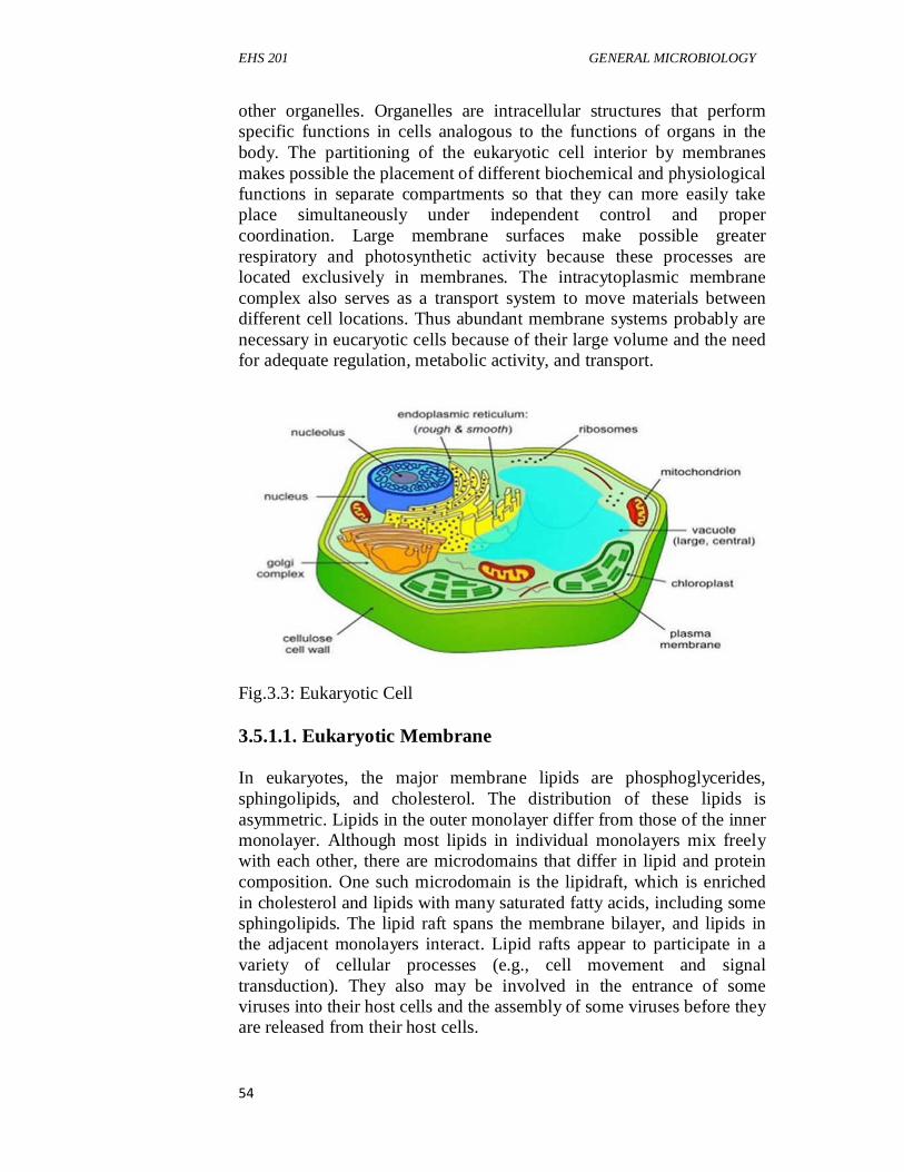
EHS 201 GENERAL MICROBIOLOGY
54
other organelles. Organelles are intracellular structures that perform specific functions in cells analogous to the functions of organs in the body. The partitioning of the eukaryotic cell interior by membranes makes possible the placement of different biochemical and physiological functions in separate compartments so that they can more easily take place simultaneously under independent control and proper coordination. Large membrane surfaces make possible greater respiratory and photosynthetic activity because these processes are located exclusively in membranes. The intracytoplasmic membrane complex also serves as a transport system to move materials between different cell locations. Thus abundant membrane systems probably are necessary in eucaryotic cells because of their large volume and the need for adequate regulation, metabolic activity, and transport.
Fig.3.3: Eukaryotic Cell 3.5.1.1. Eukaryotic Membrane In eukaryotes, the major membrane lipids are phosphoglycerides, sphingolipids, and cholesterol. The distribution of these lipids is asymmetric. Lipids in the outer monolayer differ from those of the inner monolayer. Although most lipids in individual monolayers mix freely with each other, there are microdomains that differ in lipid and protein composition. One such microdomain is the lipidraft, which is enriched in cholesterol and lipids with many saturated fatty acids, including some sphingolipids. The lipid raft spans the membrane bilayer, and lipids in the adjacent monolayers interact. Lipid rafts appear to participate in a variety of cellular processes (e.g., cell movement and signal transduction). They also may be involved in the entrance of some viruses into their host cells and the assembly of some viruses before they are released from their host cells.
EHS 201 MODULE 1
55
3.5.1.2 Eukaryotic Cytoplasm The cytoplasm is one of the most important and complex parts of a cell. It consists of a liquid component, the cytosol, in which many organelles are located. It is the location of many important biochemical processes and several physical changes seen in cells—viscosity changes, cytoplasmic streaming, and others—also are due to cytoplasmic activity. Major component of the cytoplasm is a vast network of interconnected filaments called the cytoskeleton. The cytoskeleton plays a role in both cell shape and movement. Three types of filaments form the cytoskeleton: microfilaments, microtubules, and intermediate filaments. Microfilaments are minute protein filaments, 4 to 7nm in diameter that are either scattered within the cytoplasm or organized into networks and parallel arrays. Microtubules serve at least three purposes: (1) They help maintain cell shape, (2) They are involved with microfilaments in cell movements, and (3) They participate in intracellular transport processes. Microtubules are found in long, thin cell structures requiring support such as the axopodia (long, slender, rigid pseudopodia) of protists. Microtubules also are present in structures that participate in cell or organelle movements—the mitotic spindle, cilia, and flagella. Intermediate filaments are heterogeneous elements of the cytoskeleton. They are about 10 nm in diameter and are assembled from a group of proteins that can be divided into several classes. Intermediate filaments having different functions are assembled from one or more of these classes of proteins. The role of intermediate filaments in eukaryotic microorganisms is unclear. Thus far, they have been identified and studied only in animals: some intermediate filaments have been shown to form the nuclear lamina, a structure that provides support for the nuclear envelope; and other intermediate filaments help link cells together to form tissues. 3.5.1.3 Endoplasmic Reticulum The endoplasmic reticulum (ER) is an irregular network of branching and fusing membranous tubules, around40 to 70 nm in diameter, and many flattened sacs called cisternae (s., cisterna). The nature of the ER varies with the functional and physiological status of the cell. In cells synthesizing a great deal of protein for purposes such as secretion, a
EHS 201 GENERAL MICROBIOLOGY
56
large part of the ER is studded on its outer surface with ribosomes and is called rough endoplasmicreticulum (RER). Other cells, such as those producing large quantities of lipids, have ER that lacks ribosomes. This is smooth endoplasmicreticulum (SER).The endoplasmic reticulum has many important functions.Not only does it transport proteins, lipids, and other materialsthrough the cell, it is also involved in the synthesis of many of thematerials it transports. Lipids and proteins are synthesized byER-associated enzymes and ribosomes. Polypeptide chains synthesizedon RER-bound ribosomes may be inserted either intothe ER membrane or into its lumen for transport elsewhere. TheER is also a major site of cell membrane synthesis. 3.5.1.4 Golgi Apparatus The Golgi apparatus is composed of flattened, saclike cisternae stacked on each other. These membranes, like the smooth ER, lack bound ribosomes. Usually around four to eight cisternae are in a stack, although there may be many more. Each is 15 to 20 nm thick and separated fromother cisternae by 20 to 30 nm. A complex network of tubulesand vesicles (20 to 100 nm in diameter) is located at the edgesof the cisternae. The stack of cisternae has two faces that arequite different from one another. The sacs on the cis or formingface often are associated with the ER and differ from thesacs on the trans or maturing face in thickness, enzyme content,and degree of vesicle formation. The Golgi apparatus is present in most eukaryotic cells, but many fungi and ciliate protozoa lack a well-formed structure. Sometimes the Golgi consists of a single stack of cisternae; however, many cells may contain 20 or more separate stacks. These stacks of cisternae, often called dictyosomes, can be clustered in one region or scattered about the cell. The Golgi apparatus packages materials and prepares them for secretion, the exact nature of its role varying with the organism. For instance, the surface scales of some flagellated photosynthetic and radiolarian protists appear to be constructed within the Golgi apparatus and then transported to the surface in vesicles. The Golgi often participates in the development of cell membranes and the packaging of cell products. The growth of some fungal hyphae occurs when Golgi vesicles contribute their contents to the wall at the hyphal tip. 3.5.1.5 Lysosomes Lysosomes are found in most eukaryotic organisms, including protists, fungi, plants, and animals. Lysosomes are roughly spherical and enclosed in a single membrane; they average about 500 nm in diameter but range from 50 nm to several µm in size. They are involved in
EHS 201 MODULE 1
57
intracellular digestion and contain the enzymes needed to digest all types of macromolecules. These enzymes, called hydrolases, catalyze the hydrolysis of molecules and function best under slightly acidic conditions (usually around pH 3.5 to 5.0). Lysosomes maintain an acidic environment by pumping protons into their interior. 3.6 Biosynthetic-Secretory Pathway The biosynthetic-secretory pathwayis used to move materials to lysosomes as well as from the inside of the cell to either the plasma membrane or cell exterior. The process is complex and not fully understood. The movement of proteins is of particular importance. Proteins destined for the cell membrane, lysosomes, or secretion are synthesized by ribosomes attached to the rough endoplasmic reticulum (RER). These proteins have sequences of amino acids that target them to the lumen of the RER through which they move until released in small vesicles that bud from the ER. As the proteins pass through the ER, they are often modified by the addition of sugars—a process known as glycosylation. 3.7 Endocytic Pathway Endocytosisis used to bring materials into the cell from the outside. During endocytosis, a cell takes up solutes or particles by enclosing them in vesicles pinched off from the plasma membrane. In most cases, these materials are delivered to a lysosome where they are digested. Endocytosis occurs regularly in all cells as a mechanism for recycling molecules in the membrane. In addition, some cells have specialized endocytic pathwaysthat allow them to concentrate materials outside the cell before bringing them in. Others use endocytic pathways as a feeding mechanism. Many viruses and other intracellular pathogens use endocytic pathways to enter host cells. Numerous types of endocytosis have been described. Phagocytosisinvolves the use of protrusions from the cell surface to surround and engulf particulates. It is carried out by certain immune system cells and many eucaryotic microbes. The endocytic vesicles formed by phagocytosis are called phagosomes. 3.8 Organelles Involved in Genetic Control of the Cell DNA is the molecule that houses the genetic blueprint of the cell. Eucaryotic cells differ dramatically from procaryotic cells in the way DNA is stored and used. In this section, the organelles involved with these important cellular functions are introduced.
EHS 201 GENERAL MICROBIOLOGY
58
i. Nucleus The nucleus is by far the most visually prominent organelle in eucaryotic cells. It was discovered early in the study of cell structure and was shown by Robert Brown in 1831 to be a constant feature of eucaryotic cells. The nucleusis the repository for the cell’s genetic information and its control center.
Nuclei are membrane-delimited spherical bodies about 5 to 7 µm in diameter. Dense fibrous material called chromatincan be seen within the nucleoplasm of the nucleus of a stained cell. This is the DNA-containing part of the nucleus. In non-dividing cells, chromatin is dispersed, but it condenses during cell division to become visible as chromosomes.Some chromatin, the euchromatin, is loosely organized, and it contains those genes that are actively expressed. In contrast, heterochromatin is coiled more tightly, appears darker in the electron microscope, and is genetically inactive most of the time.
ii. Eucaryotic Ribosomes
The eucaryotic ribosome (i.e., one not found in mitochondria and chloroplasts) is larger than the procaryotic 70S ribosome. It is a dimer of a 60S and a 40S subunit, is about 22 nm in diameter, and has a sedimentation coefficient of 80S and a molecular weight of 4 million. Eucaryotic ribosomes are either associated with the endoplasmic reticulum or free in the cytoplasm. When bound to the endoplasmic reticulum to form rough ER, they are attached through their 60S subunits.
Both free and ER-bound ribosomes synthesize proteins. Proteins made on the ribosomes of the RER are often secreted or are inserted into the ER membrane as integral membrane proteins. Free ribosomes are the sites of synthesis for non-secretory and non-membrane proteins. Some proteins synthesized by free ribosomes are inserted into organelles such as the nucleus, mitochondrion, and chloroplast. Proteins that are called molecular chaperones aid the proper folding of proteins after synthesis. They also assist the transport of proteins into eucaryotic organelles such as mitochondria.
3.9 Organelles Involved In Energy Conservation The two most important organelles involved in energy conservation are mitochondria and chloroplasts. They are of scientific interest not only for this role but also because of their evolutionary history. Both are
EHS 201 MODULE 1
59
thought to be derived from bacterial cells that invaded or were ingested by early ancestors of eukaryotic cells. This hypothesis, called the endosymbiotic hypothesis, is supported by several lines of evidence, including the fact that both organelles contain DNA and ribosomes. These ribosomes are the same size as bacterial ribosomes, and the 16S ribosomal RNA sequences are most similar to those of Bacteria. i. Mitochondria
Found in most eucaryotic cells, mitochondria (s., mitochondrion) frequently are called the “powerhouses” of the cell. Metabolic processes such as the tricarboxylic acid cycle and the generation of ATP, the energy currency of all life-forms, take place here. In the transmission electron microscope, mitochondria usually are cylindrical structures and measure approximately 0.3 to 1.0 µm by 5 to 10 µm. In other words, they are about the same size as procaryotic cells. Although some cells possess 1,000 or more mitochondria, others (some yeasts, unicellular algae, and trypanosome protozoa) have a single, giant, tubular mitochondrion twisted into a continuous network permeating the cytoplasm.
ii. Chloroplasts
Plastids are cytoplasmic organelles of photosynthetic protists and plants. They often possess pigments such as chlorophylls and carotenoids, and are the sites of synthesis and storage of food reserves. The most important type of plastid is the chloroplast. Chloroplasts contain chlorophyll and use light energy to convert CO2 and water to carbohydrates and O2. That is, they are the site of photosynthesis.
3.10 Overview of Fungal Structure and Function The term fungus (pl., fungi; Latin fungus, mushroom) describes eucaryotic organisms that are spore-bearing, have absorptive nutrition, lack chlorophyll, and reproduce sexually and asexually. 3.10.1 Fungal Structure The body or vegetative structure of a fungus is called a thallus (pl., thalli). It varies in complexity and size, ranging from the single-cell microscopic yeasts to multicellular molds, macroscopic puffballs, and mushrooms. The fungal cell usually is encased in a cell wall of chitin. Chitin is a strong but flexible nitrogen-containing polysaccharide consisting of N –acetylglucosamine residues.
EHS 201 GENERAL MICROBIOLOGY
60
A yeast is a unicellular fungus that has a single nucleus and reproduces either asexually by budding and transverse division or sexually through spore formation. Each bud that separates can grow into a new cell, and some group together to form colonies. Generally yeast cells are larger than bacteria, vary considerably in size, and are commonly spherical to egg shaped. They lack flagella and cilia but possess most other eukaryotic organelles. The thallus of a mold consists of long, branched, threadlike filaments of cells called hyphae (s., hypha; Greek hyphe, web) that form a mycelium (pl., mycelia), a tangled mass or tissue-like aggregation of hyphae. In some fungi, protoplasm streams through hyphae, uninterrupted by cross walls. These hyphae are called coenocyticor aseptate. The hyphae of other fungi have cross walls called septa (s., septum) with either a single pore or multiple pores that enable cytoplasmic streaming. These hyphae are termed septate. Hyphae are composed of an outer cell wall and an inner lumen, which contains the cytosol and organelles. A plasma membrane surrounds the cytoplasm and lies next to the cell wall. The filamentous nature of hyphae results in a large surface area relative to the volume of cytoplasm. This makes adequate nutrient absorption possible. 3.10.2 Fungal Reproduction Reproduction in fungi can be either asexual or sexual. Asexual reproduction is accomplished in several ways: (1) A parent cell can undergo mitosis and divide into two daughter
cells by a central constriction and formation of a new cell wall, and
(2) Mitosis in vegetative cells may be concurrent with budding to produce a daughter cell. This is very common in the yeasts.
Often accompanying asexual reproduction is the formation of asexual spores. Many asexual spores are formed as a means of dispersal. These spores are generally small and easily released from the fungus by air currents. There are many types of asexual spores, each with its own name. Arthroconidia (arthrospores) are formed when hyphae fragment through splitting of the cell wall or septum. Sporangiospores develop within a sac (sporangium; pl., sporangia) at a hyphal tip. Conidiospores are spores that are not enclosed in a sac but produced at the tips or sides of the hypha. Blastospores are produced from a vegetative mother cell by budding. Sexual reproduction in fungi involves the fusion of compatible nuclei. Homothallic fungal species are self-fertilizing and produce sexually
EHS 201 MODULE 1
61
compatible gametes on the same mycelium. Heterothallic species require outcrossing between different but sexually compatible mycelia. It has long been held that sexual reproduction must occur between mycelia of opposite mating types (MAT).However, one instance of same-sex mating was discovered following an outbreak of the pathogenic yeast Cryptococcus gatti in Canada. Depending on the species, sexual fusion may occur between haploid gametes, gamete-producing bodies called gametangia, or hyphae. Sometimes both the cytoplasm and haploid nuclei fuse immediately to produce the diploid zygote. Usually, however, there is a delay between cytoplasmic and nuclear fusion. This produces a dikaryotic stage in which cells contain two separate haploid nuclei (N + N), one from each parent. After a period of dikaryotic existence, the two nuclei fuse and undergo meiosis to yield haploid spores. Fungal spores, both asexual and sexual, are important for several reasons. They enable fungi to survive environmental stresses such as desiccation, nutrient limitation, and extreme temperatures, although they are not as stress resistant as bacterial endospores. They aid in fungal dissemination, which helps explain their wide distribution. Because spores are often small and light, they can remain suspended in air for long periods. Thus fungal spores often spread by adhering to the bodies of insects and other animals. The bright colors and fluffy textures of many molds often are due to their aerial hyphae and spores. Finally, the size, shape, color, and number of spores are useful in the identification of fungal species. 3.11 Viruses and Other Acellular Agents The microbial world consists not only of cellular organisms but also of acellular infectious agents. In this chapter, we turn our attention to these infectious agents: viruses, viroids, virusoids, and prions. These entities are composed simply of protein and nucleic acid (viruses), RNA only (viroids and virusoids), or protein only (prions). Yet they are major causes of disease. For instance, many human diseases are caused by viruses, and more are discovered every year, as demonstrated by the appearance of SARS and new avian influenza viruses. Although viruses are most often discussed in terms of their ability to cause disease, it is important to remember that viruses are significant for other reasons. Recent ecological studies have shown that viruses are important members of aquatic ecosystems. There they interact with cellular microbes and contribute to the movement of organic matter from particulate forms to dissolved forms.
EHS 201 GENERAL MICROBIOLOGY
62
They also affect population sizes of cellular microbes in these habitats. Finally, bacterial viruses transfer genes from bacterium to bacterium at a high rate, thus contributing to the evolution of bacteria. Bacterial viruses are also receiving renewed interest as therapies for bacterial infections due to the increasing number of drug-resistant bacterial pathogens. Viruses also serve as models for understanding important processes such as DNA replication, RNA synthesis, and protein synthesis. Therefore the study of viruses has contributed significantly to the discipline of molecular biology. In fact, the field of genetic engineering is based in large part on discoveries of viruses. 3.11.1 Introduction to viruses The discipline of virologystudies viruses,a unique group of infectious agents whose distinctiveness resides in their simple, acellular organization and pattern of reproduction. A complete virus particle, called a virion,consists of one or more molecules of DNA or RNA enclosed in a coat of protein. Some viruses have additional layers that can be very complex and contain carbohydrates, lipids, and additional proteins. Viruses can exist in two phases: extracellular and intracellular. They possess few, if any, enzymes and cannot reproduce outside of living cells. In the intracellular phase, viruses exist primarily as replicating nucleic acids that induce host metabolism to synthesize viral components, from which virions are assembled and eventually released. Viruses can infect either eucaryotic or procaryotic cells. Viruses that infect bacteria are called bacteriophages,or phages for short. Relatively few viruses are known to use archaea as their hosts and most have not yet been assigned to viral taxa. Despite the abundance of phages, most known viruses infect eucaryotic organisms, including plants, animals, protists, and fungi. All viruses have been classified into numerous families, based primarily on genome structure,life cycle, morphology, and genetic relatedness. These familieshave been designated by the International Committee for theTaxonomy of Viruses (ICTV), the agency responsible for standardizingthe classification of all viruses. 3.11.2 Structure of viruses Viral morphology has been intensely studied over the past decades because of the importance of viruses and the realization that their structure was simple enough to be understood in detail. Progress has come from the use of several different techniques: electron microscopy, X-ray diffraction, biochemical analysis, and immunology. Although our knowledge is incomplete due to the large number of different viruses, we can discuss the general nature of viral structure.
EHS 201 MODULE 1
63
i. Virion Size Virions range in size from about 10 to 400 nm in diameter. The smallest viruses are a little larger than ribosomes, whereas poxviruses (e.g., Variola virus, the causative agent of smallpox) are about the same size as the smallest bacteria and can be seen in the light microscope. Most viruses, however, are too small to be visible in the light microscope and must be viewed with scanning and transmission electron microscopes.
3.11.3 Types of Viral Infections 3.11.1.1 Infections of Prokaryotic Cells A virulent phage-one that has only one reproductive option: to begin multiplying immediately upon entering its host, followed by release from the host by lysis. However, many phages are temperate phages that have two reproductive options: upon entry into the host, they can reproduce like the virulent phages and lyse the host cell, or they can remain within the host without destroying it. Many temperate phages accomplish this by integrating their genome into the host cell’s chromosome. The relationship between a temperate phage and its host is called lysogeny. The form of the virus that remains within its host is called a prophage, and the infected bacteria are called lysogens or lysogenic bacteria. Lysogenic bacteria reproduce and in most other ways appear to be perfectly normal. However, they have two distinct characteristics. The first is that they cannot be reinfected by the same virus—that is, they have immunity to super infection. The second is that they can switch from the lysogenic cycle to the lytic cycle. This results in host cell lysis and release of phage particles. This occurs when conditions within the cell cause the prophage to initiate synthesis of phage proteins and to assemble new virions, a process called induction. Induction is commonly caused by changes in growth conditions or ultraviolet irradiation of the host cell. Another important outcome of lysogeny is lysogenic conversion. This occurs when a temperate phage changes the phenotype of its host. Lysogenic conversion often involves alteration in surface characteristics of the host. For example, when Salmonella is infected by epsilon phage, the phage changes the activities of several enzymes involved in construction of the carbohydrate component of the bacterium’s lipopolysaccharide. This alters the antigenic properties of the bacterium as well as eliminates the receptor for epsilon phage, so the bacterium becomes immune to infection by another epsilon phage. Many other lysogenic conversions give the host pathogenic properties. This is the
EHS 201 GENERAL MICROBIOLOGY
64
case when Corynebacterium diphtheriae, the cause of diphtheria, is infected with phage β.The phage genome encodes diphtheria toxin, which is responsiblefor the disease. Thus only those strains of C. diphtheriathat are infected by the phage (i.e., lysogens) cause disease. 3.11.1.2 Infection of Eukaryotic Cells Viruses can harm their eukaryotic host cells in many ways. An infection that results in cell death is a cytocidal infection. As with prokaryotic viruses, this can occur by lysis of the host. Viral growth does not always result in the lysis of host cells. Some viruses (e.g., herpesviruses) can establish persistent infections lasting many years. Animal viruses, in particular, can cause microscopic or macroscopic degenerative changes or abnormalities in host cells and in tissues that are distinct from lysis. These are called cytopathic effects (CPEs). Seven possible mechanisms of host cell damage are briefly described here. However, it should be emphasized that more than one of these mechanisms may be involved in any given cytopathic or cytocidal effect. 1. Many cytocidal viruses inhibit host DNA, RNA, and protein
synthesis. The mechanisms of inhibition are not yet clear. 2. Cell endosomes may be damaged, resulting in the release of
hydrolytic enzymes and cell destruction. 3. Viral infection can drastically alter plasma membranes through
he insertion of virus-specific proteins so that the infected cells are attacked by the immune system. When infected by viruses such as herpes viruses and Measles virus, as many as 50 to 100 cells may fuse into one abnormal, giant, multinucleated cell called a syncytium. HIV appears to destroy cells of the immune system called CD4 T-helper cells at least partly through its effects on their plasma membranes.
4. High concentrations of proteins from several viruses (e.g., Mumps virus and influenza virus) can have a direct toxic effect on cells and organisms.
5. Inclusion bodies that directly disrupt cell structure are formed during infections by many viruses. These intracellular structures may result from the clustering of viral components, virions, or even cell structures (e.g., ribosomes or chromatin).
6. Chromosomal disruptions result from infections by herpes viruses and others.
7. Finally, the host cell may not be directly destroyed but transformed into a malignant cell.
EHS 201 MODULE 1
65
i. Viruses and Cancer Cancer is one of the most serious medical problems in developed nations, and it is the focus of an immense amount of research. A tumor is a growth or lump of tissue resulting from neoplasia— abnormal new cell growth and reproduction due to loss of regulation. Tumor cells have aberrant shapes and altered plasma membranes that may contain distinctive tumor antigens. Their unregulated proliferation and loss of differentiation result in invasive growth that forms unorganized cell masses. This reversion to a more primitive or less differentiated state is called anaplasia.
Two major types of tumor growth patterns exist. If the tumor cells remain in place to form a compact mass, the tumor is benign. In contrast, cells from malignant or cancerous tumors actively spread throughout the body in a process known as metastasis. Some cancers are not solid but cell suspensions. For example, leukemias are composed of undifferentiated malignant white blood cells that circulate throughout the body. Indeed, dozens of kinds of cancers arise from a variety of cell types and afflict all kinds of organisms.
As one might expect from the wide diversity of cancers, cancer has many causes, only a few of which are directly related to viruses. Carcinogenesis is a complex, multistep process that involves the mutation of multiple genes. Genes involved in carcinogenesis are called oncogenes.Some oncogenes are contributed to a cell by viruses; others arise from normal genes within the cell called proto-oncogenes.Proto-oncogenes are cellular genes required for normal growth, but when mutated or overexpressed, they become oncogenes. That is, their products contribute to the malignant transformation of the cell. Many oncogenes are involved in the regulation of cell growth and signal transduction; for example, some code for growth factors that regulate cell reproduction. Proto-oncogenes can be transformed into oncogenes by spontaneous mutation or through the activity of a mutation-causing agent, called a mutagen.
4.0 CONCLUSION The structure and functions of cellular components of microorganisms are very fundamental in the study of General Microbiology. Classification of prokaryotes and eukaryotes is based on these cellular components. The functions of these organelles have shown to distinguish each class of organism from the other and conferring on each, adaptive characteristics that identifies its diversity.
EHS 201 GENERAL MICROBIOLOGY
66
5.0 SUMMARY The evaluation of the two types of cellular compositions: prokaryotic and eukaryotic cells, is fundamental in the characterization, identification and resistance to control measures like physical and chemotherapeutic agents. These characterizations may further help the student in understanding the different diversity of microorganisms, even to species level. 6.0 TUTOR-MARKED ASSIGNMENT 1. Describe the functions of a Golgi apparatus, endoplasmic
reticulum and nucleus in a cell. 2. What are lysosomes and how do they participate in intracellular
digestion? 3. Describe the biosynthetic-secretory pathway. 4. Define endocytosis. Describe the three routes that deliver
materials to lysosomes for digestion. 7.0 REFERENCES/FURTHER READING Fang, J., Chan, O., Kato, C., Sato, T., Peeples, T.,& Niggemeyer, K.
(2003). Phospholipid FA of Piezophilic Bacteria From The Deep Sea. Lipids 38:885–887.
Philippot, L., Hallin, S.& Schloter, M. (2007). Ecology of Denitrifying
Prokaryotes in Agricultural Soil. Adv Agron 96:249–305 Takacs, D. (1996). The Idea of Biodiversity: Philosophies of Paradise.
The Johns Hopkins University Press, Baltimore, p 393. Watson, R. & Blackwell, B. (2000). Purification and Characterization
of a Common Soil Component Which Inhibits the Polymerase Chain Reaction. Can J Microbiol 46:633–642
Whitman, W.B., Coleman, D.C.,& Wiebe, W.J. (1998). Prokaryotes:
The Unseen Majority. Proc Natl Acad Sci USA 95:6578–6583 Willey, J.M., Sherwood, L.M., & Woolverton, C.J. (2009). Prescott’s
Principles of ‘Microbiology. (1st ed). McGraw-Hill. pp. 1-12 Yelenik, S., Perakis, S. & Hibbs, D. (2013). Regional Constraints to
Biological Nitrogen Fixation in Post-Fire Forest Communities. Ecology 94:739–750.
EHS 201 MODULE 2
67
MODULE 2 MICROBIAL NUTRITION, GROWTH, REPRODUCTION AND CONTROL Unit 1 Microbial Nutrition Unit 2 Cell Reproduction and Microbial Growth Unit 3 Control of Microorganisms UNIT 1 MICROBIAL NUTRITION CONTENTS 1.0 Introduction 2.0 Objectives 3.0 Main Content
3.1 Nutritional Requirements of Microorganisms 3.2 Nutritional Types of Microorganism
3.3 Microbial Growth Requirements 3.4 Microbial Growth Factors 3.5 Nutrients Assimilation 3.5.1 Passive Diffusion 3.5.2 Facilitated Diffusion 3.5.3 Active Transport 3.5.4 Group Translocation 3.5.5 Iron Uptake 3.6 Culture Media 3.6.1 Chemical and Physical Types of Culture Media
3.7.2 Functional Types of Media 4.0 Conclusion 5.0 Summary 6.0 Tutor-Marked Assignment 7.0 References/Further Reading 1.0 INTRODUCTION In the course of this study, it is vital to know that microorganisms are living things that shall grow and multiply. To grow, organisms must draw from the environment all the substances they require for the synthesis of their cell materials and for the generation of energy. These substances are termed nutrients. A culture medium must therefore contain, in quantities appropriate to the specific requirements of the microorganism for which it is designed, all necessary nutrients. However, microorganisms are extraordinarily diverse in their specific physiological properties, and correspondingly in their specific nutrient requirements. Literally thousands of different media have been proposed for their cultivation, and in the descriptions of these media the reasons
EHS 201 GENERAL MICROBIOLOGY
68
for the presence of the various components are often not clearly stated. Nevertheless, the design of a culture medium can and should be based on scientific principles, the principles of nutrition, which we shall outline as a preliminary to the description of culture media. 2.0 OBJECTIVES By the end of this unit, you will be able to: • know the nutritional requirements of microorganisms • explain the nutritional types of microorganisms • discuss the microbial growth factors • explain nutrient assimilation by microorganisms • explain microbial culture media, chemical and physical types and
their functions • understand microbial growth on agar surfaces. 3.0 MAIN CONTENT The main content of this unit includes nutritional requirements of microorganisms, nutritional types of microorganisms, microbial growth factors, nutrient assimilation and culture media. 3.1 Nutritional Requirements of Microorganisms Chemical analysis of cells shows that over 95% of cell dry weight is made up of a few major elements: carbon, oxygen, hydrogen, nitrogen, sulfur, phosphorus, potassium, calcium, magnesium, and iron. These are called macroelements or macronutrients because they are required in relatively large amounts. The first six (C, O, H, N, S, and P) are components of carbohydrates, lipids, proteins, and nucleic acids. The remaining four Macro elements exist in the cell as cations and play a variety of roles. For example, potassium (K+) is required for activity by a number of enzymes, including some involved in protein synthesis. Calcium (Ca2+), among other functions, contributes to the heat resistance of bacterial endospores. Magnesium (Mg2+) serves as a cofactor for many enzymes, complexes with ATP, and stabilizes ribosomes and cell membranes. Iron (Fe2+and Fe3+) is a part of some molecules involved in the synthesis of ATP by electron transport-related processes. In addition to macroelements, all microorganisms require several nutrients in small amounts-amounts so small that in the lab they are often obtained as contaminants in water, glassware, and growth media. Usually present in adequate amounts to support the growth of microbes. These nutrients are called micronutrients or trace elements. The micronutrients-manganese, zinc, cobalt, molybdenum, nickel, and
EHS 201 MODULE 2
69
copper are needed by most cells. Micronutrients are normally a part of enzymes and cofactors, and they aid in the catalysis of reactions and maintenance of protein structure. For example, zinc (Zn2+) is present at the active site of some enzymes but can also be involved in the association of different subunits of a multimeric protein. Manganese (Mn2+) aids many enzymes that catalyze the transfer of phosphate. Molybdenum (Mo2+) is required for nitrogen fixation, and cobalt (Co2+) is a component of vitamin B12. All organisms need carbon, hydrogen, oxygen, and a source of electrons. Carbon is needed to synthesize the organic molecules from which organisms are built. Hydrogen and oxygen are also important elements found in many organic molecules. Electrons are needed for two reasons. Electrons also are needed to reduce molecules during biosynthesis (e.g., the reduction of CO2 to form organic molecules). The requirements for carbon, hydrogen, and oxygen usually are satisfied together because molecules serving as carbon sources often contribute hydrogen and oxygen as well. For instance, many heterotrophs—organisms that use reduced, preformed organic molecules as their carbon source-can also obtain hydrogen, oxygen, and electrons from the same molecules. Because the electrons provided by these organic carbon sources can be used in electron transport as well as in other oxidation reduction reactions, many heterotrophs also use their carbon source as an energy source. Indeed, the more reduced the organic carbon source (i.e., the more electrons it carries), the higher its energy content. Thus lipids have a higher energy content than carbohydrates. 3.2 Nutritional Types of Microorganism Because the need for carbon, energy, and electrons is so important, biologists use specific terms to define how these requirements are fulfilled. We have already seen that microorganisms can be classified as either heterotrophs or autotrophs with respect to their preferred source of carbon. Only two sources of energy are available to organisms: 1. Light energy 2. The energy derived from oxidizing organic or inorganic
molecules. Phototrophs: The organisms use light as their energy source; Chemotrophs: The organismsobtain energy from the oxidation of chemical compounds (eitherorganic or inorganic). Microorganisms also have only two sources for electrons. Lithotrophs (“rock-eaters”): The organisms use reduced inorganic substances as their electron source.
EHS 201 GENERAL MICROBIOLOGY
70
Organotrophs: The organismsextract electrons from reduced organic compounds. Despite the great metabolic diversity seen in microorganisms, most may be placed in one of five nutritional classes based on their primary sources of carbon, energy, and electrons. The majority of microorganisms thus far studied are either photolithoautotrophic or chemoorganoheterotrophic (Table 3.2). Photolithoautotrophs(often called simply Photoautotrophs) use light energy and have CO2 as their carbon source. Photosynthetic protists and cyanobacteria employ water as the electron donor and release oxygen. Other photolithoautotrophs, such as the purple and green sulfur bacteria cannot oxidize water but extract electrons from inorganic donors such as hydrogen, hydrogen sulfide, and elemental sulfur. Chemoorganoheterotrophs (sometimes called chemoheterotrophsor chemoorganotrophs) use organic compounds as sources of energy, hydrogen, electrons, and carbon. Frequently the same organic nutrient will satisfy all these requirements. Nearly all pathogenic microorganisms are Chemoorganoheterotrophs. The other nutritional types have fewer known microorganisms but are very important ecologically. Some photosynthetic bacteria (purple and green bacteria) use organic matter as their electron donor and carbon source. These arePhotoorganoheterotrophs. Theyare common inhabitants of polluted lakes and streams. Some of these bacteria also can grow as photolithoautotrophs with molecular hydrogen as an electron donor. Chemolithoautotrophs: Theyoxidize reduced inorganic compounds such as iron, nitrogen, or sulfur molecules to derive both energy and electrons for biosynthesis carbon dioxide is the carbon source. Chemolithoheterotrophs: Theyuse reduced inorganic molecules as their energy and electron source but derive their carbon from organic sources. Chemolithotrophs contribute greatly to the chemical transformations of elements (e.g., the conversion of ammonia to nitrate or sulfur to sulfate) that continually occur in ecosystems.
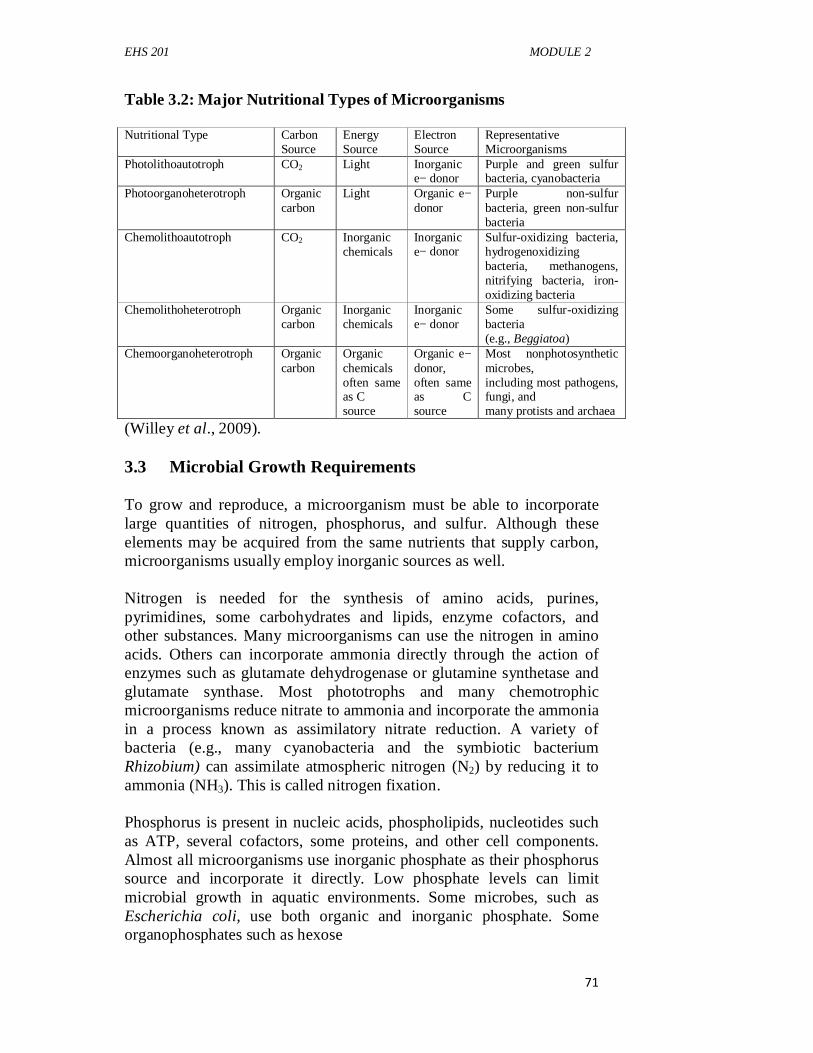
EHS 201 MODULE 2
71
Table 3.2: Major Nutritional Types of Microorganisms
Nutritional Type Carbon Source
Energy Source
Electron Source
Representative Microorganisms
Photolithoautotroph CO2 Light Inorganic e− donor
Purple and green sulfur bacteria, cyanobacteria
Photoorganoheterotroph Organic carbon
Light Organic e− donor
Purple non-sulfur bacteria, green non-sulfur bacteria
Chemolithoautotroph CO2 Inorganic chemicals
Inorganic e− donor
Sulfur-oxidizing bacteria, hydrogenoxidizing bacteria, methanogens, nitrifying bacteria, iron-oxidizing bacteria
Chemolithoheterotroph Organic carbon
Inorganic chemicals
Inorganic e− donor
Some sulfur-oxidizing bacteria (e.g., Beggiatoa)
Chemoorganoheterotroph Organic carbon
Organic chemicals often same as C source
Organic e− donor, often same as C source
Most nonphotosynthetic microbes, including most pathogens, fungi, and many protists and archaea
(Willey et al., 2009). 3.3 Microbial Growth Requirements To grow and reproduce, a microorganism must be able to incorporate large quantities of nitrogen, phosphorus, and sulfur. Although these elements may be acquired from the same nutrients that supply carbon, microorganisms usually employ inorganic sources as well. Nitrogen is needed for the synthesis of amino acids, purines, pyrimidines, some carbohydrates and lipids, enzyme cofactors, and other substances. Many microorganisms can use the nitrogen in amino acids. Others can incorporate ammonia directly through the action of enzymes such as glutamate dehydrogenase or glutamine synthetase and glutamate synthase. Most phototrophs and many chemotrophic microorganisms reduce nitrate to ammonia and incorporate the ammonia in a process known as assimilatory nitrate reduction. A variety of bacteria (e.g., many cyanobacteria and the symbiotic bacterium Rhizobium) can assimilate atmospheric nitrogen (N2) by reducing it to ammonia (NH3). This is called nitrogen fixation. Phosphorus is present in nucleic acids, phospholipids, nucleotides such as ATP, several cofactors, some proteins, and other cell components. Almost all microorganisms use inorganic phosphate as their phosphorus source and incorporate it directly. Low phosphate levels can limit microbial growth in aquatic environments. Some microbes, such as Escherichia coli, use both organic and inorganic phosphate. Some organophosphates such as hexose
EHS 201 GENERAL MICROBIOLOGY
72
6-phosphates are taken up directly by the cell. Other organophosphates are hydrolyzed in the periplasm by the enzyme alkaline phosphatase to produce inorganic phosphate, which then is transported across the plasma membrane. Sulfur is needed for the synthesis of substances such as the amino acids cysteine and methionine, some carbohydrates, biotin, and thiamine. Most microorganisms use sulfate as a source of sulfur after reducing it; a few microorganisms require a prereduced form of sulfur such as cysteine. 3.4 Microbial Growth Factors Some microorganisms lack the enzymes or biochemical pathways needed to synthesize all cell components directly from macroelements and trace elements. Therefore they must obtain these constituents or their precursors from the environment. Organic compounds that are essential cell components or precursors of such components but cannot be synthesised by the organism are called growth factors. There are three major classes of growth factors: (1) Amino acids, (2) Purines and pyrimidines, and (3) Vitamins. Amino acids are needed for protein synthesis, purines and pyrimidines for nucleic acid synthesis. Vitaminsare small organic molecules that usually make up all or part of enzyme cofactors. They are needed in only very small amounts to sustain growth. Some microorganisms require many vitamins; for example, Enterococcus faecalis needs eight different vitamins for growth. Other growth factors include heme (for the synthesis of cytochromes), which is required by Haemophilus influenzae, and cholesterol, which is needed by some mycoplasmas. Some microorganisms are able to synthesize large quantities of vitamins needed by humans. These microbes can be used to manufacture these vitamins for human use. Several water-soluble and fat-soluble vitamins are produced partly or completely using industrial fermentations. Examples of such vitamins and the microorganisms that synthesize them are riboflavin (Clostridium, Candida), coenzyme A (Brevi bacterium), vitamin B12 (Streptomyces, Propioni bacterium, and Pseudomonas), vitamin C (Glucono bacter, Erwinia, Corynebacterium), β-carotene (Dunaliella), and vitamin D (Saccharomyces). Current research focuses on improving yields and finding microorganisms that can produce large quantities of other vitamins.
EHS 201 MODULE 2
73
3.5 Nutrients Assimilation The initial step in nutrient use is their assimilation by the microbial cell. Uptake mechanisms must be specific—that is, the necessary substances, and not others, must be acquired. It does a cell no good to take in a substance that it cannot use. Because microorganisms often live in nutrient-poor habitats, they must be able to transport nutrients from dilute solutions into the cell against a concentration gradient. Finally, nutrient molecules must pass through a selectively permeable plasma membrane that prevents the free passage of most substances. In view of the enormous variety of nutrients and the complexity of the task, it is not surprising that microorganisms make use of several different transport mechanisms. The most important of these are facilitated diffusion, active transport, and group translocation. Eucaryotic microorganisms do not appear to employ group translocation but take up nutrients by the process of endocytosis. 3.5.1 Passive Diffusion A few substances, such as glycerol, can cross the plasma membrane by passive diffusion. Passive diffusion, often called diffusion or simple diffusion, is the process by which molecules move from a region of higher concentration to one of lower concentration. The rate of passive diffusion depends on the size of the concentration gradient between a cell’s exterior and its interior. 3.5.2 Facilitated Diffusion The rate of diffusion across selectively permeable membranes is greatly increased by using carrier proteins, sometimes called permeases, which are embedded in the plasma membrane. Diffusion involving carrier proteins is called facilitated diffusion. The rate of facilitated diffusion increases with the concentration gradient much more rapidly and at lower concentrations of the diffusing molecule than that of passive diffusion. 3.5.3 Active Transport Because facilitated diffusion can efficiently move molecules to the interior only when the solute concentration is higher on the outside of the cell, microbes must have transport mechanisms that can move solutes against a concentration gradient. This is important because microorganisms often live in habitats with very low nutrient concentrations. Microbes use two important transport processes in such
EHS 201 GENERAL MICROBIOLOGY
74
situations: active transport and group translocation. Both are energy-dependent processes. Active transport is the transport of solute molecules to higher concentrations, or against a concentration gradient, with the input of metabolic energy. 3.5.4 Group Translocation In active transport, solute molecules move across a membrane without modification. Another type of transport, called grouptranslocation,chemically modifies the molecule as it is brought into the cell. Group translocation is a type of active transport because metabolic energy is used during uptake of the molecule. 3.5.5 Iron Uptake Almost all microorganisms require iron for use in cytochromes and many enzymes. Iron uptake is made difficult by the extreme insolubility of ferric iron (Fe3+) and its derivatives, which leaves little free iron available for transport. Many bacteria and fungi have overcome this difficulty by secreting siderophores (Greek for ironbearers). Siderophores are low molecular weight organic moleculesthat bind ferric iron and supply it to the cell. Two examples of siderophores are ferrichrome, which is produced by many fungi, and enterobactin, which is formed by E. coli. 3.6 Culture Media Microbiology research depends largely on the ability to grow and maintain microorganisms in the laboratory, and this is possible only if suitable culture media are available. A culture medium is a solid or liquid preparation used to grow, transport, and store microorganisms. To be effective, the medium must contain all the nutrients the microorganism requires for growth. Specialized media are essential in the isolation and identification of microorganisms, the testing of antibiotic sensitivities, water and food analysis, industrial microbiology, and other activities. Although all microorganisms need sources of energy, carbon, nitrogen, phosphorus, sulfur, and various minerals, the precise composition of a satisfactory medium depends on the species one is trying to cultivate because nutritional requirements vary so greatly. Knowledge of a microorganism’s normal habitat often is useful in selecting an appropriate culture medium because its nutrient requirements reflect its natural surroundings. Frequently a medium is used to select and grow specific microorganisms or to help identify a particular species. Media can also be specifically designed to enrich for
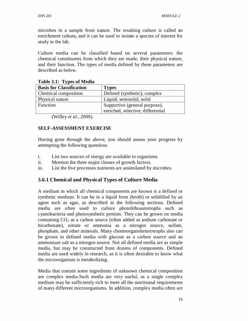
EHS 201 MODULE 2
75
microbes in a sample from nature. The resulting culture is called an enrichment culture, and it can be used to isolate a species of interest for study in the lab. Culture media can be classified based on several parameters: the chemical constituents from which they are made, their physical nature, and their function. The types of media defined by these parameters are described as below. Table 3.1: Types of Media Basis for Classification Types Chemical composition Defined (synthetic), complex Physical nature Liquid, semisolid, solid Function Supportive (general purpose),
enriched, selective, differential (Willey et al., 2009). SELF-ASSESSMENT EXERCISE Having gone through the above, you should assess your progress by attempting the following questions. i. List two sources of energy are available to organisms ii. Mention the three major classes of growth factors. iii. List the five processes nutrients are assimilated by microbes. 3.6.1 Chemical and Physical Types of Culture Media A medium in which all chemical components are known is a defined or synthetic medium. It can be in a liquid form (broth) or solidified by an agent such as agar, as described in the following sections. Defined media are often used to culture photolithoautotrophs such as cyanobacteria and photosynthetic protists. They can be grown on media containing CO2 as a carbon source (often added as sodium carbonate or bicarbonate), nitrate or ammonia as a nitrogen source, sulfate, phosphate, and other minerals. Many chemoorganoheterotrophs also can be grown in defined media with glucose as a carbon source and an ammonium salt as a nitrogen source. Not all defined media are as simple media, but may be constructed from dozens of components. Defined media are used widely in research, as it is often desirable to know what the microorganism is metabolizing. Media that contain some ingredients of unknown chemical composition are complex media.Such media are very useful, as a single complex medium may be sufficiently rich to meet all the nutritional requirements of many different microorganisms. In addition, complex media often are
EHS 201 GENERAL MICROBIOLOGY
76
needed because the nutritional requirements of a particular microorganism are unknown, and thus a defined medium cannot be constructed. Complex media are also used to culture fastidious microbes, microbes with complex nutritional or cultural requirements. Some fastidious microbes may even require a medium containing blood or serum. Most complex media contain undefined components such as peptones, meat extract, and yeast extract. Peptones are protein hydrolysates prepared by partial proteolytic digestion of meat, casein, soya meal, gelatin, and other protein sources. They serve as sources of carbon, energy, and nitrogen. Beef extract and yeast extract are aqueous extracts of lean beef and brewer’s yeast, respectively. Beef extract contains amino acids, peptides, nucleotides, organic acids, vitamins, and minerals. Yeast extract is an excellent source of B vitamins as well as nitrogen and carbon compounds. Three commonly used complex media are: (1) Nutrient broth, (2) Tryptic soy broth, and (3) MacConkey agar. Although both liquid and solidified media are routinely used, solidified media are particularly important because they can be used to isolate different microbes from each other to establish pure cultures. This is a critical step in demonstrating the relationship between a microbe and a disease using Koch’s postulates. Agaris the most commonly used solidifying agent. It is a sulfated polymer composed mainly of d-galactose, 3,6-anhydro-l-galactose, and d-glucuronic acid. It usually is extracted from red algae. Agar is well suited as a solidifying agent for several reasons. One is that it melts at about 90°C but once melted does not harden until it reaches about 45°C. Thus after being melted in boiling water, it can be cooled to a temperature that is tolerated by human hands as well as microbes. Furthermore, microbes growing on agar medium can be incubated at a wide range of temperatures. Finally, agar is an excellent hardening agent because most microorganisms cannot degrade it. 3.6.2 Functional Types of Media Media such as tryptic soy broth and tryptic soy agar are called general purpose or supportive mediabecause they sustain the growth of many microorganisms. Blood and other special nutrients may be added to supportive media to encourage the growth of fastidious microbes. These specially fortified media (e.g., blood agar) are called enriched media.
EHS 201 MODULE 2
77
Selective mediafavor the growth of particular microorganisms. Bile salts or dyes such as basic fuchsin and crystal violet favor the growth of gram-negative bacteria by inhibiting the growth of gram-positive bacteria; the dyes have no effect on gram-negative organisms. Endo agar, eosin methylene blue agar, and MacConkey agar are three media widely used for the detection of E. coli and related bacteria in water supplies and elsewhere. These media contain dyes that suppress the growth of gram-positive bacteria. MacConkey agar also contains bile salts. Bacteria also may be selected by incubation with nutrients that they specifically can use. A medium containing only cellulose as a carbon and energy source is quite effective in the isolation of cellulose-digesting bacteria from samples such as soil. Thus selective media are useful as media for enrichment cultures. The possibilities for selection are endless, and dozens of special selective media are in use. Differential media are media that distinguish among different groups of microbes and even permit tentative identification of microorganisms based on their biological characteristics. Blood agar is both a differential medium and an enriched one. It distinguishes between hemolytic and nonhemolytic bacteria. Hemolytic bacteria (e.g., many streptococci and staphylococci isolated from throats) produce clear zones around their colonies because of red blood cell destruction. MacConkey agar is both differential and selective. Since it contains lactose and neutral red dye, bacteria that catabolize lactose by fermenting it release acidic waste products that make colonies appear pink to red in color. These are easily distinguished from colonies of bacteria that do not ferment lactose. 4.0 CONCLUSION
Microbiology depends enormously on pure culture isolates and studies on microbial growth and their media composition. Understanding growth requirements is fundamental in further research studies and control of microbial growth.
5.0 SUMMARY In this unit, the student has learnt the nutritional requirements of microorganisms, nutritional types of microorganisms, microbial growth factors, nutrient assimilation systems, microbial culture media and microbial growth on agar surfaces. 6.0 TUTOR-MARKED ASSIGNMENT 1. Explain the nutritional requirements of the major nutritional
groups.
EHS 201 GENERAL MICROBIOLOGY
78
2. Briefly describe how microorganisms use the various forms of nitrogen, phosphorus, and sulfur.
3. List the growth factors produced by industrial microorganisms. 4. Define are pure cultures and state their importance. 7.0 REFERENCES/FURTHER READING Atlas, R.M. (1995). Microorganisms in Our World. Mosby Year
Book.Inc. Pelczar, M.J., Chan, E.C.S., & Krieg, R.N. (2001). Microbiology.
(5thed.).. McGraw-Hill. Willey, J.M., Sherwood, L.M. & Woolverton, C.J. (2009). Prescott’s
Principles of ‘Microbiology. (1st ed.)., McGraw-Hill. pp. 1-12.
EHS 201 MODULE 2
79
UNIT 2 CELL REPRODUCTION AND MICROBIAL GROWTH CONTENTS 1.0 Introduction 2.0 Objectives 3.0 Main Content 3.1 Cell Reproduction 3.1.1 Bacterial cell cycle 3.2 Growth Curve 3.2.1 Lag Phase 3.2.2 Exponential Phase 3.2.3 Stationary Phase 3.2.4 Senescence and Death 3.3 Factors that Influence Microbial Growth 3.3.1 Influences of Environmental Factors on Growth 3.3.2 Solutes and Water Activity 3.3.3 pH 3.3.4 Temperature 3.3.5 Oxygen Concentration 3.3.6 Pressure 3.3.7 Radiation 3.3.8 Ultraviolet (UV) Radiation 4.0 Conclusion 5.0 Summary 6.0 Tutor-Marked Assignment 7.0 References/Further Reading 1.0 INTRODUCTION In the study of General Microbiology, microbial reproduction is very vital in understanding population increase and spread in microorganisms. Cell reproduction leads to an increase in population size, and there is need to consider growth and the ways in which it can be measured.In fungi, unicellular algae, and protozoa, reproduction involves a duplication of the nucleus through the asexual process of mitosis and a splitting of the cell in cytokinesis. Reproduction can also occur by a sexual process in which haploid nuclei unite to form a diploid cell having two sets of chromosomes. Various changes then follow to yield a sexually produced offspring. Sexual reproduction has the advantage of mixing chromosomes to obtain genetic variations not possible with asexual reproduction. However, fewer individuals normally result from sexual reproduction than from asexual reproduction. Then we discuss continuous culture techniques.
EHS 201 GENERAL MICROBIOLOGY
80
An account of the influence of environmental factors on microbial growth and microbial growth in natural environments will be discussed. Growth may be defined as an increase in cellular constituents. It leads to a rise in cell number when microorganisms reproduce. Growth also results when cells simply become longer or larger. If the microorganism is coenocytic—that is, a multinucleate organism in which chromosomal replication is not accompanied by cell division— growth results in an increase in cell size but not cell number. It is usually not convenient to investigate the growth and reproduction of individual microorganisms because of their small size. Therefore, when studying growth, microbiologists normally follow changes in the total population number. 2.0 OBJECTIVES By the end of this unit, you will be able to: • explain cell reproduction • understand microbial growth curve • measure microbial growth • discuss the factors that affect microbial growth. 3.0 MAIN CONTENT The main content shall include cell reproduction, microbial growth curve, measurement of microbial growth and factors that affect microbial growth. 3.1 Cell Reproduction 3.1.1 Bacterial Cell Cycle The cell cycle is the complete sequence of events extending from the formation of a new cell through the next division. It is of intrinsic interest to microbiologists as a fundamental biological process. However, understanding the cell cycle has practical importance as well. For instance in bacteria, the synthesis of peptidoglycan is the target of numerous antibiotics. Although some procaryotes reproduce by budding, fragmentation, and other means, most procaryotes reproduce by binary fission. Binary fission is a relatively simple type of cell division: the cell elongates, replicates its chromosome, and separates the newly formed DNA molecules so there is one chromosome in each half of the cell. Finally, a septum (cross wall) is formed at midcell, dividing the parent cell into
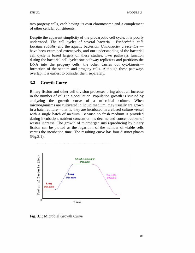
EHS 201 MODULE 2
81
two progeny cells, each having its own chromosome and a complement of other cellular constituents. Despite the apparent simplicity of the procaryotic cell cycle, it is poorly understood. The cell cycles of several bacteria— Escherichia coli, Bacillus subtilis, and the aquatic bacterium Caulobacter crescentus —have been examined extensively, and our understanding of the bacterial cell cycle is based largely on these studies. Two pathways function during the bacterial cell cycle: one pathway replicates and partitions the DNA into the progeny cells, the other carries out cytokinesis—formation of the septum and progeny cells. Although these pathways overlap, it is easiest to consider them separately. 3.2 Growth Curve Binary fission and other cell division processes bring about an increase in the number of cells in a population. Population growth is studied by analyzing the growth curve of a microbial culture. When microorganisms are cultivated in liquid medium, they usually are grown in a batch culture—that is, they are incubated in a closed culture vessel with a single batch of medium. Because no fresh medium is provided during incubation, nutrient concentrations decline and concentrations of wastes increase. The growth of microorganisms reproducing by binary fission can be plotted as the logarithm of the number of viable cells versus the incubation time. The resulting curve has four distinct phases (Fig.3.1).
Fig. 3.1: Microbial Growth Curve
EHS 201 GENERAL MICROBIOLOGY
82
3.2.1 Lag Phase When microorganisms are introduced into fresh culture medium, usually no immediate increase in cell number occurs. This period is called the lag phase.However, cells in the culture are synthesizing new components. A lag phase can be necessary for a variety of reasons. The cells may be old and depleted of ATP, essential cofactors, and ribosomes; these must be synthesized before growth can begin. The medium may be different from the one the microorganism was growing in previously. Here new enzymes would be needed to use different nutrients. Possibly the microorganisms have been injured and require time to recover. Whatever the causes, eventually the cells begin to replicate their DNA, increase in mass, and finally divide. 3.2.2 Exponential Phase During the exponential (log) phase, microorganisms are growing and dividing at the maximal rate possible given their genetic potential, the nature of the medium, and the environmental conditions. Their rate of growth is constant during the exponential phase; that is, they are completing the cell cycle and doubling in number at regular intervals. The population is most uniform in terms of chemical and physiological properties during this phase; therefore exponential phase cultures are usually used in biochemical and physiological studies. Exponential (logarithmic) growth is balanced growth.That is, all cellular constituents are manufactured at constant rates relative to each other. If nutrient levels or other environmental conditions change, unbalanced growthresults. During unbalanced growth, the rates of synthesis of cell components vary relative to one another until a new balanced state is reached. Unbalanced growth is readily observed in two types of experiments: shift-up, where a culture is transferred from a nutritionally poor medium to a richer one; and shift-down, where a culture is transferred from a rich medium to a poor one. In a shift-up experiment, there is a lag while the cells first construct new ribosomes to enhance their capacity for protein synthesis. In a shift-down experiment, there is a lag in growth because cells need time to make the enzymes required for the biosynthesis of unavailable nutrients. Once the cells are able to grow again, balanced growth is resumed and the culture enters the exponential phase. These shift-up and shift-down experiments demonstrate that microbial growth is under precise, coordinated control and responds quickly to changes in environmental conditions. When microbial growth is limited by the low concentration of a required nutrient, the final net growth or yield of cells increases with the initial amount of the limiting nutrient present. The rate of growth also
EHS 201 MODULE 2
83
increases with nutrient concentration but in a hyperbolic manner much like that seen with many enzymes. The shape of the curve seems to reflect the rate of nutrient uptake by microbial transport proteins. At sufficiently high nutrient levels, the transport systems are saturated, and the growth rate does not rise further with increasing nutrient concentration. 3.2.3 Stationary Phase In a closed system such as a batch culture, population growth eventually ceases and the growth curve becomes horizontal. This stationary phaseusually is attained by bacteria at a population level of around 10 9 cells per ml. Other microorganisms normally do not reach such high population densities. For instance, protist cultures often have maximum concentrations of about 106 cells per ml. Final population size depends on nutrient availability and other factors, as well as the type of microorganism being cultured. In the stationary phase, the total number of viable microorganisms remains constant. This may result from a balance between cell division and cell death, or the population may simply cease to divide but remain metabolically active. Microbial populations enter the stationary phase for several reasons. One obvious factor is nutrient limitation; if an essential nutrient is severely depleted, population growth will slow. Aerobic organisms often are limited by O2 availability. Oxygen is not very soluble and may be depleted so quickly that only the surface of a culture will have an O2 concentration adequate for growth. The cells beneath the surface will not be able to grow unless the culture is shaken or aerated in another way. Population growth also may cease due to the accumulation of toxic waste products. This factor seems to limit the growth of many anaerobic cultures (cultures growing in the absence of O2). For example, streptococci can produce so much lactic acid and other organic acids from sugar fermentation that their medium becomes acidic and growth is inhibited. Finally, some evidence exists that growth may cease when a critical population level is reached. Thus entrance into the stationary phase may result from several factors operating in concert. As we have seen, bacteria in a batch culture may enter stationary phase in response to starvation. This probably occurs often in nature because many environments have low nutrient levels. Procaryotes have evolved a number of strategies to survive starvation. Some bacteria respond with obvious morphological changes such as endospore formation, but many only decrease somewhat in overall size. This is often accompanied by protoplast shrinkage and nucleoid condensation. The more important changes during starvation are in gene expression and physiology. Starving bacteria frequently produce a variety of starvation
EHS 201 GENERAL MICROBIOLOGY
84
proteins,which make the cell much more resistant to damage. Some increase peptidoglycan crosslinking and cell wall strength. The Dps (DNA-binding protein from starved cells) protein protects DNA. Proteins called chaperone proteins prevent protein denaturation and renature damaged proteins. Because of these and many other mechanisms, starved cells become harder to kill and more resistant to starvation, damaging temperature changes, oxidative and osmotic damage, and toxic chemicals such as chlorine. These changes are so effective that some bacteria can survive starvation for years. There is even evidence that Salmonella enterica serovar Typhimurium (S.typhimurium) and some other bacterial pathogens become more virulent when starved. Clearly, these considerations are of great practical importance in medical and industrial microbiology. 3.2.4 Senescence and Death For many years, the decline in viable cells following the stationary phase was described simply as the “death phase.” It was assumed that detrimental environmental changes such as nutrient deprivation and the buildup of toxic wastes caused irreparable harm and loss of viability. That is, even when bacterial cells were transferred to fresh medium, no cellular growth was observed. Because loss of viability was often not accompanied by a loss in total cell number, it was assumed that cells died but did not lyse. This view is currently under debate. There are two alternative hypotheses. Some microbiologists think starving cells that show an exponential decline in density have not irreversibly lost their ability to reproduce. Rather, they suggest that microbes are temporarily unable to grow, at least under the laboratory conditions used. This phenomenon, in which the cells are called viable but nonculturable (VBNC), is thought to be the result ofa genetic response triggered in starving, stationary phase cells. Just as some bacteria form endospores as a survival mechanism, it is argued that others are able to become dormant without changes in morphology. Once the appropriate conditions are available (for instance, a change in temperature or passage through an animal), VBNC microbes resume growth. VBNC microorganisms could pose a public health threat, as many assays that test for food and drinking water safety are culture-based. The second alternative to a simple death phase is programmed cell death. In contrast to the VBN Chypothesis whereby cells are genetically programmed to survive, programmed cell death predicts that a fraction of themicrobial population is genetically programmed to die aftergrowth ceases. In this case, some cells die and the nutrientsthey leak enable the eventual growth of those cells in the populationthat did not initiate cell
EHS 201 MODULE 2
85
death. The dying cells are thus“altruistic”—they sacrifice themselves for the benefit of the larger population. Long-term growth experiments reveal that an exponential decline in viability is sometimes replaced by a gradual decline in the number of culturable cells. This decline can last months to years. During this time, the bacterial population continually evolves so that actively reproducing cells are those best able to use the nutrients released by their dying brethren and best able to tolerate the accumulated toxins. This dynamic process is marked by successive waves of genetically distinct variants. Thus natural selection can be witnessed within a single culture vessel. SELF-ASSESSMENT EXERCISE Having gone through the above, you should assess your progress by attempting the following questions. List four phases of microbial growth curve. 3.3 Factors that Influence Microbial Growth 3.3.1 Influences of Environmental Factors on Growth As we have seen, microorganisms must be able to respond to variations in nutrient levels. Microorganisms also are greatly affected by the chemical and physical nature of their surroundings. An understanding of environmental influences aids in the control of microbial growth and the study of the ecological distribution of microorganisms. 3.3.2 Solutes and Water Activity Because a selectively permeable plasma membrane separates microorganisms from their environment, they can be affected by changes in the osmotic concentration of their surroundings. If a microorganism is placed in a hypotonic solution (one with a lower osmotic concentration), water will enter the cell and cause it to burst unless something is done to prevent the influx or inhibit plasma membrane expansion. Conversely if it is placed in a hypertonic solution (one with a higher osmotic concentration), water will flow out of the cell. In microbes that have cell walls, the membrane shrinks away from the cell wall—a process called plasmolysis. Dehydration of the cell in hypertonic environments may damage the cell membrane and cause the cell to become metabolically inactive. Clearly it is important that microbes be able to respond tochanges in the osmotic concentrations of their environment.
EHS 201 GENERAL MICROBIOLOGY
86
Because the osmotic concentration of a habitat has such profound effects on microorganisms, it is useful to express quantitatively the degree of water availability. Microbiologists generally use water activity (aw)for this purpose (water availability also may be expressed as water potential, which is related to aw). The water activity of a solution is 1/100 the relative humidity of the solution (when expressed as a percent). It is also equivalent to the ratio of the solution’s vapor pressure (Psoln) to that of pure water (Pwater) at the same temperature. aw = Psoln / Pwater The water activity of a solution or solid can be determined by sealing it in a chamber and measuring the relative humidity after the system has come to equilibrium. Suppose after a sample is treated in this way, the air above it is 95% saturated—that is, the air contains 95% of the moisture it would have when equilibrated at the same temperature with a sample of pure water. The relative humidity would be 95% and the sample’s water activity, 0.95. Water activity is inversely related to osmotic pressure; if a solution has high osmotic pressure, its aw is low. 3.4.3 pH pH is a measure of the relative acidity of a solution and is defined as the negative logarithm of the hydrogen ion concentration (expressed in terms of molarity). pH = −log [H+] = log(1/[ H+]) Each species has a definite pH growth range and pH growth optimum. Acidophileshave their growth optimum between pH 0 and 5.5; Neutrophiles, between pH 5.5 and 8.0; and Alkalophiles(alkaliphiles),between pH 8.0 and 11.5. Extreme alkalophiles have growth optima at pH 10 or higher. In general, different microbial groups have characteristic pH preferences. Most bacteria and protists are neutrophiles. Most fungi prefer more acidic surroundings, about pH 4 to 6; photosynthetic protists also seem to favor slight acidity. Many archaea are acidophiles. For example, the archaeon Sulfolobus acidocaldarius is a common inhabitant of acidic hot springs; it grows well from pH 1 to 3 and at high temperatures. The archaea Ferroplasma acidarmanus and Picrophilus oshimae can actually grow very close to pH 0. Alkalophiles are distributed among all three domains of life. They include bacteria belonging to the genera Bacillus, Micrococcus, Pseudomonas, and Streptomyces; yeasts and filamentous fungi; and numerous archaea.
EHS 201 MODULE 2
87
3.4.4 Temperature Microorganisms are particularly susceptible to external temperatures because they cannot regulate their internal temperature. An important factor influencing the effect of temperature on growth is the temperature sensitivity of enzyme-catalyzed reactions. Each enzyme has a temperature at which it functions optimally. At some temperature below the optimum, it ceases to be catalytic. As the temperature rises from this low point, the rate of catalysis increases to that observed for the optimal temperature. The velocity of the reaction roughly doubles for every 10°C rise in temperature. When all enzymes in a microbe are considered together, as the rate of each reaction increases, metabolism as a whole becomes more active, and the microorganism grows faster. However, beyond a certain point, further increases actually slow growth, and sufficiently high temperatures are lethal. High temperatures denature enzymes, transport carriers, and other proteins. Temperature also has a significant effect on microbial membranes. At very low temperatures, membranes solidify. At high temperatures, the lipid bilayer simply melts and disintegrates. Thus when organisms are above their optimum temperature, both function and cell structure are affected. If temperatures are very low, function is affected but not necessarily cell chemical composition and structure. Five classes microbes based on their temperature ranges for growth: 1. Psychrophiles grow well at 0°C and have an optimum growth
temperature of 10°C or lower; the maximum is around 15°C. They are readily isolated from Arctic and Antarctic habitats. Oceans constitute an enormous habitat for psychrophiles because 90% of ocean water is 5°C or colder. The psychrophilic protist Chlamydomonasnivalis can actually turn a snowfield or glacier pink with its bright red spores. Psychrophiles are widespread among bacterial taxa and are found in such genera as Pseudomonas, Vibrio, Alcaligenes, Bacillus,Photobacterium, and Shewanella. Psychrophilic microorganisms have adapted to their environment in several ways. Their enzymes, transport systems, and protein synthetic machinery function well at low temperatures. The cell membranes of psychrophilic microorganisms have high levels of unsaturated fatty acids and remain semifluid when cold. Indeed, many psychrophiles begin to leak cellular constituents at temperatures higher than 20°C because of cell membrane disruption.
2. Psychrotrophs (facultative psychrophiles) grow at 0 to 7°C even though they have optima between 20 and 30°C, and maxima at about 35°C. Psychrotrophic bacteria and fungi are major causes of refrigerated food spoilage.
EHS 201 GENERAL MICROBIOLOGY
88
3. Mesophiles are microorganisms with growth optima around 20 to maximum is about 45°C or lower. Most microorganisms probably fall within this category. Almost all human pathogens are mesophiles, as might be expected because the human body is a fairly constant 37°C.
4. Thermophiles grow at temperatures between 55 and 85°C. Their growth minimum is usually around 45°C, and they often have optima between 55 and 65°C. The vast majority are procaryotes, although a few photosynthetic protists and fungi are thermophilic. These organisms flourish in many habitats including composts, self-heating hay stacks, hot water lines, and hot springs.
5. Hyperthermophiles have growth optima between 85°C and about 113°C. They usually do not grow well below 55°C. Pyrococcus abyssi and Pyrodictium occultum are examples of marine hyperthermophiles found in hot areas of the seafloor.
3.4.5 Oxygen Concentration The importance of oxygen to the growth of an organism correlates with its metabolism—in particular, with the processes it uses to conserve the energy supplied by its energy source. Almost all energy-conserving metabolic processes involve the movement of electrons through a series of membrane-bound electron carriers called the electron transport chain (ETC). For chemotrophs, an externally supplied terminal electron acceptor is critical to the functioning of the ETC. The nature of the terminal electron acceptor is related to an organism’s oxygen requirement. 3.4.6 Pressure Organisms that spend their lives on land or the surface of water are always subjected to a pressure of 1 atmosphere (atm) and are never affected significantly by pressure. It is thought that high hydrostatic pressure affects membrane fluidity and membrane associated function. Yet many procaryotes live in the deep sea (ocean depths of 1,000 m or more) where the hydrostatic pressure can reach 600 to 1,100 atm and the temperature is about 2 to 3°C. Many of these procaryotes are barotolerant:increased pressure adversely affects them but not as much as it does non-tolerant microbes. Some procaryotes are truly barophilic —they grow more rapidly at high pressures. Barophiles may play an important role in nutrient recycling in the deep sea. Thus far, they have been found among several bacterial genera (e.g., Photobacterium, Shewanella,Colwellia). Some archaea are thermobarophiles (e.g., Pyrococcus spp., Methanocaldococcus jannaschii).
EHS 201 MODULE 2
89
3.4.7 Radiation Our world is bombarded with electromagnetic radiation of various types. Radiation behaves as if it were composed of waves moving through space like waves traveling on the surface of water. The distance between two wave crests or troughs is the wavelength. As the wavelength of electromagnetic radiation decreases, the energy of the radiation increases; gamma rays and X rays are much more energetic than visible light or infrared waves. Electromagnetic radiation also acts like a stream of energy packets called photons, each photon having a quantum of energy whose value depends on the wavelength of the radiation. Sunlight is the major source of radiation on Earth. It includes visible light, ultraviolet (UV) radiation, infrared rays, and radio waves. Visible light is a most conspicuous and important aspect of our environment: most life depends on the ability of photosynthetic organisms to trap the light energy of the sun. Almost 60% of the sun’s radiation is in the infrared region rather than the visible portion of the spectrum. Infrared is the major source of Earth’s heat. At sea level, one finds very little ultraviolet radiation below about 290 to 300 nm. UV radiation of wavelengths shorter than 287 nm is absorbed by O2 in Earth’s atmosphere; this process forms a layer of ozone between 40 and 48 kilometers above Earth’s surface. The ozone layer absorbs somewhat longer UV rays and reforms O2. The even distribution of sunlight throughout the visible spectrum accounts for the fact that sunlight is generally “white.” Many forms of electromagnetic radiation are very harmful to microorganisms. This is particularly true of ionizing radiation,radiation of very short wavelength and high energy, which can cause atoms to lose electrons (ionize). Two major forms of ionizing radiation are: (1) X rays, which are artificially produced, and (2) Gamma rays, which are emitted during radioisotope decay. Low levels of ionizing radiation may produce mutations and may indirectly result in death, whereas higher levels are directly lethal. Although microorganisms are more resistant to ionizing radiation than larger organisms, they are still destroyed by a sufficiently large dose. Ionizing radiation can be used to sterilize items. Some procaryotes (e.g. Deinococcus radiodurans) and bacterial endospores can survive large doses of ionizing radiation. Ionizing radiation causes a variety of changes in cells. It breaks hydrogen bonds, oxidizes double bonds, destroys ring structures, and polymerizes some molecules. Oxygen enhances these destructive
EHS 201 GENERAL MICROBIOLOGY
90
effects, probably through the generation of hydroxyl radicals (OH•). Although many types of constituents can be affected, the destruction of DNA is probably the most important cause of death. 3.4.8 Ultraviolet (UV) Radiation Thiscan kill microorganisms due to its short wavelength (approximately from 10 to 400 nm) and high energy. The most lethal UV radiation has a wavelength of 260 nm, the wavelength most effectively absorbed by DNA. The primary mechanism of UV damage is the formation of thymine dimers in DNA. Two adjacent thymines in a DNA strand are covalently joined to inhibit DNA replication and function. The damage caused by UV light can be repaired by several DNA repair mechanisms, as discussed in chapter 14. Excessive exposure to UV light outstrips the organism’s ability to repair the damage and death results. Longer wavelengths of UV light (near-UV radiation; 325 to 400 nm) can also harm microorganisms because they induce the breakdown of tryptophan to toxic photoproducts. It appears that these toxic tryptophan photoproducts plus the near-UV radiation itself produce breaks in DNA strands. The precise mechanism is not known, although it is different from that seen with 260 nm UV. Even visible light, when present in sufficient intensity, can damage or kill microbial cells. Usually pigments called photosensitizers and O2 are involved. Photosensitizers include pigments such as chlorophyll, bacteriochlorophyll, cytochromes, and flavins, which can absorb light energy and become excited or activated. The excited photosensitizer (P) transfers its energy to O2, generating singlet oxygen (1O2). Singlet oxygen is a very reactive, powerful oxidizing agent that quickly
destroys a cell. Many microorganisms that are airborne or live on exposed surfaces use carotenoid pigments for protection against photooxidation. Carotenoids effectively quench singlet oxygen— that is, they absorb energy from singlet oxygen and convert it back into the unexcited ground state. Both photosynthetic and nonphotosynthetic microorganisms employ pigments in this way.
SELF-ASSESSMENT EXERCISE What are hypotonic environments and how do microorganisms adjust to it? 4.0 CONCLUSION Microbial population dynamics have been a major indices of microbial growth. Studying cell reproduction will give more information on the microbial survival and growth. The need to ascertain microbial growth
EHS 201 MODULE 2
91
requirements and factors affecting growth gives much insight into the study of microbiology.
5.0 SUMMARY At the end of this unit, the student understood cell reproduction, nutrient requirements for microbial growth, microbial growth curve, and factors affecting microbial growth among others. 6.0 TUTOR-MARKED ASSIGNMENT 1. Explain the microbial growth curve with emphasis on the
different phases of growth. 2. Define the following terms: pH, acidophile, alkalophile and
neutrophile. 3. Explain the mechanisms employed by microbes to maintain a
neutral pH. 7.0 REFERENCES/FURTHER READING Atlas, R.M. (1995). Microorganisms in Our World. Mosby Year Book.
Inc. Cappuccino, J. G., & N. Sherman. (2008). Microbiology A Laboratory
Manual, (8th ed.). Pearson/Benjamin Cummings, San Francisco, CA.
Lammert, J. M.(2007). Techniques in microbiology. A Student
Handbook. Pearson/Prentice Hall, Upper Saddle River, NJ. Pelczar, M.J., Chan, E.C.S., & Krieg, R.N. (2001). Microbiology. (5th
ed.). McGraw-Hill. Willey, J.M., Sherwood, L.M. & Woolverton, C.J. (2009). Prescott’s
Principles of ‘Microbiology. (1st ed.), McGraw-Hill. pp. 1-12
EHS 201 GENERAL MICROBIOLOGY
92
UNIT 3 CONTROL OF MICROORGANISMS CONTENTS 1.0 Introduction 2.0 Objectives 3.0 Main Content
3.1 Terms Used in the Control of Microorganisms 3.2 The Pattern of Microbial Death 3.3 Conditions Influencing the Effectiveness of Antimicrobial Agents 3.4 The Use of Physical Methods in Control 3.4.1 Heat 3.4.2 Filtration 3.4.3 Radiation 3.5 The Use of Chemical Agents in Control 3.5.1 Phenolics 3.5.2 Alcohols 3.5.3 Halogens 3.5.4 Heavy Metals 3.5.5 Quaternary Ammonium Compounds 3.5.6 Sterilizing Gases 3.6 Biological Control of Microorganisms 4.0 Conclusion 5.0 Summary 6.0 Tutor-Marked Assignment 7.0 References/Further Reading 1.0 INTRODUCTION In this chapter, we address the subject of the control and destruction of microorganisms, a topic of immense practical importance. Although most microorganisms are beneficial, some microbial activities have undesirable consequences, such as food spoilage and disease. Therefore it is essential to be able to kill a wide variety of microorganisms or inhibit their growth to minimize their destructive effects. The goal is twofold: (1) To destroy pathogens and prevent their transmission, and (2) To reduce or eliminate microorganisms responsible for the
contamination of water, food, and other substances. Thus this chapter focuses on the control of microorganisms by physical, chemical, and biological agents.
EHS 201 MODULE 2
93
3.0 MAIN CONTENT The main content shall discuss terms used in the control of microorganisms, the pattern of microbial growth, conditions influencing the effectiveness of antimicrobial agents, the use of physical and chemical agents in the control of microbial growths, and biological control of microorganisms. 3.1 Terms Used in the Control of Microorganisms Terminology is especially important when the control of microorganisms is discussed because words such as disinfectant and antiseptic often are used loosely. The situation is even more confusing because a particular treatment can either inhibit growth or kill, depending on the conditions. Sterilization (Latin sterilis, unable to produce offspring or barren) is the process by which all living cells, spores, and acellular entities (e.g., viruses, viroids, and prions) are either destroyed or removed from an object or habitat. A sterile object is totally free of viable microorganisms, spores, and other infectious agents. When sterilization is achieved by a chemical agent, the chemical is called a sterilant. In contrast, disinfection is the killing, inhibition, or removal of microorganisms that may cause disease; disinfection is the substantial reduction of the total microbial population and the destruction of potential pathogens. Disinfectants are agents, usually chemical, used to carry out disinfection and normally used only on inanimate objects. A disinfectant does not necessarily sterilize an object because viable spores and a few microorganisms may remain. Sanitization is closely related to disinfection. In sanitization, the microbial population is reduced to levels that are considered safe by public health standards. The inanimate object is usually cleaned as well as partially disinfected. For example, sanitizers are used to clean eating utensils in restaurants. It also is frequently necessary to control microorganisms on or in living tissue with chemical agents. Antisepsis (Greek anti, against, and sepsis, putrefaction) is the prevention of infection or sepsis and is accomplished with antiseptics. These are chemical agents applied to tissue to prevent infection by killing or inhibiting pathogen growth; they also reduce the total microbial population. Because they must not destroy too much host tissue, antiseptics are generally not as toxic as disinfectants.
EHS 201 GENERAL MICROBIOLOGY
94
Chemotherapy is the use of chemical agents to kill or inhibit the growth of microorganisms within host tissue. A suffix can be employed to denote the type of antimicrobial agent. Substances that kill organisms often have the suffix –cide (Latin cida, to kill); a germicide kills pathogens (and many non-pathogens) but not necessarily endospores. A disinfectant or antiseptic can be particularly effective against a specifi c group, in which case it may be called a bactericide, fungicide, or viricide. Other chemicals do not kill but rather prevent growth. If these agents are removed, growth will resume. Their names end in –static (Greek statikos, causing to stand or stopping)—for example, bacteriostatic and fungistatic. 3.2 The Pattern of Microbial Death A microbial population is not killed instantly when exposed to a lethal agent. Population death is generally exponential (logarithmic)—that is, the population will be reduced by the same fraction at constant intervals. If the logarithm of the population number remaining is plotted against the time of exposure of the microorganism to the agent, a straight-line plot will result. When the population has been greatly reduced, the rate of killing may slow due to the survival of a more resistant strain of the microorganism. It is essential to have a precise measure of an agent’s killing efficiency. One such measure is the decimal reduction time(D) or D value. The decimal reduction time is the time required to kill 90% of the microorganisms or spores in a sample under specified conditions. For example, in a semilogarithmic plot of the population remaining versus the time of heating, the D value is the time required for the line to drop by one log cycle or tenfold. The D value is usually written with a subscript to indicate the temperature for which it applies. To study the effectiveness of a lethal agent, one must be able to decide when microorganisms are dead, which may present some challenges. A microbial cell is often defined as dead if it does not grow and reproduce when inoculated into culture medium that would normally support its growth. In like manner, an inactive virus cannot infect a suitable host. This definition has flaws, however. It has been demonstrated that when bacteria are exposed to certain conditions, they can remain alive but are temporarily unable to reproduce. When in this state, they are referred to as viable but nonculturable (VBNC). In conventional tests to demonstrate killing by an antimicrobial agent, VBNC bacteria would be thought to be dead. This is a serious problem because after a period of recovery, the bacteria may regain their ability to reproduce and cause infection.
EHS 201 MODULE 2
95
3.3 Conditions Influencing the Effectiveness of Antimicrobial Agents
Destruction of microorganisms and inhibition of microbial growth are not simple matters because the efficiency of an antimicrobial agent (an agent that kills microorganisms or inhibits their growth) is affected by at least six factors. 1. Population size: Because an equal fraction of a microbial
population is killed during each interval, a larger population requires a longer time to die than a smaller one.
2. Population composition: The effectiveness of an agent varies greatly with the nature of the organisms being treated because microorganisms differ markedly in susceptibility. Bacterial spores are much more resistant to most antimicrobial agents than are vegetative forms, and younger cells are usually more readily destroyed than mature organisms. Some species are able to withstand adverse conditions better than others. For instance, Mycobacterium tuberculosis, which causes tuberculosis, is much more resistant to antimicrobial agents than most other bacteria.
3. Concentration or intensity of an antimicrobial agent: Often, but not always, the more concentrated a chemical agent or intense a physical agent, the more rapidly microorganisms are destroyed. However, agent effectiveness usually is not directly related to concentration or intensity. Over a short range, a small increase in concentration leads to an exponential rise in effectiveness; beyond a certain point, increases may not raise the killing rate much at all. Sometimes an agent is more effective at lower concentrations. For example, 70% ethanol is more bactericidal than 95% ethanol because the activity of ethanol is enhanced by the presence of water.
4. Duration of exposure: The longer a population is exposed to a microbicidal agent, the more organisms are killed. To achieve sterilization, exposure should be long enough to reduce the probability of survival to 10−6 or less.
5. Temperature: An increase in the temperature at which a chemical acts often enhances its activity. Frequently a lower concentration of disinfectant or sterilizing agent can be used at a higher temperature.
6. Local environment: The population to be controlled is not isolated but surrounded by environmental factors that may either offer protection or aid in its destruction. For example, because heat kills more readily at an acidic pH, acidic foods and beverages such as fruits and tomatoes are easier to pasteurize than more alkaline foods such as milk. A second important environmental factor is organic matter, which can protect

EHS 201 GENERAL MICROBIOLOGY
96
microorganisms against physical and chemical disinfecting agents. Biofilms are a good example. The organic matter in a biofilm protects the biofilm’s microorganisms. Furthermore, it has been clearly documented that bacteria in biofilms are altered physiologically, and this makes them less susceptible to many antimicrobial agents. Because of the impact of organic matter, it may be necessary to clean objects, especially medical and dental equipment, before they are disinfected or sterilized.
3.4 The Use of Physical Methods in Control Heat and other physical agents are normally used to control microbial growth and sterilize objects, as can be seen from the continual operation of the autoclave in every microbiology laboratory. The most frequently employed physical agents are heat, filtration, and radiation. 3.4.1 Heat Moist heat readily destroys viruses, bacteria, and fungi. Moist heat kills by degrading nucleic acids and denaturing enzymes and other essential proteins. It also disrupts cell membranes. Exposure to boiling water for 10 minutes is sufficient to destroy vegetative cells and eucaryotic spores. Unfortunately the temperature of boiling water (100°C or 212°F at sea level) is not sufficient to destroy bacterial spores, which may survive hours of boiling. Therefore boiling can be used for disinfection of drinking water and objects not harmed by water, but boiling does not sterilize. To destroy bacterial spores, moist heat sterilization must be carried out at temperatures above 100°C, and this requires the use of saturated steam under pressure. Steam sterilization is carried out with an autoclave, a device somewhat like a fancy pressure cooker. The development of the autoclave by Chamberland in 1884 tremendously stimulated the growth of microbiology. Water is boiled to produce steam, which is released into the autoclave’s chamber. The air initially present in the chamber is forced out until the chamber is filled with saturated steam and the outlets are closed. Hot, saturated steam continues to enter until the chamber reaches the desired temperature and pressure, usually 121°C and 15 pounds of pressure. At this temperature saturated steam destroys all vegetative cells and spores in a small volume of liquid within 10 to 12 minutes. Treatment is continued for at least 15 minutes to provide a margin of safety. Of course larger containers of liquid such as flasks and carboys require much longer treatment times.
EHS 201 MODULE 2
97
Autoclaving must be carried out properly or the processed materials will not be sterile. If all air has not been flushed out of the chamber, it will not reach 121°C, even though it may reach a pressure of 15 pounds. The chamber should not be packed too tightly because the steam needs to circulate freely and contact everything in the autoclave. Bacterial spores will be killed only if they are kept at 121°C for 10 to 12 minutes. When a large volume of liquid must be sterilized, an extended sterilization time is needed because it takes longer for the center of the liquid to reach 121°C; 5 liters of liquid may require about 70 minutes. In view of these potential difficulties, a biological indicator is often autoclaved along with other material. This indicator commonly consists of a culture tube containing a sterile ampule of medium and a paper strip covered with spores of Geobacillus stearothermophilus. After autoclaving, the ampule is aseptically broken and the culture incubated for several days. If the test bacterium does not grow in the medium, the sterilization run has been successful. Sometimes either special indicator tape or paper that changes color upon sufficient heating is autoclaved with a load of material. These approaches are convenient and save time but are not as reliable as the killing of bacterial spores. Many heat-sensitive substances, such as milk, are treated with controlled heating at temperatures well below boiling, a process known as pasteurizationin honor of its developer, Louis Pasteur. In the 1860s the French wine industry was plagued by the problem of wine spoilage, which made wine storage and shipping difficult. Pasteur examined spoiled wine under the microscope and detected microorganisms that looked like the bacteria responsible for lactic acid and acetic acid fermentations. He then discovered that a brief heating at 55 to 60°C would destroy these microorganisms and preserve wine for long periods. Milk, beer, and many other beverages are now pasteurized. Pasteurization does not sterilize a beverage, but it does kill any pathogens present and drastically slows spoilage by reducing the level of nonpathogenic spoilage microorganisms. Some materials cannot withstand the high temperature of the autoclave, and spore contamination precludes the use of other methods to sterilize them. For these materials, a process of intermittent sterilization, also known as tyndallization(for John Tyndall, the British physicist who used the technique to destroy heat-resistant microorganisms in dust) is used. The process also uses steam (30–60 minutes) to destroy vegetative bacteria. However, steam exposure is repeated for a total of three times with 23 to 24 hour incubations between steam exposures. The incubations permit remaining spores to germinate into heat-sensitive vegetative cells that are then destroyed upon subsequent steam exposures.
EHS 201 GENERAL MICROBIOLOGY
98
3.4.2 Filtration Filtration is an excellent way to reduce the microbial population in solutions of heat-sensitive material, and sometimes it can be used to sterilize solutions. Rather than directly destroying contaminating microorganisms, the filter simply removes them. There are two types of filters. Depth filtersconsist of fibrous or granular materials that have been bonded into a thick layer filled with twisting channels of small diameter. The solution containing microorganisms is sucked through this layer under vacuum, and microbial cells are removed by physical screening or entrapment and by adsorption to the surface of the filter material. Depth filters are made of diatomaceous earth (Berkefield filters), unglazed porcelain (Chamberlain filters), asbestos, or other similar materials. Membrane filters have replaced depth filters for many purposes. These circular filters are porous membranes, a little over 0.1 mm thick, made of cellulose acetate, cellulose nitrate, polycarbonate, polyvinylidene fluoride, or other synthetic materials. Although a wide variety of pore sizes are available, membranes with pores about 0.2 µm in diameter are used to remove most vegetative cells, but not viruses, from solutions ranging in volume from less than 1 ml to many liters. The membranes are held in special holders and are often preceded by depth filters made of glass fibers to remove larger particles that might clog the membrane filter. The solution is pulled or forced through the filter with a vacuum or with pressure from a syringe, peristaltic pump, or nitrogen gas, and collected in previously sterilized containers. Membrane filters remove microorganisms by screening them out much as a sieve separates large sand particles from small ones. These filters are used to sterilize pharmaceuticals, ophthalmic solutions, culture media, oils, antibiotics, and other heat-sensitive solutions. Air also can be sterilized by filtration. Two common examples are N-95 disposable masks used in hospitals and labs, and cotton plugs on culture vessels that let air in but keep microorganisms out. N-95 masks exclude 95% of particles that are larger than 0.3 µm. Other important examples are laminar flow biological safety cabinets, which employ high-efficiency particulate air (HEPA) filters (a type of depth filter) to remove 99.97% of particles 0.3 µm or larger. Laminar flow biological safety cabinets or hoods force air through HEPA filters, then project a vertical curtain of sterile air across the cabinet opening. This protects a worker from microorganisms being handled within the cabinet and prevents contamination of the room. A person uses these cabinets when working with dangerous agents such as M. tuberculosis and tumor viruses. They are also employed in research labs and industries, such as the pharmaceutical industry, when a sterile working environment is needed.
EHS 201 MODULE 2
99
3.4.3 Radiation Ultraviolet (UV) radiation around 260 nm is quite lethal. UV radiation causes thiamine-thiamine dimerization of the microbial DNA, preventing polymerase-mediated replication and transcription. However, UV does not penetrate glass, dirt films, water, and other substances very effectively. Because of this disadvantage, UV radiation is used as a sterilizing agent only in a few specific situations. UV lamps are sometimes placed on the ceilings of rooms or in biological safety cabinets to sterilize the air and any exposed surfaces. Because UV radiation burns the skin and damages eyes, people working in such areas must be certain the UV lamps are off when the areas are in use. Commercial UV units are available for water treatment. Pathogens and other microorganisms are destroyed when a thin layer of water ispassed under the lamps. Ionizing radiationis an excellent sterilizing agent and penetrates deep into objects. It will destroy bacterial spores and vegetative cells, both procaryotic and eucaryotic; however, ionizing radiation is not always effective against viruses. Gamma radiation from a cobalt 60 source and accelerated electrons from high-voltage electricity are used in the cold sterilization of antibiotics, hormones, sutures, and plastic disposable supplies such as syringes. Gamma radiation and electron beams have also been used to sterilize and “pasteurize” meat and other foods. Irradiation can eliminate the threat of such pathogens as Escherichia coli O157:H7, Staphylococcus aureus, and Campylobacter jejuni. Based on the results of numerous studies, both the U.S. Food and Drug Administration and the World Health Organization have approved irradiated food and declared it safe for human consumption. Currently irradiation is being used to treat poultry, beef, pork, veal, lamb, fruits, vegetables, and spices. SELF-ASSESSMENT EXERCISE Having gone through the above, you should assess your progress by attempting the following questions. i. Mention factors that affect the effectiveness of antimicrobial
agents. ii. List the physical methods used in the control of microorganisms. 3.5 The Use of Chemical Agents in Control Physical agents are generally used to sterilize objects. Chemicals, on the other hand, are more often employed in disinfection and antisepsis. The proper use of chemical agents is essential to laboratory and hospital
EHS 201 GENERAL MICROBIOLOGY
100
safety. Chemicals also are employed to prevent microbial growth in food, and certain chemicals are used to treat infectious disease. Many different chemicals are available for use as disinfectants, and each has its own advantages and disadvantages. Ideally the disinfectant must be effective against a wide variety of infectious agents (gram-positive and gram-negative bacteria, acid-fast bacteria, bacterial spores, fungi, and viruses) at low concentrations and in the presence of organic matter. Although the chemical must be toxic for infectious agents, it should not be toxic to people or corrosive for common materials. In practice, this balance between effectiveness and low toxicity for animals is hard to achieve. Some chemicals are used despite their low effectiveness because they are relatively nontoxic. The ideal disinfectant should be stable upon storage, odorless or with a pleasant odor, soluble in water and lipids for penetration into microorganisms, have a low surface tension so that it can enter cracks in surfaces, and be relatively inexpensive. One potentially serious problem is the overuse of antiseptics. For instance, the antibacterial agent triclosan is found in products such as deodorants, mouthwashes, soaps, cutting boards, and baby toys. Unfortunately, the emergence of triclosan-resistant bacteria has become a problem. For example, Pseudomonasaeruginosa actively pumps the antiseptic out of the cell. There is now evidence that extensive use of triclosan also increases the frequency of bacterial resistance to antibiotics. Thus overuse of antiseptics can have unintended harmful consequences. 3.5.1 Phenolics Phenol was the first widely used antiseptic and disinfectant. In 1867 Joseph Lister employed it to reduce the risk of infection during surgery. Today phenol and phenolics (phenol derivatives) such as cresols, xylenols, and orthophenylphenol are used as disinfectants in laboratories and hospitals. The commercial disinfectant Lysol is made of a mixture of phenolics. Phenolics act by denaturing proteins and disrupting cell membranes. They have some real advantages as disinfectants: phenolics are tuberculocidal, effective in the presence of organic material, and remain active on surfaces long after application. However, they have a disagreeable odor and can cause skin irritation. 3.5.2 Alcohols Alcohols are among the most widely used disinfectants and antiseptics. They are bactericidal and fungicidal but not sporicidal; some lipid-
EHS 201 MODULE 2
101
containing viruses are also destroyed. The two most popular alcohol germicides are ethanol and isopropanol, usually used in about 70 to 80% concentration. They act by denaturing proteins and possibly by dissolving membrane lipids. A 10 to 15 minute soaking is sufficient to disinfect small instruments. 3.5.3 Halogens A halogen is any of the five elements (fluorine, chlorine, bromine, iodine, and astatine) in group VIIA of the periodic table. They exist as diatomic molecules in the free-state and form salt-like compounds with sodium and most other metals. The halogens iodine and chlorine are important antimicrobial agents. Iodine is used as a skin antiseptic and kills by oxidizing cell constituents and iodinating cell proteins. At higher concentrations, it may even kill some spores. Iodine often has been applied as tincture of iodine, 2% or more iodine in a water-ethanol solution of potassium iodide. Although it is an effective antiseptic, the skin may be damaged, a stain is left, and iodine allergies can result. Iodine has been complexed with an organic carrier to form an iodophor. Iodophors are water soluble, stable, and non-staining, and release iodine slowly to minimize skin burns and irritation. They are used in hospitals for cleansing preoperative skin and in hospitals and laboratories for disinfecting. Some popular brands are Wescodyne for skin and laboratory disinfection and Betadine for wounds. Chlorine is the usual disinfectant for municipal water supplies and swimming pools and is also employed in the dairy and food industries. It may be applied as chlorine gas (Cl2), sodium hypochlorite (bleach, NaClO), or calcium hypochlorite [Ca(OCl)2], all of which yield hypochlorous acid (HClO) see chemical reactions below). The result is oxidation ofcellular materials and destruction of vegetative bacteriaand fungi. Cl2 + H2O →HCl + HClO NaOCl + H2O →NaOH + HClO Ca(OCl)2 + 2H2O →Ca(OH)2 + 2HClO Death of almost all microorganisms usually occurs within 30 minutes. One potential problem is that chlorine reacts with organic compounds to form carcinogenic trihalomethanes, which must be monitored in drinking water. Ozone has been used successfully as an alternative to chlorination in Europe and Canada. Chlorine is also an excellent disinfectant for individual use because it is effective, inexpensive, and easy to employ. Small quantities of drinking water can be disinfected with halazone tablets. Halazone (parasulfone dichloramidobenzoic acid) slowly releases chloride when added to water
EHS 201 GENERAL MICROBIOLOGY
102
and disinfects it in about a half hour. It is frequently used by campers lacking access to uncontaminated drinking water. 3.5.4 Heavy Metals For many years the ions of heavy metals such as mercury, silver, arsenic, zinc, and copper were used as germicides. These have now been superseded by other less toxic and more effective germicides (many heavy metals are more bacteriostatic thanbactericidal). There are a few exceptions. In some hospitals, a1% solution of silver nitrate is added to the eyes of infants toprevent ophthalmic gonorrhea. Silver sulfadiazine is used onburns. Copper sulfate is an effective algicide in lakes and swimmingpools. Heavy metals combine with proteins, often withtheir sulfhydryl groups, and inactivate them. They may alsoprecipitate cell proteins. 3.5.5 Quaternary Ammonium Compounds Quaternary ammonium compounds are detergents that have antimicrobial activity and are effective disinfectants. Detergents (Latin detergere, to wipe away) are organic cleansing agents that are amphipathic, having both polar hydrophilic and nonpolar hydrophobic components. The hydrophilic portion of a quaternary ammonium compound is a positively charged quaternary nitrogen; thus quaternary ammonium compounds are cationic detergents. Their antimicrobial activity is the result of their ability to disrupt microbial membranes; they may also denature proteins. Cationic detergents such as benzalkonium chloride and cetylpyridinium chloride kill most bacteria but not M. tuberculosis or spores. They have the advantages of being stable and nontoxic, but they are inactivated by hard water and soap. Cationic detergents are often used as disinfectants for food utensils and small instruments and as skin antiseptics. 3.5.6 Sterilizing Gases Many heat-sensitive items such as disposable plastic petri dishes and syringes, heart-lung machine components, sutures, and catheters are sterilized with ethylene oxide gas. Ethylene oxide (EtO) is both microbicidal and sporicidal. It is a very strong alkylating agent that kills by reacting with functional groups of DNA and proteins to block replication and enzymatic activity. It is a particularly effective sterilizing agent because it rapidly penetrates packing materials, even plastic wraps. Sterilization is carried out in a special ethylene oxide sterilizer,
EHS 201 MODULE 2
103
very much resembling an autoclave in appearance, that controls the EtO concentration, temperature, and humidity. Because pure EtO is explosive, it is usually supplied in a 10 to 20% concentration mixed with either CO2 or dichlorodifluoromethane. The ethylene oxide concentration, humidity, and temperature influence the rate of sterilization. A clean object can be sterilized if treated for 5 to 8 hours at 38°C or 3 to 4 hours at 54°C when the relative humidity is maintained at 40 to 50% and the EtO concentration at 700 mg/l. Because it is so toxic to humans, extensive aeration of the sterilized materials is necessary to remove residual EtO. 3.6 Biological Control of Microorganisms The emerging field of biological control of microorganisms demonstrates great promise. Scientists are learning to exploit natural control processes such as predation of one microorganism on another, viral-mediated lysis, and toxin-mediated killing. While these control mechanisms occur in nature, their approval and use by humans is relatively new. Studies evaluating control of Salmonella, Shigella, and E. coli by gram-negative predators such as Bdellovibrio suggest that poultry farms may be sprayed with the predator to reduce potential contamination. The control of human pathogens using bacteriophage is gaining wide support and appears to be effective in the eradication of a number of bacterial species by lysing the pathogenic host. This seems intuitive, knowing that the virus lyses its specific bacterial host, yet unnerving when one thinks about maybe swallowing, injecting, or applying a virus to the human body. The use of microbial toxins (such as bacteriocins) to control susceptible populations suggests yet another method for potential control of other microorganisms. 8. 4.0 CONCLUSION Microbial growth and survival in the environment have posed major environmental health challenge. The spread of microbial diseases and the consequent health implications have been of ancient concern and many previous efforts have been made in the control of microbial growth. Harnessing physical and chemical control methods with the predatory biological approach still remain to be enhance in the control of microbial growth. 5.0 SUMMARY In this unit the student learnt the terms used in control of microorganisms, pattern of microbial death, conditions influencing the effectiveness of antimicrobial agents, the use of physical and chemical control agents and biological control through microbial predatory.
EHS 201 GENERAL MICROBIOLOGY
104
6.0 TUTOR-MARKED ASSIGNMENT 1. Define the following terms: sterilisation, sterilant, disinfection,
disinfectant, sanitisation, antisepsis, antiseptic, chemotherapy, germicide.
2. Explain the difference between bactericidal and bacteriostatic? 3. How does the population size, population composition,
concentration or intensity of the agent, treatment duration, temperature, and local environmental conditions influence the effectiveness of antimicrobial agents?
4. What are the advantages and disadvantages of ultraviolet light and ionizing radiation as sterilizing agents?
7.0 REFERENCES/FURTHER READING Atlas, R., & Bartha, R. (1998). Microbial Ecology, Fundamental And
Applications, (4th ed). Addison Wesley Longman, Reading. Atlas, R.M. (1995). Microorganisms in Our World. Mosby Year Book.
Inc. Cappuccino, J. G., & N. Sherman. (2008). Microbiology a laboratory
Manual, (8th ed.) Pearson/Benjamin Cummings, San Francisco, CA.
Lammert, J. M. (2007). Techniques in microbiology. A Student
Handbook. Pearson/Prentice Hall, Upper Saddle River, NJ. Madigan, M., Martinko, J.M., Stahl, D., & Clark, D.P. (2010). Brock:
Biology of Microorganisms, (13th ed.). Pearson Benjamin-Cummings, San Francisco.
Pelczar, M.J., Chan, E.C.S. & Krieg, R.N. (2001). Microbiology. (5th
ed.). McGraw-Hill. Willey, J.M., Sherwood, L.M. & Woolverton, C.J. (2009). Prescott’s
Principles of ‘Microbiology. (1st ed). McGraw-Hill. pp. 1-12 4

EHS 201 MODULE 3
105
MODULE 3 MICROBIAL METABOLISM Unit 1 Introduction to Microbial Metabolism Unit 2 Catabolism Unit 3 Anabolism UNIT 1 INTRODUCTION TO MICROBIAL METABOLISM CONTENTS 1.0 Introduction 2.0 Objectives 3.0 Main Content 3.1 Energy and Work 3.2 Laws of Thermodynamics 3.3 Adenosine Triphosphate (ATP) 4.0 Conclusion 5.0 Summary 6.0 Tutor-Marked Assignment 7.0 References/Further Reading 1.0 INTRODUCTION Metabolism is the total of all chemical reactions occurring in the cell. Some metabolic reactions are fueling reactions. The fueling reactions are part of catabolism. They conserve energy from the organisms’ energy source, generate a ready supply of electrons (reducing power),and generate precursors for biosynthesis. The products of the fueling reactions are used in another set of metabolic reactions that build new organic molecules from smaller inorganic and organic compounds. These biosynthetic reactions are called anabolism. To understand metabolism, the nature of energy and the laws of thermodynamics must be considered. Microorganisms display an amazing array of metabolic diversity, especially in terms of the energy sources and energy-conserving processes they employ. Despite this diversity, several basic principles and processes are common to the metabolism of all microbes.
EHS 201 GENERAL MICROBIOLOGY
106
2.0 OBJECTIVES By the end of this unit, you will be able to: • Identify Microbial energy and work • Describe the Laws of thermodynamics as associated with
microbial energy utilisation • Recommend Adenosine tri-phosphate for the conservation of
energy by microorganisms. 3.0 MAIN CONTENT The main content shall discuss microbial energy at work, laws of thermodynamics in relationship with microbial energy utilization and conservation of energy by microbes by using ATP. 3.1 Energy and Work Energy may be most simply defined as the capacity to do work. This is because all physical and chemical processes are the result of the application or movement of energy. Living cells carry out three major types of work. Chemical work involves the synthesis of complex biological molecules from much simpler precursors (i.e., anabolism); energy is needed to increase the molecular complexity of a cell. Transport work requires energy to take up nutrients, eliminate wastes, and maintain ion balances. Energy input is needed because molecules and ions often must be transported across cell membranes against an electrochemical gradient. The third type of work is mechanical work, perhaps the most familiar of the three. Energy is required for cell motility and the movement of structures within cells. 3.2 Laws of Thermodynamics To understand how energy is conserved in ATP and how ATP is used to do cellular work, some knowledge of the basic principles of thermodynamics is required. The science of thermodynamics analyzes energy changes in a collection of matter (e.g., a cell or a plant) called a system. All other matter in the universe is called the surroundings. Thermodynamics focuses on the energy differences between the initial state and the final state of a system. It is not concerned with the rate of the process. For instance, if a pan of water is heated to boiling, only the condition of the water at the start and at boiling is important in thermodynamics, not how fast it is heated or on what kind of stove. Two important laws of thermodynamics must be understood. The first law of thermodynamics says that energy can be neither created nor
EHS 201 MODULE 3
107
destroyed. The total energy in the universe remains constant, although it can be redistributed, as it is during the many energy exchanges that occur during chemical reactions. For example, heat is given off by exothermic reactions and absorbed during endothermic reactions. However, the first law alone cannot explain why heat is released by one chemical reaction and absorbed by another. Explanations for this require the second law of thermodynamics and a condition of matter called entropy. Entropy is a measure of the randomness or disorder of a system. The greater the disorder of a system, the greater is its entropy. The second law states that physical and chemical processes proceed in such a way that the randomness or disorder of the universe (the system and its surroundings) increases. However, even though the entropy of the universe increases, the entropy of any given system within the universe can increase, decrease, or remain unchanged. It is necessary to specify the amount of energy used in or evolving from a particular process, and two types of energy units are employed. A calorie (cal) is the amount of heat energy needed to raise one gram of water from 14.5 to 15.5ₒC. The amount of energy also may be expressed in terms of joules (J), the units of work capable of being done. One cal of heat is equivalent to 4.1840 J of work. One thousand calories or a kilocalorie (kcal) is enough energy to boil 1.9 ml of water. A kilojoule is enough energy to boil about 0.44 ml of water or enable a person weighing 70 kg to climb 35 steps. The joule is normally used by chemists and physicists. 3.3 Adenosine Triphosphate (ATP) Considerable metabolic diversity exists in the microbial world. However, several biochemical principles are common to all types of metabolism. These are: (1) The use of ATP to conserve energy released during exergonic
reactions, so it can be used to drive endergonic reactions; (2) The organization of metabolic reactions into pathways and
cycles; (3) The catalysis of metabolic reactions by enzymes; and (4) The importance of oxidation-reduction reactions in energy
conservation. This section considers the role of ATP in metabolism.
Energy is released from a cell’s energy source in exergonic reactions (i.e., those reactions with a negative ∆Gₒ’). Rather than wasting this energy, much of it is trapped in a practical form that allows its transfer to the cellular systems doing work. These systems carry out endergonic reactions (e.g., anabolism), and the energy captured by the cell is used to
EHS 201 GENERAL MICROBIOLOGY
108
drive these reactions to completion. In living organisms, this practical form of energy is adenosine 5- triphosphate (ATP).In a sense, cells carry out certain processes so that they can “earn” ATP and carry out other processes in which they “spend” their ATP. Thus ATP is often referred to as the cell’s energy currency. In the cell’s economy, ATP serves as the link between exergonic reactions and endergonic reactions. What makes ATP suited for this role as energy currency? ATP is a high-energy molecule. That is, it breaks down or hydrolyzes almost completely to the products adenosine diphosphate (ADP)and orthophosphate (Pi) and is strongly exergonic, having a ∆Gₒ’ of -7.3 kcal/mole. ATP + H2O⇌ ADP + Pi +H +
Because ATP readily transfers its phosphate to water, it is said to have a high phosphate group transfer potential, defined as the negative of ∆Gₒ’ for the hydrolytic removal of phosphate. A molecule with a higher group transfer potential donates phosphate to one with a lower potential. Thus ATP readily donates a phosphate to molecules such as glucose and glucose 6-phosphate in reactions such as those found in some catabolic pathways. Although the free energy change for hydrolysis of ATP is quite large, metabolic reactions exist that release even greater amounts of free energy. This energy is used to resynthesize ATP from ADP and Pi during fueling reactions. Likewise, catabolism can generate molecules with a phosphate group transfer potential that is even higher than that of ATP. Phosphoenolpyruvate (PEP) and guanosine 5’-triphosphate (GTP) are two important examples. Cells use these molecules to regenerate ATP from ADP by a mechanism called substrate-level phosphorylation. Thus ATP, ADP, and Pi form an energy cycle. The fueling reactions conserve energy released from an energy source by using it to synthesize ATP from ADP and Pi. When ATP is hydrolyzed, the energy released drives endergonic processes such as anabolism, transport, and mechanical work. SELF-ASSESSMENT EXERCISE State two important laws of thermodynamics 4.0 CONCLUSION Microbial energy utilization and conservation explains the ability of microorganisms to function in an ecosystem. Therefore, to understand
EHS 201 MODULE 3
109
these energy systems, the basic principles of thermodynamics remains very vital.
5.0 SUMMARY
At the end of this unit, the student understood the microbial energy demand, utilization and conservation. 6.0 TUTOR-MARKED ASSIGNMENT . 1. List the four biochemical principles that are common to all types
of metabolism 2. Discuss the importance of energy in microorganisms. 7.0 REFERENCES/FURTHER READING Pelczar, M.J., Chan, E.C.S., & Krieg, R.N. (2001). Microbiology. (5th
ed).. McGraw-Hill. illey, J.M., Sherwood, L.M. & Woolverton, C.J. (2009). Prescott’s
Principles of ‘Microbiology.(1st ed.). McGraw-Hill. pp. 1-12.
EHS 201 GENERAL MICROBIOLOGY
110
UNIT 2 CATABOLISM CONTENTS 1.0 Introduction 2.0 Objectives 3.0 Main Content 3.1 Catabolism: Energy Release and Conservation 3.2 Anabolism: The Use of Energy in Biosynthesis 3.3 Chemoorganotrophic Fueling Processes 3.4 Aerobic Respiration 3.5 Breakdown of Glucose to Pyruvate 3.6 Anaerobic Respiration 4.0 Conclusion 5.0 Summary 6.0 Tutor-Marked Assignment 7.0 References/Further Reading 1.0 INTRODUCTION In the quest to survive in very versed environments, microbes have devised means of energy transfers and conversion. This ability to convert different energy sources to ATP has conferred different characteristics and fueling processes. 2.0 OBJECTIVES By the end of this unit, you will be able to: • explain microbial catabolism and anabolism • understand the microbial fueling process • discuss the breakdown of glucose to pyruvate • assimilate the microbial anaerobic respiration. 3.0 MAIN CONTENT The main content of this unit shall include, microbial catabolism and anabolism, microbial fueling process, microbial breakdown of molecules, and microbial anaerobic respiration. 3.1 Catabolism: Energy Release and Conservation Microbes are the most successful organisms on Earth as witnessed by their growth under almost every conceivable condition. In large part, their success results from the diversity of their fueling reactions — those
EHS 201 MODULE 3
111
reactions that convert energy from an organism’s energy source into ATP. Animals and many microbes are chemoorganoheterotrophs. These organisms use organic molecules as their source of energy, carbon, and electrons. In other words, the same molecule that supplies them with energy also supplies them with carbon and electrons. Chemoorganoheterotrophs (often simply referred to as chemoorganotrophs or chemoheterotrophs) can use one or more of the following catabolic processes: aerobic respiration, anaerobic respiration, or fermentation. Chemolithoautotrophs use CO2 as a carbon source and reduced inorganic molecules as sources of both energy and electrons. Their energy-conserving processes are sometimes referred to as respiration because they are similar to the respiratory processes carried out by chemoorganoheterotrophs. Photolithotrophic microbes use light as their source of energy and inorganic molecules as a source of electrons. Some use water as their electron source, as do plants, and release oxygen into the atmosphere in a process called oxygenic photosynthesis. Some photosynthetic bacteria use molecules such as hydrogen sulfide (H2S) rather than water as an electron source, so do not release oxygen into the atmosphere; they are called anoxygenic phototrophs. Photolithotrophs are usually autotrophic, using CO2 as a carbon source. However, some phototrophic microbes are heterotrophic. We begin our consideration of fueling reactions with an overview of the metabolism of chemoorganotrophs. This is followed by an introduction to the oxidation of carbohydrates, especially glucose, and a discussion of ATP synthesis during aerobic and anaerobic respiration. Fermentation is then described, followed by a survey of the breakdown of other carbohydrates and organic substances. 3.2 Anabolism: The Use of Energy in Biosynthesis Anabolism is the creation of order. Because a cell is highly ordered and immensely complex, much energy is required for biosynthesis. This is readily apparent from estimates of the biosynthetic capacity of rapidly growing Escherichia coli. Although most ATP dedicated to biosynthesis is employed in protein synthesis, ATP is also used to make other cell material. It is intuitively obvious why rapidly growing cells need a large supply of ATP. But even non-growing cells need energy for the biosynthetic processes they carry out. This is because non-growing cells continuously degrade and resynthesize cellular moleculesduring a process known as turnover. Thus cells are never the same from one instant to the next. Clearly metabolism must be carefully regulated if the rate of turnover is
EHS 201 GENERAL MICROBIOLOGY
112
to be balanced by the rate of biosynthesis. It must also be regulated in response to a microbe’s environment. 3.2.1 Principles Governing Biosynthesis The problem faced by all cells is how to make the many molecules they need as efficiently as possible. Cells have solved this problem by carrying out biosynthesis using a few basic principles. 1. Large molecules are made from small molecules: The
construction of large macromolecules (complex molecules) from a few simple structural units (monomers) saves much genetic storage capacity, biosynthetic raw material, and energy. A consideration of protein synthesis clarifies this. Proteins—whatever size, shape, or function—are made of only 20 common amino acids joined by peptide bonds. Different proteins simply have different amino acid sequences but not new and dissimilar amino acids. Suppose that proteins were composed of 40 different amino acids instead of 20. The cell would then need the enzymes to manufacture twice as many amino acids (or would have to obtain the extra amino acids in its diet). Genes would be required for the extra enzymes, and the cell would have to invest raw materials and energy in the synthesis of these additional genes, enzymes, and amino acids. Clearly the use of a few monomers linked together by a single type of covalent bond makes the synthesis of macromolecules a highly efficient process.
2. Many enzymes do double duty: Many enzymes are used for both catabolic and anabolic processes, saving additional materials and energy. For example, most glycolytic enzymes are involved in both the synthesis and the degradation of glucose.
3. Some enzymes function in one direction only: Although many steps of amphibolic pathways are catalyzed by enzymes that act reversibly, some are not. These steps require the use of separate enzymes: one to catalyze the catabolic reaction, the other to catalyze the anabolic reaction. The use of two enzymes allows independent regulation of catabolism and anabolism. Thus catabolic and anabolic pathways are never identical, although many enzymes are shared. Although both types of pathways can be regulated by their end products as well as by the concentrations of ATP, ADP, AMP, and NAD+, end-product regulation generally assumes more importance in anabolic pathways.
4. Anabolic pathways are irreversible: To synthesize molecules efficiently, anabolic pathways must operate irreversibly in the direction of biosynthesis. Cells achieve this by connecting some
EHS 201 MODULE 3
113
biosynthetic reactions to the breakdown of ATP and other nucleoside triphosphates. When these two processes are coupled, the free energy made available during nucleoside triphosphate breakdown drives the biosynthetic reaction to completion.
5. Catabolism and anabolism are physically separated: In eucaryotic cells, catabolic and anabolic pathways can be localized into distinct cellular compartments—a process called compartmentation. Compartmentation makes it easier for catabolic and anabolic pathways to operate simultaneously yet independently.
6. Catabolism and anabolism use different cofactors: Usually catabolic oxidations produce NADH, a substratefor electron transport. In contrast, when an electrondonor is needed during biosynthesis, NADPH serves asthe donor.
3.3 Chemoorganotrophic Fueling Processes When chemoorganotrophs oxidize an organic energy source, the electrons released are accepted by a variety of electron acceptors. When the electron acceptor is exogenous (i.e., externally supplied), the metabolic process is called respiration and may be divided into two different types. In aerobic respiration, the final electron acceptor is oxygen, whereas the terminal acceptor in anaerobic respiration is a different exogenous molecule such as NO3
_, SO4 2-, CO2, Fe3-, and SeO4
2-. Organic acceptors such as fumarate and humic acids also may be used. Respiration involves the activity of an electron transport chain. As electrons pass through the chain to the final electron acceptor, a type of potential energy called the proton motive force (PMF) is generated and used to synthesize ATP from ADP and Pi. In contrast, fermentation (Latin fermentare, to cause to rise) uses an electron acceptor that is endogenous (from within the cell) and does not involve an electron transport chain or the generation of PMF. The endogenous electron acceptor is usually an intermediate (e.g., pyruvate) of the catabolic pathway used to degrade and oxidize the organic energy source. During fermentation, ATP is synthesized only by substrate-level phosphorylation, a process in which a phosphate group is transferred to ADP from a high energy molecule (e.g., phosphoenolpyruvate) generated by catabolism of the energy source. By convention, aerobic respiration, anaerobic respiration, and fermentation are usually described with glucose as the energy source. This is done for several reasons. One is that glucose is used by many chemoorganotrophs as an energy source. But perhaps more important is the way catabolic pathways are organised. Most organisms can use a
EHS 201 GENERAL MICROBIOLOGY
114
wide variety of organic molecules as energy sources, including macromolecules such as proteins, polysaccharides, and lipids. These molecules are broken down into their constituent parts: amino acids, monosaccharides, and lipids. This is often referred to as the first stage of catabolism; it releases relatively little energy. The monomers are subsequently converted to glucose or to intermediates of the pathways used in its catabolism. Thus nutrient molecules are funneled into ever fewer metabolic intermediates. Indeed a common pathway often degrades many similar molecules (e.g., several different sugars). The existence of a few metabolic pathways, each degrading many nutrients, greatly increases metabolic efficiency by avoiding the need for a large number of less metabolically flexible pathways. The catabolic pathways of greatest importance to chemoorganotrophs are the glycolytic pathways and the tricarboxylic acid (TCA) cycle. Each pathway consists of a set of enzyme-catalyzed reactions arranged so that the product of one reaction serves as a substrate for the next. They are important not only for their role in catabolism but also for their roles in anabolism. They supply material needed for biosynthesis, including precursor metabolites and reducing power. Precursor metabolites serve as the starting molecules for biosynthetic pathways. Reducing power is used in redox reactions that reduce the precursor metabolites as they are transformed into amino acids, nucleotides, and other small molecules needed for synthesis of macromolecules. Pathways that function both catabolically and anabolically are called amphibolicpathways (Greek amphi, on both sides). Many of the reactions of amphibolic pathways are freely reversible and can be used to synthesize or degrade molecules, depending on the nutrients available and the needs of the microbe. The few irreversible catabolic steps are bypassed in biosynthesis with alternate enzymes that catalyze the reverse reaction. The presence of two separate enzymes, one catalyzing the reversal of the other’s reaction, permits independent regulation of the catabolic and anabolic functions of these amphibolic pathways. 3.4 Aerobic Respiration Aerobic respiration is a process that can completely catabolize an organic energy source to CO2 using the glycolytic pathways and TCA cycle with O2 as the terminal electron acceptor. This is accomplished in three stages. The first has already been noted: the breakdown of macromolecules into monomers. The monomers are further degraded in the second stage, which includes the glycolytic and other pathways that yield pyruvate or acetyl coenzyme A. This stage produces some ATP as well as NADH, FADH2, or both. Finally, during the third stage, partially oxidized carbon is fed into the TCA cycle and oxidized completely to
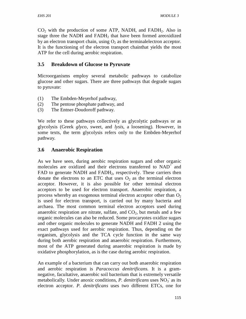
EHS 201 MODULE 3
115
CO2 with the production of some ATP, NADH, and FADH2. Also in stage three the NADH and FADH2 that have been formed areoxidized by an electron transport chain, using O2 as the terminalelectron acceptor. It is the functioning of the electron transport chainthat yields the most ATP for the cell during aerobic respiration. 3.5 Breakdown of Glucose to Pyruvate Microorganisms employ several metabolic pathways to catabolize glucose and other sugars. There are three pathways that degrade sugars to pyruvate: (1) The Embden-Meyerhof pathway, (2) The pentose phosphate pathway, and (3) The Entner-Doudoroff pathway. We refer to these pathways collectively as glycolytic pathways or as glycolysis (Greek glyco, sweet, and lysis, a loosening). However, in some texts, the term glycolysis refers only to the Embden-Meyerhof pathway. 3.6 Anaerobic Respiration As we have seen, during aerobic respiration sugars and other organic molecules are oxidized and their electrons transferred to NAD+ and FAD to generate NADH and FADH2, respectively. These carriers then donate the electrons to an ETC that uses O2 as the terminal electron acceptor. However, it is also possible for other terminal electron acceptors to be used for electron transport. Anaerobic respiration, a process whereby an exogenous terminal electron acceptor other than O2 is used for electron transport, is carried out by many bacteria and archaea. The most common terminal electron acceptors used during anaerobic respiration are nitrate, sulfate, and CO2, but metals and a few organic molecules can also be reduced. Some procaryotes oxidize sugars and other organic molecules to generate NADH and FADH 2 using the exact pathways used for aerobic respiration. Thus, depending on the organism, glycolysis and the TCA cycle function in the same way during both aerobic respiration and anaerobic respiration. Furthermore, most of the ATP generated during anaerobic respiration is made by oxidative phosphorylation, as is the case during aerobic respiration. An example of a bacterium that can carry out both anaerobic respiration and aerobic respiration is Paracoccus denitrificans. It is a gram-negative, facultative, anaerobic soil bacterium that is extremely versatile metabolically. Under anoxic conditions, P. denitrificans uses NO3
- as its electron acceptor. P. denitrificans uses two different ETCs, one for
EHS 201 GENERAL MICROBIOLOGY
116
aerobic respiration and one for anaerobic respiration. NADH transfers electrons from the microbe’s electron source to the chains. The aerobic chain has four complexes that correspond to the mitochondrial chain. When P. denitrificans grows without oxygen using NO3
- as the terminal electron acceptor, the ETC is more complex. The chain is highly branched and the cytochrome aa complex is replaced. Electrons are passed from coenzyme Q to cytochrome b for the reduction of nitrate to nitrite (catalyzed by nitrate reductase). Electrons then flow through cytochrome c for the sequential oxidation of nitrite to gaseous dinitrogen (N2). Not as many protons are pumped across the membrane during anaerobic growth, but nonetheless a PMF is established. The anaerobic reduction of nitrate makes it unavailable for assimilation into the cell. Therefore this process is called dissimilatorynitrate reduction.Nitrate reductase replaces cytochrome oxidase to catalyze the reaction: NO3
- + 2e- + 2H+ → NO2- + H2O
However, reduction of nitrate to nitrite is not a particularly efficient way of making ATP because only two electrons are accepted by nitrate when it is reduced to nitrite. Furthermore, nitrite is quite toxic. Bacteria such as P. denitrificans avoid the toxic effects of nitrite by reducing it to nitrogen gas, a process known as denitrification.By donating five electrons to a nitrate molecule, NO3
- is converted into a nontoxic product. 2NO3- + 10e- +12H+ → N2 + 6H2O SELF-ASSESSMENT EXERCISE List the three (3) pathways that degrade sugar to pyruvate 4.0 CONCLUSION The understanding of microbial energy demand, utilization and conservation has continued to contribute to the explanation of microbial environmental adaptation and the ubiquity of microbes of varying diversity. The fueling process remains unique and fundamental in the survival of microorganisms with different energy sources. 5.0 SUMMARY At the end of this unit, the student learnt the need for energy for microbial activities, microbial catabolism and anabolism, breakdown of energy sources and microbial anaerobic respiration.
EHS 201 MODULE 3
117
6.0 TUTOR-MARKED ASSIGNMENT
1. Explain aerobic respiration in microorganisms. 2. Discuss six principles employed by microorganisms in carrying
out biosynthesis. 7.0 REFERENCES/FURTHER READING Pelczar, M.J., Chan, E.C.S., & Krieg, R.N. (2001). Microbiology. 5th
ed.). McGraw-Hill. Willey, J.M., Sherwood, L.M. & Woolverton, C.J. (2009). Prescott’s
Principles of ‘Microbiology (1st ed.). McGraw-Hill. pp. 1-12.
EHS 201 GENERAL MICROBIOLOGY
118
UNIT 3 ANABOLISM CONTENTS 1.0 Introduction 2.0 Objectives 3.0 Main Content 3.1 Principles Governing Biosynthesis 3.2 Precursor Metabolites 3.3 CO2 Fixation 3.4 Calvin Cycle 3.5 Synthesis of Sugars and Polysaccharides 3.6 Synthesis of Monosaccharides 3.7 Synthesis of Polysaccharides 3.8 Synthesis of Peptidoglycan 4.0 Conclusion 5.0 Summary 6.0 Tutor-Marked Assignment 7.0 References/Further Reading 1.0 INTRODUCTION Microorganisms can obtain energy in many ways. Much of this energy is used in anabolism. During anabolism, an organism begins with simple inorganic molecules and a carbon source and constructs ever more complex molecules until new organelles and cells arise. Although there is considerably less diversity in anabolic processes as compared to catabolic processes, anabolism is amazing in its own right. From just 12 precursor metabolites, the cell is able to manufacture the myriad of molecules from which it is constructed. Furthermore, numerous antibiotics exert their control over microbial growth by inhibiting anabolic pathways. Anabolism is the creation of order. Because a cell is highly ordered and immensely complex, much energy is required for biosynthesis. This is readily apparent from estimates of the biosynthetic capacity of rapidly growing Escherichia coli. Although most ATP dedicated to biosynthesis is employed in protein synthesis, ATP is also used to make other cell material. It is intuitively obvious why rapidly growing cells need a large supply of ATP. But even non-growing cells need energy for the biosynthetic processes they carry out. This is because non-growing cells continuously degrade and resynthesize cellular molecules during a process known as turnover. Thus cells are never the same from one instant to the next. Clearly metabolism must be carefully regulated if the rate of turnover is
EHS 201 MODULE 3
119
to be balanced by the rate of biosynthesis. It must also be regulated in response to a microbe’s environment. 2.0 OBJECTIVES By the end of this unit, you will be able to: • state the principles governing biosynthesis • understand the synthesis and importance of precursor metabolites • discuss CO2 fixation and Calvin cycle • explain the synthesis of sugars and other saccharides. 3.0 MAIN CONTENT The main content shall treat precursor metabolites, CO2 fixation, Calvin cycle and synthesis of mono- and polysaccharides. 3.1 Principles Governing Biosynthesis The problem faced by all cells is how to make the many molecules they need as efficiently as possible. Cells have solved this problem by carrying out biosynthesis using a few basic principles. Six principles of biosynthesis include: 1. Large molecules are made from small molecules: The
construction of large macromolecules (complex molecules) from a few simple structural units (monomers) saves much genetic storage capacity, biosynthetic raw material, and energy. A consideration of protein synthesis clarifies this. Proteins—whatever size, shape, or function—are made of only 20 common amino acids joined by peptide bonds. Different proteins simply have different amino acid sequences but not new and dissimilar amino acids. Suppose that proteins were composed of 40 different amino acids instead of 20. The cell would then need the enzymes to manufacture twice as many amino acids (or would have to obtain the extra amino acids in its diet). Genes would be required for the extra enzymes, and the cell would have to invest raw materials and energy in the synthesis of these additional genes, enzymes, and amino acids. Clearly the use of a few monomers linked together by a single type of covalent bond makes the synthesis of macromolecules a highly efficient process.
2. Many enzymes do double duty:Many enzymes are used for both catabolic and anabolic processes, saving additional materials and
EHS 201 GENERAL MICROBIOLOGY
120
energy. For example, most glycolytic enzymes are involved in both the synthesis and the degradation of glucose.
3. Some enzymes function in one direction only:Although many steps of amphibolic pathways are catalyzedby enzymes that act reversibly, some are not. Thesesteps require the use of separate enzymes: one to catalyzethe catabolic reaction, the other to catalyze the anabolicreaction. The use of two enzymes allows independent regulation of catabolism and anabolism. Thus catabolic and anabolic pathways are never identical, although many enzymes are shared. Although both types of pathways can be regulated by their end products as well as by the concentrations of ATP, ADP, AMP, and NAD+, end-product regulation generally assumes more importance in anabolic pathways.
4. Anabolic pathways are irreversible:To synthesize molecules efficiently, anabolic pathways must operate irreversibly in the direction of biosynthesis. Cells achieve this by connecting some biosynthetic reactions to the breakdown of ATP and other nucleoside triphosphates. When these two processes are coupled, the free energy made available during nucleoside triphosphate breakdown drives the biosynthetic reaction to completion.
5. Catabolism and anabolism are physically separated: In eucaryotic cells, catabolic and anabolic pathways can be localized into distinct cellular compartments—a process called compartmentation. Compartmentation makes it easier for catabolic and anabolic pathways to operate simultaneously yet independently.
6. Catabolism and anabolism use different cofactors: Usually catabolic oxidations produce NADH, a substrate for electron transport. In contrast, when an electron donor is needed during biosynthesis, NADPH serves as the donor.
3.2 Precursor Metabolites The generation of the precursor metabolites is a critical step in anabolism, because they give rise to all other molecules made by the cell. Precursor metabolites are carbon skeletons (i.e., carbon chains) used as the starting substrates for the synthesis of monomers and other building blocks needed for the synthesis of macromolecules. Precursor metabolites are referred to as carbon skeletons because they are molecules that lack functional moieties such as amino and sulfhydryl groups; these are added during the biosynthetic process. Several things should be noted on precursors. First, all the precursor metabolites are intermediates of the glycolytic pathways (Embden-Meyerhof, Entner-Doudoroff, and the pentose phosphate pathways) and the tricarboxylic acid (TCA) cycle. Therefore these pathways play a central role in
EHS 201 MODULE 3
121
metabolism and are often referred to as the central metabolic pathways. Note, too, that most of the precursor metabolites are used for synthesis of amino acids and nucleotides. If an organism is a chemoorganotroph using glucose as its energy, electron, and carbon source (either aerobically or anaerobically), it generates the precursor metabolites as it generates ATP and reducing power. But what if the chemoorganotroph is using an amino acid as its sole source of carbon, electrons, and energy? And what about autotrophs? How do they generate precursor metabolites from CO2, their carbon source? Heterotrophs growing on something other than glucose degrade that carbon and energy source into one or more intermediates of the central metabolic pathways. From there, they can generate the remaining precursor metabolites. Autotrophs must first convert CO2 into organic carbon from which they can generate the precursor metabolites. Many of the reactions that autotrophs use to generate the precursor metabolites are reactions of the central metabolic pathways, operating either in the catabolic direction or in the anabolic direction. Thus the central metabolic pathways are important to the anabolism of both heterotrophs and autotrophs. 3.3 Co2 Fixation Autotrophs use CO2 as their sole or principal carbon source, and the reduction and incorporation of CO2 requires much energy. Many autotrophs obtain energy by trapping light during the light reactions of photosynthesis, but some derive energy from the oxidation of inorganic electron donors. Autotrophic CO2 fixation is crucial to life on Earth because it provides the organic matter on which heterotrophs depend. Four different CO2 -fixation pathways have been identified in microorganisms. Most autotrophs use the Calvin cycle, which is also called the Calvin-Benson cycle or the reductive pentose phosphate cycle. The Calvin cycle is found in photosynthetic eucaryotes and most photosynthetic bacteria. It is absent in some obligatory anaerobic and microaerophilic bacteria. Autotrophic archaea also use an alternative pathway for CO2 fixation. We consider the Calvin cycle first and then briefly introduce the three other CO2 -fixation pathways. 3.4 Calvin Cycle The Calvin cycle is also called the reductive pentose phosphate cycle because it is essentially the reverse of the pentose phosphate pathway. Thus many of the reactions are similar, in particular the sugar transformations. The reactions of the Calvin cycle occur in the chloroplast stroma of eucaryotic autotrophs. In cyanobacteria, some
EHS 201 GENERAL MICROBIOLOGY
122
nitrifying bacteria, and thiobacilli (sulfuroxidizing chemolithotrophs), the Calvin cycle is associated with inclusion bodies called carboxysomes.These polyhedral structures contain the enzyme critical to the Calvin cycle and may be the site of CO2 fixation. 3.5 Synthesis of Sugars and Polysaccharides Autotrophs using CO2 -fixation processes other than the Calvin cycle and heterotrophs growing on carbon sources other than sugars must be able to synthesize glucose. The synthesis of glucose from noncarbohydrate precursors is called gluconeogenesis.The gluconeogenic pathway shares seven enzymes with the Embden-Meyerhof pathway. However, the two pathways are not identical. Three glycolytic steps are irreversible: (1) The conversion of phosphoenolpyruvate to pyruvate, (2) The formation of fructose 1,6-bisphosphate from fructose 6-
phosphate, and (3) The phosphorylation of glucose. These must be bypassed when
the pathway is operating biosynthetically. For example, the formation of fructose 1,6-bisphosphate by phosphofructokinase is reversed by the enzyme fructose bisphosphatase, which hydrolytically removes a phosphate from fructose bisphosphate. Usually at least two enzymes are involved in the conversion of pyruvate to phosphoenolpyruvate (the reversal of the pyruvate kinase step). SELF-ASSESSMENT EXERCISE Having gone through the above, you should assess your progress by attempting the following questions. Mention three types of glycolytic pathways in microorganisms. 3.6 Synthesis of Monosaccharides As earlier explained gluconeogenesis synthesizes fructose 6-phosphate and glucose 6-phosphate. Once these two precursor metabolites have been formed, other common sugars can be manufactured. For example, mannose comes directly from fructose 6-phosphate by a simple rearrangement of a hydroxyl group. Several sugars are synthesized while attached to a nucleoside diphosphate. The most important nucleosidediphosphate sugar is uridine diphosphate glucose (UDPG), which is formed when glucose reacts with uridinetriphosphate. UDP carries glucose around the cell for participation in enzyme reactions much like ADP bears phosphate in the form of ATP. Other important
EHS 201 MODULE 3
123
uridine diphosphate sugars are UDP-galactose and UDP-glucuronic acid. 3.7 Synthesis of Polysaccharides Nucleoside diphosphate sugars also play a central role in the synthesis of polysaccharides such as starch and glycogen, both of which are long chains of glucose. Again, biosynthesis is not simply a direct reversal of catabolism. For instance, during the synthesis of glycogen and starch in bacteria and protists, adenosine diphosphate glucose (ADP-glucose) is formed from glucose 1-phosphate and ATP. It then donates glucose to the end of growing glycogen and starch chains. ATP + glucose 1-phosphate → ADP-glucose + PPi (Glucose)n + ADP-glucose → (glucose)n+1 + ADP 3.8 Synthesis of Peptidoglycan Nucleoside diphosphate sugars also participate in the synthesis of peptidoglycan. Recall that peptidoglycan is a large, complex molecule consisting of long polysaccharide chains made of alternating N-acetylmuramic acid (NAM) and N-acetylglucosamine (NAG) residues. Pentapeptide chains are attached to the NAM groups. The polysaccharide chains are connected through their pentapeptides or by interbridges. Not surprisingly, such an intricate structure requires an equally intricate biosynthetic process, especially because some reactions occur in the cytoplasm, others in the membrane, and others in the periplasmic space. Peptidoglycan synthesis involves two carriers. The first, uridine diphosphate (UDP), functions in the cytoplasmic reactions. In the first step of peptidoglycan synthesis, UDP derivatives of NAM and NAG are formed. Amino acids are then added sequentially to UDP-NAM to form the pentapeptide chain. NAM-pentapeptide is then transferred to the second carrier, bactoprenol phosphate, which is located at the cytoplasmic side of the plasma membrane. The resulting intermediate is often called Lipid I. Bactoprenol is a 55- carbon alcohol and is linked to NAM by a pyrophosphate group. Next, UDP transfers NAG to the bactoprenol-NAM-pentapeptide complex (Lipid I) to generate Lipid II. This creates the peptidoglycan repeat unit. The repeat unit is transferred across the membrane by bactoprenol. If the peptidoglycan unit requires an interbridge, it is added while the repeat unit is within the membrane. Bactoprenol stays within the membrane and does not enter the periplasmic space. After releasing the peptidoglycan repeat unit into the periplasmic space, bactoprenol-pyrophosphate is dephosphorylated and returns to the cytoplasmic side of the plasma membrane, where it can
EHS 201 GENERAL MICROBIOLOGY
124
function in the next round of synthesis. Meanwhile, the peptidoglycan repeat unit is added to the growing end of a peptidoglycan chain. The final step in peptidoglycan synthesis is transpeptidation, which creates the peptide cross-links between the peptidoglycan chains. The enzyme that catalyzes the reactionremoves the terminal d-alanine as the cross-link is formed. 4.0 CONCLUSION Microbial anabolism is the synthesis of macromolecules that serve as energy sources and for energy conservation. The system is of great importance in the survival of microorganism and the use of energy for work. 5.0 SUMMARY At the end of this unit, the student learnt the principles governing biosynthesis, the synthesis and importance of precursors, CO2 fixation and Calvin cycle. The also understood the process of synthesis of sugars and other saccharides. 6.0 TUTOR-MARKED ASSIGNMENT 1. State the three glycolytic steps are irreversible in the synthesis of
polysaccharides by microorganisms. 2. Briefly discuss the generation of precursor metabolites in
anabolism. 3. Discuss the six principles of microbial biosynthesis. 7.0 REFERENCES/FURTHER READING Pelczar, M.J., Chan, E.C.S., & Krieg, R.N. (2001). Microbiology. (5th
ed.). McGraw-Hill. Willey, J.M., Sherwood, L.M. & Woolverton, C.J. (2009). Prescott’s
Principles of ‘Microbiology (1st ed.). McGraw-Hill. pp. 1-12.
EHS 201 MODULE 4
125
MODULE 4 PREVENTION AND CONTROL OF MICROBIAL DISEASES Unit 1 Pathogenicity of Microorganisms Unit 2 Antimicrobial Chemotherapy Unit 3 Clinical Microbiology and Immunology UNIT 1 PATHOGENICITY OF MICROORGANISMS CONTENTS 1.0 Introduction 2.0 Objectives 3.0 Main Content 3.1 Host-Parasite Relationship 3.2 Pathogenesis of Viral Diseases 3.3 Survey of Bacterial Pathogenesis 3.4 Growth and Multiplication of the Bacterial Pathogen 3.5 Leaving the Host 3.6 Regulation of Bacterial Virulence Factor Expression 3.7 Toxigenicity 3.8 Exotoxins 3.8.1 Roles of Exotoxins in Disease 3.9 Endotoxins 4.0 Conclusion 5.0 Summary 6.0 Tutor-Marked Assignment 7.0 References/Further Reading 1.0 INTRODUCTION The process of parasitism is presented along with one of its possible consequences—pathogenicity. The parasitic way of life is so successful, it has evolved independently in nearly all groups of organisms. Understanding host-parasite relationships requires an interdisciplinary approach, drawing on knowledge of cell biology, microbiology, entomology, immunology, ecology, and zoology.
EHS 201 GENERAL MICROBIOLOGY
126
2.0 OBJECTIVES By the end of this unit, you will be able to: • explain host-parasite relationship • understand the pathogenesis of viral diseases • survey of bacterial pathogenesis • describe the growth and multiplication of the bacterial pathogen • explain the ability of pathogens to leave the host • describe the regulation of bacterial virulence factor expression • explain the toxigenicity of pathogens • understand exotoxins and endotoxins. 3.0 MAIN CONTENT The main content of the unit shall include the explanation of host-parasite relationship, the pathogenesis of viral and bacterial cells, the growth and multiplication of the bacterial pathogen, the ability of pathogens to leave the host, the regulation of bacterial virulence factor expression, the toxigenicity of pathogens and the exotoxins and endotoxins. 3.1 Host-Parasite Relationship Relationships between two organisms can be very complex. A larger organism that supports the survival and growth of a smaller organism is called the host. Technically, parasitesare those organisms that live on or within a host organism and are metabolically dependent on the host. Unfortunately, the term parasite has other meanings. It is often used to mean a protozoan or helminth living within a host. However, any organism that causes disease is a parasite. Even commensals, such as those associated with the gut, can become parasites when they are present in a location within the host other than the site they normally colonize. These organisms are often referred to as opportunists. Microbiologists can define infectious disease by the host-parasite relationship,which is complex and dynamic. When aparasite is growing and multiplying within or on a host, the hostis said to have an infection.The nature of an infection can varywidely with respect to severity, location, and number of organismsinvolved. An infection may or may not result inovert disease. An infectious disease is any change from a state ofhealth in which part or all of the host body is not capable of carryingon its normal functions due to the presence of a parasite orits products. Any organism or agent that produces such a diseaseis also known as a pathogen(Greek patho, disease, and gennan,to produce). Its
EHS 201 MODULE 4
127
ability to cause disease is called pathogenicity.A primary pathogenis any organism that causes disease in ahealthy host by direct interaction. Conversely, an opportunistic pathogenrefers to an organism that is part of the host’s normalmicrobiota but is able to cause disease when the host is immune-compromisedor when the organism has gained access to othertissue sites. At times an infectious organism can enter a latent statein which no shedding of the organism occurs (i.e., the organismis not infectious at that time) and no symptoms are presentwithin the host. This latency can be either intermittent or quiescent.Intermittent latency is exemplified by the herpes virus thatcauses cold sores (fever blisters). After an initial infection, thesymptoms subside. However, the virus remains in nerve tissue and can be cyclically activated weeks or years later by factorssuch as stress or sunlight. In quiescent latency, the organismpersists but remains inactive for long periods of time, usually foryears. For example, the varicella-zoster virus causes chickenpoxin children and remains after the disease has subsided. In adulthood,under certain conditions, the same virus may erupt intoa disease called shingles. The outcome of most host-parasite relationships depends on three main factors: (1) The number of microorganisms infecting the host, (2) The degree of pathogenicity (or virulence) of the organism, and (3) The host’s defenses or degree of resistance. Usually, the more pathogenic organisms within a given host, the greater the likelihood that the microbes will overcome or evade the host immune defenses and cause disease. However, a few organisms can cause disease if they are extremely virulent or if the host’s resistance is low. Such infections can be a serious problem among hospitalized patients with very low resistance. The term virulence (Latin virulentia, from virus, poison) refers to the degree or intensity of pathogenicity. As mentioned previously, pathogenicity is a general term that refers to an organism’s potential to cause disease. Various physical and chemical characteristics (such as structures that facilitate attachment and molecules that bypass host defenses) contribute to pathogenicity and thus are called virulence factors. Virulence is determined by the degree that the pathogen causes damage, including invasiveness and infectivity. Invasiveness is the ability of the organism to spread to adjacent or other tissues. Infectivityis the ability of the organism to establish a discrete, focal point of infection. Another major aspect of pathogenic potential is toxigenicity. Toxigenicityis the pathogen’s ability to produce toxins—
EHS 201 GENERAL MICROBIOLOGY
128
chemical substances that damage the host and produce disease. Virulence is often measured experimentally by determining the lethal dose 50 (LD50) or the infectious dose 50 (ID50).These values refer to the dose or number of pathogens that either kill or infect, respectively, 50% of an experimental group of hosts within a specified period. Disease can also result from causes other than toxin production. Sometimes a host triggers an exaggerated immunological response (immunopathology) upon a second or chronic exposure to a microbial antigen. These hypersensitivity reactions damage the host even though the pathogen does not produce a toxin. Tuberculosis is a good example of the involvement of hypersensitivity reactions in disease. Some diseases result from autoimmune responses. For instance, a viral or bacterial pathogen may stimulate the immune system to attack host tissues because it carries antigens that resemble those of the host, a phenomenon known as molecular mimicry. Streptococcal infections may cause rheumatic fever in this way. 3.2 Pathogenesis of Viral Diseases The fundamental process of viral infection is the expression of the viral replicative cycle in a host cell. The steps for the infectious process involving viruses usually include the following: 1. Maintain a reservoir. A reservoir is a place to live and multiply
before and after causing an infection. 2. Enter a host. 3. Contact and enter susceptible cells. 4. Replicate within the cells. 5. Release from host cells (immediately or delayed). 6. Evade the host’s immune response. 7. Spread to adjacent cells. 8. Be either cleared from the body of the host, establish a persistent
infection, or kill the host. 9. Be shed back into the environment. 3.3 Survey of Bacterial Pathogenesis The steps for infections by pathogenic bacteria usually include the following: 1. Maintain a reservoir. 2. Initial transport to and entry into the host. 3. Adhere to, colonize, or invade host cells or tissues. 4. Evade host defense mechanisms.
EHS 201 MODULE 4
129
5. Multiply (grow) or complete its life cycle on or in the host or the host’s cells.
6. Damage the host. 7. Leave the host and return to the reservoir or enter a new host. The first five factors influence the degree of infectivity and invasiveness. Toxigenicity plays a major role in the sixth. 3.4 Growth and Multiplication of the Bacterial Pathogen For a bacterial pathogen to be successful in growth and reproduction (colonization), it must find an appropriate environment (e.g., nutrients, pH, temperature, redox potential) within the host. Those areas of the host’s body that provide the most favorable conditions will harbor the pathogen and allow it to grow and multiply to produce an infection. Infectious bacteria are considered extracellular pathogensif, during the course of disease, they remain in tissues and fluids but never enter host cells. For instance, some bacteria can actively grow and multiply in the blood. The presence of viable bacteria in the bloodstream is called bacteremia. The infectious disease process caused by bacteria or their toxins in the blood is termed septicemia (Greek septikos, produced by putrefaction, and haima, blood). Yersinia pestis is a good example of an extremely virulent extracellular pathogen. Some bacteria are able to grow and multiply within various cells of a host and are called intracellular pathogens: they can be further subdivided into two groups. Facultative intracellular pathogens are those organisms that can reside within the cells of the host or in the environment. An example is Brucella abortus, which is capable of growth and replication within macrophages, neutrophils, and trophoblast cells (cells that surround the developing embryo). However, facultative intracellular pathogens can also be grown in pure culture without host cell support. In contrast, obligate intracellular pathogens are incapable of growth and multiplication outside a host cell. Examples of obligate intracellular bacterial pathogens include Chlamydia and the rickettsia. These microbes cannot be grown in the laboratory outside of their host cells. 3.5 Leaving the Host The last determinant of a successful bacterial pathogen is its ability to leave the host and enter either a new host or a reservoir. Unless a successful escape occurs, the disease cycle will be interrupted and the microorganism will not be perpetuated. Most bacteria employ passive escape mechanisms. Passive escape occurs when a pathogen or its
EHS 201 GENERAL MICROBIOLOGY
130
progeny leave the host in feces, urine, droplets, saliva, or desquamated cells. 3.6 Regulation of Bacterial Virulence Factor Expression Some pathogenic bacteria have adapted to both the free-living state and an environment within a human host. In the adaptive process, these pathogens have evolved complex signal transduction pathways to regulate the genetic expression of virulence factors only when in the host. Common factors that control virulence genes include temperature, osmolality, available iron, pH, specific ions, and nutrients. Interestingly, not all virulencefactors are encoded by genes on the bacterial chromosome.Indeed some reside on lysogenic phage genomes (prophages)or plasmids.Corynebacterium diphtheriae causes diphtheria, and the gene for diphtheria toxin is carried on the temperate B phage.Thus the toxin is produced only by strains lysogenized bythe phage. Toxin expression is regulated by iron. Expressionof the virulence genes of Bordetella pertussis, which causeswhooping cough, is enhanced when the bacteria grow at bodytemperature (37°C) and suppressed when grown at a lowertemperature. Finally, the genes for cholera toxin, produced byVibrio cholerae, are also carried on a temperate phage (CTXphage). Cholera toxin synthesis is regulated by many environmentalfactors; for instance, expression is higher at pH 6 thanat pH 8 and higher at 30°C than at 37°C. 3.7 Toxigenicity Two distinct categories of disease can be recognized based on the role of the bacteria in the disease-causing process: infections and intoxications. Host damage in an infectionresults primarily from the pathogen’s growth and reproduction (or invasiveness). Intoxicationsare diseases that result from a specifi c toxin (e.g., botulinum toxin) produced by bacteria. A toxin(Latin toxicum, poison) is a substance, such as a metabolic product of the organism that alters the normal metabolism of host cells with deleterious effects on the host. The term toxemiarefers to the condition caused by toxins that have entered the blood of the host. Some toxins are so potent that even if the bacteria that produced them are eliminated (e.g., by antibiotic chemotherapy), the disease conditions persist. Toxins produced by bacteria can be divided into two main categories: exotoxins and endotoxins.
EHS 201 MODULE 4
131
3.8 Exotoxins Exotoxinsare soluble, heat-labile proteins that usually are released into the surroundings as the bacterial pathogen grows. Most exotoxins are produced by gram-positive bacteria, although some gram-negative bacteria also make exotoxins. Often exotoxins may travel from the site of infection to otherbody tissues or target cells, where theyexert their effects. Exotoxins are usually synthesized by specific bacteria that often have plasmids or prophages bearing the toxin genes. They are associated with specific diseases and often are named for the disease they produce (e.g., the diphtheria toxin). Exotoxins are among the most lethal substances known; they are toxic in nanogram-per kilogram concentrations (e.g., botulinum toxin) but are typically heat-labile (inactivated at 60 to 80°C). Exotoxins exert their biological activity by specific mechanisms. As proteins, the toxins are highly immunogenic and can stimulate the production of neutralizing antibodies called antitoxins. The toxins can also be inactivated by formaldehyde, iodine, and other chemicals to form immunogenic toxoids(e.g., tetanus toxoid). In fact, the tetanus vaccine is a solution of tetanus toxoid. Exotoxins can be grouped into four types based on their structure and physiological activities. (1) One type is the AB toxin, which gets its name from the fact that
the B portion of the toxin binds to a host cell receptor and is separate from the A portion, which enters the cell and has enzyme activity that causes the toxicity.
(2) A second type, which also may be an AB toxin, consists of those toxins that affect a specific host site (nervous tissue [neurotoxins], the intestines [enterotoxins], general tissues [cytotoxins]).
(3) A third type does not have separable A and B portions and acts by disorganizing host cell membranes.
(4) Finally, exotoxins called superantigens act by stimulating T cells directly to release cytokines. Examples of these types are now discussed.
3.8.1 Roles of Exotoxins in Disease Humans are exposed to bacterial exotoxins in three main ways: (1) Ingestion of preformed exotoxin, (2) Bacterial colonization of a mucosal surface followed by exotoxin
production, and
EHS 201 GENERAL MICROBIOLOGY
132
(3) Colonization of a wound or abscess followed by local exotoxin production.
In the first way, the exotoxin is produced by bacteria growing in food. When food is consumed, the preformed exotoxin is also consumed. An example is staphylococcal food poisoning caused solely by the ingestion of preformed enterotoxin. Because the bacterium (Staphylococcus aureus) cannot colonize the gut, it passes through the body without producing any more exotoxin; thus, this type of bacterial disease is self-limiting. In the second way, bacteria colonize a mucosal surface but do not invade underlying tissue or enter the bloodstream. The toxin either causes disease locally or enters the bloodstream and is distributed systemically where it can cause disease at distant sites. For example cholera caused by Vibrio cholerae. Once the bacteria enter the body, they adhere to the intestinal mucosa. They are not invasive but secrete the cholera toxin, causing prolific diarrhea. The third example of exotoxins in disease pathogenesis occurs when bacteria grow in a wound or abscess. The exotoxin causes local tissue damage or kills phagocytes that enter the infected area. A disease of this type is gas gangrene in which the -toxin of Clostridium perfringens lyses red and white blood cells, induces edema, and causes tissue destruction in the wound. 3.9 Endotoxins Gram-negative bacteria have lipopolysaccharide (LPS)in the outer membrane of their cell wall that, under certain circumstances, is toxic to specific hosts. This LPS is called an endotoxinbecause it is bound to the bacterium and is released when the microorganism lyses. Some is also released during bacterial multiplication. The toxic component of the LPS is the lipid portion, called lipid A. Lipid A is not a single macromolecular structure but appears to be a complex array of lipid residues. Lipid A is heat stable and toxic in nanogram amounts but only weakly immunogenic. Unlike the structural and functional diversity of exotoxins, the lipid A of various gram-negatives produces similar systemic effects regardless of the microbe from which it is derived. These include fever (i.e., endotoxin is pyrogenic), shock, blood coagulation, weakness, diarrhea, inflammation, intestinal hemorrhage, and fibrinolysis (enzymatic breakdown of fibrin, the major protein component of blood clots). The main biological effect of lipid A is an indirect one, mediated by host molecules and systems rather than by lipid A itself. For example, endotoxins can initially activate Hageman Factor (blood clotting factor XII), which in turn activates up to four humoral systems: coagulation, complement, fibrinolytic, and kininogen systems. Endotoxins also
EHS 201 MODULE 4
133
indirectly induce a fever in the host by causing macrophages to release endogenouspyrogensthat reset the hypothalamic thermostat. One important endogenous pyrogen is the cytokine interleukin-1 (IL-1). Other cytokines released by macrophages, such as the tumor necrosis factor, also produce fever. The net effect is often called septicshock and can also be induced by certain pathogenic fungi and gram-positive bacteria. Mycotoxinsare protein toxins produced as secondary metabolites by fungi. For example, Aspergillus flavus and A. parasiticus produce aflatoxins, and Stachybotrys produces satratoxins, also known as trichothecene mycotoxins. These fungi commonly contaminate food crops and water-damaged buildings, respectively. An estimated 4.5 billion people in developing countries may be exposed chronically to aflatoxins through their diet. Exposure to aflatoxins is known to cause both chronic and acute liver disease and liver cancer. Aflatoxins are extremely carcinogenic, mutagenic, and immunosuppressive. Approximately 18 different types of aflatoxins exist. Aflatoxins are difuranocoumarins and classified in two broad groups according to their chemical structure. Aflatoxins fluoresce strongly at 365 nm (ultraviolet light) appearing blue or green, depending on the aflatoxin chemistry. The Stachybotrys trichothecene mycotoxins are potent inhibitors of DNA, RNA, and protein synthesis. They induce inflammation, disrupt surfactant phospholipids in the lungs, and may lead to pathological changes in tissues. The fungus Claviceps purpurea also produces toxic substances. The
products are generically referred to as ergots, reflecting the name of the tuberlike structure of the fungi. The ergot is a fungal resting stage and is composed of a compact mass of hyphae. The ergots from various Claviceps spp. Produce alkaloids that have varying physiological effects on humans. One such alkaloid is lysergic acid, a psychotropic hallucinogen. The ergot alkaloids have long been suspected as the cause of St. Anthony’s fi re in the eighth to sixteenth centuries in Europe and the hallucinations associated with the Salem witch trials of early American infamy.
SELF-ASSESSMENT EXERCISE Define the following: parasitic organism, infection, infectious disease, pathogenicity, virulence, invasiveness, infectivity, pathogenic potential, and toxigenicity..
4.0 CONCLUSION A very important aspect of microbiology is the understanding of microbial pathogenesis. Most breakthroughs were made through

EHS 201 GENERAL MICROBIOLOGY
134
unraveling knowledge of microbial host-parasite relationship, pathogenesis and virulence. Understanding these characteristics will assist environmental health scientists in preventing and controlling microbial diseases.
5.0 SUMMARY At the end of the unit, the student learnt host-parasite relationship, the pathogenesis of viral and bacterial cells, the growth and multiplication of the bacterial pathogen, the ability of pathogens to leave the host, the regulation of bacterial virulence factor expression, the toxigenicity of pathogens and the exotoxins and endotoxins. 6.0 TUTOR-MARKED ASSIGNMENT 1. What are the factors that influence host-parasite relationships? 2. Describe some ways in which bacterial pathogens are transmitted
to their hosts 3. Explain the general characteristics of exotoxins. 4. Explain the three main roles of exotoxins in human disease
pathogenesis. 7.0 REFERENCES/FURTHER READING Atlas, R.M. (1995). Microorganisms in Our World. Mosby Year Book.
Inc. Cappuccino, J. G., & N. Sherman. (2008). Microbiology A Laboratory
Manual, (8th ed.). Pearson/Benjamin Cummings, San Francisco,. CA.
Madigan, M., Martinko, J.M., Stahl, D. & Clark, D.P. (2010). Brock:
biology of Microorganisms, (13th ed.). Pearson Benjamin-Cummings, San Francisco.
Pelczar, M.J., Chan, E.C.S., & Krieg, R.N. (2001). Microbiology. (5th
ed.).. McGraw-Hill. Willey, J.M., Sherwood, L.M. & Woolverton, C.J. (2009). Prescott’s
Principles of ‘Microbiology. (1st ed.), McGraw-Hill. pp. 1-12
EHS 201 MODULE 4
135
UNIT 2 ANTIMICROBIAL CHEMOTHERAPY CONTENTS 1.0 Introduction 2.0 Objectives 3.0 Main Content 3.1 The Development of Chemotherapy 3.2 General Characteristics of Antimicrobial Drugs 4.0 Conclusion 5.0 Summary 6.0 Tutor-Marked Assignment 7.0 References/Further Reading 1.0 INTRODUCTION Modern medicine depends on chemotherapeutic agents—chemical agents that are used to treat disease. Ideally, chemotherapeutic agents used to treat infectious disease destroy pathogenic microorganisms or inhibit their growth at concentrations low enough to avoid undesirable damage to the host. Most of these agents are antibiotics(Greek anti, against, and bios, life), microbial products or their derivatives that can kill susceptible microorganisms or inhibit their growth. Drugs such as the sulfonamides are sometimes called antibiotics although they are synthetic chemotherapeutic agents, not microbially synthesized. This unit introduces the principles of antimicrobialchemotherapy and briefly reviews the characteristics of selected antibacterial, antifungal, anti-protozoan, and antiviral drugs. 2.0 OBJECTIVES
By the end of this unit, you will be able to: • understand the development of chemotherapy • explain the general characteristics of antimicrobial drugs. 3.0 MAIN CONTENT
The main content of this unit shall discuss the development of chemotherapy and the general characteristics of antimicrobial drugs.
3.1 The Development of Chemotherapy The modern era of chemotherapy began with the work of the German physician Paul Ehrlich(1854–1915). Ehrlich was fascinated with dyes that specifically bind to and stain microbial cells. He reasoned that one
EHS 201 GENERAL MICROBIOLOGY
136
of the dyes could be a chemical that would selectively destroy pathogens without harming human cells—a “magic bullet.” By 1904 Ehrlich found that the dye trypan red was active against the trypanosome that causes African sleeping sickness and could be used therapeutically. Subsequently Ehrlich and a young Japanese scientist named Sahachiro Hata tested a variety of arsenicals on syphilis-infected rabbits and found that arsphenamine was active against the syphilis spirochete. Arsphenamine was made available in 1910 under the trade name Salvarsan and paved the way to the testing of hundreds of compounds for their selective toxicity and therapeutic potential. In 1927 the German chemical industry giant I. G. Farbenindustrie began a long-term search for chemotherapeutic agents under the direction of Gerhard Domagk.Domagk had screened a vast number of chemicals for other “magic bullets” and discovered that Prontosil Red, a new dye for staining leather, protected mice completely against pathogenic streptococci and staphylococci without apparent toxicity. Jacques and Therese Trefouel later showed that the body metabolized the dye to sulfanilamide. Domagk received the 1939 Nobel Prize in Physiology or Medicine for his discovery of sulfonamides, or sulfa drugs. Penicillin, the first true antibiotic, was initially discovered in 1896 by a twenty-one-year-old French medical student named Ernest Duchesne. His work was forgotten until AlexanderFlemingaccidentally rediscovered penicillin in September 1928. After returning from a weekend vacation, Fleming noticed that a petri plate of Staphylococcus also had mold growing on it and there were no bacterial colonies surrounding it. Although the precise events are still unclear, it has been suggested that a Penicillium notatum spore had contaminated the petri dish before it had been inoculated with the staphylococci. The mold apparently grew before the bacteria and produced penicillin. The bacteria nearest the fungus were lysed. Fleming correctly deduced that the mold produced a diffusible substance, which he called penicillin. Unfortunately, Fleming could not demonstrate that penicillin remained active in vivo long enough to destroypathogens and thus dropped the research. In 1939 Howard Florey, a professor of pathology at Oxford University, was in the midst of testing the bactericidal activity of many substances. After reading Fleming’s paper on penicillin, one of Florey’s coworkers, Ernst Chain,obtained the Penicillium culture from Fleming and set about purifying the antibiotic. Norman Heatley,a biochemist, was enlisted to help. He devised the original assay, culture, and purification techniques needed to produce crude penicillin for further experimentation. When purified penicillin was injected into mice infected with streptococci or staphylococci, almost all the mice survived. Florey and Chain’s success
EHS 201 MODULE 4
137
was reported in 1940, and subsequent human trials were equally successful. Fleming, Florey, and Chain received the Nobel Prize in 1945 for the discovery and production of penicillin. The discovery of penicillin stimulated the search for other antibiotics. Selman Waksman,while at Rutgers University, announced in 1944 that he and his associates had found a new antibiotic, streptomycin, produced by the actinomycete Streptomycesgriseus. This discovery arose from the careful screening of about 10,000 strains of soil bacteria and fungi. The importance of streptomycin cannot be understated, as it was the first drug that could successfully treat tuberculosis. Waksman received the Nobel Prize in 1952, and his success led to a worldwide search for other antibiotic-producing soil microorganisms. Microorganisms producing chloramphenicol, neomycin, terramycin, and tetracycline were isolated by 1953. The discovery of chemotherapeutic agents and the development of newer, more powerful drugs has transformed modern medicine and greatly alleviated human suffering. Furthermore, antibiotics have proven exceptionally useful in microbiological research. 3.2 General Characteristics of Antimicrobial Drugs As Ehrlich so clearly saw, to be successful a chemotherapeutic agentmust have selective toxicity:it must kill or inhibit the microbialpathogen while damaging the host as little as possible. The degree of selective toxicity may be expressed in terms of: (1) The therapeutic dose—the drug level required for clinical
treatment of a particular infection, and (2) The toxic dose—the drug level at which the agent becomes too
toxic for the host. The therapeutic indexis the ratio of the toxic dose to the therapeutic dose. The larger the therapeutic index, the better the chemotherapeutic agent (all other things being equal).
A drug that disrupts a microbial function not found in host animal cells often has a greater selective toxicity and a higher therapeutic index. For example, penicillin inhibits bacterial cell wall peptidoglycan synthesis but has little effect on host cells because they lack cell walls; therefore penicillin’s therapeutic index is high. A drug may have a low therapeutic index because it inhibits the same process in host cells or damages the host in other ways. The undesirable effects on the host, or side effects, are of many kinds and may involve almost any organ system. Because side effects can be severe, chemotherapeutic agents should be administered with great care. Some bacteria and fungi are able to naturally produce manyof the commonly employed antibiotics. In
EHS 201 GENERAL MICROBIOLOGY
138
contrast, severalimportant chemotherapeutic agents, such as sulfonamides,trimethoprim, ciprofloxacin, isoniazid, and dapsone, are synthetic—manufactured by chemical procedures independent ofmicrobial activity. Some antibiotics are semisynthetic—naturalantibiotics that have been structurally modified by the additionof chemical groups to make them less susceptible to inactivationby pathogens (e.g., ampicillin and methicillin). In addition,many semisynthetic drugs have a broader spectrum of antibioticactivity than does their parent molecule. This is particularly trueof the semisynthetic penicillins (e.g., ampicillin, amoxycillin)versus the naturally produced penicillin G and penicillin V. Drugs vary considerably in their range of effectiveness. Many are narrow-spectrum drugs—that is, they are effective only against a limited variety of pathogens. Others are broad-spectrum drugsthat attack many different kinds of pathogens. Drugs may also be classified based on the general microbial group they act against: antibacterial, antifungal, antiprotozoan, and antiviral. A few agents can be used against more than one group; for example, sulfonamides are active against bacteria and some protozoa. Finally, chemotherapeutic agents can be either cidalor static.Static agents reversibly inhibit growth; if the agent is removed, the microorganisms will recover and grow again. SELF-ASSESSMENT EXERCISE What two ways can you express the degree of selective toxicity? 4.0 CONCLUSION Notably, antimicrobial chemotherapy has evolved through increasing research on microbial growth, multiplication, synthesis of toxic substances and pathogenicity. Host-parasite relationship has been a key factor establishing pathogenicity. Administration of chemotherapeutic agents has helped in the general control of microbial pathogenesis and toxigenicity. 5.0 SUMMARY At the end of this unit, the student has understood the development of chemotherapy and the general characteristics of antimicrobial drugs. 6.0 TUTOR-MARKED ASSIGNMENT 1. Discuss the history of the development of chemotherapy.
EHS 201 MODULE 4
139
7.0 REFERENCES/FURTHER READING Unden, G., Thines, E., & Schüffler, A. (2016). Host – Pathogen
Interaction: Microbial Metabolism, Pathogenicity and Antiinfectives. Wiley-VCH Verlag GmbH & Co.
KGaA, Boschstr. 12, 69469 Weinheim, Germany Willey, J.M., Sherwood, L.M. & Woolverton, C.J. (2009). Prescott’s
Principles of ‘Microbiology. (1st ed.), McGraw-Hill. pp. 1-12.

EHS 201 GENERAL MICROBIOLOGY
140
UNIT 3 CLINICAL MICROBIOLOGY AND IMMUNOLOGY CONTENTS 1.0 Introduction 2.0 Objectives 3.0 Main Content 3.1 Identification of Microorganisms from Specimens 3.2 Microscopy 3.3 Rapid Methods of Identification 3.4 Bacteriophage Typing 3.5 Molecular Genetic Methods 3.6 Clinical Immunology 3.6.1 Serotyping 3.6.2 Agglutination 3.6.3 Complement Fixation 3.6.4 Immunoblotting (Western Blotting) 3.6.5 Immunoprecipitation 4.0 Conclusion 5.0 Summary 6.0 Tutor-Marked Assignment 7.0 References/Further Reading 1.0 INTRODUCTION The major goal of the clinical microbiologistis to isolate and identify pathogenic microorganisms from clinical specimens rapidly. The purpose of the clinical laboratory is to provide the physician with information concerning the presence or absence of microorganisms that may be involved in the infectious disease process. Clinical microbiology is interdisciplinary, and the clinical microbiologist must have a working knowledge of microbial biochemistry and physiology, immunology, molecular biology, genomics, and the dynamics of host-parasite relationships. Importantly, tests developed to exploit the antigen-antibody binding capabilities, the focus of clinical immunology, can often detect microorganisms in specimens by identifying microbial antigens and quantifying the type and amount of responding antibody. In clinical microbiology, a clinical specimen represents a portion or quantity of human material that is tested, examined, or studied to determine the presence or absence of particular microorganisms. Safety for the patients, hospital, and laboratory staff is of utmost importance Other important concerns regarding specimens need emphasis: 1. The specimen selected should adequately represent the diseased
area and also may include additional sites (e.g., urine and blood
EHS 201 MODULE 4
141
specimens) to isolate and identify potential agents of the particular disease process.
2. A quantity of specimen adequate to allow a variety of diagnostic testing should be obtained.
3. Attention must be given to specimen collection to avoid contamination from the many varieties of microorganisms indigenous to the skin, mucous membranes, and environment.
4. The specimen should be collected in appropriate containers and forwarded promptly to the clinical laboratory.
5. If possible, the specimen should be obtained before antimicrobial agents have been administered to the patient.
Embryonated chicken eggs can be used for virus isolation. There are three main routes of egg inoculation for virus isolation as different viruses grow best on different cell types: (1) The allantoic cavity, (2) The amniotic cavity, and (3) The chorioallantoic membrane. Egg tissues are inoculated with clinical specimens to determine the presence of virus; virus is revealed by the development of pocks on the chorioallantoic membrane, by the development of hemagglutinins in the allantoic and amniotic fluid, and by death of the embryo. 2.0 OBJECTIVES By the end of this unit, you will be able to: • identification of microorganisms from specimens • carry out microscopy on specimen microorganisms • apply Rapid Methods of Identification on specimens • carry out bacteriophage typing on specimen samples • understand molecular genetic methods in specimen samples • understand clinical immunology. 3.0 MAIN CONTENT The main content of this unit include the identification of microorganisms from specimens, microscopy of specimen microorganisms, application of Rapid methods of Identification on specimens, bacteriophage typing on specimens, molecular genetic methods in specimens, and clinical immunology.
EHS 201 GENERAL MICROBIOLOGY
142
3.1 Identification of Microorganisms from Specimens The clinical microbiology laboratory can provide preliminary or definitive identification of microorganisms based on: (1) Microscopic examination of specimens, (2) Study of the growth and biochemical characteristics of isolated
microorganisms (pure cultures), (3) Immunologic tests that detect antibodies or microbial antigens, (4) Bacteriophage typing (restricted to research settings and the
CDC), and (5) Molecular methods. 3.2 Microscopy Wet-mount, heat-fixed, or chemically fixed specimens can be examined with an ordinary bright-field microscope. Examination can be enhanced with either phase-contrast or dark-field microscopy. The latter is the procedure of choice for the detection of spirochetes in skin lesions associated with early syphilis or Lymedisease. The fluorescence microscope can be used to identify certainacid-fast microorganisms (Mycobacterium tuberculosis)after they are stained with fluorochromes such as auraminerhodamine. Direct microscopic examination of most specimens suspected of containing fungi can be made as well. Identification of hyphae in clinical specimens is a presumptive positive result for fungal infection. Definitive identification of most fungi is based on the morphology of reproductive structures (spores). Indirect immunofluorescence is used to detect the presence of antibodies in serum following an individual’s exposure to microorganisms. In this technique, a known antigen is fixed onto a slide. The test antiserum is then added, and if the specific antibody is present, it reacts with antigen to form a complex. When fluorescein-labeled antibodies are added, they react with the fixed antibody. After incubation and washing, the slide is examined with the fluorescence microscope. The occurrence of fluorescence shows that antibody specific to the test antigen is present in the serum. Indirect immunofluorescence is used to identify the presence of Treponema pallidum antibodies in the diagnosis of syphilis as well as antibodies produced in response to other microorganisms.
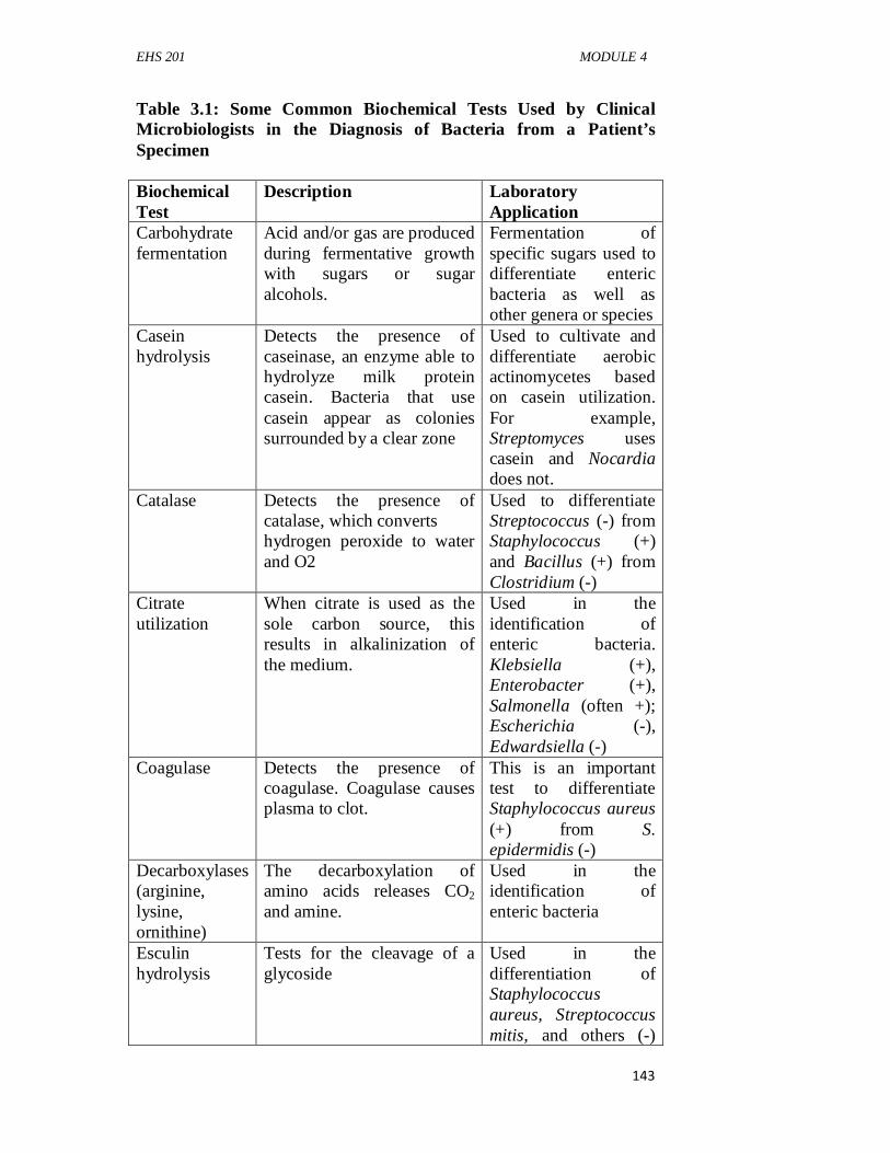
EHS 201 MODULE 4
143
Table 3.1: Some Common Biochemical Tests Used by Clinical Microbiologists in the Diagnosis of Bacteria from a Patient’s Specimen Biochemical Test
Description Laboratory Application
Carbohydrate fermentation
Acid and/or gas are produced during fermentative growth with sugars or sugar alcohols.
Fermentation of specific sugars used to differentiate enteric bacteria as well as other genera or species
Casein hydrolysis
Detects the presence of caseinase, an enzyme able to hydrolyze milk protein casein. Bacteria that use casein appear as colonies surrounded by a clear zone
Used to cultivate and differentiate aerobic actinomycetes based on casein utilization. For example, Streptomyces uses casein and Nocardia does not.
Catalase Detects the presence of catalase, which converts hydrogen peroxide to water and O2
Used to differentiate Streptococcus (-) from Staphylococcus (+) and Bacillus (+) from Clostridium (-)
Citrate utilization
When citrate is used as the sole carbon source, this results in alkalinization of the medium.
Used in the identification of enteric bacteria. Klebsiella (+), Enterobacter (+), Salmonella (often +); Escherichia (-), Edwardsiella (-)
Coagulase Detects the presence of coagulase. Coagulase causes plasma to clot.
This is an important test to differentiate Staphylococcus aureus (+) from S. epidermidis (-)
Decarboxylases (arginine, lysine, ornithine)
The decarboxylation of amino acids releases CO2 and amine.
Used in the identification of enteric bacteria
Esculin hydrolysis
Tests for the cleavage of a glycoside
Used in the differentiation of Staphylococcus aureus, Streptococcus mitis, and others (-)
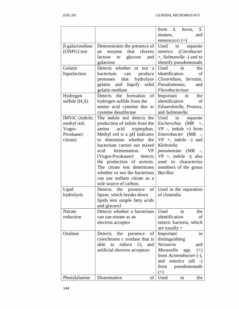
EHS 201 GENERAL MICROBIOLOGY
144
from S. bovis, S. mutans, and enterococci (+)
β-galactosidase (ONPG) test
Demonstrates the presence of an enzyme that cleaves lactose to glucose and galactose
Used to separate enterics (Citrobacter +, Salmonella -) and to identify pseudomonads
Gelatin liquefaction
Detects whether or not a bacterium can produce proteases that hydrolyze gelatin and liquify solid gelatin medium
Used in the identification of Clostridium, Serratia, Pseudomonas, and Flavobacterium
Hydrogen sulfide (H2S)
Detects the formation of hydrogen sulfide from the amino acid cysteine due to cysteine desulfurase
Important in the identification of Edwardsiella, Proteus, and Salmonella
IMViC (indole; methyl red; Voges-Proskauer; citrate)
The indole test detects the production of indole from the amino acid tryptophan. Methyl red is a pH indicator to determine whether the bacterium carries out mixed acid fermentation. VP (Voges-Proskauer) detects the production of acetoin. The citrate test determines whether or not the bacterium can use sodium citrate as a sole source of carbon.
Used to separate Escherichia (MR +, VP -, indole +) from Enterobacter (MR -, VP +, indole -) and Klebsiella pneumoniae (MR -, VP +, indole -); also used to characterize members of the genus Bacillus
Lipid hydrolysis
Detects the presence of lipase, which breaks down lipids into simple fatty acids and glycerol
Used in the separation of clostridia
Nitrate reduction
Detects whether a bacterium can use nitrate as an electron acceptor
Used in the identification of enteric bacteria, which are usually +
Oxidase Detects the presence of cytochrome c oxidase that is able to reduce O2 and artificial electron acceptors
Important in distinguishing Neisseria and Moraxella spp. (+) from Acinetobacter (-), and enterics (all -) from pseudomonads (+)
Phenylalanine Deamination of Used in the
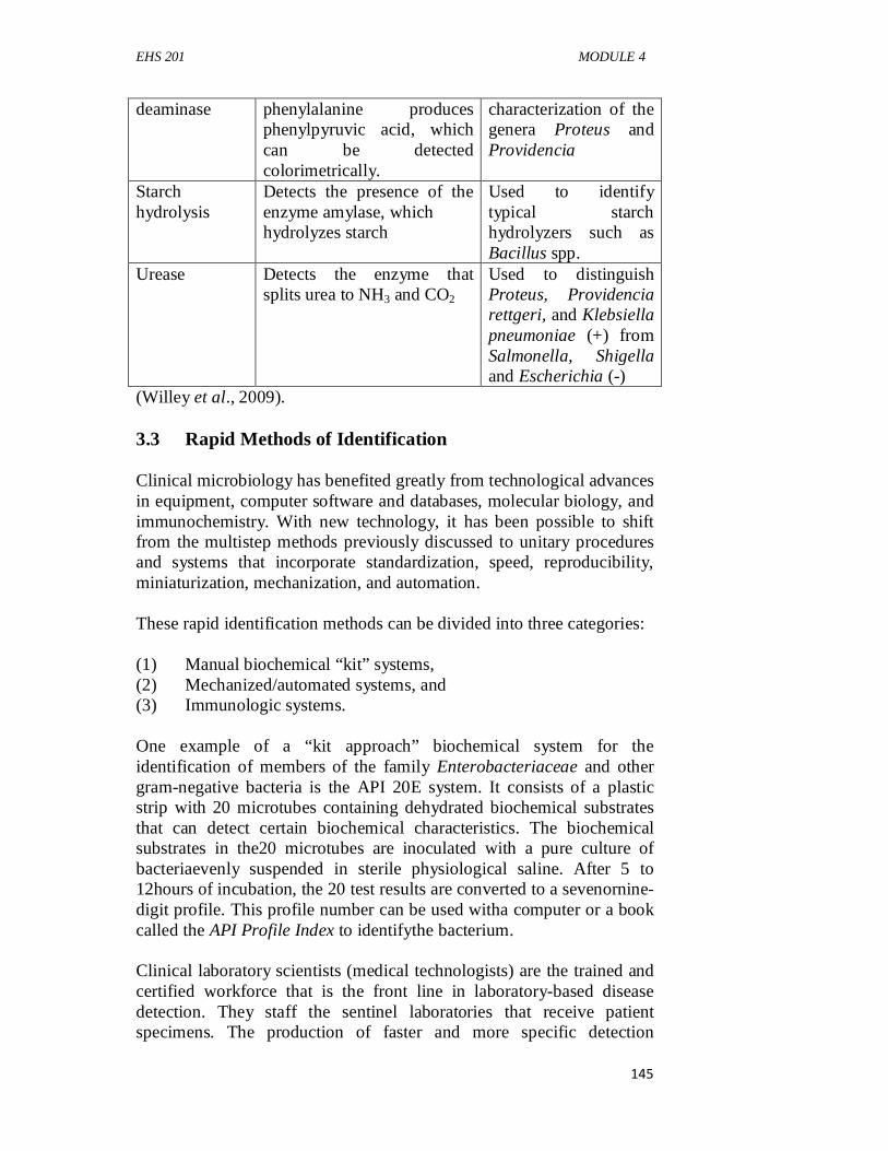
EHS 201 MODULE 4
145
deaminase phenylalanine produces phenylpyruvic acid, which can be detected colorimetrically.
characterization of the genera Proteus and Providencia
Starch hydrolysis
Detects the presence of the enzyme amylase, which hydrolyzes starch
Used to identify typical starch hydrolyzers such as Bacillus spp.
Urease Detects the enzyme that splits urea to NH3 and CO2
Used to distinguish Proteus, Providencia rettgeri, and Klebsiella pneumoniae (+) from Salmonella, Shigella and Escherichia (-)
(Willey et al., 2009). 3.3 Rapid Methods of Identification Clinical microbiology has benefited greatly from technological advances in equipment, computer software and databases, molecular biology, and immunochemistry. With new technology, it has been possible to shift from the multistep methods previously discussed to unitary procedures and systems that incorporate standardization, speed, reproducibility, miniaturization, mechanization, and automation. These rapid identification methods can be divided into three categories: (1) Manual biochemical “kit” systems, (2) Mechanized/automated systems, and (3) Immunologic systems. One example of a “kit approach” biochemical system for the identification of members of the family Enterobacteriaceae and other gram-negative bacteria is the API 20E system. It consists of a plastic strip with 20 microtubes containing dehydrated biochemical substrates that can detect certain biochemical characteristics. The biochemical substrates in the20 microtubes are inoculated with a pure culture of bacteriaevenly suspended in sterile physiological saline. After 5 to 12hours of incubation, the 20 test results are converted to a sevenornine-digit profile. This profile number can be used witha computer or a book called the API Profile Index to identifythe bacterium. Clinical laboratory scientists (medical technologists) are the trained and certified workforce that is the front line in laboratory-based disease detection. They staff the sentinel laboratories that receive patient specimens. The production of faster and more specific detection
EHS 201 GENERAL MICROBIOLOGY
146
technologies has enabled the rapid and accurate identification of disease agents. However, the bioterror incidents of 2001 spawned a renewed demand for “better, faster, and smarter” microbial detection and identification technologies. While nucleic acid–based detection systems, such as PCR, have garnered much attention as the basis of newer detection systems, antibody-based identification technologies are still considered more flexible and easier to modify. Traditional antibody-based detection technologies are being linked to sophisticated reporting systems that provide “med techs” with an ever-increasing array of cutting-edge technology. Examples of more recent microbial identification technologies include biosensorsbased on: (1) Microfluidic antigen sensors, (2) Real time (20-minute) PCR, (3) Highly sensitive spectroscopy systems, and (4) Liquid crystal amplification of microbial immune complexes. Some of these technologies are being used as part of military sentinel detection programs; others are awaiting approval by various licensing agencies before being deployed in clinical laboratories. Additional technologies are expected as the demand for immediate, highly sensitive microbial detection increases globally. Thus the rapidly growing discipline of immunology has greatly aided the clinical microbiologist. Numerous technologies now exist that exploit the specificity and sensitivity of monoclonal antibodies to detect and identify microorganisms. 3.4 Bacteriophage Typing Bacteriophages are viruses that attack members of a particular bacterial species or strains within a species. Bacteriophage (phage) typing is based on the specificity of phage surface proteins for cell surface receptors. Only those bacteriophages that can attach to these surface receptors can infect bacteria and cause lysis. On a petri dish culture, lytic bacteriophages cause plaques on lawns of sensitive bacteria. These plaques represent infection by the virus. 3.5 Molecular Genetic Methods Some of the most accurate approaches to microbial identification are through the analysis of proteins and nucleic acids. Examples include comparison of proteins; physical, kinetic, and regulatory properties of microbial enzymes; nucleic acid–base composition; nucleic acid hybridization; and nucleic acid sequencing. Other molecular methods being widely used are nucleic acid probes, gas-liquid chromatography, and DNA fingerprinting.
EHS 201 MODULE 4
147
3.6 Clinical Immunology The culturing of certain viruses, bacteria, fungi, and protozoa from clinical specimens may not be possible because the methodology remains undeveloped (e.g., Treponema pallidum; hepatitis A, B, C; and Epstein-Barr virus), is unsafe (rickettsias and HIV), or is impractical for all but a few microbiology laboratories (e.g., mycobacteria, strict anaerobes, Borrelia). Cultures also may be negative because of prior antimicrobial therapy. Under these circumstances, detection of antibodies or antigens may be quite valuable diagnostically. Immunologic systems for the detection and identification of pathogens from clinical specimens are easy to use, give relatively rapid reaction end points, and are sensitive and specific with a low percentage of false positives and negatives. Dramatic advances in clinical immunology have given rise to a marked increase in the number, sensitivity, and specificity of serological tests. This increase reflects a better understanding of: (1) Immune cell surface antigens (CD antigens), (2) Lymphocyte biology, (3) The production of monoclonal antibodies, and (4) The development of sensitive antibody-binding reporter systems. For a number of reasons, the utility of these tests depends on proper test selection and timing of specimen collection. For instance, each individual’s immunologic response to a microorganism is quite variable, making the interpretation of immunologic tests potentially difficult. For example, a single, elevated IgM titer does not distinguish between active and past infections. Rather an elevated IgG titer typically indicates an active infection, especially when subsidence of symptoms correlates with a fourfold (or greater) decrease in antibody titer. Furthermore, a lack of a measurable antibody titer may reflect an organism’s lack of immunogenicity or insufficient time for an antibody response to develop following the onset of the infectious disease. Some patients are also immunosuppressed due to other disease processes or treatment procedures (e.g., cancer and AIDS patients) and therefore do not respond. 3.6.1 Serotyping Serum is the liquid portion of blood (devoid of clotting factors) that contains many different components, especially the immunoglobulins or antibodies. Serotypingrefers to the use of serum (antibodies) to
EHS 201 GENERAL MICROBIOLOGY
148
specifically detect and identify other molecules. Serotyping can be used to identify specific white blood cells or the proteins on cell surfaces. Serotyping can also be used to differentiate strains (serovars or serotypes) of microorganisms that differ in the antigenic composition of a structure or product. The serological identification of a pathogenic strain has diagnostic value. Often the symptoms of infections depend on the nature of the cell products released by the pathogen. Therefore it is sometimes possible to identify a pathogen serologically by testing for cell wall antigens. For example, there are 90 different strains of Streptococcus pneumoniae, each unique in the nature of its capsular material. These differences can be detected by antibody-induced capsular swelling, termed the Quellung reaction,when the appropriate antiserum for a specific capsular type is used. 3.6.2 Agglutination An agglutination reactionoccurs when an immune complex is formed by cross-linking cells or particles with specific antibodies. Agglutination reactions usually form visible aggregates or clumps, called agglutinatesthat can be seen with the unaided eye. Direct agglutination reactions are very useful in the diagnosis of certain diseases. For example, the Widal testis a reaction involving the agglutination of typhoid bacilli when they are mixed with serum containing typhoid antibodies from an individual who has typhoid fever. Techniques have also been developed that employ microscopic synthetic latex spheres coated with antigens. These coated microspheres are extremely useful in diagnostic agglutination reactions. For example, microspheres are used in common pregnancy tests that detect the elevated level of human chorionic gonadotropin (hCG) hormone, which occurs in a woman’s urine and blood early in pregnancy. Latex agglutination tests are also used to detect antibodies that develop during certain mycotic, helminthic, and bacterial infections, as well as in drug testing. 3.6.3 Complement Fixation When complement binds to an antigen-antibody complex, it becomes “fixed” and “used up.” Complement fixation tests are very sensitive and can be used to detect extremely small amounts of an antibody for a suspect microorganism in an individual’s serum. A known antigen is mixed with test serum lacking complement. When immune complexes have had time to form, complement is added to the mixture. If immune complexes are present, they will fix and consume complement. Afterward, sensitized indicator cells, usually sheep red
EHS 201 MODULE 4
149
blood cells previously coated with complement-fixing antibodies, are added to the mixture. If specific antibodies are present in the test serum and complement is consumed by the immune complexes, insufficient amounts of complement will be available to lyse the indicator cells. On the other hand, in the absence of antibodies, complement remains and lyses the indicator cells. Thus absence of lysis shows that specific antibodies are present in the test serum. Complement fixation was once used in the diagnosis of syphilis (the Wassermann test) and is still used as a rapid, inexpensive screening method in the diagnosis of certain viral, fungal, rickettsial, chlamydial, and protozoan diseases. 3.6.4 Immunoblotting (Western Blotting) Another immunologic technique used in the clinical microbiology laboratory is immunoblotting, also known as Western blotting. Immunoblottinginvolves polyacrylamide gel electrophoresis of a protein specimen followed by transfer of the separated proteins to sheets of nitrocellulose or polyvinyl difluoride. Protein bands are then visualized by treating the nitrocellulose sheets with solutions of enzyme-tagged antibodies. This procedure demonstrates the presence of common and specific proteins among different strains of microorganisms. Immunoblotting also can be used to show strain-specific immune responses to microorganisms, to serve as an important diagnostic indicator of a recent infection with a particular strain of microorganism, and to allow for prognostic implications with severe infectious diseases. 3.6.5 Immunoprecipitation The immunoprecipitationtechnique detects soluble antigens that react with antibodies called precipitins.The precipitin reaction occurs when bivalent or multivalent antibodies and antigens are mixed in the proper proportions. The antibodies link the antigen to form a large antibody-antigen network or lattice that settles out of solution when it becomes sufficiently large. Immunoprecipitation reactions occur only at the equivalence zone when there is an optimal ratio of antigen to antibody so that an insoluble lattice forms. If the precipitin reaction takes place in a test tube, a precipitation ring forms in the area in which the optimal ratio or equivalence zone develops. SELF-ASSESSMENT EXERCISE Mention four more recent microbial identification technologies.

EHS 201 GENERAL MICROBIOLOGY
150
4.0 CONCLUSION Clinical microbiology and immunology are very important aspects of microbiology. Host-parasite relationships, pathogenicity and virulence of microorganism are better understood through clinical studies of specimen samples. Immunological responses to pathogens and toxins remain fundamental in this unit. 5.0 SUMMARY At the end of this unit, the student learnt the identification of microorganisms from specimens, microscopy of specimen microorganisms, application of Rapid methods of Identification on specimens, bacteriophage typing on specimens, molecular genetic methods in specimens, and clinical immunology. 6.0 TUTOR-MARKED ASSIGNMENT 1. What is serum? Explain serological typing. 2. List some of the most accurate approaches to microbial
identification through the analysis of proteins and nucleic acids. 3. List five preliminary or definitive identification of
microorganisms that clinical microbiology laboratory can provide.
7.0 REFERENCES/FURTHER READING Fredriksson-Ahomaa, M. (2012). Isolation of enteropathogenic Yersinia
From Nonhuman Sources. Adv. Exp. Med. Biol., 954, 97–105. Fredriksson-Ahomaa, M., Stolle, A., & Korkeala, H. (2006) Molecular
epidemiology of Yersinia enterocolitica infections. FEMS Immunol. Med.Microbiol., 47, 315–329.
Koornhof, H., Smego, R., & Nicol, M. (1999). Yersiniosis. II: the
pathogenesis of Yersinia infections. Eur. J. Clin. Microbiol. Infect. Dis., 18, 87–112.
Unden, G., Thines, E., & Schüffler, A. (2016). Host – Pathogen
Interaction: Microbial Metabolism, Pathogenicity and Antiinfectives. Wiley-VCH Verlag GmbH & Co.
KGaA, Boschstr. 12, 69469 Weinheim, Germany. Willey, J.M., Sherwood, L.M. & Woolverton, C.J. (2009). Prescott’s
Principles of ‘Microbiology (1st ed.), McGraw-Hill. pp. 1-12.
EHS 201 MODULE 5
151
MODULE 5 MICROBES IN THE ENVIRONMENT, AGRICULTURE, AND INDUSTRY Unit 1 Microbiology in Food And Industry Unit 2 Environmental Microbiology Unit 3 Microbiology in Agriculture UNIT 1 MICROBIOLOGY IN FOOD AND INDUSTRY CONTENTS 1.0 Introduction 2.0 Objectives 3.0 Main Content
3.1 Microorganism Growth in Foods 3.1.1 Intrinsic Factors 3.1.1 Extrinsic Factors 3.2 Microbial Growth and Food Spoilage 3.3 Controlling Food Spoilage 3.3.1 Removal of Microorganisms 3.3.2 Low Temperature 3.3.3 High Temperature 3.3.4 Water Availability 3.3.5 Chemical-Based Preservation 3.3.6 Radiation 3.3.7 Microbial Product–Based Inhibition 3.4 Food-borne Diseases 3.4.1 Food-Borne Infection 3.4.2 Food-Borne Intoxications 3.5 Industrial Microbiology 3.5.1 Microbiology of Fermented Foods 3.5.2 Fermented Milks 3.5.3 Mesophilic Fermentations 3.5.4 Thermophilic Fermentations 3.5.5 Probiotics 3.5.6 Yeast-Lactic Fermentation 3.5.7 Mold-Lactic Fermentation 3.5.8 Cheese Production 3.5.9 Meat and Fish 3.5.10 Wines and Champagnes 3.5.11 Beers and Ales 3.5.12 Distilled Spirits 3.5.13 Production of Breads 3.5.14 Microorganisms as Foods and Food Amendments 4.0 Conclusion
EHS 201 GENERAL MICROBIOLOGY
152
5.0 Summary 6.0 Tutor-Marked Assignment 7.0 References/Further Reading 1.0 INTRODUCTION Foods, microorganisms, and humans have had an interesting association that developed long before recorded history. On the one hand, microorganisms can be used to transform raw foods into gastronomic delights including chocolate, cheeses, pickles, sausages, soy sauce, wines, and beers. On the other hand, microorganisms can degrade food quality and lead to spoilage. Sometimes a fine line exists between the microbial enhancement of foods and degradation and potential disease transmission. The detection and control of pathogens and food spoilage microorganisms are important parts of food microbiology. 2.0 OBJECTIVES By the end of this unit, you will be able to: • understand microbial growth in foods, • explain microbial growth and food spoilage, • learn how to control food spoilage, • survey food-borne diseases • discuss the application of microbiology in the industrial. 3.0 MAIN CONTENT The main content of this unit shall include discussions on microbial growth in food, microbial growth and food spoilage, control of food spoilage, food-borne diseases and industrial microbiology. 3.1 Microorganism Growth in Foods Foods, because they are nutrient-rich, are excellent environments for the growth of microorganisms. Microbial growth is controlled by factors related to the food itself, called intrinsic factors, and to the environment where the food is stored, described as extrinsic factors. 3.1.1 Intrinsic Factors Food composition is a critical intrinsic factor that influences microbial growth. If a food consists primarily of carbohydrates, fungal growth
EHS 201 MODULE 5
153
predominates and spoilage does not result in major odors. Thus foods such as breads, jams, and some fruits first show spoilage by fungal growth. In contrast, when foods contain large amounts of proteins or fats (e.g., meat and butter), bacterial growth can produce a variety of foul odors—think of rotting eggs. The anaerobic breakdown of proteins is called putrefaction. It yields foul-smelling amine compounds such as cadaverine (imagine the origin of that name) and putrescine. Degradation of fats ruins food as well. The production of shortchained fatty acids from fats renders butter rancid and foul smelling. Thus the major substrate present in a food helps determine the type of spoilage that may occur. The presence and availability of water also affect the ability of microorganisms to colonize foods. Simply by drying a food, one can control or eliminate spoilage processes. Water can bemade less available by adding solutes such as sugar and salt.Water availability is measured in terms of water activity(aw).This represents the ratio of relative humidity of the air over a testsolution compared with that of distilled water, which has an aw ofone. When large quantities of salt or sugar are added to food,most microorganisms are dehydrated by the hypertonic conditionsand cannot grow. Even under these adverseconditions, osmophilic and xerophilic microorganisms may spoilfood. Osmophilic (Greek osmus, impulse, and philein, to love)microbes grow best in or on media with a high osmotic concentration(e.g., jams and jellies), whereas xerophilic (Greek xerosis,dry, and philein, to love) microorganisms prefer a low aw environment(e.g., dried fruits, cereals) and may not grow under highaw conditions. The pH and oxidation-reduction (redox) potential of a food also are critical. A low pH favors the growth of yeasts and molds. In neutral or alkaline pH foods, such as meats, bacteria are more dominant in spoilage and putrefaction. Furthermore, when meat products, especially broths, are cooked, they often have lower oxidation-reduction potentials—that is, they present a reducing environment for microbial growth. These products, with their readily available amino acids, peptides, and growth factors, are ideal media for the growth of anaerobes, including Clostridium. The physical structure of a food also can affect the course and extent of spoilage. The grinding and mixing of foods such as sausage and hamburger increase the food surface area and distribute contaminating microorganisms throughout the food. This can result in rapid spoilage if such foods are stored improperly. In addition, some spoilage microorganisms have specialized enzymes that help them penetrate protective peels and rinds, especially after the fruits and vegetables have been bruised.
EHS 201 GENERAL MICROBIOLOGY
154
Many foods contain natural antimicrobial substances, including complex chemical inhibitors and enzymes. Coumarins found in fruits and vegetables exhibit antimicrobial activity. Cow’s milk and eggs also contain antimicrobial substances. Eggs are rich in the enzyme lysozyme that can lyse the cell walls of contaminating gram-positive bacteria. Herbs and spices often possess significant antimicrobial substances; generally fungi are more sensitive than most bacteria. Sage and rosemary are two of the most antimicrobial spices. Aldehydic and phenolic compounds that inhibit microbial growth are found in cinnamon, mustard, and oregano. Other important inhibitors are garlic, which contains allicin; cloves, which have eugenol; and basil, which contains rosmarinic acid. Nonetheless, spices can sometimes contain pathogenic and spoilage organisms. Enteric bacteria, Bacillus cereus, Clostridium perfringens, and Salmonella species have been detected in spices. Microorganisms can be eliminated or reduced by ethylene oxide sterilization. This treatment can result in Salmonella -free spices and herbs and a 90% reduction in the levels of general spoilage organisms. 3.1.1 Extrinsic Factors Temperature and relative humidity are important extrinsic factors in determining whether a food will spoil. At higher relative humidities, microbial growth is initiated more rapidly, even at lower temperatures (especially when refrigerators are not maintained in a defrosted state). When drier foods are placed in moist environments, moisture absorption can occur on the food surface, promoting microbial growth. The atmosphere in which food is stored also is important. This is especially true with shrink-wrapped foods because many plastic films allow oxygen diffusion, which results in increased growth of surface-associated microorganisms. Excess CO2 can decrease the solution pH, inhibiting microbial growth. Storing meat in a high CO2 atmosphere inhibits gram-negative bacteria, resulting in a population dominated by the lactobacilli. The observation that food storage atmosphere is important has led to the development of modified atmosphere packaging(MAP).Modern shrink-wrap materials and vacuum technology make it possible to package foods with controlled atmospheres. These materials are largely impermeable to oxygen. This prolongs shelf life by a factor of two to five times compared to the same product packaged in air. With a CO2 content of 60% or greater in the atmosphere surrounding a food, spoilage fungi will not grow, even if low levels of oxygen are present. Recently it has been found that high-oxygen MAP also may be effective.
EHS 201 MODULE 5
155
This is due to the formation of the superoxide (O2-) anion inside cells under these conditions. The superoxide anion is then transformed to highly toxic peroxide and hydroxyl radical, resulting in antimicrobial effects. Some products currently packaged using MAP technology include delicatessen meats and cheeses, pizza, grated cheese, some bakery items, and dried products such as coffee. 3.2 Microbial Growth and Food Spoilage Meat and dairy products, with their high nutritional value and easily metabolized carbohydrates, fats, and proteins, provide ideal environments for microbial spoilage. Proteolysis and putrefaction are typical results of microbial spoilage of such high-protein materials. Unpasteurized milk undergoes a predictablefour-step microbial succession during spoilage: acidproduction by Lactococcus lactis subsp. lactis is followed byadditional acid production associated with the growth of moreacid-tolerant organisms such as Lactobacillus. At this pointyeasts and molds become dominant and degrade the accumulatedlactic acid, and the acidity gradually decreases. Eventuallyprotein-digesting bacteria become active, resulting in a putridodor and bitter flavor. The milk, originally opaque, eventuallybecomes clear. In comparison with meat and dairy products, most fruits and vegetables have a much lower protein and fat content and undergo a different kind of spoilage, which often is initiated by molds. These organisms have enzymes that contribute to the weakening and penetration of the protective outer skin. The readily degradable carbohydrates within favor spoilage by bacteria, especially bacteria that cause soft rots, such as Erwinia carotovora, which produces hydrolytic enzymes. The lack of reduced conditions enables aerobes and facultative anaerobes to contribute to the decomposition processes. Molds can rapidly grow on grains and corn when these products are stored in moist conditions. The green growth most likely is Penicillium; the black growth is characteristic of Rhizopus stolonifer. Contamination of grains by the ascomycete Clavicepspurpura causes ergotism, a toxic condition. Hallucinogenic alkaloids produced by this fungus can lead to altered behavior, abortion, and death if infected grains are eaten. Molds are also a special problem for tomatoes. Even the slightest bruising of the tomato skin, exposing the interior, will result in rapid fungal growth. This affects the quality of tomato products, including tomato juices and ketchups. Fungus-derived carcinogens include the aflatoxins and fumonisins. Aflatoxinsare produced most commonly in moist grains and nut products. Aflatoxins were discovered in 1960, when 100,000 turkey poults died from eating fungus-infested peanut meal. Aspergillus flavus
EHS 201 GENERAL MICROBIOLOGY
156
was found in the infected peanut meal, together with alcohol-extractable toxins termed aflatoxins. These flat-ringed planar compounds intercalate with nucleic acids and act as potent frame-shift mutagens and carcinogens. This occurs primarily in the liver, where they are converted to unstable derivatives. At the present time, a total of 18 aflatoxins are known. Of these, aflatoxin B1 is the most common and the most potent carcinogen. Aflatoxins B1 and B2, after ingestion by lactating animals, are modified in the animal body to yield the aflatoxins M1 and M2. If cattle consume aflatoxin-contaminated feeds, aflatoxins also can appear in milk and dairy products. Besides their importance in grains, they have also been found in beer, cocoa, raisins, and soybean meal. 3.3 Controlling Food Spoilage With the beginning of agriculture and a decreasing dependence on hunting and gathering, the need to preserve surplus foods became essential to human survival. The use of salt as a meat preservative, the production of wines, and the preservation of fish and meat by smoking were introduced in Near Eastern civilization as early as 3000 BCE. But it was not until the nineteenth century that the microbial spoilage of food was studied systematically. Louis Pasteur established the modern era of food microbiology in 1857, when he showed that microorganisms cause milk spoilage. Pasteur’s work in the 1860s proved that heat could be used to control spoilage organisms in wines and beers. Foods can be preserved by a variety of methods. The goal ofeach method is to eliminate or reduce the populations of spoilageand disease-causing microorganisms while maintaining foodquality. 3.3.1 Removal of Microorganisms Microorganisms can be removed from water, wine, beer, juices, soft drinks, and other liquids by filtration. This can keep bacterial populations low or eliminate them entirely. Removal of large particulates by prefiltration and centrifugation maximizes filter life and effectiveness. Several major brands of beer are filtered rather than pasteurized to better preserve the flavor and aroma of the original product. 3.3.2 Low Temperature Refrigeration at 5°C retards microbial growth, although with extended storage, microorganisms eventually grow and produce spoilage. Slow microbial growth at temperatures below 10°C has been described, particularly with fruit juice concentrates, ice cream, and some fruits. Of particular concern is the growth of Listeria, which can grow at temperatures used for refrigeration. It should be kept in mind that

EHS 201 MODULE 5
157
refrigeration slows the metabolic activity of most microbes, but it does not lead to significant decreases in overall microbial populations. 3.3.3 High Temperature Controlling microbial populations in foods by means of high temperatures can significantly limit disease transmission and spoilage. Heating processes, first used by Nicholas Appert in 1809, provide a safe means of preserving foods, particularly when carried out in commercial canning operations. Canned food is heated in special containers called retorts at about 115°C for intervals ranging from 25 to over 100 minutes. The precise time and temperature depend on the nature of the food. Sometimes canning does not kill all microorganisms but only those that will spoil the food (e.g., remaining bacteria are unable to grow due to acidity of the food). After heat treatment, the cans are cooled as rapidly as possible, usually with cold water. Quality control and processing effectiveness are sometimescompromised, however, in home processing of foods,especially with less acidic (pH values greater than 4.6) productssuch as green beans or meats. Despite efforts to eliminate spoilage microorganisms during canning, canned foods may become spoiled. This may be due to spoilage before canning, underprocessing during canning, and leakage of contaminated water through seams during cooling. Spoiled food can be altered in such characteristics as color, texture, odor, and taste. Organic acids, sulfides, and gases (particularly CO2 and H2S) may be produced. If spoilage microorganisms produce gas, both ends of the can will bulge outward. Sometimes the swollen ends can be moved by thumb pressure (soft swells); in other cases, the gas pressure is so great that the ends cannot be dented by hand (hard swells). However, swelling is not always due to microbial spoilage; acid in low pH foods may react with iron in the can to release hydrogen and generate a hydrogen swell. Pasteurizationinvolves heating food to a temperature that kills disease-causing microorganisms and substantially reduces the levels of spoilage organisms. When processing milk, beers, and fruit juices by conventional low-temperature holding (LTH) pasteurization, the liquid is maintained at 62.8°C for 30 minutes. Products can also be held at 72°C for 15 seconds, a high-temperature, short-time (HTST) process; milk can be treated at 138°C for 2 seconds for ultra-high-temperature (UHT) processing. Shorter-term processing results in improved flavor and extended product shelf life. The duration of pasteurization is based on the statistical probability that the number of remaining viable microorganisms will be below a certain level after a particular heating time at a specific temperature.
EHS 201 GENERAL MICROBIOLOGY
158
3.3.4 Water Availability Dehydration, such as lyophilization to produce freeze-dried foods, is a common means of eliminating microbial growth. The modern process is simply an update of older procedures in which grains, meats, fish, and fruits were dried. The combination of free-water loss with an increase in solute concentration in the remaining water makes this type of preservation possible. 3.3.5 Chemical-Based Preservation Various chemical agents can be used to preserve foods, and these substances are closely regulated by the U.S. Food and Drug Administration and are listed as being “generally recognized as safe” or GRAS. They include simple organic acids, sulfite, ethylene oxide as a gas sterilant, sodium nitrite, and ethyl formate. These chemical agents may damage the microbial plasma membrane or denature various cell proteins. Other compounds interfere with the functioning of nucleic acids, thus inhibiting cell reproduction. Sodium nitrite is an important chemical used to help preserve ham, sausage, bacon, and other cured meats by inhibiting the growth of Clostridium botulinum and the germination of its spores. This protects against botulism and reduces the rate of spoilage. Besides increasing meat safety, nitrite decomposes to nitric acid, which reacts with heme pigments to keep the meat red in color. Concern about nitrite arises from the observation that it can react with amines to form carcinogenic nitrosamines. Low pH can also be used to hinder microbial spoilage. For example, acetic and lactic acids inhibit listerial growth. Organic acids (1–3%) can be used to treat meat carcasses, and poultry can be cleansed with 10% lactic acid/sodium lactate buffer (pH 3) prior to packaging. In addition, low pH can increase the activity of other chemical preservatives. Sodium propionate is most effective at lower pH values, where it is primarily undissociated. Breads, with their low pH values, often contain sodium propionate as a preservative. 3.3.6 Radiation The major method used for radiation sterilization of food is gamma irradiation from a cobalt-60 source; however, cesium-137 is used in some facilities. Gamma radiation has excellent penetrating power but must be used with moist foods because radiation is effective only if it can generate peroxides from water in the microbial cells, resulting in oxidation of sensitive cellular constituents. This process of
EHS 201 MODULE 5
159
radappertization,named after Nicholas Appert, can extend the shelf life of seafoods, fruits, and vegetables. To sterilize meat products, commonly 4.5 to 5.6 megarads are used. Electron beams can also be used to irradiate foods. The electrons are generated electrically, so unlike gamma radiation, they can be turned on only when needed. Also, this approach does not generate radioactive waste. On the other hand, electron beams do not penetrate food items as deeply as does gamma radiation. It is important to note that regardless of the radiation source (gamma rays or electron beams), the food itself does not become radioactive. 3.3.7 Microbial Product–Based Inhibition Interest is increasing in the use of bacteriocins for the preservationof foods. Bacteriocins are bactericidal proteins active againstclosely related bacteria, which bind to specific sites on the celland often affect cell membrane integrity and function. The onlycurrently approved product is nisin, a small amphiphilic peptideproduced by some strains of Lactococcus lactis. It is nontoxic to humans and affects other gram-positive bacteria by binding tothe lipid II portion of the growing peptidoglycan and formingpores in the plasma membrane. Nisin can beused particularly in low-acid foods to improve inactivation ofClostridium botulinum during the canning process or to inhibitgermination of any surviving spores. 3.4 Food-Borne Diseases Food-borne illnesses impact the entire world. In the United States, based on recent information from the Centers for Disease Control and Prevention, annual incidences of food-related diseases involve 76 million cases, of which only 14 million can be attributed to known pathogens. Food-borne diseases result in 325,000 hospitalizations and at least 5,000 deaths per year. Since 1942 the number of recognized food-borne pathogens has increased over fivefold. In most cases, these “new” pathogens are simply agents that now can be described, based on an improved understanding of microbial diversity. Recent estimates indicate that noroviruses, Campylobacter jejuni, and Salmonella are the major causes of food-borne diseases. In addition, Escherichia coli O157:H7 and Listeria are important food related pathogens. There are two primary types of food-related diseases: 1. Food-borne infections, and 2. Food intoxications.
EHS 201 GENERAL MICROBIOLOGY
160
All of these food-borne diseases are associated with poor hygienic practices. Whether by water or food transmission, the fecal-oral route is key, with the food providing the vital link between hosts. Fomites, such as sink faucets, drinking cups, and cutting boards, also play a role in the maintenance of the fecal-oral route of contamination. 3.4.1 Food-Borne Infection A food-borne infectioninvolves the ingestion of the pathogen, followed by growth in the host, including tissue invasion or the release of toxins. Salmonellosisresults from ingestion of a variety of Salmonellaserovars, particularly Typhimurium and Enteritidis. Gastroenteritis is the disease of most concern in relation to foods such as meats,poultry, and eggs, and the onset of symptoms occurs after an incubationtime as short as 8 hours. Salmonella infection can arisefrom contamination by workers in food-processing plants and restaurants,as well in canning processes. Campylobacter jejuni is considered a leading cause of acute bacterial gastroenteritis in humans. This important pathogen is often consumed in under- or uncooked poultry products. For example, transmission often occurs when kitchen utensils and containers are used for chicken preparation and then for salads. Contamination with as few as 10 viable C. jejuni cells can lead to the onset of diarrhea. C. jejuni also is transmitted by raw milk, and the organism has been found on various red meats. Thorough cooking of food prevents its transmission. Listeriosis,caused by Listeria monocytogenes, was responsible for the largest meat recall in U.S. history. In 2002 a seven-state listeriosis outbreak was linked to deli meats and hot dogs produced at a single meat-processing plant in Pennsylvania. Pregnant women, the young and the old, and immunocompromised individuals are especially vulnerable to L. monocytogenes infections. Escherichia coli is an important food-borne disease organism. Enteropathogenic, enteroinvasive, and enterotoxigenic types can cause diarrhea. E. coli O157:H7, with its specific LPS O-antigen (O) and flagellar (H) antigen, is thought to have acquired enterohemorrhagic genes from Shigella, including the genes for shigalike toxins. This produced a pathogenic strain, first discovered in 1982 that is transmitted by the fecaloral route. Its infectious dose appears to be only 500 bacteria. Enterohemorrhagic E. coli has been found in meat products such as hamburger and salami, in unpasteurized fruit drinks, on fruits and vegetables, and in untreated well water. Prevention of food contamination by E. coli O157:H7 is essential from the time of production until consumption. Hygiene must be monitored carefully in larger-volume slaughterhouses, where contact of meat with fecal material can occur, and fruits and vegetables should be handled with

EHS 201 MODULE 5
161
care. Caution also is essential at the point of use. For example, utensils used with raw foods should not contact cooked food; proper cleaning of cutting boards and utensils minimizes contamination. Virus contamination is always a potential problem. This is based on transmission by water or by lack of hygiene in food preparation and direct contamination by food processors and handlers. Similar situations occur with protozoan pathogens. Virus contamination has become a severe problem on many cruise ships, where noroviruses have been involved in outbreaks, with person-to-person contact and possibly foods implicated in these avoidable occurrences. Foods that are transported and consumed in an uncooked state are an increasingly important source of food-borne infection. The problem becomes more serious with rapid movement of people and products around the world. International trade in uncooked foods, aided by rapid air transport, provides many opportunities for disease transmission. As examples we discuss sprouts, seafood, and raspberries. Sprouts, a popular and attractive garnish, are sometimes germinated and grown in waters containing pathogens. Contaminated alfalfa, bean, watercress, mungbean, mustard, and soybean sprouts can be major sources of typhoid and cholera. 3.4.2 Food-Borne Intoxications Microbial growth in food products also can result in food intoxication. Intoxication produces symptoms shortly after the food is consumed because growth of the disease-causing microorganism is not required. Toxins produced in the food can be associated with microbial cells or can be released from the cells. Most Staphylococcus aureus strains cause a staphylococcalenteritis related to the synthesis of extracellular toxins. Theseare heat-resistant proteins, so heating does not usually renderthe food safe. The effects of the toxins are quickly felt, withdisease symptoms occurring within 2 to 6 hours. The main reservoirof S. aureus is the human nasal cavity. Frequently S.aureus is transmitted to a person’s hands and then is introduced into food during preparation. Growth and enterotoxin production usually occur when contaminated foods are held at room temperature for several hours. Three gram-positive rods are known to cause food intoxications: Clostridium botulinum, C. perfringens, and Bacillus cereus. However, here we note that baked potatoes served in aluminum foil can, even after washing, be contaminated with C. botulinum, which naturally occurs in the soil. If the foil-covered potatoes are not heated sufficiently in the
EHS 201 GENERAL MICROBIOLOGY
162
baking process, surviving clostridia can proliferate after removal of the potatoes from the oven and rapidly produce toxins. 3.5 Industrial Microbiology 3.5.1 Microbiology of Fermented Foods Fermentation has been a major way of preserving food for thousands of years. Microbial growth, either of natural or inoculated populations, causes chemical or textural changes to form a product that can be stored for extended periods. The fermentation process also is used to create new, pleasing food flavors and odors—such as chocolate. The major fermentations used in food microbiology are the lactic, propionic, and alcoholic fermentations. These fermentations are carried out with a wide range of microbes, many of which have not been characterized. 3.5.2 Fermented Milks Throughout the world, at least 400 different fermented milks are produced. The majority of fermented milk products rely on lactic acid bacteria (LAB),which include species belonging to the genera Lactobacillus, Lactococcus, Leuconostoc, and Streptococcus. These are low G + C grampositive bacteria that tolerate acidic conditions, are nonsporing, and are aerotolerant with a strictly fermentative metabolism. The art of fermentation developed long before the science, and fermented milks were produced for thousands of years before Louis Pasteur discovered lactic acid fermentation. 3.5.3 Mesophilic Fermentations Mesophilic milk fermentations result from similar manufacturing techniques, in which acid produced through microbial activity causes protein denaturation. To carry out the process, milk is typically inoculated with the desired starter culture—a carefully selected group of microbes used to initiate the fermentation. It is then incubated at optimum temperature (approximately 20 to 30°C). Microbial growth is stopped by cooling, and Lactobacillus spp. and Lactococcus lactis cultures are used for aroma and acid production. The organism Lactococcus lactis subspecies diacetilactis converts milk citrate to diacetyl, which gives a richer flavor to the finished product. The use of these microorganisms with skim milk produces cultured buttermilk, and when cream is used, sour cream is the result.
EHS 201 MODULE 5
163
3.5.4 Thermophilic Fermentations Thermophilic fermentations are carried out at temperatures around 45°C. An important example is yogurt production. Yogurt is one of the most popular fermented milk products in the United States and Europe. In commercial production, nonfat or low-fat milk is pasteurized, cooled to 43°C or lower, and inoculated with a 1:1 ratio of Streptococcus salivarius subspecies thermophilus (S. thermophilus) and Lactobacillusdelbrueckii subspecies bulgaricus (L. bulgaricus). S. thermophiles grows more rapidly at first and renders the milkanaerobic and weakly acidic. L. bulgaricus then acidifies themilk even more. Acting together, the two species fermentalmost all of the lactose to lactic acid and flavor the yogurtwith diacetyl (S. thermophilus) and acetaldehyde (L. bulgaricus). Fruits or fruit flavors are pasteurized separately and thencombined with the yogurt. Freshly prepared yogurt containsabout 109 bacteria per gram. 3.5.5 Probiotics The health benefits of fermented foods such as yogurt have been touted for a great number of years. However, only recently have rigorous studies explored the effects of certain bacteria that are either commensals or mutualists in the human intestine. Microorganisms such as Lactobacillus and Bifi dobacterium are being used in the rapidly developing area of probiotics,the addition of microorganisms to the diet to provide health benefits beyond basic nutritive value. The possible health benefits of the use of such microbial dietary adjuvants include immunomodulation, control of diarrhea, anticancer effects, and possible improvement of Crohn’s disease (inflammatory bowel disease). These bacteria may also influence antigen presentation, uptake, and possible degradation. Probiotics have become a more attractive treatment option because the rate of antibiotic resistance among pathogens continues to climb. In addition, disease ecologists have come to recognize that intestinal microflora can be a contributing factor for certain conditions (e.g., Crohn’s disease). Acidophilus milk is produced by using Lactobacillus acidophilus. L. acidophilus may modify the microbial flora in the lower intestine, thus improving general health, and it often is used as a dietary adjunct, especially for lactose-intolerant persons. Many microorganisms in fermented dairy products stabilize the bowel microflora, and some appear to have antimicrobial properties. The exact nature and extent of health benefits of consuming fermented milks may involve minimizing lactose intolerance, lowering serum cholesterol, and possibly exhibiting anticancer activity. Several lactobacilli have antitumor compounds in their cell walls. Such findings suggest that diets including lactic acid bacteria, especially L. acidophilus, may contribute to the prevention of colon cancer.
EHS 201 GENERAL MICROBIOLOGY
164
3.5.6 Yeast-Lactic Fermentation Yeast-lactic fermentations include kefir, a product with an ethanol concentration of up to 2%. This unique fermented milk originated in the Caucasus Mountains and is produced east into Mongolia. Kefir products tend to be foamy and frothy, due to active carbon dioxide production. This process is based on the use of kefir “grains” as an inoculum. These are coagulated lumps of casein that contain yeasts, lactic acid bacteria, and acetic acid bacteria. In this fermentation, the grains are used to inoculate the fresh milk and then recovered at the end of the fermentation. 3.5.7 Mold-Lactic Fermentation Mold-lactic fermentation results in a unique Finnish fermented milk called viili . The milk is placed in a cup and inoculated with a mixture of the fungus Geotrichium candidum and lactic acid bacteria. The cream rises to the surface, and after incubation at 18 to 20°C for 24 hours, lactic acid reaches a concentration of 0.9%. The fungus forms a velvety layer across the top of the final product, which also can be made with a bottom fruit layer. 3.5.8 Cheese Production Cheese is one of the oldest foods, probably developed roughly 8,000 years ago. About 2,000 distinct varieties of cheese are produced throughout the world, representing approximately 20 general types. Often cheeses are classified based on texture or hardness as soft cheeses (cottage, cream, Brie), semisoft cheeses (Muenster, Limburger, blue), hard cheeses (cheddar, Colby, Swiss), or very hard cheeses (Parmesan). All cheese results from a lactic acid fermentation of milk, which results in coagulation of milk proteins and formation of a curd. Rennin, an enzyme from calf stomachs but now produced by genetically engineered microorganisms, can also be used to promote curd formation. After the curd is formed, it is heated and pressed to remove the watery part of the milk (called the whey), salted, and then usually ripened. The cheese curd can be packaged for ripening with or without additional microorganisms. 3.5.9 Meat and Fish A variety of meat products can be fermented: sausage, country cured hams, salami, cervelat, Lebanon bologna, fish sauces (processed by halophilic Bacillus species), izushi, and katsuobushi. Pediococcus acidilactici and Lactobacillus plantarum are most often involved in sausage fermentations. Izushi is based on the fermentation of fresh fish,

EHS 201 MODULE 5
165
rice, and vegetables by Lactobacillus spp.; katsuobushi results from the fermentation of tuna by Aspergillus glaucus. These fermentations originated in Japan. 3.5.10 Wines and Champagnes Wine production, or the focus of enology(Greek oinos, wine, and ology, the science of), starts with the collection of grapes, continues with their crushing and the separation of the liquid, called must, before fermentation, and concludes with a variety of storage and aging steps. All grapes have white juices. To make a red wine from a red grape, the grape skins are allowed to remain in contact with the must before fermentation to release their skin-coloring components. Wines can be produced by using the natural grape skin microorganisms, but this natural mixture of bacteria and yeasts gives unpredictable fermentation results. To avoid this, fresh must is treated with a sulfur dioxide fumigant and a desired strain of the yeast Saccharomyces cerevisiae or S. ellipsoideus is added. After inoculation, the juice is fermented for 3 to 5 days at temperatures between 20 and 28°C. Depending on the alcohol tolerance of the yeast strain (the alcohol eventually kills the yeast that produced it), the final product may contain 10 to 14% alcohol. Clearing and development of flavor occur during the aging process. The malolactic fermentation is an important part of wine production. Grape juice contains high levels of organic acids, including malic and tartaric acids. If the levels of these acids are not decreased during the fermentation process, the wine will be too acidic and have poor stability and “mouth feel.” This essential fermentation is carried out by the bacteriaLeuconostoc oenos, L. plantarum, L. hilgardii, L. brevis, andL. casei. The activities of these microbes transform malic acid(a four-carbon tricarboxylic acid) to lactic acid (a three-carbonmonocarboxylic acid) and carbon dioxide. This results in deacidification, improvement of flavor stability, and, in somecases, the possible accumulation of bacteriocins in thewines. 3.5.11 Beers and Ales Beer and ale production uses cereal grains such as barley, wheat, and rice. The complex starches and proteins in these grains must be hydrolyzed to a more readily usable mixture of simpler carbohydrates and amino acids. This process, known as mashing, involves germination of the barley grains and activation of their enzymes to produce a malt. The malt is then mixed with water and the desired grains, and the mixture is transferred to the mash tun or cask in order to hydrolyze the starch to usable carbohydrates. Once this process is completed, the mashis heated with hops(dried flowers of the female vine
EHS 201 GENERAL MICROBIOLOGY
166
Humulus lupulis), which were originally added to the mash to inhibit spoilage microorganisms. The hops also provide flavor and assist in clarification of the wort. In this heating step, the hydrolytic enzymes are inactivated and the wort can be pitched—inoculated—with the desired yeast. Most beers are fermented with bottom yeasts, related to Saccharomyces pastorianus, which settle at the bottom of the fermentation vat. The beer flavor also is influenced by the production of small amounts of glycerol and acetic acid. Bottom yeasts require 7 to 12 days of fermentation to produce beer with a pH of 4.1 to 4.2. With a top yeast, such as Saccharomyces cerevisiae, the pH is lowered to 3.8 to produce ales. Freshly fermented (green) beers are aged or lagered, and when they are bottled, CO2 is usually added. Beer can be pasteurized at 140°F or higher or sterilized by passage through membrane filters to minimize flavor changes. 3.5.12 Distilled Spirits Distilled spirits are produced by an extension of beer production processes. The fermented liquid is boiled, and the volatile components are condensed to yield a product with a higher alcohol content than beer. Rye and bourbon are examples of whiskeys. Rye whiskey must contain at least 51% rye grain, and bourbon must contain at least 51% corn. Scotch whiskey is made primarilyof barley. Usually a sour mashis used; the mash is inoculatedwith a homolactic (lactic acid is the major fermentation product)bacterium such as Lactobacillus delbrueckii subspecies bulgaricus, which can lower the mash pH to around 3.8 in6 to 10 hours. This limits the development of undesirable organisms.Vodka and grain alcohols are also produced by distillation.Gin is vodka to which resinous flavoring agents—often juniperberries—have been added to provide a unique aroma and flavor. 3.5.13 Production of Breads Bread is one of the most ancient of human foods. The use of yeasts to leaven bread is carefully depicted in paintings from ancient Egypt, and a bakery at the Giza Pyramid area, from the year 2575 BCE, has been excavated. In breadmaking, yeast growth is carried out under aerobic conditions. This results in increased CO2 production and minimum alcohol accumulation. The fermentation of bread involves several steps: α- and β-amylases present in the moistened dough release maltose and sucrose from starch. Then a baker’s strain of the yeast Saccharomyces cerevisiae, which produces maltase, invertase, and zymase enzymes, is added. The CO2 produced by the yeast results in the light texture of many breads, and traces of fermentation products contribute to the final flavor. Usually bakers add sufficient yeast to allow the bread to rise within 2 hours—the longer the rising time, the more additional growth
EHS 201 MODULE 5
167
by contaminating bacteria and fungi can occur, making the product less desirable. For instance, bread products can be spoiled by Bacillus species that produce ropiness. If the dough is baked after these organisms have grown, stringy and ropy bread will result, leading to decreased consumer acceptance. 3.5.14 Microorganisms as Foods and Food Amendments A variety of bacteria, yeasts, and other fungi have been used as animal
and human food sources. Mushrooms (e.g., Agaricusbisporus) are one of the most important fungi used directly as a food source. Large caves provide optimal conditions for their production. Another popular microbial food supplement is the cyanobacterium Spirulina. It is used as a food source in Africa and is sold in North American health food stores as a dried cake or powdered product. Probiotic microbes can also be used as food amendments.Such microbes, primarily Lactobacillus acidophilus, are used inbeef cattle feed. When the bacteria are sprayed on feed, the cattlethat eat it appear to have markedly lower (60% in some experiments)carriage of the toxic E. coli strain O157:H7. This canmake it easier to produce beef that will meet current standards formicrobiological quality at the time of slaughter.
SELF-ASSESSMENT EXERCISE
Explain the intrinsic and extrinsic factors that influence food
spoilage. 4.0 CONCLUSION The importance of microorganisms in food production, their involvement in food spoilage and food diseases has remained of major component of microbiology. 5.0 SUMMARY At the end of this unit, the student must have learnt microbial growth in food, microbial growth and food spoilage, control of food spoilage, food-borne diseases and industrial microbiology. 6.0 TUTOR-MARKED ASSIGNMENT 1. Describe the major approaches used in food preservation. 2. What is fermentation and the major types of milk fermentations? 3. Describe the major steps are used to produce cheese.
EHS 201 GENERAL MICROBIOLOGY
168
7.0 REFERENCES/FURTHER READING
Unden, G., Thines, E., & Schüffler, A. (2016). Host – Pathogen
Interaction: Microbial Metabolism, Pathogenicity and Antiinfectives. Wiley-VCH Verlag GmbH & Co.
KGaA, Boschstr. 12, 69469 Weinheim, Germany Willey, J.M., Sherwood, L.M. & Woolverton, C.J. (2009). Prescott’s
Principles of ‘Microbiology. (1st ed.), McGraw-Hill. pp. 1-12
EHS 201 MODULE 5
169
UNIT 2 ENVIRONMENTAL MICROBIOLOGY CONTENTS 1.0 Introduction 2.0 Objectives 3.0 Main Content 3.1 Water Purification and Sanitary Analysis 3.2 Sanitary Analysis of Waters 3.3 Wastewater Treatment 3.3.1 Wastewater Treatment Processes 3.3.2 Home Treatment Systems 3.3.3 Measuring Water Quality 3.4 Biodegradation and Bioremediation by Natural Communities 3.4.1 Biodegradation and Bioremediation Processes 3.4.2 Bioaugmentation 4.0 Conclusion 5.0 Summary 6.0 Tutor-Marked Assignment 7.0 References/Further Reading 1.0 INTRODUCTION Environmental microbiology is referred to as the use of microbes in their natural environment to perform processes useful to humankind. Such processes include wastewater treatment and bioremediation. In developed countries, the processes and products of applied microbiology are taken for granted, but this is not true globally. For instance, clean drinking water and sanitary treatment of contaminated water are beyond reach for an alarmingly high number of people. According to the World Health Organization, over 1 billion people worldwide do not have access to safe, drinkable water and about 40% of the world’s population lacks basic sanitation. We begin this chapter by presenting ways in which water can be purified so that it can be consumed without fear of disease transmission. Several approaches have been described in the treatment of wastewater, and to keep our rivers, streams, lakes, and groundwater—often the source of drinking water—clean. This discussion focuses on the use of microbes as tools to clean up toxic chemicals. 2.0 OBJECTIVES At the end of this unit, you will be able to: • explain water purification and sanitary analysis • understand sanitary analysis of waters
EHS 201 GENERAL MICROBIOLOGY
170
• explain wastewater treatment • application of biodegradation and bioremediation by natural
communities. 3.0 MAIN CONTENT
The main content of this unit shall include water purification and sanitary analysis, sanitary analysis of waters, wastewater treatment, and biodegradation and bioremediation by natural communities.
3.1 Water Purification and Sanitary Analysis Many important human pathogens are maintained in association with living organisms other than humans, including many wild animals and birds. Some of these bacterial and protozoan pathogens can survive in water and infect humans. When waters are used for recreation or are a source of food, the possibility for disease transmission exists. In many cases, such waters are the source of drinking water. Water purification is a critical link in controlling diseasetransmission in waters. Usually municipal water supplies are purified by a process that consists of at least three or four steps. If the raw water contains a great deal of suspended material, it often is first routed to a sedimentation basinand held so that sand and other very large particles can settle out. The partially clarified water is then moved to a settling basinand mixed with chemicals such as alum (aluminium sulfate) and lime to facilitate further precipitation. This procedure is called coagulation or flocculation and removes microorganisms, organic matter, toxic contaminants, and suspended fi ne particles. The water is further purified by passing it through rapid sand filters to physically trap fi ne particles and flocs. This removes up to 99% of the bacteria. After filtration the water is disinfected. This step usually involves chlorination, but ozonation is becoming increasingly popular. When chlorination is employed, the chlorine dose must be large enough to leave residual free chlorine at a concentration of 0.2 to 2.0 mg/l. A concern is the creation of disinfection by-products (DBPs)such as trihalomethanes (THMs), formed when chlorine reacts with organic matter. Some DBPs are carcinogens. This purification process removes or inactivates disease causing bacteria and indicator organisms (coliforms). Unfortunately, the use of coagulants, rapid filtration, and chemical disinfection often does not remove Giardia intestinalis cysts,Cryptosporidium oocysts, Cyclospora, and viruses. Giardia, a cause of human diarrhea, is now recognised as the most commonly identified waterborne pathogen in the United States. More consistent removal of Giardia cysts, which are about 7 to 10 by 8 to 12 µm in size, can be achieved with slowsand filters.This treatment
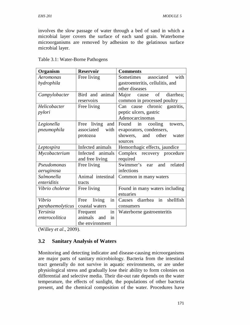
EHS 201 MODULE 5
171
involves the slow passage of water through a bed of sand in which a microbial layer covers the surface of each sand grain. Waterborne microorganisms are removed by adhesion to the gelatinous surface microbial layer. Table 3.1: Water-Borne Pathogens Organism Reservoir Comments Aeromonas hydrophila
Free living Sometimes associated with gastroenteritis, cellulitis, and other diseases
Campylobacter Bird and animal reservoirs
Major cause of diarrhea; common in processed poultry
Helicobacter pylori
Free living Can cause chronic gastritis, peptic ulcers, gastric Adenocarcinomas
Legionella pneumophila
Free living and associated with protozoa
Found in cooling towers, evaporators, condensers, showers, and other water sources
Leptospira Infected animals Hemorrhagic effects, jaundice Mycobacterium Infected animals
and free living Complex recovery procedure required
Pseudomonas aeruginosa
Free living Swimmer’s ear and related infections
Salmonella enteriditis
Animal intestinal tracts
Common in many waters
Vibrio cholerae Free living Found in many waters including estuaries
Vibrio parahaemolyticus
Free living in coastal waters
Causes diarrhea in shellfish consumers
Yersinia enterocolitica
Frequent in animals and in the environment
Waterborne gastroenteritis
(Willey et al., 2009). 3.2 Sanitary Analysis of Waters Monitoring and detecting indicator and disease-causing microorganisms are major parts of sanitary microbiology. Bacteria from the intestinal tract generally do not survive in aquatic environments, or are under physiological stress and gradually lose their ability to form colonies on differential and selective media. Their die-out rate depends on the water temperature, the effects of sunlight, the populations of other bacteria present, and the chemical composition of the water. Procedures have
EHS 201 GENERAL MICROBIOLOGY
172
been developed to attempt to “resuscitate” these stressed coliforms using selective and differential media. A wide range of viral, bacterial, and protozoan diseases result from the contamination of water with human and other animal fecal wastes. Although many of these pathogens can be detected directly, environmental microbiologists generally use indicator organismsas an index of possible water contamination by human pathogens. Researchers are still searching for the “ideal” indicator organism to use in sanitary microbiology. Among the suggested criteria for such an indicator are: 1. The indicator bacterium should be suitable for the analysis of all
types of water: tap, river, ground, impounded, recreational, estuary, sea, and waste.
2. It should be present whenever enteric pathogens are present. 3. It should survive longer than the hardiest enteric pathogen. 4. It should not reproduce in the contaminated water, thereby
producing an inflated value. 5. It should be harmless to humans. 6. Its level in contaminated water should have some direct
relationship to the degree of fecal pollution. 7. The assay procedure for the indicator should have great
specificity; in other words, other bacteria should not give positive results. In addition, the procedure should have high sensitivity and detect low levels of the indicator.
8. The testing method should be easy to perform. Coliforms,including Escherichia coli, are members of the family Enterobacteriaceae. These bacteria make up about 10% of the intestinal microorganisms of humans and other animals and have found widespread use as indicator organisms. They lose viability in freshwater at slower rates than most of the major intestinal bacterial pathogens. When such “foreign” enteric indicator bacteria are not detectable in a specific volume (100 ml) of water, the water is considered potable. 3.3 Wastewater Treatment Waters often contain high levels of organic matter from industrial, agricultural, and human wastes, which can be removed by the process of wastewater treatment.Depending on the effort given to this task, it may still produce waters containing nutrients and some microorganisms, which can be released to rivers and streams. Thus, the process of wastewater treatment, when performed at a municipal level, must be monitored to ensure that waters released into the environment do not pose environmental and health risks. Our discussion of wastewater treatment must therefore begin with the means by which water quality is
EHS 201 MODULE 5
173
monitored. This topic discussed large-scale wastewater treatment processes and home treatment systems. 3.3.1 Wastewater Treatment Processes Wastewater treatment involves a numberof steps that are spatially segregated. The first threesteps are called primary, secondary, and tertiary treatment. At the end of the process, the water is usually chlorinated(itself an emerging environmental and human health problem)before it is released. Primary treatmentphysically removes 20 to 30% of theBOD present in particulate form. In this treatment, particulatematerial is removed by screening, precipitation of small particulates,and settling in basins or tanks. The resulting solid materialis usually called sludge. Secondary treatmentpromotes the biological transformationof dissolved organic matter into microbial biomass andcarbon dioxide. About 90 to 95% of the BOD and many bacterialpathogens are removed by this process. Several approaches canbe used in secondary treatment to remove dissolved organic matter.All of these techniques involve similar microbial activities.Under oxic conditions, dissolved organic matter will be transformedinto additional microbial biomass plus carbon dioxide.When microbial growth is completed, under ideal conditions themicroorganisms will aggregate and form stable flocs that settle.Minerals in the water also may be tied up in microbial biomass. A healthy settleable floc is compact. In contrast, poorly formed flocs have a network of filamentous microbes that retard settling. When these processes occur with lower O2 levels or with a microbial community that is too young or too old, unsatisfactory floc formation and settling can occur. The result is a bulking sludge,caused by the massive development of filamentous bacteria such as Sphaerotilus and Thiothrix, together with many poorly characterized filamentous organisms. These important filamentous bacteria form flocs that do not settle well and thus produce effluent quality problems. An aerobic activated sludgesystem involves the horizontal flow of materials with recycling of sludge—the active biomass that is formed when organic matter is oxidized and degraded by microorganisms. Activated sludge systems can be designed with variations in mixing. In addition, the ratio of organic matter added to the active microbial biomass can be varied. A low rate system (low nutrient input per unit of microbial biomass), with slower growing microorganisms, will produce an effluent with low residual levels of dissolved organic matter. A high-rate system (high nutrient input per unit of microbial biomass), with
EHS 201 GENERAL MICROBIOLOGY
174
faster growing microorganisms, will remove more dissolved organic carbon per unit time but produce a poorer quality effluent. Aerobic secondary treatment also can be carried out with a trickling filter. The waste effluent is passed over rocks or other solid materials upon which microbial biofilms have developed, and the microbial community degrades the organic waste. A sewage treatment plant can be operated to produce less sludge by employing the extended aerationprocess. Microorganisms grow on the dissolved organic matter, and the newly formed microbial biomass is eventually consumed to meet maintenance energy requirements. This requires extremely large aeration basins and long aeration times. In addition, with the biological self-utilization of the biomass, minerals originally present in the microorganisms are again released to the water. All aerobic processes produce excess microbial biomass, or sewage sludge, which contains many recalcitrant organics. Often the sludge from aerobic sewage treatment, together with the materials settled out in primary treatment, are further treated by anaerobic digestion. Anaerobic digestersare large tanks designed to operate with continuous input of untreated sludge and removal of the final,stabilized sludge product. Methane is vented and often burned forheat and electricity production. This digestion process involvesthree steps: (1) The fermentation of the sludge components to form organic
acids, including acetate; (2) Production of the methanogenic substrates: acetate, CO2, and
hydrogen; and finally, (3) Methanogenesis by the methane producers. These methanogenic processes, involve critical balances between electron acceptors and donors. To function most efficiently, the hydrogen concentration must be maintained at a low level. If hydrogen and organic acids accumulate, methane production can be inhibited, resulting in a stuck digester. Anaerobic digestion has many advantages. Most of the microbial biomass produced in aerobic growth is used for methane production in the anaerobic digester. Also, because the process of methanogenesis is energetically very inefficient, the microbes must consume about twice the nutrients to produce an equivalent biomass as that of aerobic systems. Consequently, less sludge is produced and it can be easily dried. Dried sludge removed from well-operated anaerobic systems can even be sold as organic garden fertilizer. However, sludge can be dangerous if the system is not properly managed because heavy metals and other environmental contaminants may be concentrated in it.
EHS 201 MODULE 5
175
Tertiary treatmentfurther purifies wastewaters. It is particularly important to remove nitrogen and phosphorus compounds that can promote eutrophication. Organic pollutants can be removed with activated carbon filters. Phosphate usually is precipitated as calcium or iron phosphate (e.g., by the addition of lime). To remove phosphorus, oxic and anoxic conditions are used alternately in a series of treatments, and phosphorus accumulates in microbial biomass as polyphosphate. Excess nitrogen may be removed by “stripping,” volatilization of NH3 at high pHs. Ammonia itself can be chlorinated to form dichloramine, which is then converted to molecular nitrogen. In some cases, microbial processes can be used to remove nitrogen and phosphorus. A widely used process for nitrogen removal is denitrification. Here, nitrate, produced by microbes under aerobic conditions, is used as an electron acceptor under conditions of low oxygen with organic matter added as an energy source. Nitrate reduction yields nitrogen gas (N2) and nitrous oxide (N2O) as the major products. In addition to denitrification, the anammox process is also important. In this reaction, ammonium ion (used as the electron donor) is reacted with nitrite (the electron acceptor) produced by partial nitrification (i.e., the oxidation of ammonium to nitrite). The anammox process can convert up to 80% of the beginning ammonium ion to N2 gas. Tertiary treatment is expensive and is usually not employed except where necessary to prevent obvious ecological disruption. Wetlands are a vital natural resource and a critical part of our environment, and increasingly efforts are being made to protect these fragile aquatic communities from pollution. A major means of wastewater treatment is the use of constructed wetlands, where the basic components of natural wetlands (soils, aquatic plants, waters) are used as a functional waste treatment system. Constructed wetlands now are increasingly employed in the treatment of liquid wastes and for bioremediation. This system uses floating, emergent, or submerged plants. The aquatic plants provide nutrients in the root zone, which support microbial growth. Especially with emergent plants, the root zone can be maintained in an anoxic state in which sulfide, produced by Desulfovibrio using root zone organic matter as an energy source, can trap metals. Constructed wetlands also are being used to treat acid mine drainage and industrial wastes in many sparts of the world. 3.3.2 Home Treatment Systems Groundwater—the water in gravel beds and fractured rocks below the surface soil—is a widely used but often unappreciated resource. In the United States, groundwater supplies at least 100 million people with drinking water, and in rural and suburban areas beyond municipal water
EHS 201 GENERAL MICROBIOLOGY
176
distribution systems, 90 to 95% of all drinking water comes from this source. Unfortunately our dependence on groundwater has not resulted in a corresponding understanding of microorganisms and microbiological processes that occur in this environment. Pathogenic microorganisms and dissolved organic matter are removed from water during subsurface passage through adsorption and trapping by fine sandy materials, clays, and organic matter. Microorganisms associated with these materials—including predators such as protozoa—can use the trapped pathogens as food. This results in purified water with a lower microbial population. This combination of adsorption and biological predation is used in home treatment systems. Conventional septictanksystems include an anaerobic liquefaction and digestion step that occurs in the septic tank, which functions as a simple anaerobic digester. This is followed by organic matter adsorption and entrapment of microorganisms in an aerobic leach field where A septic tank may not operate correctly for several reasons.If the retention time of the waste in the septic tank is too short,undigested solids move into the leach field, plugging the system.If the leach field floods and becomes anoxic, biological oxidationdoes not occur, and effective treatment ceases. Otherproblems can occur, especially when a suitable soil is not presentand the septic tank outflow from a conventional systemdrains too rapidly to the deeper subsurface. Fractured rocks andcoarse gravel materials provide little effective adsorption or filtration.This may result in the contamination of well water withpathogens and the transmission of disease. In addition, nitrogenand phosphorus from the waste can pollute the groundwater.This leads to nutrient enrichment of ponds, lakes, rivers, andestuaries as the subsurface water enters these environmentallysensitive ecosystems. Domestic and commercial on-site septic systems are now being designed with nitrogen and phosphorus removal steps. Nitrogen is usually removed by nitrification and denitrification, with organic matter provided by sawdust or a similar material. For phosphorus removal, a reductive iron dissolution process can be used. With the need to control nitrogen and phosphorus releases from septic systems, there is an increased emphasis on use of these and similar technologies. Subsurface zones also can become contaminated with pollutants from other sources. Land disposal of sewage sludges, illegal dumping of septic tank pumpage, improper toxic waste disposal, and runoff from agricultural operations all contribute to groundwater contamination with chemicals and microorganisms. Many pollutants that reach the subsurface will persist and may affect the quality of groundwater for
EHS 201 MODULE 5
177
extended periods. Much research is being conducted to find ways to treat groundwater in place—in situ treatment. As earlier explained, microorganisms and microbial processes are critical in many of these remediation efforts. 3.3.3 Measuring Water Quality Carbon removal during wastewater treatment can be measured several ways, including: (1) As total organic carbon (TOC), (2) As chemically oxidizable carbon by the chemical oxygen demand
(COD)test, or (3) As biologically usable carbon by the biochemical oxygen demand
(BOD)test. The TOC includes all carbon, whether or not it can be used by microorganisms. It is measured by oxidizing the organic matter in a sample to CO2 at high temperature in an oxygen stream. The resultant CO2 is measured by infrared or potentiometric techniques. The COD gives a similar measurement, except that lignin often will not react with the oxidizing chemical, such as permanganate, that is used in this procedure. 3.4 Biodegradation and Bioremediation by Natural Communities The metabolic activities of microbes can also be exploited in complex natural environments such as waters, soils, or high organic matter–containing composts where the physical and nutritional conditions for microbial growth cannot be completely controlled and a largely unknown microbial community is present. Examples are: (1) The use of microbial communities to carry out biodegradation,
bioremediation, and environmental maintenance processes; and (2) The addition of microorganisms to soils or plants for the
improvement of crop production. 3.4.1 Biodegradation and Bioremediation Processes Before discussing biodegradation processes carried out by natural microbial communities, it is important to consider definitions. Biodegradationhas at least three outcomes: (1) A minor change in an organic molecule leaving the main
structure still intact,
EHS 201 GENERAL MICROBIOLOGY
178
(2) Fragmentation of a complex organic molecule in such a way that the fragments could be reassembled to yield the original structure, and
(3) Complete mineralization, which is the transformation of organic molecules to inorganic forms.
The removal of toxic industrial products in soils and aquatic environments has become a daunting and necessary task. Compounds such as perchloroethylene (PCE), trichloroethylene (TCE), and polychlorinated biphenyls (PCBs) are common contaminants. These compounds adsorb onto organic matter in the environment, making decontamination using traditional approaches difficult or ineffective. The use of microbes to transform these contaminants to nontoxic degradation products is called bioremediation.To understand how bioremediation takes place at the level of an ecosystem, you first must consider the biochemistry of biodegradation. Degradation of complex compounds requires several discrete stages, usually performed by different microbes. Initially contaminants are converted to less-toxic compounds that are more readily degraded. The first step for many contaminants, including organochloride pesticides, alkyl solvents, and aryl halides, is reductivedehalogenation.This is the removal of a halogen substituent (e.g., chlorine, bromine, fluorine) while at the same time adding electrons to the molecule. This can occur in two ways. In hydrogenolysis, the halogen substituent is replaced by a hydrogen atom. Alternatively, dihaloelimination removes two halogen substituents from adjacent carbons while inserting an additional bond between the carbons. Both processes require an electron donor. The dehalogenation of PCBs uses electrons derived from water; alternatively hydrogen can be the electron donor for the dehalogenation of different chlorinated compounds. Major genera that carry out this process include Desulfi tobacterium, Dehalospirillum, and Desulfomonile. 3.4.2 Bioaugmentation The acceleration of microbiological processes by the addition of known active microorganisms to soils, waters, or other complex systems is called bioaugmentation.For example, commercial culture preparations are available to facilitate silage formation and to improve septic tank performance. While such additions usually led to short-term increases in rates of the desired activity, after a few days the microbial community responses were similar in treated and control systems. The lack of effectiveness of such added cultures was due to at least three factors:
EHS 201 MODULE 5
179
(1) The attractiveness of laboratory-grown microorganisms as a food source for predators such as soil protozoa,
(2) The inability of these added microorganisms to contact the compounds to be degraded, and
(3) The failure of the added microorganisms to survive and compete with indigenous microorganisms.
Such a modified microorganism may be less fit to compete and survive because of the additional energetic burden required to maintain the extra DNA. 4.0 CONCLUSION Environmental microbiology is a novel area of microbiology, which has improved wastewater treatment processes, encouraging water reuse and conservation, preventing the spread of water-borne diseases and ensuring good health of man. More sustainable environmental management approaches include bioremediation of contaminated environments and biodegradation of environmental pollutants. 5.0 SUMMARY At the end of this unit, the student must have learntwater purification and sanitary analysis, sanitary analysis of waters, wastewater treatment, and biodegradation and bioremediation by natural communities. 6.0 TUTOR-MARKED ASSIGNMENT 1. What is an indicator organism, and their properties? 2. Explain primary, secondary, and tertiary treatments of
wastewater. 3. What are the three major outcomes of biodegradation? 4. In bioaugmentation process, what are the three factors
responsible for lack of effectiveness? 7.0 REFERENCES/FURTHER READING Atlas, R., & Bartha, R. (1998). Microbial Ecology, Fundamental and
Applications, (4th ed.). Addison Wesley Longman, Reading Bertrand, J., Caumette, P., Lebaron, P., Matheron, R., & Normand, P.
(2011). Environmental Microbiology: Fundamentals and Applications. Universities Press

EHS 201 GENERAL MICROBIOLOGY
180
Fewtrell, L., & Bartram, J. (2001) Water quality: guidelines, standards and health. World Health Organisation Water Series. IWA Publishing, London.
Willey, J.M., Sherwood, L.M. & Woolverton, C.J. (2009). Prescott’s
Principles of ‘Microbiology. (1st ed.), McGraw-Hill. pp. 1-12.
EHS 201 MODULE 5
181
UNIT 3 MICROORGANISMS IN AGRICULTURE CONTENTS 1.0 Introduction 2.0 Objectives 3.0 Main Content 3.1 The Soil Plant Microorganisms 3.1.1 Soil Fertility 3.1.2 Ammonification in Soil 3.1.3 Nitrification in Soil 3.1.4 Nitrogen-Fixing Bacteria 3.1.5 Rhizosphere Environment 4.0 Conclusion 5.0 Summary 6.0 Tutor-Marked Assignment 7.0 References/Further Reading 1.0 INTRODUCTION Microbes are the tiny living organisms not visible to naked eye. However, they play a very important role in Nature and contribute a lot to plants, animals, and human beings. In general, they represent the groups, prokaryotes and eukaryotes. Bacteria, actinomycetes, and blue-green algae are the sole representatives of prokaryotes. Among eukaryotes, algae, protozoa, and fungi are placed as microbes. Farmers and ranchers often think of microbes as pests that are destructive to their crops or animals (as well as themselves), but many microbes are beneficial. Soil microbes (bacteria and fungi) are essential for decomposing organic matter and recycling old plant material. Some bacteria and fungiform relationships with plant roots that provide important nutrients such as nitrogen and phosphorus. Fungi can colonize upper parts of plants and provide many benefits, including drought and heat tolerance and resistance to insects plant diseases. In agriculture, fungi, bacteria, algae, and viruses are important with respect to their contribution in the form of either loss or gain in the production of grains, fruits, vegetables, oil, milk, poultry, fodder, and livestock. Most of the nutrients, both minor and major, present in the soil are managed by microorganisms through integrated nutrient management. It has been observed that the efficiency of utilization of nutrients of high grade complex fertilizers in terms of biological chemicals and agronomic/economic efficiency has dropped from 20 to below 9 in terms of kg grain/kg of NPK. It is therefore important to manage the nutrients in a proper way.

EHS 201 GENERAL MICROBIOLOGY
182
2.0 OBJECTIVES By the end of this unit. you will be able to: • understand the interactions between soil, plant and
microorganisms target improving soil fertility • understand the ammonification in soil and the nitrification in soil • know the nitrogen fixation by bacteria • list the activities in the rhizosphere environment.
3.0 MAIN CONTENT 3.1 The Soil Plant Microorganisms Microorganisms are known to be very important for plant growth. They multiply and actively help in making essential nutrients available for the plant through a symbiotic process by releasing the “locked-up” nutrients to be ready for uptake and utilization. Microorganisms have an active role in protecting plants against soil-borne diseases. It seems obvious that microorganisms are in the soil because there is food. Soil is an excellent culture media for the growth of many types of organisms, including bacteria, fungi, algae, protozoa, and viruses. In addition, various nematodes, insects, and so on are also present. A spoonful of soil contains billions of microorganisms. In general, the majority of microbial population is found in the upper 6–12 in of soil and the number decreases with depth. Higher number occurs in the organically rich surface layers than in the underlying mineral soils. Particularly, high numbers of microbes occur in association with plant roots. Fungal populations are favored in soils of low pH, and bacteria tend to occur in higher numbers in those of higher pH. There is about two and a half times more carbon in the soil than there is in the atmosphere. Unseen soil microbes respond to and influence global climate change. It has been noted that the respiration of soil microbes returns to normal after a number of years under heated conditions. It was further argued that the microbes consumed so much of the available food under heated conditions that future levels of decomposition were reduced because of food scarcity. Some soil microbes are also adapted to the changed environment and reduce their respiration accordingly. The abundance of soil microbes decreased under warm conditions. Various types of organisms and microorganisms live in soil. Some microorganisms also burrow and channel through soil, which improves soil structure and aggregation, while other microorganisms have the ability to break down resistant organic matter such as lignin, toxins, and pesticides. Microorganisms also have the ability to protect plants from antagonistic pathogens, and some can dissolve minerals, making nutrients available to plants. Fungi are able to break down resistant
EHS 201 MODULE 5
183
materials such as cellulose, gums, and lignin. They dominate in acidic, sandy soils and in fresh organic matter. Actionomycetes also are able to decompose resistant substances in soil. One type, Frankia, helps plants to get nutrients needed from the air by breaking triple bond nitrogen to ammonium that plants can use. Antibiotics are made from soil actinomycetes. Bacteria decompose a wider range of earth material than any other microbe group. Heterotrophs gain their energy and carbon from other organisms, while autotrophs synthesize their own energy from light or by chemical oxidation. Some bacteria can fix nitrogen into forms that plants can use. How quickly decomposition of dead organic matter occurs depends on soil temperature and soil moisture. Without the microorganisms, dead organic matter would pile high on Earth’s surface. 3.1.1 Soil Fertility Soil fertility is the capacity to supply proper amounts of different nutrients in the appropriate proportion for the growth of crops. The availability of both inorganic and organic matter determines the soil fertility. The inorganic matter of soil comprises the entire essential and trace minerals present in the soil in the form of salts (acidic and basic). The inorganic element either gets adsorbed onto the clay particles or gets dissolved in the soil water. Soil fertility refers to the amount of nutrients in the soil, which is sufficient to support plant life. It must contain organic matter and a relatively low pH value. The soil must also contain micro- and macroorganisms, and it must be well drained. The organic matter in the soil exists mainly as humus or as partially decomposed (plant and animal tissues). It is also prepared artificially as farmyard manure, green manure/green-leaf manure, compost, vermicompost, biofertilizers, and so on. The four strategies used by farmers for soil management show that they had a good empirical understanding of soil fertility and degradation but that this understanding was incomplete: they were entirely unaware of crucial soil processes occurring underneath the soil surface. On the whole, the balanced availability of both inorganic and organic matter in the soil determines the soil fertility. Indirectly, these organic and inorganic matters help in theproliferation of various qualitative microflora that play a very vital role in maintaining the nutritional balance of the soil. Thus, microorganisms have a great role to play in determining soil fertility, for without a proper distribution of microflora, no soil can support plant growth which speaks of its fertility. Microorganisms in soil affect the fertility of soil by means of physical or chemical changes.
EHS 201 GENERAL MICROBIOLOGY
184
Soil fertility is critical in sustainable farming and needs to be considered not only for crop productivity but also for the protection of aquatic environments. Fertile soil has an abundance of plant nutrients including nitrogen, phosphorus, and potassium; an abundance of minerals; as well as an abundance of organic matter. Mycorrhizae is being used to enhance the uptake of nutrients and water for establishment of seedlings on degraded lands. This will, however, not lead to improvement of soil fertility by mycorrhizae as such, although the success of plant growth will eventually lead to reclamation of degraded lands. However, nitrogen-fixing bacteria such as Rhizobium or the actinomycete genus Frankia can be used to induce nodule formation in a variety of plant species, so that they can be used for improving soil fertility of degraded lands. This nodule formation can be induced both in leguminous and nonleguminous plant species comprising annuals (cereal and legume crops) and perennials (trees). Efforts are underway to manipulate the genes of both host and rhizobia to obtain maximum efficiency of nodule formation. Strains are also being tailored for unusual soil environments representing degraded lands. Most of the plant nutrients, besides carbon, hydrogen, and oxygen, originate from the soil. The soil system is viewed by the soil scientists as a triple-phased system of solid, liquid, and gaseous phases. These phases are physically separable. The plant nutrients are based in the solid phase, and their usual pathway to the plant system is through the surrounding liquid phase, the soil solution, and then to the plant root and plant cells. This pathway may be written in the form of an equation as: M (solid) → M (solution) → M (plant root) → (plant top), Where “M” is the plant nutrient element in continual movement through the soil-plant system. The operation of the above system is dependent on the solar energy through photosynthesis and metabolic activities. Soil is a mosaic of dynamic microenvironment that differs in physical, biological, and chemical properties. Hence, the microbial communities that govern ecosystem C and N cycling are spatially and temporally variable. It has been observed that both fungi and earthworms, which are known to enhance the formation of soil aggregates, directly control the formation of microaggregates (within macroaggregates). In addition, several studies have indicated that the microaggregate structure creates an operationally definable microenvironment for microorganisms; that is, the differences in microbial community are greater between macroaggregates andmicroaggregates within a soil type than among different soil types. This microenvironment is characterized by low predation pressure, relatively stable water potential, low oxygen

EHS 201 MODULE 5
185
availability, and low accessibility for exogenous toxic elements. As a result, the spatial compartmentalization associated with these microenvironments protects microbes from contaminants, fosters a unique microbial community structure, and also reduces the activity level of the microflora. The last function of microaggregates directly induces the stabilization and storage of soil C and N. C, and especially C derived from fungal and bacterial cell wall components, is preferentially sequestered within microaggregates occluded within macroaggregates. 3.1.2 Ammonification in Soil The nitrogen in most plants and animals exists in the form of protein. Most of the nitrogen in soil exists in the form of organic molecules, mostly proteins derived from the decomposition of dead plant and animal tissue. When an organism dies, its proteins are attacked by the proteases of soil bacteria to produce polypeptides (peptones) and amino acids (C2H4NO2-R). This process is called peptonization. Then, the amino groups on the amino acids are removed by a process called deamination, producing ammonia (NH3). In most soils, the ammonia dissolves in water to form ammonium ions (NH4
+). The processof the production of ammonia from organic compounds is called ammonification. Inaddition to the ammonification of amino acids, other compounds such as nucleic acids,urea, and uric acid go through the ammonification process. The bacteria that accomplishthe process (Bacillus, Clostridium, Proteus, Pseudomonas, and Streptomyces) are called ammonifying bacteria. Ammonification of organic compounds is a very important step in the cycling of nitrogen in soil, since most autotrophs are unable to assimilate amino acids, nucleic acids, urea, and uric acid and use them for their own enzyme and protoplasm construction. Ammonification is an important stage in the nitrogen cycle, a natural cycle that makes the Earth’s supply of nitrogen available to organisms that need it, such as plants. Like many other natural cycles, the nitrogen cycle can be disrupted by human activities, which can lead to imbalances at various stages, sometimes causing environmental problems. Ammonification can be a major problematic area in the nitrogen cycle when human intervention occurs, as buildups of ammonia can cause health problems and environmental issues. One of the most elementary of the ammonification reactions is the oxidation of the simple organic compound urea (CO(NH2)2), to ammonia through the action of a microbial enzyme known as urease (two units of ammonia are produced for every unit of urea that is oxidized). Urea is a commonly utilized agricultural fertilizer, used to supply ammonia or ammonium for direct
EHS 201 GENERAL MICROBIOLOGY
186
uptake by plants, or as a substrate for the microbial production of nitrate through nitrification. 3.1.3 Nitrification in Soil The biological conversion of ammonium to nitrate nitrogen is called nitrification. Nitrification is a two-step process. Nitrosomonas convert ammonia and ammoniumto nitrite. Bacteria called Nitrobacter finish the conversion of nitrite to nitrate.The reactionsare generally coupled and proceed rapidly to the nitrate form; therefore, nitrite levels at any given time are usually low. These bacteria known as nitrifiers are strict “aerobes,” meaning they must have free dissolved oxygen (DO) to perform their work. Nitrification occurs only under aerobic conditions at DO levels of 1 mg/l or more. At DO concentrations less than 0.5 mg/l, the growth rate is minimal. Nitrification requires a long retention time, a low food-to-microorganism ratio (F/M), a high mean cell residence time (measured as MCRT or sludge age), and adequate buffering (alkalinity). A plug-flow extended aeration tank is ideal. Temperature, as discussed below, is also important, but not really. The nitrification process produces acid. This acid formation lowers the pH of the biological population in the aeration tank and can cause a reduction in the growth rate of nitrifying bacteria. The optimum pH for Nitrosomonas and Nitrobacter is between 7.5 and 8.5; most treatment plants are able to effectively nitrify with a pH of 6.5–7.0. 3.1.4 Nitrogen-Fixing Bacteria Nitrogen is one of the most important chemical elements for plants. If there is not enough nitrogen available in the soil, plants look pale and their growth is stunted. Nitrogen-fixing plants are called legumes. All peas and beans are legumes. The plants work together with nitrogen-fixing bacteria called rhizobia to “fix” nitrogen. The rhizobia chemically convert the nitrogen from the air to make it available to the plant. Leguminous plants live in a symbiotic relationship with the nitrogen-fixing bacteria. Rhizobia live in nodules in the plant’s root. This way the plant can look after its own nitrogen needs and fertilizer is not required. In addition, when the crop is harvested and the plant cut back to ground level, the root nodules should release all the valuable fixed nitrogen. Nitrogen-fixing leguminous plants have the unique ability to fix atmospheric nitrogen in the ground and make their own fertilizers. Actually, these plants do not pull off this feat on their own. They owe partial credit for this effort to their symbiotic relationship with nitrogen-fixing bacteria. The leguminous plants provide nutrients to the bacteria in return for which the bacteria fix atmospheric nitrogen through anaerobic processes (processes that work
EHS 201 MODULE 5
187
without oxygen). The primary function of nitrogen-fixing bacteria is “survival,” and in their efforts to survive, they enter into a symbiotic relationship with leguminous plants or some survive on their own. As a part of their metabolic cycle, they fix nitrogen. The enzyme that nitrogen-fixing bacteria use is called nitrogenase. It is a chemical responsible for nitrogen fixation and without which, this process is impossible. The process at chemical level that enables nitrogen fixation can be summarized in the following way: N2+ 6H+ + 6e− → 2NH3 3.1.5 Rhizosphere Environment The rhizosphere is the zone of the soil surrounding a plant root where the biology and chemistry of the soil are influenced by the root. This zone is about 1mm wide but has nodistinct edge. Rather, it is an area of intense biological and chemical activity influenced by compounds exuded by the root and by microorganisms feeding on the compounds. As plant roots grow through soil, they release water-soluble compounds such as amino acids, sugars, and organic acids that supply food to the microorganisms. The food supply means microbiological activity in the rhizosphere, which is much greater in soil away from plant roots. In return, the microorganisms provide nutrients to the plants. All this activity makes the rhizosphere the most dynamic environment in the soil. Because roots are underground, rhizosphere activity has been largely overlooked, and it is only now that we are starting to unravel the complex interactions that occur. For this reason, the rhizosphere has been called the last frontier in agricultural science. Practically all ecological interactions, such as symbiosis, syntrophism, synergism, commensalism, and antagonism, between plants and microorganisms, and among different microorganisms, are found in this region. SELF-ASSESSMENT EXERCISE What are the importance of microorganisms in the soil? 4.0 CONCLUSION Soil does not only serve as the habitat for soil living organisms but is involved in a sustainable interaction between plants and microorganisms. Most biogeochemical cycles occur in the soil and mediated by soil microorganisms. Nutrient cycling has remained a natural means improving soil fertility and providing soil nutrients. In all these and more, soil microorganisms are very vital component of the soil.
EHS 201 GENERAL MICROBIOLOGY
188
5.0 SUMMARY At the end of this unit the student has understood the interactions between soil, plant and microorganisms targeted at improving soil fertility, ammonification in soil, nitrification in soil, nitrogen fixation by bacteria, and activities in the rhizosphere environment. 6.0 TUTOR-MARKED ASSIGNMENT 1. Explain the importance of soil fertility in the sustenance of
agriculture. 2. What is Rhizosphere and explain the activities of microorganisms
in the rhizosphere. 7.0 REFERENCES/FURTHER READING Atlas, R.,& Bartha, R. (1998). Microbial Ecology, Fundamental And
Applications, (4th ed). Addison Wesley Longman, Reading. Bertrand, J., Caumette, P., Lebaron, P., Matheron, R., & Normand, P.
(2011). Environmental Microbiology: Fundamentals and Applications. Universities Press.
Willey, J.M., Sherwood, L.M. & Woolverton, C.J. (2009). Prescott’s
Principles of ‘Microbiology.(1st ed)., McGraw-Hill. pp. 1-12.