Embed Size (px)
Citation preview
Academic Journal of Animal Diseases 9(1): 19-32, 2020ISSN 2079-200X© IDOSI Publications, 2020DOI: 10.5829/idosi.ajad.2020.19.32
Corresponding Author: Dereje Tulu, Ethiopian Institute of Agricultural Research, Tepi Agricultural Research Center,P.O. Box: 34, Tepi, Ethiopia. Tel: +251920654572.
19
Epidemiology and Zoonotic Implication ofLeptospirosis in Domestic Animals in Ethiopia
Dereje Tulu
Ethiopian Institute of Agricultural Research,Tepi Agricultural research center, P.O. Box: 34, Tepi, Ethiopia
Abstract: Leptospirosis is a re-emerging zoonotic disease of worldwide public health significance that affectsdomestic animals and humans. The disease is caused by various serovars of leptospira interrogans that belongto the genus Leptospira. Pomona, Canicola, Bratislava, Grippotyphosa, Hardjo and Icterohemorrhagiae are thecommon serovars of L. interrogans. Leptospirosis contains a large spectrum of the host that harbor andexcretes the agent from their renal tubules. The principal reservoir hosts for many Leptospira serovars arerodents. However, cattle, sheep, goats, dogs, horses and pigs are reservoir hosts among domestic animals andthey act as a carrier for several months. Leptospirosis occurs as acute, sub-acute and chronic forms. It ischaracterized by septicemia, hemorrhagic syndrome, abortion storm and stillbirth. Leptospirosis can betransmitted through direct and indirect contact. The urine of diseased or carrier animals, contaminates water,mud feed, aborted fetus and uterine discharge are the major sources of leptospirosis. Abattoir workers, sewageworkers, veterinarians and recreational activities such as water sports and white rafting are the major risk groupsof leptospirosis. Laboratory tests used for the detection of leptospira are microscope evaluation, culture,molecular method, serology and anima inoculation. The occurrence of leptospirosis is affected by factors relatedto management; host and environmental. Leptospirosis in a domestic animal might be controlled throughvaccination, prophylactic treatment of exposed animals with antibiotics; quarantine introduced new animals,rodent management and improved environmental hygiene. As a result, it is important to conduct applicablecontrol techniques and increasing the public awareness about zoonotic transmission of leptospirosis isrecommended. Besides, any investigation and control effort should be conducted by collaboration amonghuman, animal and environmental health professions.
Key words: Domestic Animals Epidemiology Leptospirosis Zoonosis
INTRODUCTION Leptospirosis has occurred as acute, sub-acute and
Leptospirosis is a worldwide important zoonotic hemorrhagic syndrome, abortion storm, stillbirth anddisease caused by genus Leptospira [1]. Leptospira reduced milk production [4]. The diagnosis ofinterrogans are pathogenic species that cause leptospirosis is based on the availability of the sampleleptospirosis whereas L. biflexa is nonpathogenic [2]. and the temporal stage of the disease. Laboratory testsLeptospirosis is most common in temperate regions used for the detection of leptospira are microscopeduring late summer and early rainfall and in tropical evaluation, culture, molecular method, serology and animaregions during rainy seasons [3]. The disease can affect inoculation [5]. Leptospirosis can be treated by antibioticsboth humans and various animals’ species resulting in such as tetracycline, penicillin, ampicillin, doxycycline,morbidity and mortality. It can be directly transmitted streptomycin and erythromycin [6], while prevention isthrough interaction with secretions, blood or urine of characterized by sanitary control and decrease in thediseased animals or indirectly through water contaminated risk of infection occurring due to interaction withmainly with the urine of reservoir animals [1]. contaminated environments, infected wild animals as well
chronic forms. It is characterized by septicemia,
Acad. J. Anim. Diseases 9(1): 19-32, 2020
20
as with synanthropic animals and rodents [3]. Control The prioritization was done based on the severity inmeasures of leptospirosis are aimed at limiting the humans, the proportion of human diseases attributed tooccurrence of clinical disease based on integrated action animal exposure, the impact of animal disease at thein several links of the transmission chain [7]. household level, the availability of intervention methods
Leptospirosis is recognized as a re-emerging global and the existence of collaboration among the sector.public health problem due to the increased incidence in Leptospirosis is one of the first five selected diseasesboth developing and developed countries [8, 9]. to be tackled through the establishment of the OneGlobal warming that leads to extreme weather events Health-focused zoonotic diseases in the coming fivesuch as cyclones and floods, increased rainfall and years in the country [21]. Even though, there is fewincreased population and urbanization are considered as documented information concerning the occurrencethe factors associated with the upsurge in the incidence of leptospirosis in domestic animals in Ethiopia,of leptospirosis as well as the magnitude of outbreaks socioeconomic, warm and humid conditions of the areas[10]. The global burden of leptospirosis is unknown due are favorable for the survival and transmission of theto the paucity of data [11]. Although the incidence of pathogen [22].leptospirosis appears to have decreased in developed Leptospirosis is one of the zoonotic diseases that cancountries, it is rising as a major public health problem in easily be transmitted through contact with contaminateddeveloping countries [12]. areas by rodent urine and other excreta, where there is
In tropical areas where humans and animals live in easy access of the communities to such areas with noclose interaction, warm and humid conditions favor knowledge about its transmission and preventionenvironmental survival and transmission of the pathogen mechanism. Its investigation and creating awareness[10]. Leptospirosis is recognizing as an important cause of about its risk to exposed groups is very important torenal failure and febrile disease in south-east Asia and safeguard the public health and livelihoods of society.Latin America [13]. However, in Africa there is no or very Therefore, this paper aimed to review the epidemiologylittle is understood concerning the extent of human and zoonotic implication of Leptospira infection inzoonosis or the epidemiology of Leptospira infection in domestic animals.several animal species [14]. This is why it remains as oneof the neglected tropical zoonotic diseases and further Leptospirosis in Domestic Animalsevaluating its impact on livestock health and productivity Etiology: Leptospirosis is caused by pathogenicneeds priority for prospective study in Africa [15]. spirochaetes of genus Leptospira occurring in almost allGenerally, the battle against leptospirosis can be the mammalian species [23]. Genus Leptospira isconsidered an excellent example of ‘One Health', where the categorizing under order Spirochaetales, familyrelationship between humans, animals and ecosystems Leptospiraceae, class Spirochaetes and it is dividing intoneed to better understand and manage the disease. two species: Leptospira interrogans, including allSubsequently, any study and control effort requires a pathogenic strains and Leptospira biflexa involving thetruly multi-disciplinary and coordinated approach [16]. saprophytic strains isolated from the environments [2].
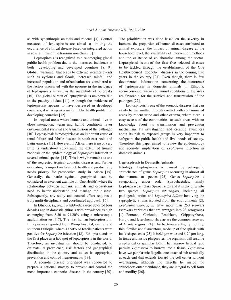
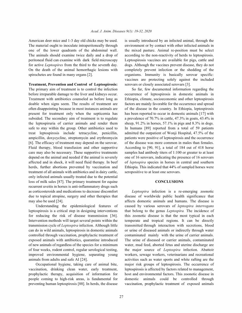
In Ethiopia, Leptospira antibodies were detected four Leptospira interrogans have more than 250 serovarsdecades ago in domestic animals with prevalence as high (serovars varieties) that are arranged into 23 serogroupsas ranging from 8.30 to 91.20% using a microscopic [1]. Pomona, Canicola, Bratislava, Grippotyphosa,agglutination test [17]. The first human leptospirosis in Hardjo and Icterohemorrhagiae are the common serovarsEthiopia was reported from Wonji hospital, central and of L. interrogans [24]. The bacteria are highly motility,southern Ethiopia, where 47.50% of febrile patients were thin, flexible and filamentous, made up of fine spirals withpositive for Leptospira infection [18]. Ethiopia stands in hook-shaped ends [25]. It is 0.1 µm wide and 6-20 µm long.the first place as a hot spot of leptospirosis in the world. In tissue and inside phagocytes, the organism will assumeTherefore, an investigation should be conducted, to a spherical or granular look. Their narrow helical typeestimate its prevalence, risk factors and geographical permits Leptospira to burrow into a tissue. Leptospiradistribution in the country and to set its appropriate have two periplasmic flagella, one attached sub terminallyprevention and control measurements [19]. at each end that extends toward the cell center without
A zoonotic disease prioritized was conducted to overlapping, although the flagella lie inside theprepare a national strategy to prevent and control the spirochaete outer membrane, they are integral to cell formmost important zoonotic disease in the country [20]. and motility [26].
Acad. J. Anim. Diseases 9(1): 19-32, 2020
21
Fig. 1: Leptospira species showing characteristic helical shape, periplasmic flagella and outer membrane [25]
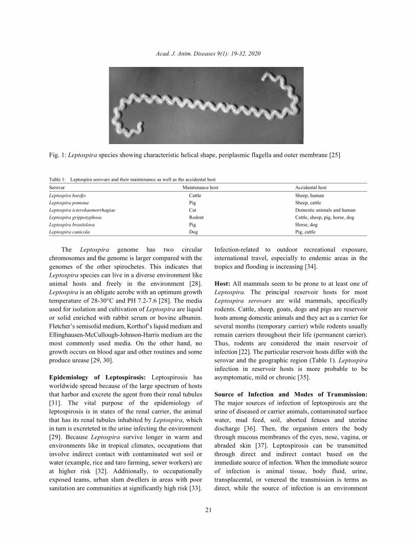
Table 1: Leptospira serovars and their maintenance as well as the accidental host Serovar Maintenance host Accidental hostLeptospira hardjo Cattle Sheep, humanLeptospira pomona Pig Sheep, cattleLeptospira icterohaemorrhagiae Cat Domestic animals and humanLeptospira grippotyphosa Rodent Cattle, sheep, pig, horse, dogLeptospira brastislava Pig Horse, dogLeptospira canicola Dog Pig, cattle
The Leptospira genome has two circular Infection-related to outdoor recreational exposure,chromosomes and the genome is larger compared with the international travel, especially to endemic areas in thegenomes of the other spirochetes. This indicates that tropics and flooding is increasing [34].Leptospira species can live in a diverse environment likeanimal hosts and freely in the environment [28]. Host: All mammals seem to be prone to at least one ofLeptospira is an obligate aerobe with an optimum growth Leptospira. The principal reservoir hosts for mosttemperature of 28-30°C and PH 7.2-7.6 [28]. The media Leptospira serovars are wild mammals, specificallyused for isolation and cultivation of Leptospira are liquid rodents. Cattle, sheep, goats, dogs and pigs are reservoiror solid enriched with rabbit serum or bovine albumin. hosts among domestic animals and they act as a carrier forFletcher’s semisolid medium, Korthof’s liquid medium and several months (temporary carrier) while rodents usuallyEllinghausen-McCullough-Johnson-Harris medium are the remain carriers throughout their life (permanent carrier).most commonly used media. On the other hand, no Thus, rodents are considered the main reservoir ofgrowth occurs on blood agar and other routines and some infection [22]. The particular reservoir hosts differ with theproduce urease [29, 30]. serovar and the geographic region (Table 1). Leptospira
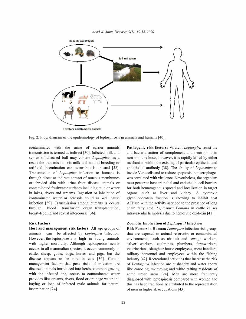
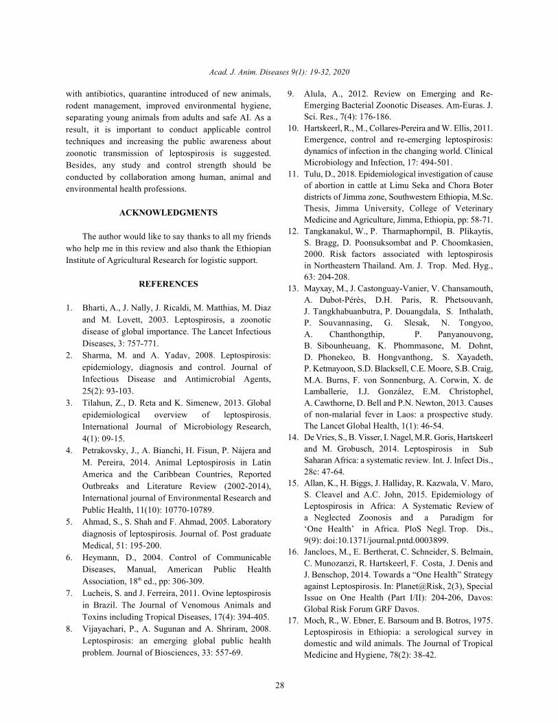
Epidemiology of Leptospirosis: Leptospirosis has asymptomatic, mild or chronic [35].worldwide spread because of the large spectrum of hoststhat harbor and excrete the agent from their renal tubules Source of Infection and Modes of Transmission:[31]. The vital purpose of the epidemiology of The major sources of infection of leptospirosis are theleptospirosis is in states of the renal carrier, the animal urine of diseased or carrier animals, contaminated surfacethat has its renal tubules inhabited by Leptospira, which water, mud feed, soil, aborted fetuses and uterinein turn is excreteted in the urine infecting the environment discharge [36]. Then, the organism enters the body[29]. Because Leptospira survive longer in warm and through mucous membranes of the eyes, nose, vagina, orenvironments like in tropical climates, occupations that abraded skin [37]. Leptospirosis can be transmittedinvolve indirect contact with contaminated wet soil or through direct and indirect contact based on thewater (example, rice and taro farming, sewer workers) are immediate source of infection. When the immediate sourceat higher risk [32]. Additionally, to occupationally of infection is animal tissue, body fluid, urine,exposed teams, urban slum dwellers in areas with poor transplacental, or venereal the transmission is terms assanitation are communities at significantly high risk [33]. direct, while the source of infection is an environment
infection in reservoir hosts is more probable to be
Acad. J. Anim. Diseases 9(1): 19-32, 2020
22
Fig. 2: Flow diagram of the epidemiology of leptospirosis in animals and humans [40].
contaminated with the urine of carrier animals Pathogenic risk factors: Virulent Leptospira resist thetransmission is termed as indirect [30]. Infected milk and anti-bacteria action of complement and neutrophils insemen of diseased bull may contain Leptospira; as a non-immune hosts, however, it is rapidly killed by eitherresult the transmission via milk and natural breeding or mechanism within the existing of particular epithelial andartificial insemination can occur but is unusual [38]. endothelial antibody [38]. The ability of Leptospira toTransmission of Leptospira infection to humans is invade Vero cells and to reduce apoptosis in macrophagesthrough direct or indirect contact of mucous membranes was correlated with virulence. Nevertheless, the organismor abraded skin with urine from disease animals or must penetrate host epithelial and endothelial cell barrierscontaminated freshwater surfaces including mud or water for both hematogenous spread and localization in targetin lakes, rivers and streams. Ingestion or inhalation of organs, such as liver and kidney. A cytotoxiccontaminated water or aerosols could as well cause glycolipoprotein fraction is showing to inhibit hostinfection [39]. Transmission among humans is occurs ATPase with the activity ascribed to the presence of longthrough blood transfusion, organ transplantation, chain fatty acid. Leptospira Pomona in cattle causesbreast-feeding and sexual intercourse [36]. intravascular hemolysis due to hemolytic exotoxin [41].
Risk Factors Zoonotic Implication of Leptospiral InfectionHost and management risk factors: All age groups ofanimals can be affected by Leptospira infection.However, the leptospirosis is high in young animalswith higher morbidity. Although leptospirosis nearlyoccurs in all mammalian species, it occurs commonly incattle, sheep, goats, dogs, horses and pigs, but thedisease appears to be rare in cats [36]. Certainmanagement factors that pose risks of infection arediseased animals introduced into herds, common grazingwith the infected one, access to contaminated waterprovides like streams, rivers, flood or drainage water andbuying or loan of infected male animals for naturalinsemination [24].
Risk Factors in Human: Leptospira infection risk groupsthat are exposed to animal reservoirs or contaminatedenvironments, such as abattoir and sewage workers,salver workers, coalmines, plumbers, farmworkers,veterinarians, slaughter house employees, meat handlers,military personnel and employees within the fishingindustry [42]. Recreational activities that increase the riskof Leptospira infection are husbandry and water sportslike canoeing, swimming and white rafting residents ofsome urban areas [24]. Men are more frequentlydiagnosed with leptospirosis compared with women andthis has been traditionally attributed to the representationof men in high-risk occupations [43].
Acad. J. Anim. Diseases 9(1): 19-32, 2020
23
Pathogenesis and Clinical Signs of Leptospirosis: high fever and anorexia, petechiation of mucosa,The Leptospira organism enters the body through depression and acute hemolytic anemia withmucous membranes (mouth, nose, eyes and vagina) or hemoglobinuria, jaundice and pallor of the mucosa [29]. skin with lesions and scratches [31]. Via lymphatic vessels Clinical signs related to chronic infections in animalsfrom the infection site, the Leptospira enter the are generally associated with reproductive losses throughbloodstream. In the bloodstream, the bacteria will multiply abortion, stillbirth, infertility and mastitis and milk dropand spread to organs such as kidneys, spleen, central syndrome. Abortion usually occurs during the lastnervous system, liver, eyes and reproductive organs. trimester of pregnancy [47, 48]. Infertility and milk dropThere are 3 attainable pathways once the systemic happens solely in pregnant or lactating cows as a resultcirculation. If the animal has a high and adequate of Leptospira organisms like the pregnant uterus andantibody titer, the body will clear from Leptospira and no lactating mammary gland to proliferate [24]. A suddenclinical signs can be seen. An animal with a moderate drop in milk production may affect up to 50% of the cowantibody can present with a mild or short leptospiremia at one time and precipitate fall in the herds' milk yield, thefollowed by mild clinical signs. The Leptospira are then decline may last for up to 8 weeks but individual cow'seliminating through the kidneys and after the elimination, milk production will return to normal with 1-14 days [38].the animals will not continue to shed Leptospira. If the Leptospira infection in goat and sheep will be severeanimal has a low or absent antibody titer there will be a or subclinical and will manifest as reproductive problemsmultiplication of Leptospires in the bloodstream [44]. like infertility, abortion and stillbirth [49, 50, 51]. In several
The endothelium will be damaged that can result study anorexia, lethargy and vomiting were the three mostin ischemia in different organs such as the kidneys common clinical signs of leptospirosis in the dog.(renal tubular necrosis), liver (hepatocellular damage) or Besides, weight loss, polyuria, diarrhea, abdominal orlungs. Neutrophils and thrombocytes are stimulated by lumbar pain, musculoskeletal pain and dehydration werelipopolysaccharides (LPS) in the outer membrane of the also common [52]. The clinical features of equineLeptospira and this contributes to inflammation and leptospirosis are similar to those detected in other animalscoagulatory abnormalities. The LPS can contribute to such as cattle, with low-grade fever, listlessness andrenal and hepatic damage. Meningitis can develop if the anorexia the most common presentation in milder disease.Leptospira enter the nervous system or cerebral spinal In more severe forms leptospirosis a range of typical signsfluid in the acute phase of the disease. If Leptospira might occur, together with conjunctiva suffusion,organism persists despite the antibody response, then jaundice, anemia, petechial hemorrhages on the mucosaimmune-complex-mediated meningitis occurs. When this and general depression. Renal failure may also occur,phenomenon occurs in the eyes it causes uveitis [29]. particularly in foals. The infection of pregnant mares may
The incubation period of leptospirosis is based on cause placentitis, abortion and stillbirths [53]. dose, infectious strain and host but is on average 7-14 The majority of cases may have a subclinicaldays [45]. Antibodies become detectable 5-7 days once disease or show very mild symptoms and do not requireinfection [36]. It takes concerning 2 weeks for the medical treatment [26]. The mild symptoms ofLeptospira to reach proximal tubular cells and also the leptospirosis in both animals and humans are nottubular lumen within the kidneys. In the best-case disease-specific. For instance, clinical symptoms inscenario, the antibodies will clear the blood and tissues animals mimic other infectious abortifacient diseasesfrom Leptospira. The bacteria can also become eliminated (brucellosis, neosporosis, bovine virus diarrhea,from the kidneys and no Leptospira shed in the urine. porcine circa virus) [10], while clinical symptoms inIn some animals, despite an increased antibody liter, the humans mimic many other diseases with febrilebacteria can replicate and persist in the renal tubular cell. syndromes (dengue fever, influenza, hepatic disease,This may result in chronic shedding of Leptospira in the Hantavirus infections) [26]. Thus, leptospirosis is oftenurine for days to months, even years [46]. misdiagnosed, which contributes to the underestimation
Leptospirosis is characterized by a broad range of of the occurrence of the disease [54].clinical symptoms in animals with slight variations amongspecies: acute, sub-acute or chronic. Clinical signs of Diagnosis of Leptospirosis: Because of the severalacute or sub-acute disease are detected in the clinical and often "flu-like" symptoms, humanleptospiremic phase and it is characterized by septicemia, leptospirosis is frequently undiagnosed or misdiagnosed
Acad. J. Anim. Diseases 9(1): 19-32, 2020
24
as other diseases with febrile syndromes (aseptic sent to a laboratory without delay), the requirement of ameningitis, influenza, hepatic disease, Hantavirus high level of operator skill and this method provides noinfections) [26]. This is also the case in domestic animals, information of infecting serovars [1]. A variety of stainingwhere most cases are difficult to diagnosis clinically, due methods either Geimsa stain or silver impregnation onto non-specific clinical presentation or unapparent clinical air-dried smears have been used to increase thesigns with host-adapted serovars [29]. Hence, the sensitivity of direct microscopic examination ofdiagnosis of leptospirosis in both humans and animals Leptospira in veterinary specimens, includingcannot be made with confidence without laboratory immunofluorescence staining of bovine urine [52].confirmation [55].
Leptospirosis is, as usual, a biphasic disease, with Culture: Leptospira organisms could be isolated from thethe acute phase about 4-7 days. During this phase, body fluid, mainly urine. However, tissue from deadleptospiremia occurs and Leptospira can be present in animals is giving a better chance of successful isolation,high numbers in multiple body fluids including blood and if target tissue is not autolyzed. Such target tissue is thecerebrospinal fluid [56]. The immune (convalescent) phase kidney, liver, lungs and brain. If the agent is suspected forusually occurs during the second week after onset of abortions, isolation could be attempted from nonsymptoms and is characterizing by excretion of autolyzed abortion materials or tissue samples from aLeptospira in the urine and appearance of antibodies in freshly aborted fetus. Isolation of Leptospira organismsthe serum [36]. Antibodies usually reach maximum levels from tissue (kidney, liver, lungs) confirms maternalwithin 2-3 weeks then slowly withdraw but may remain infection [29]. Isolation needs expensive and properlydetectable for 2-10 years in humans and similar period or prepared and kept culture media. Inoculated media arefor a lifetime at low levels (particularly in reservoir hosts) incubated at 28-30°C for several weeks or months.in animals [57]. Cultures are incubated in a dark and quiet environment.
Several laboratory tests described for the detection of Time of incubation based on the serovar such as PomonaLeptospira include; direct examination of clinical and grippotyphosa require the least time incubation upspecimens for organisms (microscopic evaluation), the to 10 days. Regardless of the time required for isolation,culture of Leptospira from clinical specimens, detecting the inoculated culture media should be protectedthe presence of Leptospiral DNA (molecular method), from contamination, thus require the addition ofdetection of Leptospira antibodies (serology) and animal antimicrobial agents selected to inhibit the growth ofinoculation [58]. The tests available have different contamination [31].capacities for diagnosis and this is based on the kind ofspecimen that is available, the course of the disease and Molecular methodthe purpose for testing [59]. Polymerase Chain Reaction (PCR): The PCR can be
Direct Examination for Leptospires and Antigen serum, urine and aqueous humor. The PCR involves theDark Field Microscopic Examination: Dark field enzymatic amplification of target deoxyribonucleic acidmicroscopic examination (DFM) of body fluids like blood, sequences specific to the organism through a series ofurine, CSF and dialysate fluid will use to identify rapidly polymerizations that are applied by heat-stable DNAthe presence of Leptospira [55]. This method can be polymerase enzymes using primers, which are short DNAapplied to tissues removed for surgical or experimental fragments and they bind specifically to the sequence ofreasons from animals, or necropsy specimens, tissues interest. The amplified DNA produced by this reaction isfrom carcasses or abortion products [57]. Although the visualizing agarose gel electrophoresis. Modern methodsdark field microscope is useful in situations where such as fragment length polymorphism (FLP), pulse fieldlaboratory resources are limited, this method has low gel electrophoresis (PFGE) and other methods aresensitivity and specificity [57]. The risk of false positives currently being assessed [5].due to misinterpretation of fibrin or protein threads, cell Compared with culture, PCR assays are reported todebris and other artifacts can be high, even for experts be more sensitive for detecting Leptospira in clinical[51]. Dark field microscope also suffers from other material from both humans and animals [63]. The highdisadvantages like technical with obtaining suitable sensitivity of PCR assays may be due to the fact thatspecimens (specimens should be taken aseptically and these assays detect both viable and dead bacteria, while
detecting Leptospira DNA in clinical samples like the
Acad. J. Anim. Diseases 9(1): 19-32, 2020
25
culture requires sufficient numbers of viable bacteria for IgM antibodies can identify infections at an earlier stagesample preparation (which is difficult to ensure due to the than those for the detection of IgG antibodies. Tests forlimited survival time of Leptospira) and may be affected the detection of IgG antibodies are more appropriate forby poor sample quality [64]. The PCR assays also provide detecting residual antibodies from past infections.a considerable time advantage, compared with the longincubation time for culture and the long reporting times Microscopic Agglutination Test (MAT): Microscopicfor MAT (involves diagnosis with paired sera) [65]. agglutination test (MAT) is the standard method for theThe use of PCR for early leptospirosis diagnosis, that is serological diagnosis of leptospirosis [76]. It determinesbefore antibodies appear in the blood, can provide a major agglutinating antibodies within the blood serum of aimprovement in disease management, since treatment patient by mix it in varied dilutions with life or killedshould be started as soon as possible after disease onset. formalized Leptospira. An anti-leptospiral antibody
Compared with the tests like MAT and ELISA that presence within the blood serum causes Leptospira toidentify antibodies in blood serum instead of the presence stay along to create clumps. This agglutination isof Leptospira directly, the direct detection of Leptospira observed by using dark field microscopy [45].DNA by PCR can differentiate current infection from past Agglutinating antibodies can be either IgM or IgG.exposure. For animals, the PCR has additional advantages Approximately 7 to 10 days after the onset of symptoms,over serological tests, such as avoiding false-positive antibodies can also be detected by the MAT [77]. serological results due to vaccination induces antibodies For each human and animal cases, the qualityand PCR informs the carrier status of the host [66]. criterion for confirming clinical Leptospira infection
The most recognized limitation for the PCR - based cases by the MAT is seroconversion or a fourfold ormostly diagnosing of Leptospira infection is that the additional increase in antibody titer between paired serainability of most assays to detect the infecting serovar [1]. The specified interval between paired sera to detect[67]. Though this is often not crucial for individual rising titers depends on the delay between onset ofpatient/animal management, the identity of the infecting symptoms and presentation of the individual. If the firstserovar has necessary epidemiological and public health serum is collected while the symptoms of overtworth. Strategies designed to overcome this drawback leptospirosis are present, an interval of three to five daysinclude restriction endonuclease digestion of PCR may be adequate to detect rising titers. Although theproducts, analyzing the amplification products by melting MAT is in common use for leptospirosis diagnosis incurves and direct sequencing of amplification [68]. animals, this test detects antibodies in serum, rather
Moreover, DNA extracted from clinical specimens than the presence of Leptospira directly in urine andmay contain inhibitors to PCR and lead to false-negative kidney and therefore does not reflect the carrier status ofresults. In blood samples, substances such as urea, the host [78]. creatinine and hemoglobin derivatives are likely to inhibit The MAT will offer a general impression of thatDNA amplification [69]. The chemical components in the serogroups/serovars are circulating a population and isblood collection systems may also interfere with PCR. taken into account because the most acceptable test toA study comparing the results from some standard blood employ in epidemiological sero surveys. The titer cut-offcollection systems demonstrated that the collection tubes for determining exposure to Leptospira is different fromcontaining lithium heparin interfered with the PCR [70]. that determining clinical disease. It is suggested that a
Serology Techniques: Serology is the most frequently to Leptospira spp. for both humans and animals [79].used diagnostic approach for leptospirosis [71]. When applied to early infections, the high degree ofThe serological tests available are mainly base on the cross-reaction that occurs between serovars fromdetection of immunoglobulin IgM and IgG class the same or different serogroups prevents MAT fromantibodies. The response patterns of IgM and IgG class being a reliable test to predict the infectingantibodies found in human and animal cases are similar serogroup/serovar [80]. [72, 73]. IgM antibodies appear first (as early as the The MAT is extremely specific, however, has thesecond day of onset of symptom) during the infection, subsequent disadvantages: (i) facilities for culturing andfollowed by IgG class antibodies [74]. Both IgM and IgG maintaining live Leptospira are needed; (ii) the method isantibodies commonly persist after infection, but the technically strict and long, notably once the panel ispersistence of IgM antibodies is generally shorter than large; (iii) antibodies might not be detectable once theIgG antibodies [75]. Therefore, tests for the detection of causative strain is not represented within the panel or
titer cut-off of 50 should be used to indicate exposure
Acad. J. Anim. Diseases 9(1): 19-32, 2020
26
solely low titer is found with a serovar that antigenically are detected earlier in the acute phase of infection andresembles the absent causative serovar (the finding of no persisted for shorter periods than IgG, the IgM-ELISA isor low titer within the MAT does not exclude Leptospira deemed as a more suitable method for detecting acuteinfection in these circumstances, it is never potential to infection in humans and animals and is more commonlymake sure that the panel is complete since new used [1].unidentifiable Leptospira could cause disease and for this The IgM-ELISA is less specific than MAT test,reason, it is ought to contain a genus-specific screening hindering its use as a single test of diagnosis oftest, like ELISA using a generally reactive antigen); (iv) leptospirosis. A limitation of the use of single serumthe MAT can't be standardized consequences of live samples for the IgM-ELISA test is the persistence of IgMLeptospira are used as antigen [81]. antibodies. Compared with the MAT, false positive
Enzyme Linked Immuno Sorbent Assay (ELISA): IgM-ELISA may happen with higher frequency. Due toConventional microtitre plate ELISA and dot-ELISA can the lower specificity of ELISA compared with the MAT,detect IgM-class antibody in the early phase of the a positive result from a single sample can only bedisease, 24-48 hours before it can be detected by considered as presumptive evidence of infection.MAT, so that current or recent infections may be Subsequent confirmation of a positive test is required byindicated. Whereas, whenever no antibody is detected or testing a convalescent sample with an alternative method,low titer is found, a second sample should examine for preferably MAT [70].seroconversion or a significant rise in titer. The test Most of the ELISA assays use whole-cell lysates,(antigen) can be standardized and commercial kits are usually the saprophytic strain L. biflexa serovar Patocavailable so there is no need for facilities for the culture of because the antigen, which shares many surface antigensLeptospira in local laboratories to provide antigen [81]. with infective strains [67]. Recently, a recombinantThis ELISA works on the principle that any Leptospira lipoprotein-based ELISA test has been accessed, withIgM antibodies present in patient serum will bind to the improved specificity and reproducibility. RecombinantLeptospira antigen attached to the polystyrene surface of cell-surface lipoprotein antigen lipL32 has proved to be athe micro wells. The residual serum is removed from these helpful antigen for the enzyme-linked-immunosorbentmicro wells by washing with 1% buffer (provided in the serologic assay test in humans, cattle and dogs [85].kit). The peroxidase-conjugated anti-human IgM is there The enzyme-linked-immunosorbent serologic assayafter added to the wells and the plate is re-incubated has the subsequent disadvantages: (i) solely one antigenallowing the bound antigen antibody complexes to is employed particularly the genus-specific antigen that isbind to the conjugate. Wells are washed again and a shared by infective and saprophytic Leptospira alike; (ii)colorless substrate system, tetramethylbenzidine since it is based on genus-specific antigen, the enzyme-hydrogen peroxide added. The substrate is hydrolyzed linked-immunosorbent serologic assay test does notand the chromogen turns blue. The TMB turns yellow provide a sign of infecting serovar [86].once the reaction is to stop using phosphoric acid.Color development indicates the presence of IgM Other Serological Methods: Several other serologicalantibodies to Leptospira in the serum sample [81]. tests have been used as screening tests for antibodies,
Enzyme-linked immunosorbent assay (ELISA) is often including macro-agglutination, complement fixationused as an alternative to MAT for screening for reaction, indirect immunofluorescence, indirectLeptospira infection in both humans and animals [83]. hemagglutination assay, lepto dipstick test, lateral flowAs well as being easier to perform, ELISA is inexpensive, assay test and latex agglutination. Nevertheless, thesesafer (as it uses killed antigen and therefore reduces the tests are rarely used due to their lack of sensitivity orrisk of infection for laboratory personnel) and gives a less specificity [67].subjective result than MAT [84].
Another advantage of ELISA over MAT is the Animal Inoculation: Laboratory animals are useful forserological response of IgM and IgG can detect isolating the organisms from contaminated material andseparately. The earliest time post-infection that the for maintaining recent isolates and may be used to recoverantibody may be detected by ELISA is affected by which a single serotype from a mixed culture. Young animalsclass of antibody the ELISA is testing [55]. The IgG ideally weanlings ought to be used that should beantibody was detected regarding constant time as IgM, free from endemic Leptospira infection; guinea pigs,however, persisted for much longer. As IgM antibodies hamsters, gerbils, young rabbits, Swiss white mice, albino
reactions detected in animals due to vaccination by
Acad. J. Anim. Diseases 9(1): 19-32, 2020
27
American deer mice and 1-3 day old chicks may be used. is usually introduced by an infected animal, through theThe material ought to inoculate intraperitoneally through environment or by contact with other infected animals inone of the lower quadrants of the abdominal wall. the mixed pasture. Animal re-position must be selectThe animals should examine twice daily and a drop of according to the non-reactivity of herds to leptospirosis.peritoneal fluid can examine with dark field microscopy Leptospirosis vaccines are available for pigs, cattle andfor active Leptospires from the third to the seventh day. dogs. Although the vaccines prevent disease, they do notOn the death of the animal hemorrhagic lesions with completely prevent infection or the shedding of thespirochetes are found in many organs [2]. organisms. Immunity is basically serovar specific:
Treatment, Prevention and Control of Leptospirosis: serovars or closely associated serovars [3].The primary aim of treatment is to control the infection So far, few documented information regarding thebefore irreparable damage to the liver and kidneys occur. occurrence of leptospirosis in domestic animals inTreatment with antibiotics counseled as before long as Ethiopia, climate, socioeconomic and other leptospirosisdoable when signs seem. The results of treatment are factors are mainly favorable for the occurrence and spreadoften disappointing because in most instances animals are of the disease in the country. In Ethiopia, leptospirosispresent for treatment only when the septicemia has has been reported to occur in domestic animals [17] withsubsided. The secondary aim of treatment is to regulate a prevalence of 70.7% in cattle, 47.3% in goats, 43.4% inthe leptospiruria of carrier animals and render them sheep, 91.2% in horses, 57.1% in pigs and 8.3% in dogs.safe to stay within the group. Other antibiotics used to In humans [89] reported from a total of 59 patientstreat leptospirosis include tetracycline, penicillin, admitted the outpatient of Wonji Hospital, 47.5% of theampicillin, doxycycline, streptomycin and erythromycin patients were positive of leptospirosis and the occurrence[6]. The efficacy of treatment may depend on the serovar. of the disease was more common in males than females.Fluid therapy, blood transfusion and other supportive According to [90, 91], a total of 184 out of 418 horsecare may also be necessary. These supportive treatments samples had antibody titers of 1:100 or greater to at leastdepend on the animal and needed if the animal is severely one of 16 serovars, indicating the presence of 16 serovarsaffected and in shock, it will need fluid therapy. In beef of leptospira species in horses in central and southernherds, further abortions prevented by vaccination and Ethiopia. This indicated that 44% of sampled horses weretreatment of all animals with antibiotics and in dairy cattle, seropositive to at least one serovars.only infected animals usually treated due to the potentialloss of milk sales [87]. The primary treatment for equine CONCLUSIONSrecurrent uveitis in horses is anti-inflammatory drugs suchas corticosteroids and medications to decrease discomfort Leptospira infection is a re-emerging zoonoticdue to topical atropine, surgery and other therapies that disease of worldwide public health significance thatmay also be used [24]. affects domestic animals and humans. The disease is
Understanding the epidemiological features of caused by various serovars of leptospira interrogansleptospirosis is a critical step in designing interventions that belong to the genus Leptospira. The incidence offor reducing the risk of disease transmission [36]. this zoonotic disease is that the most typical in eachIntervention methods will target several points within the temperate and tropical regions. It can be directlytransmission cycle of Leptospira infection. Although little transmitted through interaction with secretions, bloodcan do in wild animals, leptospirosis in domestic animals or urine of diseased animals or indirectly through watercontrolled through vaccination, prophylactic treatment of contaminated mainly with the urine of carrier animals.exposed animals with antibiotics, quarantine introduced The urine of diseased or carrier animals, contaminatedof new animals of regardless of the species for a minimum water, mud feed, aborted fetus and uterine discharge areof four weeks, rodent control, regular serological testing, the major source of Leptospira infection. Abattoirimproved environmental hygiene, separating young workers, sewage workers, veterinarians and recreationalanimals from adults and safe AI [24]. activities such as water sports and white rafting are the
Occupational hygiene, taking care of animal bite, major risk groups of leptospirosis. The occurrence ofvaccination, drinking clean water, early treatment, leptospirosis is affected by factors related to management,prophylactic therapy, acquisition of information for host and environmental factors. This zoonotic disease inpeople coming to high-risk areas, is fundamental for domestic animals could be controlled throughpreventing human leptospirosis [88]. In herds, the disease vaccination, prophylactic treatment of exposed animals
vaccines are protecting solely against the included
Acad. J. Anim. Diseases 9(1): 19-32, 2020
28
with antibiotics, quarantine introduced of new animals, 9. Alula, A., 2012. Review on Emerging and Re-rodent management, improved environmental hygiene,separating young animals from adults and safe AI. As aresult, it is important to conduct applicable controltechniques and increasing the public awareness aboutzoonotic transmission of leptospirosis is suggested.Besides, any study and control strength should beconducted by collaboration among human, animal andenvironmental health professions.
ACKNOWLEDGMENTS
The author would like to say thanks to all my friendswho help me in this review and also thank the EthiopianInstitute of Agricultural Research for logistic support.
REFERENCES
1. Bharti, A., J. Nally, J. Ricaldi, M. Matthias, M. Diazand M. Lovett, 2003. Leptospirosis, a zoonoticdisease of global importance. The Lancet InfectiousDiseases, 3: 757-771.
2. Sharma, M. and A. Yadav, 2008. Leptospirosis:epidemiology, diagnosis and control. Journal ofInfectious Disease and Antimicrobial Agents,25(2): 93-103.
3. Tilahun, Z., D. Reta and K. Simenew, 2013. Globalepidemiological overview of leptospirosis.International Journal of Microbiology Research,4(1): 09-15.
4. Petrakovsky, J., A. Bianchi, H. Fisun, P. Nájera andM. Pereira, 2014. Animal Leptospirosis in LatinAmerica and the Caribbean Countries, ReportedOutbreaks and Literature Review (2002-2014),International journal of Environmental Research andPublic Health, 11(10): 10770-10789.
5. Ahmad, S., S. Shah and F. Ahmad, 2005. Laboratorydiagnosis of leptospirosis. Journal of. Post graduateMedical, 51: 195-200.
6. Heymann, D., 2004. Control of CommunicableDiseases, Manual, American Public HealthAssociation, 18 ed., pp: 306-309.th
7. Lucheis, S. and J. Ferreira, 2011. Ovine leptospirosisin Brazil. The Journal of Venomous Animals andToxins including Tropical Diseases, 17(4): 394-405.
8. Vijayachari, P., A. Sugunan and A. Shriram, 2008.Leptospirosis: an emerging global public healthproblem. Journal of Biosciences, 33: 557-69.
Emerging Bacterial Zoonotic Diseases. Am-Euras. J.Sci. Res., 7(4): 176-186.
10. Hartskeerl, R., M., Collares-Pereira and W. Ellis, 2011.Emergence, control and re-emerging leptospirosis:dynamics of infection in the changing world. ClinicalMicrobiology and Infection, 17: 494-501.
11. Tulu, D., 2018. Epidemiological investigation of causeof abortion in cattle at Limu Seka and Chora Boterdistricts of Jimma zone, Southwestern Ethiopia, M.Sc.Thesis, Jimma University, College of VeterinaryMedicine and Agriculture, Jimma, Ethiopia, pp: 58-71.
12. Tangkanakul, W., P. Tharmaphornpil, B. Plikaytis,S. Bragg, D. Poonsuksombat and P. Choomkasien,2000. Risk factors associated with leptospirosisin Northeastern Thailand. Am. J. Trop. Med. Hyg.,63: 204-208.
13. Mayxay, M., J. Castonguay-Vanier, V. Chansamouth,A. Dubot-Pérès, D.H. Paris, R. Phetsouvanh,J. Tangkhabuanbutra, P. Douangdala, S. Inthalath,P. Souvannasing, G. Slesak, N. Tongyoo,A. Chanthongthip, P. Panyanouvong,B. Sibounheuang, K. Phommasone, M. Dohnt,D. Phonekeo, B. Hongvanthong, S. Xayadeth,P. Ketmayoon, S.D. Blacksell, C.E. Moore, S.B. Craig,M.A. Burns, F. von Sonnenburg, A. Corwin, X. deLamballerie, I.J. González, E.M. Christophel,A. Cawthorne, D. Bell and P.N. Newton, 2013. Causesof non-malarial fever in Laos: a prospective study.The Lancet Global Health, 1(1): 46-54.
14. De Vries, S., B. Visser, I. Nagel, M.R. Goris, Hartskeerland M. Grobusch, 2014. Leptospirosis in SubSaharan Africa: a systematic review. Int. J. Infect Dis.,28c: 47-64.
15. Allan, K., H. Biggs, J. Halliday, R. Kazwala, V. Maro,S. Cleavel and A.C. John, 2015. Epidemiology ofLeptospirosis in Africa: A Systematic Review ofa Neglected Zoonosis and a Paradigm for‘One Health’ in Africa. PloS Negl. Trop. Dis.,9(9): doi:10.1371/journal.pntd.0003899.
16. Jancloes, M., E. Bertherat, C. Schneider, S. Belmain,C. Munozanzi, R. Hartskeerl, F. Costa, J. Denis andJ. Benschop, 2014. Towards a “One Health” Strategyagainst Leptospirosis. In: Planet@Risk, 2(3), SpecialIssue on One Health (Part I/II): 204-206, Davos:Global Risk Forum GRF Davos.
17. Moch, R., W. Ebner, E. Barsoum and B. Botros, 1975.Leptospirosis in Ethiopia: a serological survey indomestic and wild animals. The Journal of TropicalMedicine and Hygiene, 78(2): 38-42.
Acad. J. Anim. Diseases 9(1): 19-32, 2020
29
18. Yimer, E., S. Koopman, T. Messele, D. Wolday, 29. Adler, B. and A. Moctezuma, 2010. Leptospira andB. Newayeselassie, N. Gessese, B. Degefe andE. Sandres, 2004. Human leptospirosis in Ethiopia: apilot study in Wonji. Ethiopian Journal of HealthDevelopment, 18(1): 48-51. doi:10.4314/ejhd.v18i1.9866
19. Grace, D., F. Mutua, P. Ochungo, R. Kruska, K. Jones,L. Brierley, L. Lapar, M. Said, M. Herrero andP. Phuc, 2012. Mapping of poverty and likelyzoonoses hotspots. Zoonoses Project 4, report toDepartment for International Development, UK.International research Institute, July.
20. ELMP, 2015. Ethiopia Livestock Master Plan.Road maps for growth and transformation. Acontribution to The Growth and TransformationPlan II (2015-2020). ILRI Editorial and PublishingServices: Addis Ababa, Ethiopia.
21. Pieracci, E.G., J.H. Aron, G. Radhika, H. Abraham,W. Elias, D. Asefa, B. Getahun, K. Meron, W. Henryand B. Ermias, 2016. Prioritizing zoonotic diseases inEthiopia using a one health approach. One Health,2: 131-135.
22. Waktole, Y., G. Bashahun and A. Nejash, 2016.Leptospirosis in animal and its Public HealthImplications, a review. World Appl. Sci. J.,34(6): 845-853. doi: 10.5829/idosi.wasj.2016.34.6.103113.
23. Dhanze, H., M.K. Suman and B. Mane, 2013.Epidemiology of leptospirosis, An Indianperspective. Journal of Food Borne and ZoonoticDiseases, 1(1): 6-13.
24. Radostits, O., C. Gay, K. Hinchcliff and P. Constable,2006. Veterinary Medicine: A textbook of thediseases of cattle, horses, sheep, pigs and goats.London, Saunders, Elsevier Health Sciences,pp: 1094-1110.
25. Doern, G., 2000. Detection of selected fastidiousbacteria. Journal of Clinical Infectious Diseases,30: 166-173.
26. Plank, R. and D. Dean, 2000. Overview of theepidemiology, microbiology and pathogenesis ofLeptospira spp. in humans. Journal of Microbes andInfection, 2(10): 1265-1276.
27. Carlton, L., F. John, J. Prescott, S. Glenn andO. Charles, 2004. Pathogenesis of Bacterial Infectionsin Animals, 3 ed., USA, Blackwell, pp: 386.rd
28. Fraser, C., S. Norris and G. Weinstock, 1998.Complete genome sequence of Treponema pallidum,the syphilis spirochete. Sci., 281: 375-88.
Leptospirosis. Veterinary Microbiology, 140: 287-296.30. Sehgal, S., 2006. Epidemiological patterns of
leptospirosis. Indian Journal of MedicalMicrobiology, 24(4): 310-311.
31. Ko, A., C. Goarant and M. Picardeau, 2009.Leptospira, the dawn of the molecular genetics era foran emerging zoonotic pathogen. Journal of NatureReviews Microbiology, 7: 736-47.
32. Musso, D. and B. La Scola, 2013. Laboratorydiagnosis of leptospirosis: A challenge. Journal ofMicrobiology, Immunology and Infection, 46: 245-52.
33. Abela-Ridder, B., R. Sikkema and R. Hartskeerl, 2010.Estimating the burden of human leptospirosis.International Journal of Antimicrobial Agents, 36: 5-7.
34. Lau, C., L. Smythe, S. Craig and P. Weinstein, 2010.Climate change, flooding, urbanization andleptospirosis: fuelling the fire? Transactions of theRoyal Society of Tropical Medicine and Hygiene,104: 631-38.
35. Office International des Epizooties (OIE), 2005.Institute for International Cooperation in AnimalBiology. Iowa State University, College of VeterinaryMedicine, Web: http://www.cfsph.iastate.edu.
36. Levett, P., 2001. Leptospirosis, Clinical MicrobiologyReviews. Journal of American Society ofMicrobiology, 14(2): 296-326.
37. Thayaparan, S., I. Robertson, A. Fairuz, L. Suut andM. Abdullah, 2013. Leptospirosis, an emergingzoonotic disease in Malaysia. Malaysian Journal ofPathology, 35(2): 123-32.
38. Fentahun, T. and M. Alemayehu, 2012. Leptospirosisand its public health significance, a review. EuropeanJournal of Applied Sciences, 4(6): 238-44.
39. Meites, E., M. Jay, S. Deresinski, W. Shieh, S. Zaki,L. Tompkins and D. Smith, 2004. Reemergingleptospirosis, California. Emerg Infect Dis.,10(3): 406-12.
40. Fang, F., 2014. Leptospirosis diagnostics andexposure at the human and animal interface in NewZealand. Unpublished thesis (PhD), MasseyUniversity.
41. Craig, E., J. Greene, A. Sykes, Cathy andK. Hartmann, 2006. Infectious disease of the dog andcat, 3 ed., Canada: Saunders, pp: 402-417.rd
42. Katz, A., V. Ansdell, P. Effler, C. Middleton andD. Sasaki, 2002. Leptospirosis in Hawaii, 1974-1998,Epidemiologic analysis of 353 laboratory-confirmedcases. American Journal of Tropical MedicineHygiene, 66:61.
Acad. J. Anim. Diseases 9(1): 19-32, 2020
30
43. Pavli, A., Z. Helena and C. Maltezou, 2008. 55. O'Keefe, J., 2002. A brief review on the laboratoryTravel-Acquired Leptospirosis. Journal of TravelMedicine, 15(6): 447-453.
44. Langston, C. and K. Heuter, 2003. Leptospirosis,A re-emerging zoonotic disease, Veterinary Clinics ofNorth America. Journal of Small Animal Practice,33(4): 791-807.
45. Sykes, J., K. Hartmann, K. Lunn, G. Moore, R. andR. Stoddard, 2011. ACVIM small animal consensusstatement on leptospirosis: diagnosis, epidemiology,treatment and prevention. Journal of VeterinaryInternal Medicine, 25(1): 1-13.
46. Greene, C., J. Sykes, R. Moore, G. Goldstein andR. Schultz, 2006. Chapter 42, Leptospirosis, InfectiousDiseases of the dog and cat, 4 ed., St Louis,th
Saunders, Elsevier, pp: 431-447.47. Tulu, D., B. Deresa, F. Begna and A. Gojam, 2018.
Review of common causes of abortion in dairy cattlein Ethiopia. Journal of Veterinary Medicine andAnimal Health, 10(1): 1-13.
48. Zacarias, F., S. Vasconcellos, E. Anzai, N. Giraldi,J. Freitas and R. Hartskeerl, 2008. Isolation ofleptospira serovars Canicola and Copenhagen fromcattle urine in the state of Parana, Brazil. Brazilian,Journal of Microbiology, 39(4): 744-748.
49. Tulu, D. and S. Gebeyehu, 2018. Prevalence of MajorReproductive Problem and Associated Risk Factor inDairyCattle of Jimma Horro District in Kelem WollegaZone, Western Ethiopia. International Journal ofResearch in Agricultural Sciences, 5(5): 2348-3997.
50. Tulu, D. and S. Gebeyehu, 2018. Prevalence ofAbortion and Associated Risk Factor in Dairy Cattleof Jimma Horro District in Kellem Wollega Zone,Western Ethiopia. Journal of Veterinary Science &Technology, 9: 564.
51. Tulu, D., A. Gojam and B. Deresa, 2019.Case-controlstudy of risk factors associated with brucellosis inaborted breeding sheep in Jimma zone, Ethiopia,Journal of Reproduction and Infertility, 10(3): 70-77.
52. Greenlee, J., C. Bolin, D. Alt, N. Cheville andC. Andreasen, 2004. Clinical and pathologiccomparison of acute leptospirosis in dogs caused bytwo strains of Leptospira kirschneri serovargrippotyphosa. American Journal of VeterinaryResearch, 65(8): 1100-1107.
53. Verma, A., B. Stevenson and B. Adler, 2013.Leptospirosis in horses. Journal of VeterinaryMicrobiology, 167(1): 61-66.
54. Yang, C., 2007. Leptospirosis in Taiwan-anunderestimated infectious disease. Chang GungMedical Journal, 30: 109-15.
diagnosis of leptospirosis. New Zealand VeterinaryJournal, 50: 9-13.
56. Segura, E., C. Ganoza, K. Campos, J. Ricaldi,S. Torres, H. Silva, M. Céspedes, M. Matthias,M. Swancutt, R. López, E. Gotuzzo, H. Guerra,R. Gilman and J. Vinetz, 2005. Clinical spectrum ofpulmonary involvement in leptospirosis in a region ofendemicity, with quantification of leptospiral burden.Clinical Infectious Diseases, 40: 343-351.
57. Faine, S., B. Adler, C. Bolin and P. Perolat, 1999.Leptospira and Leptospirosis, 2 ed., medisci,nd
Melbourne, Australia.58. Ahmad, N., M. Devi, L. Valverde, P. Vijayachari,
R. Machang'u, W. Ellis and R. Hartskeerl, 2006.Multilocus sequence typing method for identificationand genotypic classification of pathogenicLeptospira species. Annals of Clinical Microbiologyand Antimicrobials, 5: 28.
59. Jouglard, S., S. Simionatto, F. Seixas, F. Nassi andO. Dellagostin, 2006. Nested polymerase chainreaction for detection of pathogenic leptospires.Canadian Journal of Microbiology, 52: 747-752.
60. Zuerner, R., 2010. Genus Leptospira, Bergey’sManual of Systematic Bacteriology, 2 ed., Newnd
York: Springer, pp: 232-242.61. Vijayachari, P., A. Sugunan, T. Umapathi and
S. Sehgal, 2001. Evaluation of dark groundmicroscopy as a rapid diagnostic procedure inleptospirosis. The Indian Journal of MedicalResearch, 114: 54-58.
62. Cerqueira, M. and M. Picardeau, 2009. A century ofLeptospira strain typing. Infection, Genetics andEvolution, 9: 760-768.
63. Wagenar, J., R. Zuerner, D. Alt and C. Bolin, 2000.Comparison of polymerase chain reaction assayswith bacteriologic culture, immunofluorescenceand nucleic acid hybridization for detection ofLeptospira borgpetersenii serovar hardjo in urine ofcattle. American Journal of Veterinary Research,61: 316-320.
64. Brown, P., D. Carrington, C. Gravekamp, H. van deKemp, C. Edwards, S. Jones, P. Prussia, S. Garriques,W. Terpstra and P. Levett, 2003. Direct detection ofleptospiral material in human postmortem samples.Research in Microbiology, 154: 581-586.
65. Ootemanm, M., A. Vago and M. Koury, 2006.Evaluation of MAT, IgM ELISA and PCR methodsfor the diagnosis of human leptospirosis. Journal ofMicrobiological Methods, 65: 247-257.
Acad. J. Anim. Diseases 9(1): 19-32, 2020
31
66. Lilenbaum, W., R. Varges, P. Ristow, A. Cortez, 76. Subharat, S., P. Wilson, C. Heuer and J. Collins-S. Souza, L. Richtzenhain and S. Vasconcellos, 2009. Emerson, 2011. Evaluation of a SYTO9 real-timeIdentification of Leptospira spp. carriers among polymerase chain reaction assay to detect andseroreactive goats and sheep by polymerase chain identify pathogenic Leptospira species in kidneyreaction. Research in Veterinary Science, 87: 16-19. tissue and urine of New Zealand farmed deer.
67. Picardeau, M., 2013. Diagnosis and epidemiology of Journal of Veterinary Diagnostic Investigation,leptospirosis. Médecine et Maladies Infectieuses, 23: 743-752. 43: 1-9. 77. Dutta, T. and M. Christopher, 2005. Leptospirosis-an
68. Merien, F., D. Portnoi, P. Bourhy, F. Charavay, overview. Journal of the Association of Physicians ofA. Berlioz-Arthaud and G. Baranton, 2005. Arapid India, 53: 545-551.and quantitative method for the detection of 78. Thiermann, A., 1984. Isolation of leptospires inLeptospira species in human leptospirosis. FEMS diagnosis of leptospirosis. Modern VeterinaryMicrobiology Letters, 249: 139-147. Practice, 65: 758-759.
69. Perez, J. and C. Goarant, 2010. Rapid Leptospira 79. Shivakumar, S. and B. Krishnakumar, 2006.identification by direct sequencing of the diagnostic Diagnosis of Leptospirosis-Role of MAT. Journal ofPCR products in New Caledonia. BMC Microbiology, Association of Physicians of India, 54: 338-339.10: 325. 80. Miller, M., K. Annis, M., Lappin and K. Lunn,
70. De Abreu Fonseca, C., V. Teixeira de Freitas, E. Caló 2011. Variability in results of the microscopicRomero, C. Spinosa, M. Arroyo Sanches, M. da Silva agglutination test in dogs with clinicaland M. Shikanai-Yasuda, 2006. Polymerase chain leptospirosis and dogs vaccinated againstreaction in comparison with serological tests for early leptospirosis. Journal of Veterinary Internaldiagnosis of human leptospirosis. Tropical Medicine Medicine, 25: 426-342.& International Health, 11: 1699-1707. 81. Cumberland, P., C. Everard and P. Levett, 1999.
71. Smythe, L., I. Smith, G. Smith, M. Dohnt, Assessment of the efficacy of an IgM-ELISA andM. Symonds, L. Barnett and D. McKay, 2002. microscopic agglutination test (MAT) in theA quantitative PCR (TaqMan) assay for diagnosis of acute leptospirosis. Am J. Trop. Med.pathogenic Leptospira spp. BMC Infectious Hyg., 61: 731-734.Diseases, 2: 13. 82. Winslow, W., D. Merry, M. Pirc and P. Devine, 1997.
72. Toyokawa, T., M. Ohnishi and N. Koizumi, 2011. Evaluation of a commercial enzyme-linkedDiagnosis of acute leptospirosis. Expert Review of immunosorbent assay for detection ofAnti-infective Therapy, 9: 111-121. immunoglobulin M antibody in diagnosis of human
73. Adler, B., A. Murphy, S. Locarnini and S. Faine, leptospiral infection. Journal of Clinical1980. Detection of specific anti-leptospiral Microbiology, 35(8): 1938-1942.immunoglobulins M and G in human serum by solid- 83. Aslanta, O., 2005. Determination of thephase enzyme-linked immunosorbent assay. Journal Seroprevalence of Leptospirosis in Cattle by MATof Clinical Microbiology, 11: 452-457. and ELISA in Hatay, Turkey. Turkish Journal of
74. Silva, M., L E. Camargo, A. Batista, A. Vaz, Brandao, Veterinary and Animal Sciences, 29: 1019-1024.P. Nakamura and J. Negrao, 1995. Behavior of specific 84. Sakhaee, E., R. Abdollahpour, M. Bolourchi,IgM, IgG and IgA class antibodies in human A. Hasani Tabatabayi and S. Sattari Tabrizi, 2007.leptospirosis during the acute phase of the disease Serologic and bacteriologic diagnosis of bovineand during convalescence. The Journal of Tropical leptospirosis in Tehran suburb dairy farms. IranianMedicine and Hygiene, 98: 268-272. Journal of Veterinary Research, University of Shiraz,
75. Cumberland, P., C. Everard, J. Wheeler and P. Levett, 8: 325-332.2001. Persistence of anti-leptospiral IgM, IgG 85. Bomfim, M., A. Ko and M. Koury, 2005. Evaluation ofand agglutinating antibodies in patients the recombinant LipL32 in enzyme-linkedpresenting with acute febrile illness in Barbados immunosorbent assay for the serodiagnosis of1979-1989. European Journal of Epidemiology, bovine leptospirosis. Veterinary Microbiology,17: 601-608. 109: 89-94.
Acad. J. Anim. Diseases 9(1): 19-32, 2020
32
86. Safiullah, S., A. Ahmed and M. Shaila, 2009. 90. Tsegaye, K., A. Potts, N. Aklilu, C. Lotter andLaboratory Methods for Diagnosing Leptospirosis: B. Gummow, 2016. Circulating serovars of LeptospiraA Review. Bangladesh J. Med. Microbiol., 3(1): 39-43. in cart horses of central and southern Ethiopia and
87. Guidugli, F., A. Castro and A. Atallah, 2006. associated risk factors. Journal of PreventiveAntibiotics for Leptospirosis. The Cochrane Veterinary Medicine, 125: 106-115.Database of Systematic Reviews 2006. Issue 2. 91. Adugna, S., 2016. A Review of Bovine Leptospirosis,Abstract retrieved June 20. European Journal of Applied Sciences, 8(6): 347-355.
88. Wale, F.T., 2017. Household Knowledge, Attitudesand Practices Related to Pet Contact and AssociatedZoonosis in Bishoftu, Ethiopia, Global Veterinaria,18(4): 277-285.
89. Eshetu, Y.K., M. Simone, W., Tsehaynesh, N. Dawit,G. Bethelehem, D. Neway, J. Belachew andE. Sanders, 2004. Human leptospirosis in Ethiopia,a pilot study in Wonji, Ethiopian Journal of HealthDevelopment, 18(1): 48-51.