Embed Size (px)
Citation preview
Evaluation of Focal Cortical Dysplasia andMixed Neuronal and Glial Tumors inPediatric Epilepsy Patients Using 18F-FDGand 11C-Methionine PET
Ji Hoon Phi1,2, Jin Chul Paeng3, Hyo Sang Lee3, Kyu-Chang Wang1,2, Byung-Kyu Cho1,2, Ji-Yeoun Lee1,2,Sung-Hye Park4, Joongyub Lee5, Dong Soo Lee3, and Seung-Ki Kim1,2
1Division of Pediatric Neurosurgery, Seoul National University Children’s Hospital, Seoul National University College of Medicine,Seoul, Korea; 2Pediatric Clinical Neuroscience Center, Seoul National University Children’s Hospital, Seoul, Korea; 3Department ofNuclear Medicine, Seoul National University College of Medicine, Seoul, Korea; 4Department of Pathology, Seoul NationalUniversity College of Medicine, Seoul, Korea; and 5Medical Research Collaborating Center, Seoul National University Hospital,Seoul, Korea
Focal cortical dysplasia (FCD) and mixed neuronal and glial tu-mors share many clinical characteristics; therefore, the presurgi-cal differential diagnosis of these diseases using MRI is difficult insome cases. The aim of this study was to determine whether 11C-methionine PET, compared with 18F-FDG PET, was useful for theevaluation of these diseases. Methods: The clinical and imagingdata of 30 pediatric lesional epilepsy patients pathologicallydiagnosed with FCD, dysembryoplastic neuroepithelial tumor(DNT), or ganglioglioma were reviewed. Eleven patients hadFCD, 8 patients had a DNT, and 11 patients had a ganglioglioma.18F-FDG and 11C-methinine PET scans were obtained from 25patients and 15 patients, respectively. Visual grading analysisand quantitative assessment of 18F-FDG and 11C-methioninePET, represented as a lesion–to–gray matter ratio (LGR), wereperformed. Results: In the visual grading analysis, both18F-FDG PET and 11C-methionine PET detected a significant dif-ference among the 3 disease groups (P 5 0.033 and P 5 0.016,respectively), but discrimination of FCD from mixed neuronal andglial tumors was possible only with 11C-methionine PET. Themean LGR of 18F-FDG PET was 0.502 6 0.119 for FCD, 0.631 6
0.107 for DNTs, and 0.620 6 0.196 for gangliogliomas; therewas no significant difference in LGR among the groups (P 5
0.111). However, the mean LGR of 11C-methionine PET was1.078 6 0.182 for FCD, 1.564 6 0.368 for DNT, and 2.114 6
0.723 for gangliogliomas; there was a significant difference inLGR among the groups (P 5 0.014). Post hoc analysis revealedthat the LGR of FCD was significantly different from that of DNTsand gangliogliomas. The mean LGR value of DNTs fell betweenthose of FCD and gangliogliomas. Conclusion: Although 18F-FDG plays a major role in the preoperative work-up of epilepsysurgery patients, it appears from this study that 18F-FDG does
not contribute to the differential diagnosis and that another tracersuch as 11C-methinine is required. 11C-methinine PET results cor-related well with the pathologic spectrum in pediatric lesionalepilepsy patients.
Key Words: methionine; PET; focal cortical dysplasia; dysem-bryoplastic neuroepithelial tumor; ganglioglioma
J Nucl Med 2010; 51:728–734DOI: 10.2967/jnumed.109.070920
Any lesions involving the brain cortex can causeepileptic seizures. Malformation of cortical developmentis a broad term for a variety of developmental corticalanomalies frequently associated with epilepsy (1). Focalcortical dysplasia (FCD) is the major type of malformationof cortical development and represents a large proportion ofthe pathologic diagnosis in pediatric epilepsy patients (2).Because they usually appear as a discrete lesion on MRI,many FCDs, especially of high pathologic grades, can bediagnosed preoperatively using high-resolution MRI (3).
Low-grade brain tumors are another important cause ofepilepsy in children and adolescents. Mixed neuronal andglial tumors (MNGTs), such as dysembryoplastic neuro-epithelial tumors (DNTs) and gangliogliomas, are the mostcommon brain tumors encountered in epilepsy surgeryseries (4). Although FCD and MNGTs are obviouslydifferent from each other in their pathologic characteristics,the clinical and imaging features can be similar in somecases, especially in cases of masslike FCD (5). Bothdiseases are diagnosed predominantly in children andadolescents and are associated with intractable epilepsy.The temporal lobe is the most frequently affected site inboth diseases, which involve the brain cortex and adjacent
Received Sep. 20, 2009; revision accepted Feb. 12, 2010.For correspondence or reprints contact either of the following: Seung-
Ki Kim, Division of Pediatric Neurosurgery, Seoul National UniversityChildren’s Hospital, 101 Daehangno, Jongno-gu 110-744, Seoul, Korea.
E-mail: [email protected] Chul Paeng, Department of Nuclear Medicine, Seoul National
University College of Medicine, 101 Daehangno, Jongno-gu 110-744,Seoul, Korea.
E-mail: [email protected] ª 2010 by the Society of Nuclear Medicine, Inc.
728 THE JOURNAL OF NUCLEAR MEDICINE • Vol. 51 • No. 5 • May 2010
by on May 7, 2018. For personal use only. jnm.snmjournals.org Downloaded from
subcortical regions. Although ganglioglioma typicallyshows contrast enhancement on MRI—a sign indicativeof a brain tumor—the same tumor frequently shows noenhancement when situated in the mesial temporal lobe,making differential diagnosis using MRI complicated.
Nonetheless, preoperative differential diagnosis of FCDand MNGTs is important clinically. First, although manycontroversies exist regarding this subject, the treatmentpolicy differs between these diseases. In the case of FCD,wide cortical resection over the MRI-delineated lesionusing invasive electroencephalography is frequently rec-ommended for adequate seizure control, because thehistologic boundaries of FCD are not clear in many cases(6). In contrast, recent studies on tumor-associated epilepsyemphasize that total resection of the tumor is sufficient forseizure control in many patients (7,8). Second, the outcomeand prognosis after surgery vary significantly between thesediseases. The seizure-free rates after surgery are reportedlyaround 70%280% for tumor-associated epilepsy (4,8),whereas the seizure-free rates after surgery for FCD, forall surgical techniques, fall around 40%250% (2,9).
Although 18F-FDG PET has been extensively applied toepilepsy surgery, this technique has been focused on themetabolic suppression around the epileptogenic zone,rather than on the differential diagnosis of each lesion.18F-FDG PET is useful for the detection of high-grademalignant brain tumors but is less effective in the diagnosisof low-grade tumors (10). In contrast, 11C-methinine PET,which visualizes amino acid metabolism, may be a moreeffective method for the differential diagnosis of tumorousand nontumorous conditions, because it is both sensitiveand specific for the detection of brain tumors (11–13). Todate, however, the usefulness of 11C-methinine PET inepilepsy surgery has not received much attention.
In this study, the findings of 18F-FDG and 11C-methininePET studies in pediatric epilepsy patients who werepathologically diagnosed with FCD or MNGTs werecompared, and the effectiveness of 11C-methionine PETin pediatric epilepsy surgery was investigated.
MATERIALS AND METHODS
PatientsPatients who underwent epilepsy surgery and were finally
diagnosed as having FCD, DNT, or ganglioglioma—accordingto the operation database of the Division of Pediatric Neurosur-gery of the Seoul National University Children’s Hospital—werereviewed retrospectively. For FCD, patients with normal imaging,subtle gyral thickening, and hemimegalencephaly were excludedbecause a brain tumor cannot be suspected as the pathologicdiagnosis in such cases. Only patients with a masslike lesion wereincluded. On preoperative MRI, the lesions showed low signalintensity in T1-weighted images and high signal intensity in T2-weighted images. Among them, we selected patients who un-derwent either preoperative 18F-FDG PET or 11C-methinine PET.If a patient underwent repeated operations or PET scans, only theinitial operation and PET scan were included in the analysis.
Thirty pediatric patients (13 men and 17 women) were eligiblefor this study, and the median age at surgery was 10 y (range,11 mo to 17 y). All patients presented with epileptic seizures, andthe median duration of epilepsy was 2 y (range, 1 mo to 10 y) atthe time of surgery. All patients had a focal masslike lesion onpreoperative MRI. The lesions were located in the mesial temporallobe in 15 patients, frontal lobe in 5 patients, lateral temporal lobein 4 patients, occipital lobe in 3 patients, parietal lobe in 2patients, and temporoparietal lobe in 1 patient. 18F-FDG PETscans were obtained for most of the patients as part of ourpresurgical evaluations for epilepsy. In cases for which a tumorouscondition, such as a DNT or ganglioglioma, was highly suspected,18F-FDG PET was combined with or replaced by 11C-methininePET. 18F-FDG PET was performed in 25 patients, and 11C-methinine PET was performed in 15 patients. All patients un-derwent lesionectomy with or without extended cortisectomy.
Pathologic examinations revealed FCD in 11 patients (type 1a,3 patients; type 1b, 2 patients; type 2a, 2 patients; and type 2b,4 patients, according to the classification of Palmini et al. (14)),DNT in 8 patients, and ganglioglioma in 11 patients. Clinicalinformation on the patients is summarized in Table 1.
18F-FDG and 11C-Methionine PET18F-FDG PET was performed at a median of 5 d (range, 1 d to
32 mo) before surgery, and 11C-methionine PET was performed ata median of 6 d (range, 1 d to 6 mo) before surgery. In the 10 casesin which both scans were obtained for the same patient, themedian interval between the scans was 1 d (range, 0–5 d). For PETscans, patients fasted for at least 6 h, and 7.4 MBq of 18F-FDG perkilogram or 5.55 MBq of 11C-methionine per kilogram wereinjected into each patient. Transmission scans were obtained usinga 68Ge rod source or a CT scanner, with the patient resting supine;the emission scan was started 40 min after injection in the case of18F-FDG and 10 min after injection in the case of 11C-methionine.Scan images were acquired for 20 min using a dedicated PETscanner (ECAT EXACT 47; Siemens) or PET/CT scanners(Gemini [Philips] or Biograph 40 [Siemens]).
Image Analysis of PET ResultsThe overall quality of the PET images was initially assessed by
2 nuclear medicine physicians. For visual grading analyses, MRimages were reviewed simultaneously with the PET images. Theuptake of 18F-FDG or 11C-methionine was assessed on the PETimage slice, which was matched with the MR image slice of thelesion, using a 7-grade system. In the grading system, a lesion thatshowed uptake of no significant difference from that of the con-tralateral area was defined as a normometabolic lesion. Lesions ofincreased or decreased uptake were classified as mild, moderate,and marked hyper- or hypometabolic lesions. All gradings wereperformed by agreement of 2 nuclear medicine physicians whowere unaware of the pathologic results.
For quantitative assessment, a circular region of interest (ROI)was drawn for the lesion designated on MRI, with the minimalsize including the whole lesion. The maximal count of the lesion(Lmax) was measured in the ROI of a lesion. Subsequently, 3circular ROIs of a predetermined size were drawn on the graymatter of the contralateral frontal, parietal, and temporal lobes,which were used as reference sites (Supplemental Fig. 1; supple-mental materials are available online only at http://jnm.snmjournals.org). The mean value of the 3 ROIs was used as the meancontralateral gray matter uptake (Gmean). The lesion–to–gray matter
18F-FDG AND METHIONINE PET IN EPILEPSY • Phi et al. 729
by on May 7, 2018. For personal use only. jnm.snmjournals.org Downloaded from
ratio (LGR) was defined as Lmax/Gmean and represented quantitativetumor uptake.
Statistical AnalysesThe patients were classified into 3 pathologic diagnosis
groups: FCD, DNT, and ganglioglioma. The Kruskal–Wallis testwas performed to compare the visual grading among the 3groups and to compare the distribution of LGR among the
groups. We also performed a post hoc analysis in cases for whichthe Kruskal–Wallis test yielded a significant result. All P valueswere tested as 2-sided, and significance was set at a P value lessthan 0.05. SPSS (version 17.0; SPSS Inc.) and MedCalc (version11.0; MedCalc Software) software were used for the statisticalanalyses.
RESULTS
Among the 11 patients with FCD, preoperative 18F-FDGPET was performed in 9 patients and 11C-methinine PETwas performed in 4 patients (Fig. 1). Among the 8 patientswith DNT, 18F-FDG PET was performed in 5 patients and11C-methinine PET was performed in 5 patients (Fig. 2).Among the 11 patients with ganglioglioma, all patientsunderwent 18F-FDG PET and 6 patients underwent 11C-methinine PET (Fig. 3).
In visual grading analyses, 18F-FDG PET showed hypo-metabolic lesions predominantly, regardless of the type ofpathologic entity. Only 2 patients (one with FCD and theother with ganglioglioma) showed mild and moderatehypermetabolism, respectively, in their lesions (Table 2).There was a significant difference in the visual gradingbetween the 3 groups (P 5 0.033). Post hoc analysisshowed that the visual grades of FCD were significantlydifferent from those of gangliogliomas but that there was nodifference in the grades of DNTs from those of FCD or
TABLE 1. Clinical Information of 30 Patients
Patient
no. Sex
Age at
surgery
Duration of
epilepsy Location
Pathologic
diagnosis
1 F 10 y 10 y Frontal FCD Ia
2 M 6 y 1 y Mesial
temporal
FCD Ia
3 M 16 y 4 y Occipital FCD Ia
4 F 2 y 1 mo Mesial
temporal
FCD, Ib
5 M 8 y 2 y Mesialtemporal
FCD, Ib
6 M 13 y 6 y Mesial
temporal
FCD, IIa
7 M 13 y 9 mo Lateraltemporal
FCD, IIa
8 F 5 y 3 y Frontal FCD, IIb
9 M 8 y 4 m Frontal FCD, IIb
10 M 4 y 3 y Lateraltemporal–
parietal
FCD, IIb
11 F 4 y 1 y Parietal FCD, IIb12 M 15 y 2 y Frontal DNT
13 F 12 y 2 y Mesial
temporal
DNT
14 F 11 mo 2 mo Mesialtemporal
DNT
15 F 11 y 5 mo Mesial
temporal
DNT
16 M 9 y 6 y Mesialtemporal
DNT
17 F 13 y 2 y Lateral
temporal
DNT
18 F 5 y 3 y Occipital DNT
19 M 17 y 8 y Parietal DNT
20 F 9 y 7 mo Frontal Ganglioglioma
21 F 10 y 7 y Mesialtemporal
Ganglioglioma
22 F 10 y 9 y Mesial
temporal
Ganglioglioma
23 M 1 y 1 y Mesialtemporal
Ganglioglioma
24 F 1 y 2 mo Mesial
temporal
Ganglioglioma
25 F 11 y 2 mo Mesialtemporal
Ganglioglioma
26 F 11 y 7 y Mesial
temporal
Ganglioglioma
27 F 6 y 5 y Mesial
temporal
Ganglioglioma
28 M 15 y 1 mo Lateral
temporal
Ganglioglioma
29 M 10 y 5 mo Lateral
temporal
Ganglioglioma
30 F 12 y 3 mo Occipital Ganglioglioma
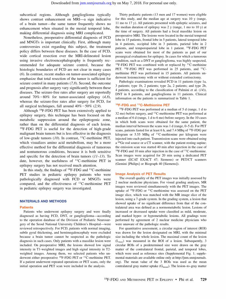
FIGURE 1. PET images of 4-y-old girl with 1-y history ofepilepsy (patient 11) showed masslike lesion (arrows) withhigh signal intensity in right parietal lobe on fluid-attenuatedinversion recovery MR image (A). No contrast enhancementwas observed on gadolinium-enhanced T1-weighted MRimage (B). Lesion was markedly hypometabolic on 18F-FDGPET (C) and normometabolic on 11C-methinine PET (D). Aftersurgery, lesion was identified as FCD, of type 2b.
730 THE JOURNAL OF NUCLEAR MEDICINE • Vol. 51 • No. 5 • May 2010
by on May 7, 2018. For personal use only. jnm.snmjournals.org Downloaded from
gangliogliomas. Therefore, discrimination of FCD fromMNGTs was not possible by visual grading of 18F-FDGPET data.
In contrast, FCD and MNGTs exhibited different resultson 11C-methinine PET. None of the FCD patients showedhypermetabolism in their lesions, and none of the DNT andganglioglioma patients showed hypometabolism in theirlesions (Table 3). The grades of 11C-methionine uptakewere significantly different among the 3 groups (P 5
0.016). Post hoc analysis revealed that the visual gradesof FCD were significantly different from those of DNTsand gangliogliomas.
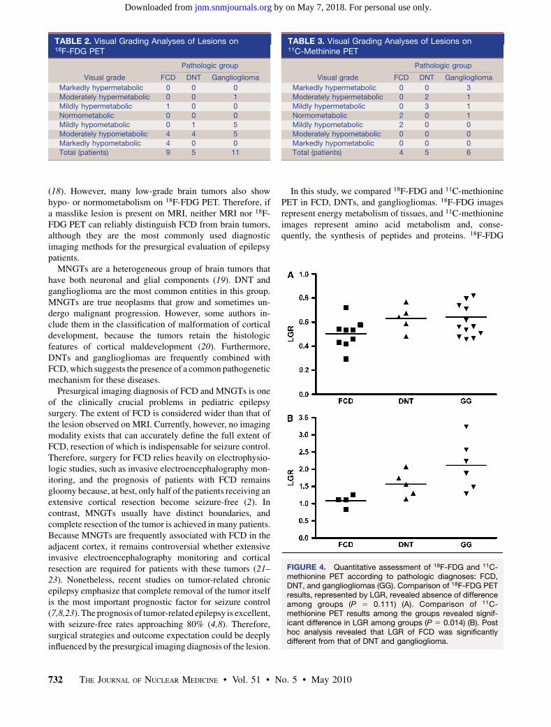
The quantitative assessment of 18F-FDG and 11C-methi-onine PET, represented by LGR, revealed that the meanLGR of 18F-FDG uptake was 0.502 6 0.119 for FCD,0.631 6 0.107 for DNT, and 0.620 6 0.196 for ganglio-glioma; there was no significant difference in LGR amongthe groups (P 5 0.111) (Fig. 4). However, the mean LGRof 11C-methionine uptake was 1.078 6 0.182 for FCD,1.564 6 0.368 for DNT, and 2.114 6 0.723 for ganglio-gliomas; there was a significant difference in LGR amongthe groups (P 5 0.014). Post hoc analysis revealed that theLGR of the FCD patients was significantly different fromthat of the DNT and ganglioglioma patients. Although nosignificant difference between the LGR of DNT andganglioglioma was observed, the mean LGR value ofDNT lay between that of FCD and gangliogliomas.
DISCUSSION
Preoperative imaging diagnosis of pathologic character-istics is important in epilepsy surgery. Most surgicaldecisions including the selection of surgical candidates,determination of when and how to operate, and outcomeexpectation depend heavily on the pathologic diagnosis.Although MRI has revolutionized the field of preoperativediagnosis, some lesions resist differentiation from otherentities, even using high-resolution MRI. FCD and MNGTsare typical examples that confuse clinicians.
Since Taylor’s classic description of the imaging andpathologic features of FCD (15), this disease has been wellcharacterized as a distinct clinicopathologic entity. Theimaging features of FCD are quite diverse, ranging from theabsence of abnormality or subtle gyral thickening toa tumorlike lesion (5). Although FCD usually lacks contrastenhancement on MRI, there are some exceptions (16,17).Many DNTs show minimal or no contrast enhancement onMRI. Gangliogliomas usually show contrast enhancement,but gangliogliomas located in the mesial temporal lobe tendto show no contrast enhancement. Therefore, it is oftendifficult to make a preoperative diagnosis for a masslikelesion in epilepsy patients (16).
18F-FDG PET is an effective diagnostic imaging methodfor epilepsy and brain tumors. In addition, FCD exhibitsmetabolic suppression during the interictal period and focalor lobar hypometabolism is demonstrated on 18F-FDG PET
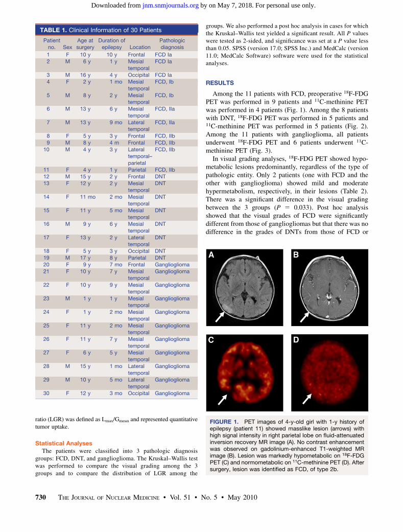
FIGURE 2. PET images of 13-y-old girl with 2-y history ofepilepsy (patient 17) showed masslike lesion (arrows) withhigh signal intensity in right fusiform gyrus on T2-weightedMR image (A). Lesion had no contrast enhancement ongadolinium-enhanced T1-weighted MR image (B). Lesionwas moderately hypometabolic on 18F-FDG PET (C) andmildly hypermetabolic on 11C-methinine PET (D). Aftersurgery, lesion was identified as DNT.
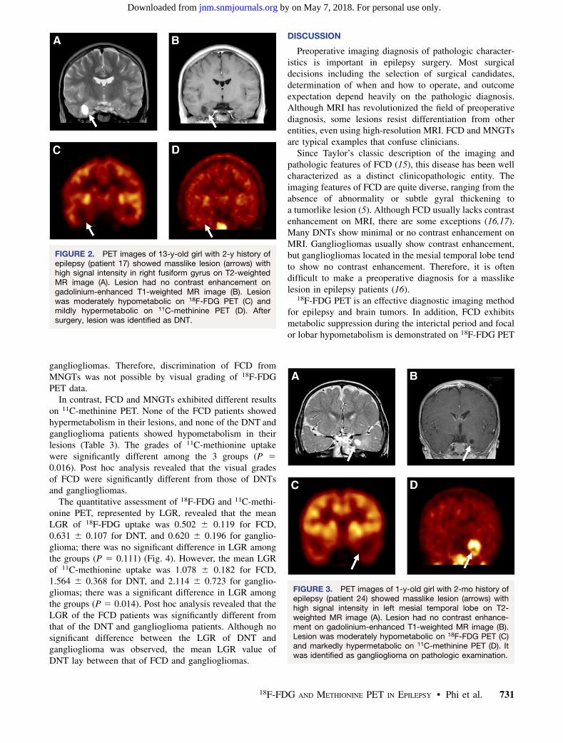
FIGURE 3. PET images of 1-y-old girl with 2-mo history ofepilepsy (patient 24) showed masslike lesion (arrows) withhigh signal intensity in left mesial temporal lobe on T2-weighted MR image (A). Lesion had no contrast enhance-ment on gadolinium-enhanced T1-weighted MR image (B).Lesion was moderately hypometabolic on 18F-FDG PET (C)and markedly hypermetabolic on 11C-methinine PET (D). Itwas identified as ganglioglioma on pathologic examination.
18F-FDG AND METHIONINE PET IN EPILEPSY • Phi et al. 731
by on May 7, 2018. For personal use only. jnm.snmjournals.org Downloaded from
(18). However, many low-grade brain tumors also showhypo- or normometabolism on 18F-FDG PET. Therefore, ifa masslike lesion is present on MRI, neither MRI nor 18F-FDG PET can reliably distinguish FCD from brain tumors,although they are the most commonly used diagnosticimaging methods for the presurgical evaluation of epilepsypatients.
MNGTs are a heterogeneous group of brain tumors thathave both neuronal and glial components (19). DNT andganglioglioma are the most common entities in this group.MNGTs are true neoplasms that grow and sometimes un-dergo malignant progression. However, some authors in-clude them in the classification of malformation of corticaldevelopment, because the tumors retain the histologicfeatures of cortical maldevelopment (20). Furthermore,DNTs and gangliogliomas are frequently combined withFCD, which suggests the presence of a common pathogeneticmechanism for these diseases.
Presurgical imaging diagnosis of FCD and MNGTs is oneof the clinically crucial problems in pediatric epilepsysurgery. The extent of FCD is considered wider than that ofthe lesion observed on MRI. Currently, however, no imagingmodality exists that can accurately define the full extent ofFCD, resection of which is indispensable for seizure control.Therefore, surgery for FCD relies heavily on electrophysio-logic studies, such as invasive electroencephalography mon-itoring, and the prognosis of patients with FCD remainsgloomy because, at best, only half of the patients receiving anextensive cortical resection become seizure-free (2). Incontrast, MNGTs usually have distinct boundaries, andcomplete resection of the tumor is achieved in many patients.Because MNGTs are frequently associated with FCD in theadjacent cortex, it remains controversial whether extensiveinvasive electroencephalography monitoring and corticalresection are required for patients with these tumors (21–23). Nonetheless, recent studies on tumor-related chronicepilepsy emphasize that complete removal of the tumor itselfis the most important prognostic factor for seizure control(7,8,23). The prognosis of tumor-related epilepsy is excellent,with seizure-free rates approaching 80% (4,8). Therefore,surgical strategies and outcome expectation could be deeplyinfluenced by the presurgical imaging diagnosis of the lesion.
In this study, we compared 18F-FDG and 11C-methioninePET in FCD, DNTs, and gangliogliomas. 18F-FDG imagesrepresent energy metabolism of tissues, and 11C-methionineimages represent amino acid metabolism and, conse-quently, the synthesis of peptides and proteins. 18F-FDG
TABLE 2. Visual Grading Analyses of Lesions on18F-FDG PET
Pathologic group
Visual grade FCD DNT Ganglioglioma
Markedly hypermetabolic 0 0 0
Moderately hypermetabolic 0 0 1
Mildly hypermetabolic 1 0 0
Normometabolic 0 0 0Mildly hypometabolic 0 1 5
Moderately hypometabolic 4 4 5
Markedly hypometabolic 4 0 0Total (patients) 9 5 11
TABLE 3. Visual Grading Analyses of Lesions on11C-Methinine PET
Pathologic group
Visual grade FCD DNT Ganglioglioma
Markedly hypermetabolic 0 0 3
Moderately hypermetabolic 0 2 1
Mildly hypermetabolic 0 3 1
Normometabolic 2 0 1Mildly hypometabolic 2 0 0
Moderately hypometabolic 0 0 0
Markedly hypometabolic 0 0 0Total (patients) 4 5 6
FIGURE 4. Quantitative assessment of 18F-FDG and 11C-methionine PET according to pathologic diagnoses: FCD,DNT, and gangliogliomas (GG). Comparison of 18F-FDG PETresults, represented by LGR, revealed absence of differenceamong groups (P 5 0.111) (A). Comparison of 11C-methionine PET results among the groups revealed signif-icant difference in LGR among groups (P 5 0.014) (B). Posthoc analysis revealed that LGR of FCD was significantlydifferent from that of DNT and ganglioglioma.
732 THE JOURNAL OF NUCLEAR MEDICINE • Vol. 51 • No. 5 • May 2010
by on May 7, 2018. For personal use only. jnm.snmjournals.org Downloaded from
has a limited sensitivity in the evaluation of brain tumorsbecause many low-grade tumors have low-energy metabo-lism and hardly any uptake of 18F-FDG. In contrast, 11C-methionine PET is more sensitive for brain tumors than 18F-FDG because of low background activity in the normalbrain tissue. Furthermore, 11C-methionine is expected to beeffective also in the discrimination of low-grade braintumors from nontumorous lesions such as FCD, becausepeptide synthesis can be a specific biomarker for tumors,even if the energy metabolism is low. In this study, 11C-methionine PET revealed a significantly higher uptake inDNTs and gangliogliomas than in FCD, whereas 18F-FDGPET did not reveal any differences among the groups in thequantitative analysis, as expected. These results are con-sistent with previous studies showing that 11C-methioninePET has a higher sensitivity and specificity for the di-agnosis of brain tumors than does 18F-FDG PET (11–13).This result suggests that 11C-methionine PET is effective inthe differential diagnosis of pediatric lesional epilepsy.
This study showed that the average LGR of DNTs on11C-methionine PET fell between those of FCD andgangliogliomas. This finding is reminiscent of the patho-logic conjecture that DNT lies between FCD and bona fidebrain tumors. DNT is an extremely benign brain tumor withexcellent long-term oncologic outcome, whereas ganglio-gliomas are associated with a more aggressive astroglialcomponent and their oncologic prognosis is worse than thatof DNT. Also noteworthy is that the individual LGR valuesof DNTs and gangliogliomas were distributed over a ratherwide range. This may reflect the pathologic and clinicalheterogeneity of DNTs and gangliogliomas. The widehistologic spectrum of DNTs remains poorly understood,and gangliogliomas exhibit diverse features, from a well-differentiated tumor to an anaplastic variant (19). Althoughit has long been held that DNTs do not recur, even afterincomplete resection (24), recurrence and progression ofsome DNTs have been reported (8,25). The clinicalbehavior of gangliogliomas is also unpredictable, withmalignant progression occurring in some patients (26,27).A study including additional patients may reveal whetherthe distinction of DNTs from gangliogliomas and theclinical stratification of these tumors using 11C-methioninePET are possible.
CONCLUSION
Preoperative imaging diagnosis of FCD and MNGTs isimportant for the determination of treatment strategies andexpectation of outcome in pediatric epilepsy. Whereas 18F-FDG PET did not contribute to the differentiation of FCDand MNGTs, 11C-methinine PET identified a significantdifference among these diseases. Specifically, DNTs andgangliogliomas showed a higher 11C-methinine uptake thanFCD. Moreover, the average LGR of DNTs on 11C-methionine PET fell between that of FCD and ganglio-glioma. These findings suggest that 11C-methinine PET
results correlated well with the pathologic spectrum ofpediatric lesional epilepsy.
ACKNOWLEDGMENTS
This work was supported in part by the Korea Scienceand Engineering Foundation (KOSEF) grant funded by theKorean government (MEST) (to Seung-Ki Kim, R01-2008-000-20268-0) and by a National Research Foundation ofKorea grant funded by the Korean government (to Kyu-Chang Wang, 2009-007-6743).
REFERENCES
1. Sisodiya SM. Malformations of cortical development: burdens and insights from
important causes of human epilepsy. Lancet Neurol. 2004;3:29–38.
2. Hader WJ, Mackay M, Otsubo H, et al. Cortical dysplastic lesions in children
with intractable epilepsy: role of complete resection. J Neurosurg. 2004;100:
110–117.
3. Colombo N, Tassi L, Galli C, et al. Focal cortical dysplasias: MR imaging,
histopathologic, and clinical correlations in surgically treated patients with
epilepsy. AJNR. 2003;24:724–733.
4. Luyken C, Blumcke I, Fimmers R, et al. The spectrum of long-term epilepsy-
associated tumors: long-term seizure and tumor outcome and neurosurgical
aspects. Epilepsia. 2003;44:822–830.
5. Raybaud C, Shroff M, Rutka JT, Chuang SH. Imaging surgical epilepsy in
children. Childs Nerv Syst. 2006;22:786–809.
6. Krsek P, Maton B, Jayakar P, et al. Incomplete resection of focal cortical
dysplasia is the main predictor of poor postsurgical outcome. Neurology. 2009;
72:217–223.
7. Zaatreh MM, Firlik KS, Spencer DD, Spencer SS. Temporal lobe tumoral
epilepsy: characteristics and predictors of surgical outcome. Neurology. 2003;61:
636–641.
8. Phi JH, Kim SK, Cho BK, et al. Long-term surgical outcomes of temporal
lobe epilepsy associated with low-grade brain tumors. Cancer. 2009;115:
5771–5779.
9. Kim DW, Lee SK, Chu K, et al. Predictors of surgical outcome and pathologic
considerations in focal cortical dysplasia. Neurology. 2009;72:211–216.
10. Padma MV, Said S, Jacobs M, et al. Prediction of pathology and survival by FDG
PET in gliomas. J Neurooncol. 2003;64:227–237.
11. Chung JK, Kim YK, Kim SK, et al. Usefulness of 11C-methionine PET in the
evaluation of brain lesions that are hypo- or isometabolic on 18F-FDG PET. Eur
J Nucl Med Mol Imaging. 2002;29:176–182.
12. Pirotte B, Goldman S, Massager N, et al. Comparison of 18F-FDG and 11C-
methionine for PET-guided stereotactic brain biopsy of gliomas. J Nucl Med.
2004;45:1293–1298.
13. Wong TZ, van der Westhuizen GJ, Coleman RE. Positron emission tomography
imaging of brain tumors. Neuroimaging Clin N Am. 2002;12:615–626.
14. Palmini A, Najm I, Avanzini G, et al. Terminology and classification of the
cortical dysplasias. Neurology. 2004;62:S2–S8.
15. Taylor DC, Falconer MA, Bruton CJ, Corsellis JA. Focal dysplasia of the
cerebral cortex in epilepsy. J Neurol Neurosurg Psychiatry. 1971;34:369–387.
16. Bronen RA, Vives KP, Kim JH, Fulbright RK, Spencer SS, Spencer DD. Focal
cortical dysplasia of Taylor, balloon cell subtype: MR differentiation from low-
grade tumors. AJNR. 1997;18:1141–1151.
17. Urbach H, Scheffler B, Heinrichsmeier T, et al. Focal cortical dysplasia of
Taylor’s balloon cell type: a clinicopathological entity with characteristic
neuroimaging and histopathological features, and favorable postsurgical out-
come. Epilepsia. 2002;43:33–40.
18. Goffin K, Dedeurwaerdere S, Van Laere K, Van Paesschen W. Neuronuclear
assessment of patients with epilepsy. Semin Nucl Med. 2008;38:227–239.
19. Louis DN, Ohgaki H, Wiestler OD, Cavenee WK, eds. WHO Classification of
Tumours of the Central Nervous System. Lyon, France: International Agency for
Research on Cancer; 2007.
20. Barkovich AJ, Kuzniecky RI, Jackson GD, Guerrini R, Dobyns WB. A
developmental and genetic classification for malformations of cortical de-
velopment. Neurology. 2005;65:1873–1887.
21. Cataltepe O, Turanli G, Yalnizoglu D, Topcu M, Akalan N. Surgical management
of temporal lobe tumor-related epilepsy in children. J Neurosurg. 2005;102:
280–287.
18F-FDG AND METHIONINE PET IN EPILEPSY • Phi et al. 733
by on May 7, 2018. For personal use only. jnm.snmjournals.org Downloaded from
22. Lombardi D, Marsh R, de Tribolet N. Low grade glioma in intractable epilepsy:
lesionectomy versus epilepsy surgery. Acta Neurochir Suppl. 1997;68:70–74.
23. Giulioni M, Galassi E, Zucchelli M, Volpi L. Seizure outcome of lesionectomy in
glioneuronal tumors associated with epilepsy in children. J Neurosurg. 2005;102:
288–293.
24. Daumas-Duport C, Scheithauer BW, Chodkiewicz JP, Laws ER Jr, Vedrenne C.
Dysembryoplastic neuroepithelial tumor: a surgically curable tumor of young
patients with intractable partial seizures: report of thirty-nine cases. Neurosur-
gery. 1988;23:545–556.
25. Rushing EJ, Thompson LD, Mena H. Malignant transformation of a dysembryo-
plastic neuroepithelial tumor after radiation and chemotherapy. Ann Diagn Pathol.
2003;7:240–244.
26. Majores M, von Lehe M, Fassunke J, Schramm J, Becker AJ, Simon M. Tumor
recurrence and malignant progression of gangliogliomas. Cancer. 2008;113:
3355–3363.
27. El Khashab M, Gargan L, Margraf L, et al. Predictors of tumor progression
among children with gangliogliomas: clinical article. J Neurosurg Pediatr. 2009;
3:461–466.
734 THE JOURNAL OF NUCLEAR MEDICINE • Vol. 51 • No. 5 • May 2010
by on May 7, 2018. For personal use only. jnm.snmjournals.org Downloaded from
Doi: 10.2967/jnumed.109.070920Published online: April 15, 2010.
2010;51:728-734.J Nucl Med. Joongyub Lee, Dong Soo Lee and Seung-Ki KimJi Hoon Phi, Jin Chul Paeng, Hyo Sang Lee, Kyu-Chang Wang, Byung-Kyu Cho, Ji-Yeoun Lee, Sung-Hye Park,
C-Methionine PET11F-FDG and 18Pediatric Epilepsy Patients Using Evaluation of Focal Cortical Dysplasia and Mixed Neuronal and Glial Tumors in
http://jnm.snmjournals.org/content/51/5/728This article and updated information are available at:
http://jnm.snmjournals.org/site/subscriptions/online.xhtml
Information about subscriptions to JNM can be found at:
http://jnm.snmjournals.org/site/misc/permission.xhtmlInformation about reproducing figures, tables, or other portions of this article can be found online at:
(Print ISSN: 0161-5505, Online ISSN: 2159-662X)1850 Samuel Morse Drive, Reston, VA 20190.SNMMI | Society of Nuclear Medicine and Molecular Imaging
is published monthly.The Journal of Nuclear Medicine
© Copyright 2010 SNMMI; all rights reserved.
by on May 7, 2018. For personal use only. jnm.snmjournals.org Downloaded from