Embed Size (px)
Citation preview
J Int Adv Otol 2016; 12(2): 161-5 • DOI: 10.5152/iao.2016.2439
Original Article
INTRODUCTIONAs the most common pathology among congenital neural pathologies, hearing loss may cause various problems in every stage of life [1]. Hearing loss affects 1–3 of every 1,000 infants and 11% of school-aged children [2]. Regarding unilateral sensorineural hearing loss (USNHL), the condition affects 3% of school-aged children [3]. As neonatal screening has become widespread, more realistic results pertaining to the incidence of hearing loss have been obtained. In a study conducted in Finland, the prevalence of USNHL was found to be 1.7 per 1,000 infants [4]. The incidence of bilateral hearing loss in infants was found to be 3.32% and that of unilateral hearing loss was found to be 1.40% in a study conducted at Eskişehir Turkey [5].
Bilateral hearing loss shows its signs in the early stages because of the impairment of language development; however, because the child continues to communicate through the intact ear, the diagnosis of USNHL may be delayed until the primary school period [6]. Children with USNHL do not have the advantage of binaural hearing and thus have a difficulty with auditory perception. Because the vestibular system may also be affected in cases with USNHL, imbalance and vertigo may also be present [1]. All of these negative effects may manifest in low academic performance, difficulties in social life, and lack of self-confidence [3, 7-11].
Although many examinations are conducted in cases of SNHL to determine the etiology, it is not always possible to obtain a definite result. Because inner ear anomalies and mass lesions are more common risk factors for USNHL than bilateral hearing loss, computed tomography (CT) and magnetic resonance imaging (MRI) investigations are necessary [12].
In unilateral hearing loss, the vestibular system is generally affected in addition to the cochlea. Clinical findings may vary, but the vestibular impairment may be asymptomatic because of central compensation. When the vertigo becomes symptomatic, the phys-ical and emotional health of the child is adversely affected [13].
Corresponding Address: Leman Birdane E-mail: [email protected]
Submitted: 28.03.2016 Accepted: 01.04.2016©Copyright 2016 by The European Academy of Otology and Neurotology and The Politzer Society - Available online at www.advancedotology.org
Evaluation of the Vestibular System and Etiology in Children with Unilateral Sensorineural Hearing Loss
OBJECTIVE: The aim of this study was to evaluate the vestibular system of children with unilateral sensorineural hearing loss (USNHL), investigate the etiological factors of USNHL and analyze whether a genetic predisposition exists.
MATERIALS and METHODS: Thirty-three children aged less than 18 years with USNHL, who visited the ear, nose, and throat (ENT) department between January 2004 and December 2012, were included in this study. Cases with conductive hearing loss were excluded from the study. The patients were subjected to etiologic, genetic, and ophthalmologic evaluation; radiologic imaging; electronystagmography (ENG); and vestibular evoked myogenic potential (VEMP) tests. The control group, which included 25 healthy children (13 males and 12 females), had undergone audi-ological assessment and were subjected to ENG and VEMP tests.
RESULTS: All of the patients had severe-to-profound hearing loss. Mumps immunoglobulin G was positive in 22 (66.7%) of 33 patients. The 35delG mutation was not found in any of the patients. All of the patients underwent temporal computed tomography (CT) and magnetic resonance im-aging (MRI). Inner ear anomaly was present in 51.5% of the patients. Overall, 21 of 31 ENG patients had canal paresis in the affected ear. The VEMP response was absent on the affected side in three patients. The n23 latency average of the patient group was longer than that of the control group.
CONCLUSION: Because USNHL causes irreversible problems in children, early diagnosis and auditory rehabilitation are very important. As USNHL is accompanied by inner ear anomaly, children with USNHL should undergo temporal bone CT and MRI. To evaluate the vestibular system, ENG and VEMP are non-invasive and diagnostic tests.
KEYWORDS: Children with unilateral hearing loss, electronystagmography, vestibular evoked myogenic potentials
Leman Birdane, Armağan İncesulu, Erkan Özüdoğru, Cemal Cingi, Hamdi Caklı, Melek Kezban Gürbüz, Baki AdapınarDepartment of Otorhinolaryngology, Yunus Emre State Hospital, Eskişehir, Turkey (LB)Department of Otorhinolaryngology, Eskişehir Osmangazi University School of Medicine, Eskişehir, Turkey (Aİ, EÖ, CC, HC, MKG)Department of Radiology, Eskişehir Osmangazi University School of Medicine, Eskişehir, Turkey (BA)
161
Our objective in this study was to evaluate the vestibular system in child-hood USNHL and to determine the incidence of abnormalities on imag-ing studies, risk factors, and whether there is a genetic predisposition.
MATERIALS and METHODSThis study was conducted with 33 patients aged less than 18 years (14 males and 19 females; average age: 12.2±3.68 years) who visited our clinic with a complaint of unilateral hearing loss. The control group comprised 25 volunteer children (13 males and 12 females; average age: 12.4±4.21 years) with normal hearing. Patients with conductive hearing loss and chronic otitis media were excluded from the study.
A detailed history was obtained from the patients and their families, including with respect to consanguineous marriage, hearing loss, and family history of the syndrome. Antenatal or perinatal infec-tions, premature birth, ototoxic drug use, noise exposure, and trau-ma were assessed. After otological examination, all of the patients underwent vestibular evaluation tests. In addition to undergoing the Romberg test, Unterberger stepping test, head shake test, and head impulse test, they were examined by a pediatric neurologist and ophthalmologist. The patient and control groups were also sub-jected to pure tone threshold and suprathreshold audiometry using the AC-40 audiometry device (Interacoustics; Assens, Denmark). The middle ear pressure and stapedius reflex were measured using the AZ-7 device (Interacoustics). The complete blood count, antinuclear antibody (ANA) level, antinuclear cytoplasmic antibody level, human leukocyte antigen (HLA) type, sedimentation rate, thyroid hormone level, and urea and creatinine levels with respect to renal function were evaluated in all of the patients. Mumps immunoglobulin (Ig) M and IgG levels were measured in all of the patients, and they all un-derwent electrocardiography (ECG). The ECG results were evaluated by a cardiologist; particularly, QT intervals were measured. All of the patients underwent high-resolution CT (HRCT) and MRI for radiolog-ical monitoring, and the images were evaluated by a neuroradiolo-gist at our university hospital. The inner ear anomaly, diameter and size of the semicircular canals, and width of the vestibular aqueduct and internal auditory canal were particularly evaluated by CT scans. The mass lesions and presence of the eighth nerve were evaluated by MRI. The patient and control groups were both subjected to the electronystagmography (ENG) test using the Chart Medical ENG de-vice (ICS Co. Ltd.; Iwate, Japan). The ENG test battery was composed of a saccadic test, a gaze (fixation) test, an optokinetic test, a tracking (smooth pursuit) test, a caloric test, and positional tests. The water used in the bithermal caloric test was maintained at 30°C and 44°C. When the difference between the sums of the nystagmus durations of the horizontal canals generated with hot and cold water was more than 20%, it was accepted as canal paresis.
The vestibular evoked myogenic potential (VEMP) records were tak-en with the Medelec Synergy version 10 (VIASYS Health Care UK; Sur-rey, UK) device that is used in our ENT department. First, the skin of the patient was cleaned with alcohol to prevent artefacts. The active electrode was replaced at the middle-third of the sternocleidomas-toid muscle (SCM), the reference electrode was replaced at the ster-nal notch, and the earth electrode was replaced at the middle of the forehead. It was demanded from the patient that he/she turn his/her head to the opposite side to that of the stimulus (during the “click” stimulus). In the meantime, the muscle action potential from the
SCM was recorded. The test was repeated twice for both sides. The first positive wave was detected as p13, and the first negative wave was detected as n23. The latencies of p13 and n23 and the amplitude values between the two waves (p13–n23) were measured. The nor-mative data from the control group were compared with the latency and amplitude values of the patient group. The waves, in which p13 and n23 latencies were longer than the average latency of the con-trol group and the peak-to-peak amplitude value was lower than that of the control group, were determined as impaired.
Genetic examination of the patients was performed at Ankara Uni-versity School of Medicine, Department of Medical Genetics. The connexin 26 gene of the samples was examined for the presence of the 35delG mutation. DNA isolation was performed using the clas-sic phenol/chloroform method. Blood samples (9 mL) were collected from the patients into polyethylene tubes (Sigma-Aldrich; St. Louis, MO, USA) containing 1 mL of 0.5 M ethylenediaminetetraacetic acid (EDTA). After the blood samples had been obtained, the PCR prod-ucts were evaluated in 2% agarose gels, and enzyme cuts of the c.35delG mutation were evaluated in 3% agarose gels. The 35delG mutation was examined via the polymerase chain reaction-restric-tion fragment length polymorphism (PCR/RFLP) method.
Statistical evaluation was performed using the Statistical Package for the Social Sciences (SPSS) for Windows software (SPSS Inc.; version 15.0, Chicago, IL, USA). The data were compared by t-test and Fisher’s exact test. A p value less than 0.05 was deemed to indicate statistical significance.
The study was approved (date: 19.11.2009; number: 23) by the Med-ical Ethics Committee of our school. Informed consent was obtained from the parents of all of the patients and volunteers. RESULTSIn total, 33 patients (14 males and 19 females) and 25 healthy chil-dren (13 males and 12 females) were included in this study. The aver-age age of the patients was 12.2±3.68 years (range: 5–18 years) and that of the control group was 12.4±4.21 years (range: 4–18 years). There was no statistically significant difference in the average age between the groups. The main complaint of the patients was unilat-eral hearing loss.
The average age at which hearing loss was first diagnosed was 9.06 years. None of the patients visited the clinic with a complaint of sud-den hearing loss. There were 20 patients with right-sided hearing loss and 13 with left-sided hearing loss. All of the patients had stable, uni-lateral, severe-to-profound sensorineural hearing loss.
Nine of the patients had a history of mumps parotitis, of whom six had hearing loss in the first week after recovering from mumps. One of the patients had a diagnosis of bilateral horseshoe kidney. Two pa-tients had neuromotor retardation up to 2 years of age, and six had stayed in an incubator for a period of 1.5 months. One of the patients had a history of tricyclic antidepressant drug abuse at 3 years of age. Five of the patients had a history of febrile convulsion; however, their pediatric neurology consultation was normal, and there was no ab-normality on electroencephalography (EEG). No risk factors were identified in any of the histories of any of the other nine patients. The
162
J Int Adv Otol 2016; 12(2): 161-5
family history of hearing loss in all patients was negative (Table 1). The middle ear pressure of all of the patients was normal, and the acous-tic stapedius reflex was absent on the affected side. The bilateral hearing level and results of immittance testing were normal in the control group.
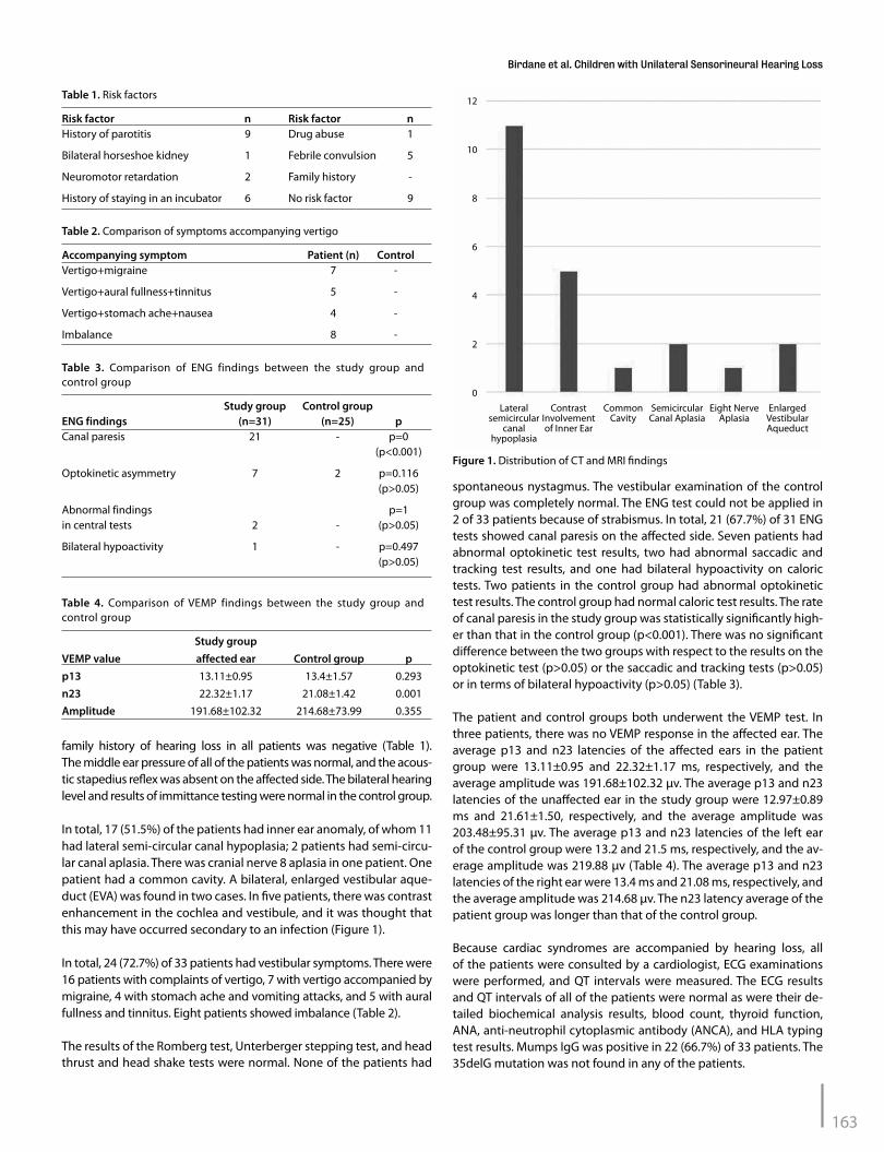
In total, 17 (51.5%) of the patients had inner ear anomaly, of whom 11 had lateral semi-circular canal hypoplasia; 2 patients had semi-circu-lar canal aplasia. There was cranial nerve 8 aplasia in one patient. One patient had a common cavity. A bilateral, enlarged vestibular aque-duct (EVA) was found in two cases. In five patients, there was contrast enhancement in the cochlea and vestibule, and it was thought that this may have occurred secondary to an infection (Figure 1).
In total, 24 (72.7%) of 33 patients had vestibular symptoms. There were 16 patients with complaints of vertigo, 7 with vertigo accompanied by migraine, 4 with stomach ache and vomiting attacks, and 5 with aural fullness and tinnitus. Eight patients showed imbalance (Table 2).
The results of the Romberg test, Unterberger stepping test, and head thrust and head shake tests were normal. None of the patients had
spontaneous nystagmus. The vestibular examination of the control group was completely normal. The ENG test could not be applied in 2 of 33 patients because of strabismus. In total, 21 (67.7%) of 31 ENG tests showed canal paresis on the affected side. Seven patients had abnormal optokinetic test results, two had abnormal saccadic and tracking test results, and one had bilateral hypoactivity on caloric tests. Two patients in the control group had abnormal optokinetic test results. The control group had normal caloric test results. The rate of canal paresis in the study group was statistically significantly high-er than that in the control group (p<0.001). There was no significant difference between the two groups with respect to the results on the optokinetic test (p>0.05) or the saccadic and tracking tests (p>0.05) or in terms of bilateral hypoactivity (p>0.05) (Table 3).
The patient and control groups both underwent the VEMP test. In three patients, there was no VEMP response in the affected ear. The average p13 and n23 latencies of the affected ears in the patient group were 13.11±0.95 and 22.32±1.17 ms, respectively, and the average amplitude was 191.68±102.32 μv. The average p13 and n23 latencies of the unaffected ear in the study group were 12.97±0.89 ms and 21.61±1.50, respectively, and the average amplitude was 203.48±95.31 μv. The average p13 and n23 latencies of the left ear of the control group were 13.2 and 21.5 ms, respectively, and the av-erage amplitude was 219.88 μv (Table 4). The average p13 and n23 latencies of the right ear were 13.4 ms and 21.08 ms, respectively, and the average amplitude was 214.68 μv. The n23 latency average of the patient group was longer than that of the control group.
Because cardiac syndromes are accompanied by hearing loss, all of the patients were consulted by a cardiologist, ECG examinations were performed, and QT intervals were measured. The ECG results and QT intervals of all of the patients were normal as were their de-tailed biochemical analysis results, blood count, thyroid function, ANA, anti-neutrophil cytoplasmic antibody (ANCA), and HLA typing test results. Mumps IgG was positive in 22 (66.7%) of 33 patients. The 35delG mutation was not found in any of the patients.
Study group Control groupENG findings (n=31) (n=25) pCanal paresis 21 - p=0 (p<0.001)
Optokinetic asymmetry 7 2 p=0.116 (p>0.05)
Abnormal findings p=1 in central tests 2 - (p>0.05)
Bilateral hypoactivity 1 - p=0.497 (p>0.05)
Table 3. Comparison of ENG findings between the study group and control group
Study groupVEMP value affected ear Control group pp13 13.11±0.95 13.4±1.57 0.293n23 22.32±1.17 21.08±1.42 0.001Amplitude 191.68±102.32 214.68±73.99 0.355
Table 4. Comparison of VEMP findings between the study group and control group
Figure 1. Distribution of CT and MRI findings
Lateral semicircular
canal hypoplasia
12
10
8
6
4
2
0Contrast
Involvement of Inner Ear
Common Cavity
Semicircular Canal Aplasia
Eight Nerve Aplasia
Enlarged Vestibular Aqueduct
Risk factor n Risk factor nHistory of parotitis 9 Drug abuse 1
Bilateral horseshoe kidney 1 Febrile convulsion 5
Neuromotor retardation 2 Family history -
History of staying in an incubator 6 No risk factor 9
Table 1. Risk factors
Accompanying symptom Patient (n) ControlVertigo+migraine 7 -
Vertigo+aural fullness+tinnitus 5 -
Vertigo+stomach ache+nausea 4 -
Imbalance 8 -
Table 2. Comparison of symptoms accompanying vertigo
163
Birdane et al. Children with Unilateral Sensorineural Hearing Loss
DISCUSSIONBecause unilateral hearing in early childhood cannot be verbally expressed by children, such that their families are unaware of any impairment, early treatment is not possible. Patients typically real-ise they have hearing loss in primary school with the help of their teacher, or while talking on the phone or listening to music using earphones. Besides having low academic performance in school, af-fected children may also show difficulties in understanding conver-sations in a noisy environment [7-9].
Unilateral sensorineural hearing loss is often dismissed as an envi-ronmental entity, and the etiological basis has not been explored deeply. As the etiology of sensorineural hearing loss varies widely, we performed most of the recommended laboratory tests. To define autoimmune-mediated inner ear disease, HLA typing, ANA levels, and anti DNA levels were investigated [14]. However, we obtained no positive result. The most common cause of viral neuritis and co-chleitis in childhood is mumps. Hearing loss is seen in approximately 0.005%–0.3% of all post-mumps cases and is generally unilateral and profound [15-17]. Mumps vaccination has been made mandatory since 2007 in Turkey; however, it remains a common cause of hearing loss. In our study, 22 of 33 patients had a positive mumps IgG value, and none of these patients had been vaccinated against mumps.
The most common genetic cause of congenital hearing loss in Tur-key is the mutation of 35delG. In one of the most recent studies, it was reported that there may be a mutation in the GJB3, TECTA, and COCH genes in USNHL [18]. None of the patients had a mutation in the 35delG gene.
Many studies have shown that USNHL might be accompanied by in-ner ear anomaly. Compared with bilateral cases, inner ear anomaly and mass lesions are more frequently seen in unilateral hearing loss. In the study by Laury et al. [12] 8 of 11 patients had cochlear nerve aplasia, and 2 patients had a mass lesion. In another study, the rate of inner ear anomaly was found to be 66.7% [19]. In our study, 17 (51.5%) of 33 pa-tients had inner ear anomaly in the vestibular or cochlear region. Elev-en patients had lateral semicircular canal hypoplasia, two had bilateral EVAs, two had bilateral semicircular aplasia, one had no cranial nerve 8, and one had vestibulocochlear anomaly (Figure 1). It should be noted that both the child and parents were informed that it was necessary to avoid contact sports and be careful to avoid head trauma to preserve the unaffected ear. The gold standard methods for detectinginner ear anomalies and mass lesions are CT and MRI [20-23]. It is necessary to per-form imaging procedures on children who have USNHL to relieve the anxiety of their families and to eliminate the possibility of unknown pathologies that could affect the healthy ear.
Most patients with inner ear anomalies also show vestibular symp-toms [24]. Grimmer et al. [25] reviewed 21 pediatric patients with EVAs and found that the incidence of vestibular symptoms was 48.0%; 6 patients (28.6%) had episodic vertigo, whereas 1 patient showed im-balance. In our study, 24 (72.7%) of 33 patients had vestibular symp-toms, of whom 16 had episodic vertigo attacks. Vertigo attacks in sev-en of these patients were accompanied by migraine. Four children had vertigo accompanied by stomach ache, nausea, and vomiting. Vertigo attacks in five patients were accompanied by aural fullness, tinnitus, and nausea. Eight patients had complaints of dizziness. After
detailed vestibular testing, all of the patients underwent ENG, which can record and evaluate the nystagmic response and allow central and peripheral vertigo to be distinguished. The pediatric patients could tolerate ENG easily [26]. Canal paresis was found in 21 patients after ENG. One patient had a bilateral hypoactive caloric test. The ab-normal ENG results and vestibular complaint rates were similar. In our study, cooperation during ENG, even with a 4-year-old patient, was acceptable, and there were no problems after the test.
Another test that has been frequently used in recent years for the differential diagnosis of patients with vertigo is VEMP. This test can be performed in two different ways: ocular or cervical. Our patients were subjected to the cervical VEMP test, which provides information about the sacculus and inferior vestibular nerve using the sacculo-colic reflex arc. The VEMP test is easy to implement and tolerable by children [27, 28]. Our objective in using the VEMP test was to determine its effectiveness in measuring the vestibular function in children and to ascertain whether the factor that causes hearing loss also dam-ages inner ear dynamics. Regarding the pathologies relevant to the vestibular system in children, besides medical history, physical ex-amination, audiography, and hematological evaluation, the ENG and VEMP tests also provide important information to clinicians for the differential diagnosis [29]. In the study by Yulian et al. [30] on athletes with congenital hearing loss, 75% of the patients had normal VEMP waves. In contrast, Kegel et al. [31] found a high incidence of VEMP response absence among hearing-impaired children. In our study, the VEMP response was absent unilaterally in three patients (9.09%). VEMP responses were in the normal range in the control group. The n23 latency of the affected ear in the study group was longer than that in the control group, and the difference was statistically sig-nificant. It was assumed that the vestibular anomaly or etiological factors of hearing loss caused the absence of the VEMP wave. These findings are valuable for the future evaluation of young children with vestibular symptoms.
Children with vertigo should undergo ophthalmological examina-tions. In the study by Haffey et al. [6] one-third of patients with uni-lateral hearing loss had a refractive defect. All of the patients in the study group underwent an ophthalmological examination in our study and one patient had uveitis. Four patients had a refraction de-fect, but this impairment was not related to vertigo (as assessed in the Department of Ophthalmology).
Although it is commonly accepted that unilateral hearing loss causes only auditory perception problems, Bess et al. [3] suggested that chil-dren with USNHL greater than 45 dB since early childhood may show retarded language development and behavioral disorders. They also remarked that 35% of the children in their study had failed at least once during their school careers, 13% needed private tutoring at their home, and 20% had behavioral disorders [3]. It is well known that such children have problems and difficulties in their academic life. Language development depends on healthy binaural hearing. Even children with asymmetric or mild hearing loss have difficulties in learning at school; therefore, their cognitive abilities are poor. Chil-dren with USNHL also have problems in sound localization, and the IQs of children with unilateral hearing loss since early childhood have been found to be lower than those of children with binaural hearing; therefore, the former group needs additional academic support [7, 9].
164
J Int Adv Otol 2016; 12(2): 161-5
The school performance of 14 of the patients in our study was poor; six of these children had failed at least once during their school ca-reers. Twelve patients remarked that their school performance was moderate, and seven children indicated they were successful at school. It is of utmost importance that these children should receive auditory rehabilitation to ensure psychosocial development and ac-ademic success. For these children, hearing aids such as contralateral routing of signals (CROS) or bone-anchored hearing aid (BAHA) can be used, or cochlear implantation can be performed.
Because sensorineural hearing loss is an irremediable pathology, early diagnosis is vital. Screening of newborns for hearing ability allows di-agnosis in the early period of life. Radiological imaging, ENG, and VEMP should be a part of the routine evaluation of USNHL. Auditory and ves-tibular rehabilitation is the main objective of treatment to improve the social functioning and communication of affected children.
Ethics Committee Approval: Ethics committee approval was received for this study from the ethics committee of Eskişehir Osmangazi University (No: 23/19.11.2009).
Informed Consent: Written informed consent was obtained from patients’ parents who participated in this study.
Peer-review: Externally peer-reviewed.
Author Contributions: Concept - A.İ., L.B.; Design - L.B., C.C., B.A.; Supervision - A.İ.; Resources - L.B.; Materials - L.B., B.A., H.Ç.; Data Collection and/or Pro-cessing - L.B., M.K.G., E.Ö., H.Ç.; Analysis and/or Interpretation - A.İ.; Literature Search - L.B., A.İ.; Writing Manuscript - L.B.; Critical Review - A.İ.
Conflict of Interest: No conflict of interest was declared by the authors.
Financial Disclosure: The authors declared that this study has received no financial support.
REFERENCES1. Propst E, Greinwald J, Schmithorst V. Neuroanatomic differences in chi-
dren with unilateral sensorineural hearing loss detected using functional magnetic resonance imaging. Arch Otolaryngol Head Neck Surg 2010; 136: 22-6. [CrossRef]
2. Erenberg A, Lemons J, Sia C, Trunkel D, Ziring P. Newborn and İnfant hearing loss: detection and intervention, American Academy of Pediat-rics. Task Force on Newborn and İnfant Hearing, 1998-1999. Pediatrics 1999; 103: 527-30.
3. Bess FH, Dodd-Murphy J, Parker RA. Children with minimal sensorineural hearing loss: prevalence, educational performance, and functional sta-tus. Ear Hear 1998; 19: 339-54. [CrossRef]
4. Vartiainen E, Karjalainen S. Prevalence and etiology of unilateral sensori-neural hearing impairment in a Finnish childhood population. Int J Pedi-atr Otorhinolaryngol 1998; 43: 253-9. [CrossRef]
5. Serin G, Gurbuz MK, Kecik C, Incesulu A, Tekin N. Newborn auditory screening programme of newborns with risk and well babies in Turkey. Int Adv Otol 2011; 7: 351-6.
6. Haffey T, Fowler N, Anne S. Evaluation of unilateral sensorineural hearing loss in the pediatric patient. Int J Pediatr Otorhinolaryngol 2013; 77: 955-8. [CrossRef]
7. Ead B, Hale S, Dealwis D, Lieu JE. Pilot study of cognition in children with unilateral hearing loss. Int J Pediatr Otorhinolaryngol 2013; 77: 1856-60. [CrossRef]
8. Martinez-Cruz C.F, Poblano A, Conde-Reyes M. Cognitive performance of school children with unilateral sensorineural hearing loss. Arch Med Res 2009; 40: 374-9. [CrossRef]
9. Niedzielski A, Humeniuk E, Blaziak P, Gwisda G. İntellectual efficiency of children with unilateral hearing loss. Int J Pediatr Otorhinolaryngol 2006; 70: 1529-32. [CrossRef]
10. Lieu J. Speech-language ad educational consequences of unilateral hearing loss in children. Arch Otolaryngol Head Neck Surg 2004; 130: 524-30. [CrossRef]
11. Brookhouser PE, Worthington DW, Kelly WJ. Unilateral Hearing Loss in Children. Laryngoscope 1991; 101: 1264-72. [CrossRef]
12. Laury AM, Casey S, McKay S, Germiller J. Etiology of unilateral neural hearing loss in children. Int J Pediatr Otorhinolaryngol 2009; 73: 417-7. [CrossRef]
13. Casani AP, Navari E, Sellari Franceschini S, Cerchiai N. Vertigo in child-hood: proposal for a diagnostic algorithm based upon clinical experi-ence Acta Otolaryngol 2015; 35: 180-5.
14. Simons JP, Mandell DL, Arjmand EM. Computed tomography and magnet-ic resonance imaging in pediatric unilateral and asymmetric sensorineural hearing loss. Arch Otolaryngol Head Neck Surg 2006; 132: 186-92. [CrossRef]
15. Tsubota M, Shojaku H, Ishimaru H, Fujisaka M, Watanabe Y. Mumps virus may damage the vestibular nerve as well as the inner ear. Acta Otolaryn-gol 2008; 128: 644-7. [CrossRef]
16. Kaaijk P, Gouma S, Hulscher HI, Han WG, Kleijne DE, van Binnendijk RS, et al. Dynamics of the serologic response in vaccinated and unvaccinated mumps cases during an epidemic. Hum Vaccin Immunother 2015; 11: 1754-61. [CrossRef]
17. El-Badry MM, Abousetta A, Kader RMA. Vestibular dysfunction in patients with post-mumps sensorineural hearing loss. J Laryngol Otol 2015; 129: 337-41. [CrossRef]
18. Dodson KM, Georgolios A, Barr N, Nguyen B, Sismanis A, Arnos KS, et al. Etiology of unilateral hearing loss in a national hereditary deafness repository. Am J Otolaryngol 2012; 33: 590-4. [CrossRef]
19. Masuda S, Usui S, Matsunaga T. High prevalance of inner ear and/or inter-nal auditory canal malformations in children with unilateral sensorineural hearing loss. Int J Pediatr Otorhinolaryngol 2013; 77: 228-32. [CrossRef]
20. Cama E, Inches I, Muzzi E, Sadushi O, Santarelli R, De Colle W, et al. Tem-poral Bone High resolution computed tomography in non-syndromic unilateral hearing loss in Children. ORL J Otorhinolaryngol Relat Spec 2012; 74: 70-7. [CrossRef]
21. Coticchia JM, Gokhale A, Waltonen J, Sumer B. Characteristics of sensori-neural hearing loss in children with inner ear anomalies. Am J Otolaryn-gol 2006; 27: 33-8. [CrossRef]
22. Song JJ, Choi HG, Seung HO, Chang SO, Kim CS, Lee JH. Unilateral Sen-sorineural Hearing Loss in Children: The İmportance of temporal bone computed tomography and audiometric follow-up. Otol Neurotol 2009; 30: 604-8. [CrossRef]
23. Bamiou DE, Savy L, Mahoney C, Phelps P, Sirimanna T. Unilateral senso-rineural hearing loss and its aetiology in chidhood: the contribution of computerised tomography in aetiological diagnosis and management. Int J Pediatr Otorhinolaryngol 1999; 51: 91-9. [CrossRef]
24. Zalewski CK, Chien WW, King KA, Muskett JA, Baron RE, Butman JA, et al. Vestibular Dysfunction in Patients with enlarged vestibular aqueduct. Otolaryngol Head Neck Surg 2015; 153: 257-62. [CrossRef]
25. Grimmer J, Hedlund G. Vestibular symptoms in children with enlarged vestibular aqueduct anomaly. Int J Pediatr Otorhinolaryngol 2007; 71: 275-82. [CrossRef]
26. Salami A, Dellepiane M, Mora R, Taborelli G, Jankowska B. Electronystag-mography finding in children with peripheral and central vestibular dis-orders. Int J Pediatr Otorhinolaryngol 2006; 70: 13-8. [CrossRef]
27. Colebatch JG, Halmagyi GM, Skuse NF. Myogenic potentials generated by a click-evoked vestibulocollic reflex. J Neurol Neurosurg Psychiatry 1994; 57: 190-7. [CrossRef]
28. Kelsch TA, Schaefer LA, Esquivel CR. Vestibular evoked myogenic poten-tials in young children: test parameters and normative data. Laryngo-scope 2006; 116: 895-900. [CrossRef]
29. Phillips JO, Backous DD. Evaluation of vestibular function in young chil-dren. Otolaryngol Clin North Am 2002; 35: 765-90. [CrossRef]
30. Yulian J, Munetaka U, Hayası A, Takegoshı H. Vestibular myogenic poten-tials of athletes for the Deaf Olympic Games with congenital profound hearing loss. Acta Oto-Laryngologica 2010: 1-7.
31. Kegel A, Maes L, Baetens T, Dhooge I, Van Waelvelde H. The influence of a Vestibular Dysfunction on the motor development of Hearing-Impaired Children. Laryngoscope 2012; 122: 2837-43. [CrossRef]
165
Birdane et al. Children with Unilateral Sensorineural Hearing Loss


![Electrical Vestibular Stimulation after Vestibular ......electrical stimulation of the vestibular system to one ear [4,5,9]. However studies have also reported vestibular responses](https://img.pdfslide.net/doc/110x75/60f6b0762ca1b41e91018b73/electrical-vestibular-stimulation-after-vestibular-electrical-stimulation.jpg)