Embed Size (px)
Citation preview
Abstract
Recent trends in multidetector computed
tomography (MDCT), such as increasing scan
speed, coverage, and resolution, have enabled
a wide range of clinical advancements in one of
the most useful diagnostic devices of modern
medicine. However, for some routine and
advanced applications, optimization of image
quality and other factors — such as radiation
dose and workflow — can vary considerably in
radiological practice, and could be simplified for
application to broader patient populations.
To address this, many years of Philips research
and development for the Brilliance CT platform
have culminated in a set of scalable innovations
for present and future scanners, referred to
collectively as Essence technology. These
innovations include multiple enhancements of the
X-ray tube, detector, and image reconstruction
system, that were designed to deliver a new level
of MDCT image quality optimization. In addition,
Essence technology is seamlessly integrated
with dose-management techniques and a rich
suite of advanced image analysis and enterprise
applications to streamline workflow.
Phantom and other scan-related measurements
demonstrate the application-specific and
intrinsically-available performance gains of Essence
technology that ease the optimization of image
quality along with other imaging factors. Clinical
results illustrate how this platform synergistically
provides significant imaging benefits for challenging
applications. The scalable Essence technology
platform has extra capacity that holds promise to
enable MDCT results not previously possible and
to facilitate improved clinical decisions and patient
care.
Evolve to EssenceEssence technology, a new scalable platform for optimizing image quality
2
Beginning with the introduction of the Twin detector
CT scanner in 1992 (Philips), trends in MDCT have
continually increased the number and diversity of
emerging examinations while improving imaging
performance for routine applications (Table 1).
These trends include adopting diagnostic and therapeutic
procedures in earlier stages of the care cycle over a
wider range of disease conditions and across broader
patient populations. In addition, MDCT has been
employed for some examinations, such as angiography,
that were formerly performed with other invasive
imaging modalities.
With this expansion, unique challenges and
opportunities for routine image quality optimization
remain for many applications, body regions, and patient
populations (Table 2). Accordingly, optimization of
patient-specific imaging factors without trading off the
as low as reasonably achievable (ALARA) principle and
other factors can vary in radiological practice.
Scalable enhancements to the main components of
the MDCT imaging chain, referred to as “Essence
technology,” were designed to meet these challenges
and to further the expansion of MDCT (Figure 1).
The synergistic set of scalable Essence technology
components provide application-specific and intrinsically
available performance enhancements that optimize
image quality across broader populations, as well
as streamline workflow and simplify X-ray dose
management. Ultimately — and most importantly
— these performance enhancements have extra
capacity to facilitate improved clinical results for both
today’s challenging routine applications and for future
applications.
Table 1: Increasing MDCT Trends
coverage per second
longitudinal resolution
temporal resolution
rotation speed
tube power
number of channels
reconstruction performance
Table 2
Patient Population or Body Region
Challenge
Cardiac To image small and rapidly moving coronary arteries with a low X-ray dose.
Abdominal To image subtle changes in low contrast lesions, such as the monitoring of response to therapy.
Pulmonary To image dyspneic patients with small lesions and rapidly distributing contrast agents at high resolution with a low X-ray dose.
Pediatric To image restless patients with a low X-ray dose.
Bariatric To maintain image quality for thoracic and abdominal scans.
Introduction
3
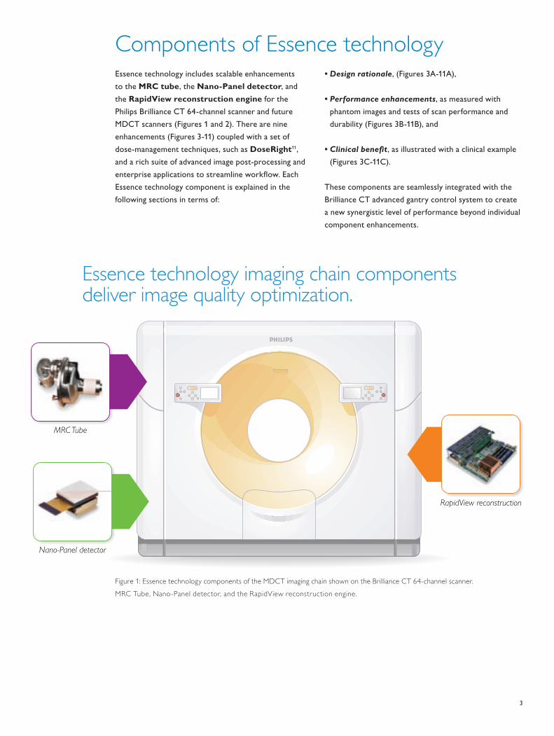
Essence technology includes scalable enhancements
to the MRC tube, the Nano-Panel detector, and
the RapidView reconstruction engine for the
Philips Brilliance CT 64-channel scanner and future
MDCT scanners (Figures 1 and 2). There are nine
enhancements (Figures 3-11) coupled with a set of
dose-management techniques, such as DoseRight11,
and a rich suite of advanced image post-processing and
enterprise applications to streamline workflow. Each
Essence technology component is explained in the
following sections in terms of:
•Designrationale, (Figures 3A-11A),
•Performanceenhancements, as measured with
phantom images and tests of scan performance and
durability (Figures 3B-11B), and
•Clinicalbenefit, as illustrated with a clinical example
(Figures 3C-11C).
These components are seamlessly integrated with the
Brilliance CT advanced gantry control system to create
a new synergistic level of performance beyond individual
component enhancements.
Nano-Panel detector
MRC Tube
Essence technology imaging chain components deliver image quality optimization.
Figure 1
RapidView reconstruction
Components of Essence technology
Figure 1: Essence technology components of the MDCT imaging chain shown on the Brilliance CT 64-channel scanner.
MRC Tube, Nano-Panel detector, and the RapidView reconstruction engine.
4
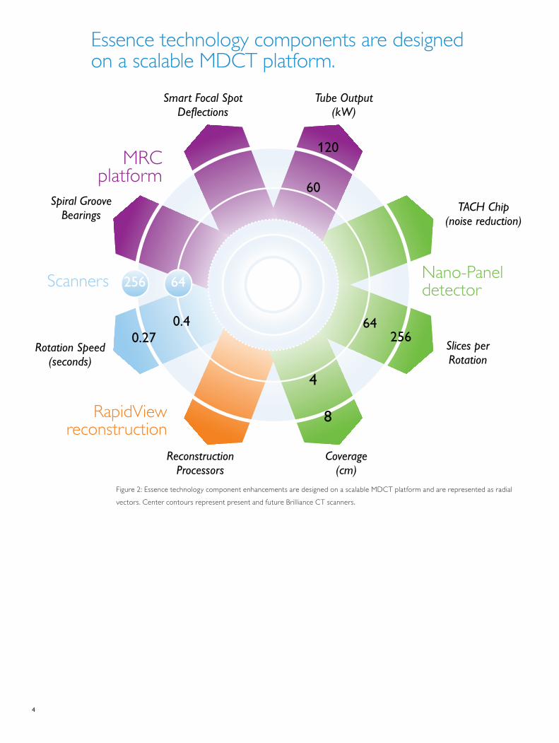
Essence technology components are designed on a scalable MDCT platform.
Spiral Groove Bearings
Smart Focal Spot Deflections
Tube Output(kW)
TACH Chip(noise reduction)
Rotation Speed (seconds)
Slices perRotation
Coverage(cm)
Reconstruction Processors
0.2764
256
4
8
Figure 2
0.4
120
60
256 64Scanners
MRC platform
Nano-Panel detector
RapidViewreconstruction
Figure 2: Essence technology component enhancements are designed on a scalable MDCT platform and are represented as radial
vectors. Center contours represent present and future Brilliance CT scanners.
5
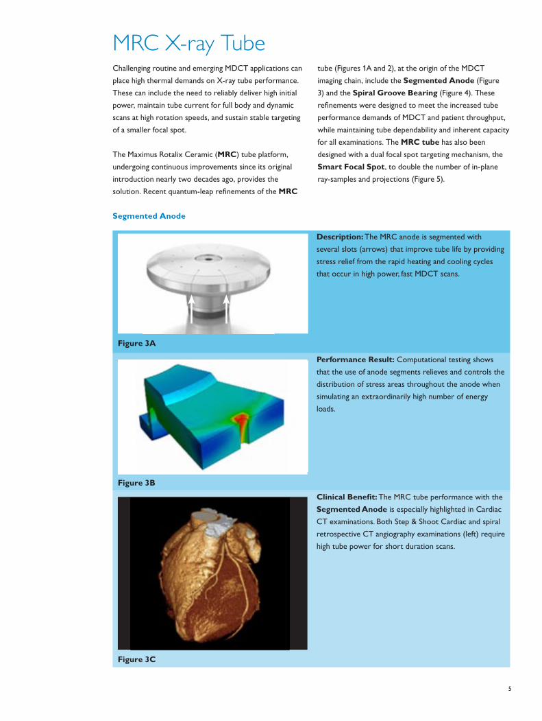
Challenging routine and emerging MDCT applications can
place high thermal demands on X-ray tube performance.
These can include the need to reliably deliver high initial
power, maintain tube current for full body and dynamic
scans at high rotation speeds, and sustain stable targeting
of a smaller focal spot.
The Maximus Rotalix Ceramic (MRC) tube platform,
undergoing continuous improvements since its original
introduction nearly two decades ago, provides the
solution. Recent quantum-leap refinements of the MRC
tube (Figures 1A and 2), at the origin of the MDCT
imaging chain, include the Segmented Anode (Figure
3) and the Spiral Groove Bearing (Figure 4). These
refinements were designed to meet the increased tube
performance demands of MDCT and patient throughput,
while maintaining tube dependability and inherent capacity
for all examinations. The MRC tube has also been
designed with a dual focal spot targeting mechanism, the
Smart Focal Spot, to double the number of in-plane
ray-samples and projections (Figure 5).
MRC X-ray Tube
Segmented Anode
Figure 3A
Description: The MRC anode is segmented with
several slots (arrows) that improve tube life by providing
stress relief from the rapid heating and cooling cycles
that occur in high power, fast MDCT scans.
Figure 3B
Performance Result: Computational testing shows
that the use of anode segments relieves and controls the
distribution of stress areas throughout the anode when
simulating an extraordinarily high number of energy
loads.
Figure 3C
Clinical Benefit: The MRC tube performance with the
Segmented Anode is especially highlighted in Cardiac
CT examinations. Both Step & Shoot Cardiac and spiral
retrospective CT angiography examinations (left) require
high tube power for short duration scans.
6
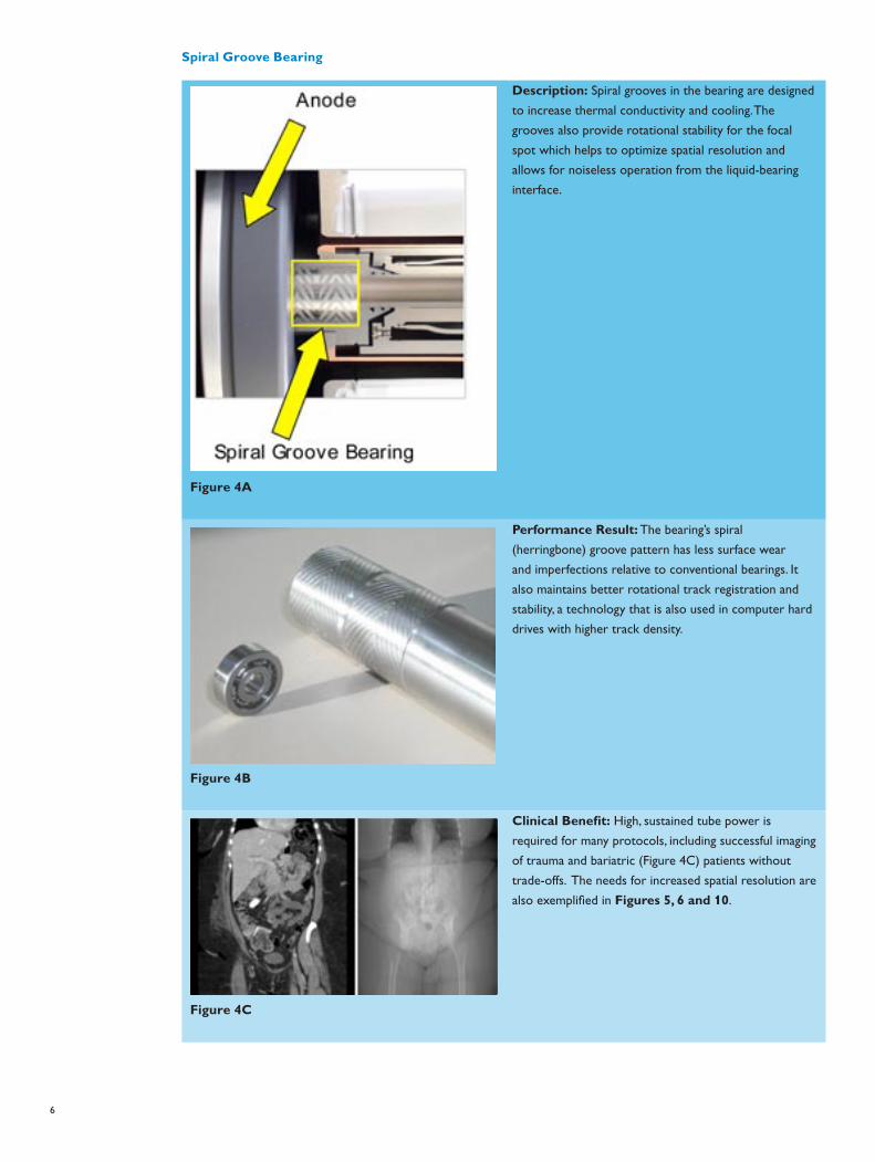
Spiral Groove Bearing
Figure 4A
Description: Spiral grooves in the bearing are designed
to increase thermal conductivity and cooling. The
grooves also provide rotational stability for the focal
spot which helps to optimize spatial resolution and
allows for noiseless operation from the liquid-bearing
interface.
Figure 4B
Performance Result: The bearing’s spiral
(herringbone) groove pattern has less surface wear
and imperfections relative to conventional bearings. It
also maintains better rotational track registration and
stability, a technology that is also used in computer hard
drives with higher track density.
Figure 4C
Clinical Benefit: High, sustained tube power is
required for many protocols, including successful imaging
of trauma and bariatric (Figure 4C) patients without
trade-offs. The needs for increased spatial resolution are
also exemplified in Figures 5, 6 and 10.
7
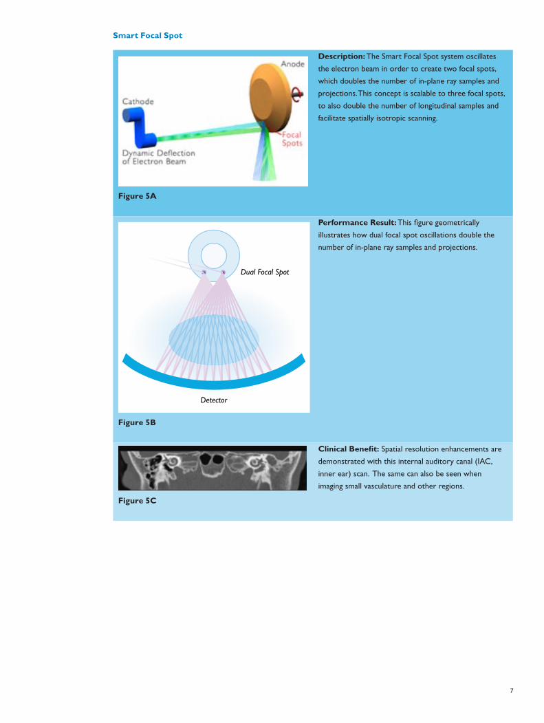
Smart Focal Spot
Figure 5A
Description: The Smart Focal Spot system oscillates
the electron beam in order to create two focal spots,
which doubles the number of in-plane ray samples and
projections. This concept is scalable to three focal spots,
to also double the number of longitudinal samples and
facilitate spatially isotropic scanning.
Figure 5b
Dual Focal Spot
Detector
Figure 5B
Performance Result: This figure geometrically
illustrates how dual focal spot oscillations double the
number of in-plane ray samples and projections.
Figure 5C
Clinical Benefit: Spatial resolution enhancements are
demonstrated with this internal auditory canal (IAC,
inner ear) scan. The same can also be seen when
imaging small vasculature and other regions.
8
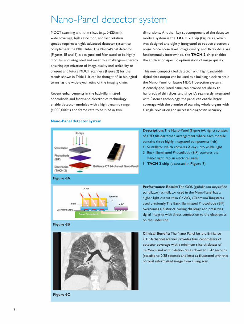
MDCT scanning with thin slices (e.g., 0.625mm),
wide coverage, high resolution, and fast rotation
speeds requires a highly advanced detector system to
complement the MRC tube. The Nano-Panel detector
(Figures 1B and 6) is designed and fabricated to be highly
modular and integrated and meet this challenge— thereby
ensuring optimization of image quality and scalability to
present and future MDCT scanners (Figure 2) for the
trends shown in Table 1. It can be thought of, in biological
terms, as the wide-eyed retina of the imaging chain.
Recent enhancements in the back-illuminated
photodiode and front-end electronics technology
enable detector modules with a high dynamic range
(1,000,000:1) and frame rate to be tiled in two
dimensions. Another key subcomponent of the detector
module system is the TACH 2 chip (Figure 7), which
was designed and tightly-integrated to reduce electronic
noise. Since noise level, image quality, and X-ray dose are
fundamentally intertwined, the TACH 2 chip enables
the application-specific optimization of image quality.
This new compact tiled detector with high bandwidth
digital data output can be used as a building block to scale
the Nano-Panel for future MDCT detection systems.
A densely-populated panel can provide scalability to
hundreds of thin slices, and since it’s seamlessly integrated
with Essence technology, the panel can enable larger
coverage with the promise of scanning whole organs with
a single revolution and increased diagnostic accuracy.
Nano-Panel detector system
Nano-Panel detector system
Figure 6A
Description: The Nano-Panel (Figure 6A, right) consists
of a 2D tile-patterned arrangement where each module
contains three highly integrated components (left):
1. Scintillator which converts X-rays into visible light
2. Back-Illuminated Photodiode (BIP) converts the
visible light into an electrical signal
3. TACH 2 chip (discussed in Figure 7).
Conductive Epoxy
X-rays
Printed Circuit Board
ASIC
Scintillator
Light
Figure 6b
Figure 6B
Performance Result: The GOS (gadolinium oxysulfide
scintillator) scintillator used in the Nano-Panel has a
higher light output than CdWO4 (Cadmium Tungstate)
used previously. The Back Illuminated Photodiode (BIP)
overcomes a historical wiring challenge and preserves
signal integrity with direct connection to the electronics
on the underside.
Figure 6C
Clinical Benefit: The Nano-Panel for the Brilliance
CT 64-channel scanner provides four centimeters of
detector coverage with a minimum slice thickness of
0.625mm and with rotation times down to 0.42 seconds
(scalable to 0.28 seconds and less) as illustrated with this
coronal reformatted image from a lung scan.
9
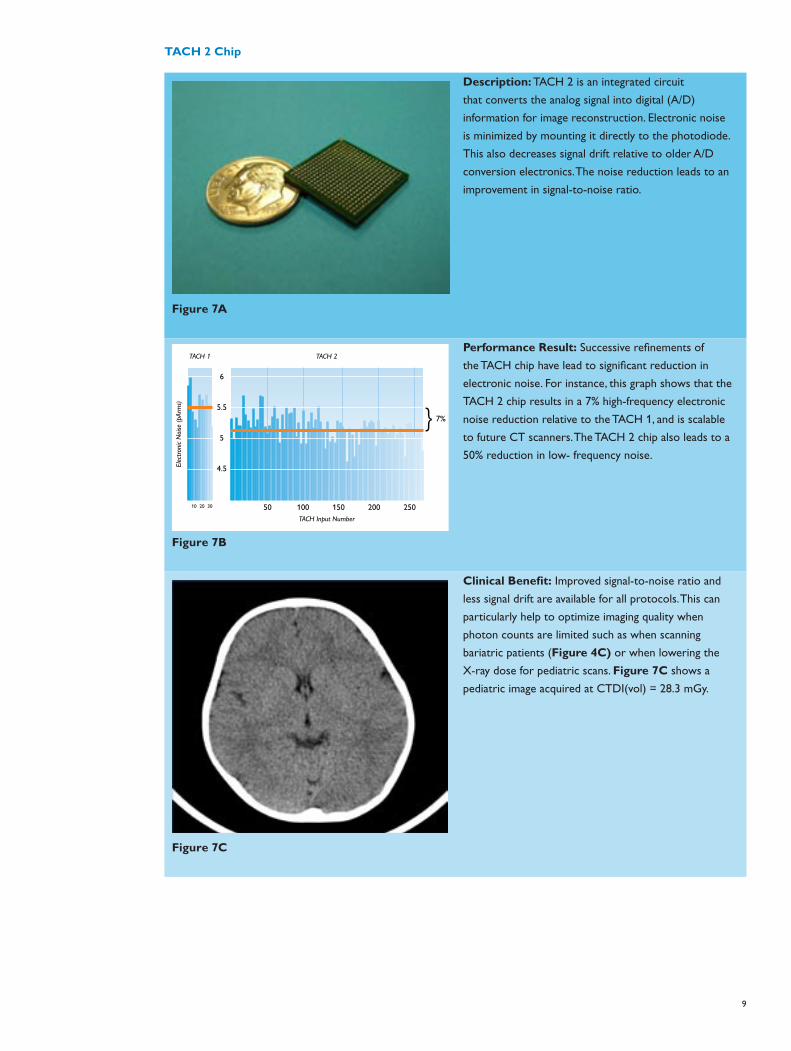
TACH 2 Chip
Figure 7A
Description: TACH 2 is an integrated circuit
that converts the analog signal into digital (A/D)
information for image reconstruction. Electronic noise
is minimized by mounting it directly to the photodiode.
This also decreases signal drift relative to older A/D
conversion electronics. The noise reduction leads to an
improvement in signal-to-noise ratio.
Figure 7b
TACH Input Number
50 100 150 200 25010 20 30
6
5.5
5
4.5Elec
tron
ic N
oise
(pA
rms)
TACH 2TACH 1
7%
Figure 7B
Performance Result: Successive refinements of
the TACH chip have lead to significant reduction in
electronic noise. For instance, this graph shows that the
TACH 2 chip results in a 7% high-frequency electronic
noise reduction relative to the TACH 1, and is scalable
to future CT scanners. The TACH 2 chip also leads to a
50% reduction in low- frequency noise.
Figure 7C
Clinical Benefit: Improved signal-to-noise ratio and
less signal drift are available for all protocols. This can
particularly help to optimize imaging quality when
photon counts are limited such as when scanning
bariatric patients (Figure 4C) or when lowering the
X-ray dose for pediatric scans. Figure 7C shows a
pediatric image acquired at CTDI(vol) = 28.3 mGy.
10
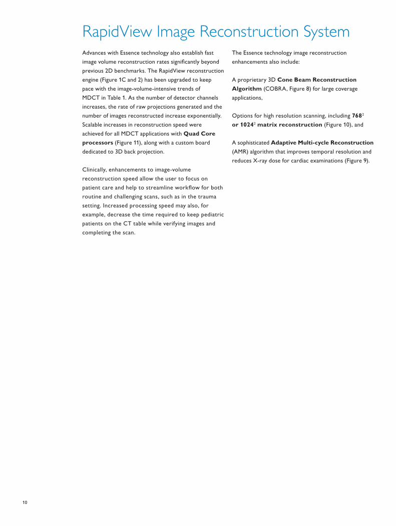
Advances with Essence technology also establish fast
image volume reconstruction rates significantly beyond
previous 2D benchmarks. The RapidView reconstruction
engine (Figure 1C and 2) has been upgraded to keep
pace with the image-volume-intensive trends of
MDCT in Table 1. As the number of detector channels
increases, the rate of raw projections generated and the
number of images reconstructed increase exponentially.
Scalable increases in reconstruction speed were
achieved for all MDCT applications with Quad Core
processors (Figure 11), along with a custom board
dedicated to 3D back projection.
Clinically, enhancements to image-volume
reconstruction speed allow the user to focus on
patient care and help to streamline workflow for both
routine and challenging scans, such as in the trauma
setting. Increased processing speed may also, for
example, decrease the time required to keep pediatric
patients on the CT table while verifying images and
completing the scan.
The Essence technology image reconstruction
enhancements also include:
A proprietary 3D Cone Beam Reconstruction
Algorithm (COBRA, Figure 8) for large coverage
applications,
Options for high resolution scanning, including 7682
or 10242 matrix reconstruction (Figure 10), and
A sophisticated Adaptive Multi-cycle Reconstruction
(AMR) algorithm that improves temporal resolution and
reduces X-ray dose for cardiac examinations (Figure 9).
RapidView Image Reconstruction System
11
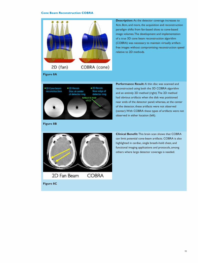
Cone Beam Reconstruction COBRA
Figure 8A
Description: As the detector coverage increases to
4cm, 8cm, and more, the acquisition and reconstruction
paradigm shifts from fan-based slices to cone-based
image volumes. The development and implementation
of a true 3D cone beam reconstruction algorithm
(COBRA) was necessary to maintain virtually artifact-
free images without compromising reconstruction speed
relative to 2D methods.
Figure 8B
Performance Result: A thin disc was scanned and
reconstructed using both the 3D COBRA algorithm
and an entirely 2D method (right). The 2D method
had obvious artifacts when the disk was positioned
near ends of the detector panel; whereas, at the center
of the detector, these artifacts were not observed
(center). With COBRA these types of artifacts were not
observed in either location (left).
Figure 8C
Clinical Benefit: This brain scan shows that COBRA
can limit potential cone-beam artifacts. COBRA is also
highlighted in cardiac, single breath-hold chest, and
functional imaging applications and protocols, among
others where large detector coverage is needed.

12
Adaptive Multi-cycle Reconstruction
Figure 9A
Description: One way to improve temporal resolution
is by combining projection data from consecutive
cardiac cycles. Dedicated AMR cardiac techniques1
significantly improve temporal resolution by combining
data from as many cycles as are available, thus optimizing
coronary image quality at high heart rates. Shown here
is an example of AMR combining data from 90-degree
segments obtained from two consecutive cardiac cycles.
Combined with Philips patented Beat-to-Beat delay
algorithm2,3 which tracks a desired physiological cardiac
phase of interest, consistent high-quality coronary
imaging can be obtained.
Figure 9B
Performance Result: AMR optimizes the temporal
resolution by adaptively combining projection data from
consecutive cardiac cycles. Figure 9B on the left is a plot
displaying optimization of the temporal resolution (in
blue) for varying heart rate (yellow) during a cardiac
acquisition.
Figure 9C
Clinical Benefit: Retrospective ECG-gated cardiac CT
with AMR maintains image quality over a wide range of
heart rates. Shown on the left (Figure 9C) are volume
rendered images obtained from scans of patients with
low, medium, and high variable heart rates.
13
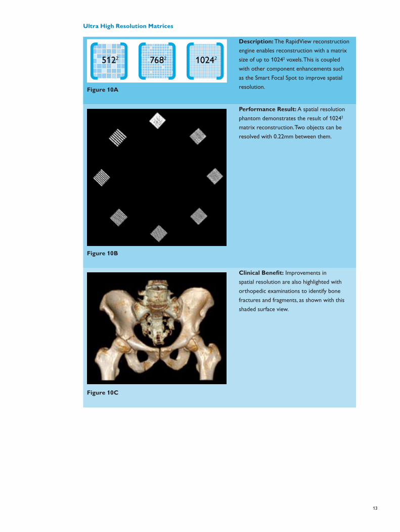
Ultra High Resolution Matrices
5122 7682 10242
Figure 10a
Figure 10A
Description: The RapidView reconstruction
engine enables reconstruction with a matrix
size of up to 10242 voxels. This is coupled
with other component enhancements such
as the Smart Focal Spot to improve spatial
resolution.
Figure 10B
Performance Result: A spatial resolution
phantom demonstrates the result of 10242
matrix reconstruction. Two objects can be
resolved with 0.22mm between them.
Figure 10C
Clinical Benefit: Improvements in
spatial resolution are also highlighted with
orthopedic examinations to identify bone
fractures and fragments, as shown with this
shaded surface view.
14
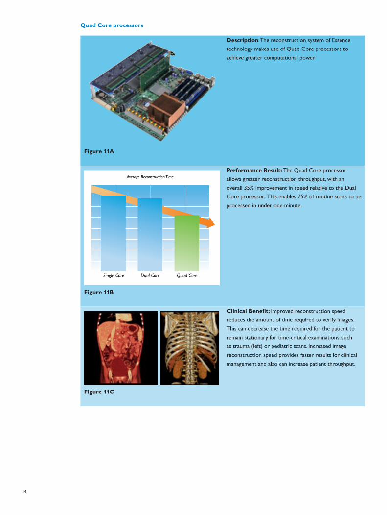
Quad Core processors
Figure 11A
Description: The reconstruction system of Essence
technology makes use of Quad Core processors to
achieve greater computational power.
Figure 11b
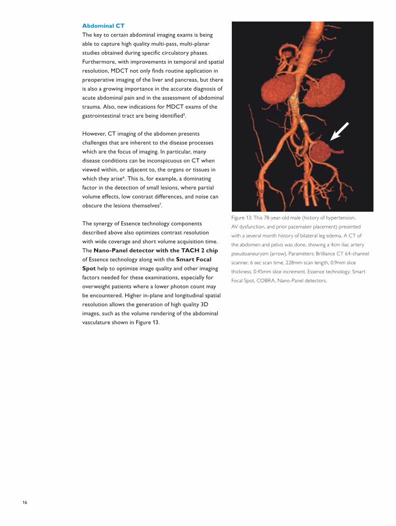
Average Reconstruction Time
Single Core Dual Core Quad Core
Figure 11B
Performance Result: The Quad Core processor
allows greater reconstruction throughput, with an
overall 35% improvement in speed relative to the Dual
Core processor. This enables 75% of routine scans to be
processed in under one minute.
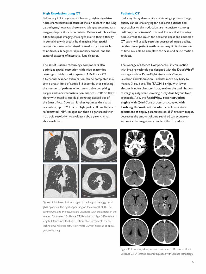
Figure 11C
Clinical Benefit: Improved reconstruction speed
reduces the amount of time required to verify images.
This can decrease the time required for the patient to
remain stationary for time-critical examinations, such
as trauma (left) or pediatric scans. Increased image
reconstruction speed provides faster results for clinical
management and also can increase patient throughput.

15
MDCT applications can have unique imaging
requirements depending on anatomical region and
reason for scanning, and therefore consistent image
quality optimization can be challenging, especially for
those procedures listed in Table 2. Essence technology
can meet these challenges with synergistic performance
enhancements of the entire imaging chain provided
by the aforementioned components. Four clinical
applications are presented below to illustrate how
this can lead to application-specific optimization of
image quality without trading-off other factors, as well
as highlight the benefits of Essence technology for all
routine applications, as summarized in Figure 16.
Cardiac CT
All aspects of Essence technology are leveraged to
enhance the challenging needs of cardiac CT imaging
as it becomes accessible to broader populations earlier
in the care cycle. Increases in detector coverage, tube
power, rotation speed, spatial and temporal resolution,
along with improvements in cone beam reconstruction
techniques, have significantly improved the image
quality and robustness of cardiac scanning. Artifacts
are prevented in the scanning and 3D reconstruction
process, instead of attempting to correct the
reconstructed images as with other approaches in the
industry.
As part of Essence technology and the Rate Responsive
Toolkit, dedicated AMR algorithms are employed
in retrospective ECG-gated spiral scans and offer
significant improvements in temporal resolution. These
advanced algorithms, together with scalable gantry
rotation speed (up to .27 sec and faster), and the
patented Beat-to-Beat variable delay algorithm that
tracks the quiet physiological cardiac phase, permit
consistent, high quality coronary imaging.
A new prospective, ECG-triggered acquisition method,
Step & Shoot Cardiac10, is also enabled by wide Nano-
Panel detector coverage and the fast acquisition time
of Essence technology. The wide coverage Nano-Panel
detector enables four-beats whole heart acquisition on
the Brilliance CT 64-channel scanner and is scalable to
less beats in the future. Step & Shoot Cardiac has also
enabled motion artifact suppression and an industry-
leading reduction in radiation dose of up to 80 percent
without compromising cardiac image quality.
Figure 12: Clinical History: This asymptomatic 59-year-old male
had a strong family history of coronary artery disease (CAD). A
cardiac CT examination was performed for further evaluation.
There is a lesion with non-calcified plaque (60% area stenosis)
present in the proximal left circumflex artery. Parameters:
Brilliance CT 64-channel scanner, 14 sec scan time, 140mm scan
length, 0.9mm slice thickness, 0.5mm slice increment. Essence
technology: AMR, Segmented Anode, Nano-Panel detectors.
Optimizing the Image Quality of Applications with Essence technology
16
Abdominal CT
The key to certain abdominal imaging exams is being
able to capture high quality multi-pass, multi-planar
studies obtained during specific circulatory phases.
Furthermore, with improvements in temporal and spatial
resolution, MDCT not only finds routine application in
preoperative imaging of the liver and pancreas, but there
is also a growing importance in the accurate diagnosis of
acute abdominal pain and in the assessment of abdominal
trauma. Also, new indications for MDCT exams of the
gastrointestinal tract are being identified5.
However, CT imaging of the abdomen presents
challenges that are inherent to the disease processes
which are the focus of imaging. In particular, many
disease conditions can be inconspicuous on CT when
viewed within, or adjacent to, the organs or tissues in
which they arise6. This is, for example, a dominating
factor in the detection of small lesions, where partial
volume effects, low contrast differences, and noise can
obscure the lesions themselves7.
The synergy of Essence technology components
described above also optimizes contrast resolution
with wide coverage and short volume acquisition time.
The Nano-Panel detector with the TACH 2 chip
of Essence technology along with the Smart Focal
Spot help to optimize image quality and other imaging
factors needed for these examinations, especially for
overweight patients where a lower photon count may
be encountered. Higher in-plane and longitudinal spatial
resolution allows the generation of high quality 3D
images, such as the volume rendering of the abdominal
vasculature shown in Figure 13.
Figure 13: This 78-year-old male (history of hypertension,
AV dysfunction, and prior pacemaker placement) presented
with a several month history of bilateral leg edema. A CT of
the abdomen and pelvis was done, showing a 4cm iliac artery
pseudoaneurysm (arrow). Parameters: Brilliance CT 64-channel
scanner, 6 sec scan time, 228mm scan length, 0.9mm slice
thickness, 0.45mm slice increment. Essence technology: Smart
Focal Spot, COBRA, Nano-Panel detectors.
17
High Resolution Lung CT
Pulmonary CT images have inherently higher signal-to-
noise characteristics because of the air present in the lung
parenchyma; however, there are challenges to pulmonary
imaging despite this characteristic. Patients with breathing
difficulties pose imaging challenges due to their difficulty
in complying with breath-hold imaging. High spatial
resolution is needed to visualize small structures such
as nodules, sub-segmental pulmonary emboli, and the
textural patterns of interstitial lung diseases.
The set of Essence technology components also
optimizes spatial resolution with wide anatomical
coverage at high rotation speeds. A Brilliance CT
64-channel scanner examination can be completed in a
single breath-hold of about 5-8 seconds, thus reducing
the number of patients who have trouble complying.
Larger and finer reconstruction matrices, 7682 or 10242,
along with stability and dual-targeting capabilities of
the Smart Focal Spot can further optimize the spatial
resolution, up to 24 Lp/cm. High quality, 3D multiplanar
reformatted (MPR) images can then be generated with
isotropic resolution to evaluate subtle parenchymal
abnormalities.
Figure 14: High resolution images of the lungs showing ground
glass opacity in the right upper lung on the coronal MPR. The
parenchyma and the fissures are visualized with great detail in the
images. Parameters: Brilliance CT, Resolution: High, 327mm scan
length, 0.8mm slice thickness, 0.4mm slice increment Essence
technology: 768 reconstruction matrix, Smart Focal Spot, spiral
groove bearing.
Pediatric CT
Reducing X-ray dose while maintaining optimum image
quality can be challenging for pediatric patients and
approaches to this reduction are inconsistent among
radiology departments4. It is well known that lowering
tube current too much for pediatric chest and abdomen
CT scans will usually result in decreased image quality.
Furthermore, patient restlessness may limit the amount
of time available to complete the scan and cause motion
artifacts.
The synergy of Essence Components - in conjunction
with imaging technologies designed with the DoseWise11
strategy, such as DoseRight Automatic Current
Selection and Modulation – enables more flexibility to
manage X-ray dose. The TACH 2 chip, with lower
electronic noise characteristics, enables the optimization
of image quality while lowering X-ray dose beyond fixed
protocols. Also, the RapidView reconstruction
engine with Quad Core processors, coupled with
Evolving Reconstruction which enables real-time
adjustment of display parameters on 2562 preview images,
decreases the amount of time required to reconstruct
and verify the images and complete the procedure.
Figure 15: Low X-ray dose pediatric brain scan of 11 month old with
Brilliance CT 64-channel scanner equipped with Essence technology.
18
Essence technology raises MDCT to a new volume imaging
standard, ensuring that the number and diversity of diagnostic
and therapeutic applications will continue to expand. The
full scalability of Essence technology will enable further
technical advances in all aspects of the imaging chain, including
increases in scan speed, volume coverage, and all measures
of resolution. These new levels of scan performance will
continue to optimize image quality at a lower X-ray dose to
improve clinical results for broader patient populations.
The scalable Essence platform will ultimately lead to the
routine adoption of today’s emerging MDCT examinations
and further broaden the spectrum of new applications. This
includes, for example, isotropic volume functional imaging,
dynamic visualization of whole organs, and fast imaging
of multiple parenchyma and vascular regions at a lower
X-ray dose and with less contrast material. Furthermore,
seamless integration of ultra-fast image reconstruction,
automated image analysis, and enterprise distribution can
further streamline workflow, patient throughput, and
physician access to easily manage images.
In summary, Essence technology supports advances in
imaging today, while providing the continuity for MDCT
platform growth in the future. This scalable platform
provides for optimum CT image quality and also enables
better dose management and workflow solutions. Most
importantly, Essence technology will ultimately facilitate
scanning earlier in the care cycle and increased diagnostic
accuracy, and holds promise for expanding horizons in
MDCT imaging with improved patient care.
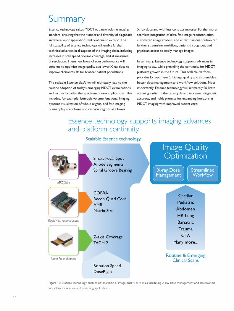
Summary
Figure 16: Essence technology enables optimization of image quality as well as facilitating X-ray dose management and streamlined
workflow for routine and emerging applications.
Essence technology supports imaging advances and platform continuity.
Scalable Essence technology
COBRARecon Quad CoreAMRMatrix Size
RapidView reconstruction
Z-axis CoverageTACH 2
Nano-Panel detector
Smart Focal SpotAnode SegmentsSpiral Groove Bearing
MRC Tube
Routine & EmergingClinical Scans
Cardiac
Pediatric
Abdomen
HR Lung
Bariatric
Trauma
CTA
Many more...
Image Quality Optimization
X-ray Dose Management
Streamlined Workflow
Figure 15
Rotation SpeedDoseRight
19
1 Manzke R, Grass M, Nielsen T, Shechter T, Hawkes D.
Adaptive temporal resolution optimization in
helical cardiac cone beam CT reconstruction.
Med Phys 2003; 30:3072-3080.
2 Heuscher DJ, Chandra S. Multi-phase cardiac imager.
US patent number 6,510,337. 2003
3 Vembar M, Garcia MJ, Heuscher DJ, Haberl R, Matthews
D, Boehme GE, Greenberg NL. A dynamic approach to
identifying desired physiological phases for cardiac
imaging using multislice spiral CT. Med Phys 2003;
30:1683-1693.
4 Karabulut N, Arıyürek M. Low Dose CT: Practices And
Strategies Of Radiologists In University Hospitals,
Diagn Interv Radiol 2006; 12:3-8
5 Aschoff AJ. MDCT of the abdomen, European Radiology
Supplement 2006; 16: M54-M57
6 Breen DJ. Gastrointestinal and abdominal radiology,
Clinical Radiology 2004; 59: 709-712
7 Prokop M, Galanski M, van der Molen AJ, Schaefer-Prokop
CM. Spiral and multislice computed tomography of
the body, Thieme 2003
8 Schmidt T, Behling R. “MRC: a successful platform for
future X-ray tube development”, Medica Mundi, vol. 44,
no. 2, pp.50–55, November 2000.
9 Luhta R, Chappo M, Harwood B, Mattson R, Salk D,
Vrettos C. A new 2D-tiled detector for multislice CT,
Proceedings of SPIE – Volume 6142, Medical Imaging 2006:
Physics of Medical Imaging, Michael J. Flynn, Jiang Hsieh,
Editors, 61420U (Mar. 2, 2006)
10 Philips Healthcare CT Marketing;”Step & Shoot Cardiac -
Low-dose cardiac imaging”; Koninklijke Philips Electronics
N.V.; Whitepaper, 2007.
11 http://www.medical.philips.com/main/products/xray/
dosewise/
Jim Adams
Christopher Bauer
Ronda Bruce
Linda Carney
Sandy Cassell
Michael Chilbert, Ph.D.
Joao Correa
Ekta Dharaiya
Mike Hayden
Peter Johnson
Paul Klahr
Randy Luhta, Ph.D.
Dhruv Mehta
James Meier
Kim Miles
Mark E. Olszewski, Ph.D.
Scott Pohlman
Robert Popilock
Katrina Read
Paul Seltzer
John Steidley, Ph.D.
Jeffrey Studenka
Mani Vembar
Manfred Wachtel
References Acknowledgment of Contributors
For more information, visit: www.philips.com/CT
© 2008 Koninklijke Philips Electronics N.V.All rights are reserved.
Philips Medical Systems Nederland B.V. reserves the right to make changes in specifications and/or to discontinue any product at any time without notice or obligation and will not be liable for any consequences resulting from the use of this publication.
Printed in The Netherlands.4522 962 34201/728 * MAY 2008
Philips Healthcare is part of
Royal Philips Electronics
How to reach us
www.philips.com/healthcare
+31 40 27 64 887
Asia
+852 2821 5888
Europe, Middle East, Africa
+49 7031 463 2254
Latin America
+55 11 2125 0764
North America
+1 425 487 7000
+1 800 285 5585 (toll free, US only)
Philips Healthcare
Global Information Center
P.O. Box 1286
5602 BG Eindhoven
The Netherlands