Embed Size (px)
Citation preview

Biomedical Reviews 1999; 10:55-68.
Dedicated to Rita Levi-Montalcini
©The Bulgarian-American Center, Varna, Bulgaria
ISSN1310-392X
EXPRESSION OFTHETRKNEUROTROPH1N RECEPTORS IN NONNEUROWAL TISSUES
AND CANCER
Hirotaka Koizumi
Department of Pathology, St Marianna University School of Medicine, Kawasaki, Japan
In this article, detailed immunolocalization of the expression of receptor tyrosine kinase (Trk) in a wide range of normal and cancer
nonneuronal tissues is described. Trk immunoreactivity is detected at various levels in all tissues examined, except for the heart and
liver. The gastric parietal cells reveal strong TrkA and TrkC expression and all of the Trk immunoreactivities are expressed in the
putative intestinal neuroendocrine cells. In the pancreas, TrkA and TrkC are detected in the subintralobular ducts, while TrkB is
found specifically in the pancreatic beta cells. The lymph node and spleen exhibit TrkB in monocytes/macrophages. The adrenal
cortex show selective TrkA with TrkC in the medulla. In the reproductive system, TrkA immunoreactivity is detected in the prostatic
epithelial cells, TrkC in the ovarian theca and granulosa cells, TrkA and TrkC in the secretory-phase endometrium, and TrkA in the
mammary ducts. The kidney reveal strong TrkA and TrkC in its tubules, but no Trk receptors are present in the glomeruli. In the
skin, TrkA and TrkB/TrkC are expressed in the basal and granular layers of the epidermis, respectively. The expression patterns
of TrkA in 337 nonneuronal invasive carcinomas of 15 different human tissues are also demonstrated iminunohistochemically.
Overall, 133 (39%), 101(30%) and 103 (31%) tumors exhibited strong, moderate and no TrkA immunoreactivity, respectively.
Esophageal and thyroid carcinomas expressed high levels of TrkA, whereas the levels in gastric and colon cancers are low. TrkA
expression is detected not only in carcinomas originating from TrkA-positive normal counterpart tissues, including the esophagus,
breast, lung and uterus, but also in those from TrkA-negative cells of the thyroid, liver and ovary. Immunostainingforbeta-NGF, the
specific UgandforTrkA, inesophagealandbreast carcinomas demonstrates its immunoreactivity in stromalfibroblasts and some TrkA-
expressing tumor cells. These results suggest that paracrine and autocrine regulation via stromal/tumoral NGF-tumoral TrkA
interaction may be involved in growth of certain nonneuronal carcinomas. Bioined Rev 1999; 10: 55-68.
INTRODUCTION
The Trk family of receptor tyrosine protein kinase (receptozyme),
TrkA, TrkB, and TrkC, play significant roles in the development
and maintenance of neural tissue by mediating the trophic
effects of the nerve growth factor (NGF) family of neurotrophins
(1-4). NGF specifically recognizes TrkA, a receptor identified in
all major NGF targets, including sympathetic, trigeminal, and
dorsal root ganglia as well as in cholinergic neurons of the basal
forebrain and striatum (5-7). Brain-derivedneurotrophic factor
(BDNF) and neurotrophin-4/5 (NT-4/5) activate the TrkB receptor
(8-11), while a higher concentration of NT-4/5 can also activate
the TrkA receptor (12). TrkB transcripts encoding this receptor
are found throughout the central and peripheral nervous system.
Neurotrophin-3 (NT-3) primarily activates the TrkC whose gene is
also expressed widely in the mammalian neural tissues (13).
Unlike the other neurotrophins, NT-3 appears to be somewhat
nonspecific, since it can activate TrkA and TrkB receptors in
Received 17 October 1999 and accepted 30 November 1999.
Correspondence and reprint requests to Dr Hirotaka Koizumi. Department of Pathology. StMariannaUniversity School of Medicine,
Miyamae,Kawasaki216-8511, Japan. Tel: 81449778111, Fax: 81449777827,E-mail:[email protected]

certain cell systems (1). When ectopically expressed in fibro-
blasts, activation of the signal transduction cascade initiated by
each of the Trk receptors results in a similar biological
consequence (10,14). Thus, it would appear likely that the
neuro-trophin requirements of distinct neuronal populations
are dictated by highly specific regulation of Trkreceptor
expression. Recently, we and others have demonstrated
immunocy-tologically or biochemically that various
nonneuronal tissues also express Trks (15-17). We carried out
immunostaining analysis for Trks in 24 types of normal
nonneuronal tissue from adult humans and found that, except for
heart, thyroid and liver, they all expressed these receptors (15),
partly consistent with the results of RNAse protection assay
in rat tissues (17). Moreover, our results demonstrated highly
specific Trk expression by distinct nonneuronal cell populations:
for instance, esophageal and epidermal basal cells selectively
expressed TrkA, gastric parietal cells TrkA and TrkC, pancreatic
beta cell TrkB, adrenal cortical and medullary cell TrkA and
TrkC, respectively, prostatic epithelial cell TrkA and
monocytes/ macrophages of the lymph node and spleen TrkB.
As some of these tissues are known to produce the
corresponding neurotrophins, e.g., the skin (NGF) (18,19),
adrenal glands (NGF andNT-3) (20,21), prostate (NGF) (22,23),
andpancreas, lymph nodes and spleen (BDNF) (16,24), theTrk-
neurotrophin system may be involved in the regulation of specific
functions, such as hormone secretion, of the individual receptor-
expressing cells (15). Furthermore, the fact that Trks are
expressed by a broad spectrum of nonneuronal cells suggests
that they also play roles in phenomena common to such cells,
i.e., cell growth and differentiation, as established for nervous
system (1-4). Indeed, ectopic expression of Trk receptors in
fibroblasts has been shown to result in ligand-mediated cell
growth (10,14), and NGF stimulated nonneuronal cell proliferation
(22,23,25), (26), through TrkA phosphorylation (25). In addition
to the normal tissues, the immunohistochemical expression of TrkA
in over 300 invasive
nonneuronal carcinomas of diverse histologic types is analyzed
(27). TrkA expression is detected not only in carcinomas
originating from TrkA-positi ve normal counterpart tissues, but
also in those from TrkA-negative tissues or cells. These results
suggest that TrkA expression is characteristic of specific subsets of
epithelial malignancies.
In this review, I describe detailed immunolocalization of Trk
receptor family in normal nonneuronal tissues and the expression
profiles of TrkA in nonneuronal carcinomas.
CELLULAR LOCALIZATION OF TRK RECEPTORS IN NORMAL NONNEURONAL TISSUES
Gastrointestinal system
Trk immunostaining is detected in various cell types within the
gastrointestinal system. In the salivary glands, the serous
acinar cells showed TrkB immunoreactivity (IR), whereas the
ductal epithelium stained for both TrkA and TrkC and no Trk IRs
were detected in the mucinous acinar cells (Fig. 1A). In the
esophagus, TrkA immunostaining only was detected in the
basal layers of the stratified squamous epithelium (Fig. 1B,C). In
the stomach, the parietal cells, located in the lower half of the
gastric glands, showed strong IR for TrkA and TrkC, but not for
TrkB (Fig. ID, left). In addition to subepithelial connective
tissue fibroblasts andlymphocytes, no othercellular components of
the gastric mucosa showed Trk immunostaining (Table 1). In the
small intestine and colon, there is little or no staining in the
epithelial cells for Trk proteins, but small cells, located along the
glandular basement membrane show intense immunostaining
for all three Trk receptors (Fig. ID, right). The morphology,
immunoreactivity and distribution of the Trk- positivecells were
consistent with those of the intestinal neuroendocrine cells.
Weak TrkA and TrkC staining was detected in all the muscle
layers and in the ganglion cells present in Meissner's and
Auerbach' s plexuses throughout the gastrointestinal tract (Table
Figure 1. The immunolocalization of Trk receptors in the human nonneuronal tissues. Immunostaining result(s) showing only the
representative type(s) of Trk receptor in each tissue. A. TrkB IR in the serous acinar cells of the salivary glands. B. TrkA
immunoreactivity in the basal layers of the esophageal squamous epithelium. C. No TrkB immunoreactivity in the esophagus. D, left.
Strong TrkA immunoreactivity in the parietal cells of the stomach. D, right. TrkB immuno reactive cells in the small intestine. TrkB
immunoreactivity was detected in small cells present along the glandular basement membrane (arrows). E. Strong TrkB
immunoreactivity in the pancreatic islet. F. Glucagon-immunoreactive beta-islet cells in the serial section. The distribution of
TrkB-positive and glucagon-positive cells was virtually identical. G. TrkB-immunoreactive cells present around the white pulp of
the spleen, which showed a nearly identical distribution pattern to lysozyme-positive monocytes/macrophages in the serial section
(H. left, TrkB; right, lysozyme). I. TrkA immunoreactivity in the adrenal cortex (upper panel) and TrkC in the medulla (lower
panel). J. TrkA immunoreactivity in the prostatic epithelial cells, which was clearly present in luminal secretory cells, but faintly in
the basal cells K. Trk immuno reactivities in the secretory-phase endometrium. K. left. TrkC immunoreactivity at the luminal
surface of the endometrial epithelium. K. right. TrkA immunoreactivity in the glandular epithelium. L. TrkC immunoreactivity
in the mammary duct. Dot-like immunoreactivity was apparent in the luminal epithelium as well as in the intraductal phagocytes.
Very weak immunoreactivity was present in the my o epithelium. M. Strong TrkC immunoreactivity in the collecting tubules of kidney.
The distal tubules showed weaker immunostaining. N. Immunoreactivitiesfor TrkA (upper panel) and TrkB (lower panel) in the skin
epidermis. O. TrkA immunoreactivity in the skeletal muscle. Immunoperoxidase staining.
Biomed Rev 10, 1999
Koizumi 56

Biomed Rev 10, 1999
Trk neurotrophin receptors 57

1). No Trk IR is found in the hepatocytes, Kupffer cells, sinusoi-dal cells or bile ductal epithelium of the liver.
In the exocrine pancreas, moderate to strong TrkA and TrkC IRs are present in the intercalated and intralobular ducts, but they were absent from the interlobular and main pancreatic ducts and the acini (15). In the endocrine pancreas, very intense TrkB IR is detected in cells preferentially located around the periphery of the islets (Fig. IE), their localization is consistent with that of glucagon-secreting cells (28). In serial sections, immunolocalization of TrkB is nearly identical with that of glucagon (Fig. IF), but not with those of insulin. No TrkA and TrkC immunostaining is seen in the pancreatic islets.
Cardiopulmonary system
In the lung, low levels of TrkA and TrkC are present in some alveolar cells and in the bronchial gland mucinous cells. No Trk immunoreactivity is present in the cardiac muscles (Table 1).
Hematolymphoid system
Only TrkB is detectable in the lymph node and spleen. The TrkB immunoreactive cells are moderate- to large-sized, cytoplasm rich, and of irregular shape, consistent with the morphological features of monocytes and macrophages. They resided preferentially in the subcapsular and interfollicular sinuses of the node and around the white pulp of the spleen (Fig. 1G). Double immunostaining showed that the nodal TrkB-positive cells are only colabeled with CD 68, the marker for monocytic lineage (29), and not with other markers for hematopoietic cells, such as CDS, CD20, CD45 and immunoglobulin light chains (15). In addition, the splenic TrkB-positive cells show a virtually identical distribution pattern to that of lysozyme-containing monocytes/macrophages on serial sections (Fig. 1H). Like these hematolymphoid tissues, the bone marrow show selective TrkB immunostaining in hematopoietic cells (Table 1). Considering that BDNF and NT-3 are expressed by lymph node and spleen (16), our data indicate that monocytes/macrophages may be the selective targets of BDNF and NT-3 in these tissues. In the thymus, intense TrkA staining was seen in the subcapsular epithelial cells, and the thymocytes themselves stained occasionally for this protein (15). Since the thy mic subcapsular epithelium is a region of known p75 neurotrophin receptor (p75
NTR) localization (30), the coexpression of low- and high-
affinity NGF receptors by these epithelial cells might be related to their well-characterized neuroendocrine phenotype (31) and to their ability to synthesize different thymic hormones (30).
Endocrine system
In endocrine tissues, the thyroid follicular cells reveal no Trk immunostaining, exceptforsomecellsinphysiologicallyatrophic follicles that showed TrkA and TrkC immunostaining (Table 1). TrkA IR was detected throughout the adrenal cortex, although several cells stained quite intensely. Virtually no TrkB or TrkC immunostaining is seen in the cortex. The medulla expresses
only for TrkC IR (Fig. II). The distinct distribution of TrkA and TrkC in the adrenal glands indicates that endogenous NGF and NT-3 (20,21) might be capable of exerting selective regulatory effects on the secretion of cortical and medullary adrenal hormones, respectively. As mentioned above, the pancreatic alpha cells are TrkB-positive.
Reproductive system
In the male reproductive system, selective immunostaining of TrkA is found in the epithelial cells of the prostate; it was clearly present in the luminal secretory cells, but not or only faintly in the basal cells (Fig. 1J). Pflugetal also reported TrkA expression in the human prostate using a pan-Trk antibody in conjunction with Scatchard plot and receptor phosphorylation analyses (25). Since prostatic stromal cells produce NGF that has been shown to bind to prostatic epithelial cells (25), this strongly suggests that the growth of the latter is regulated by a paracrine mechanism. In the testis, the fibrous septa surrounding the seminiferous epithelium also showed TrkA IR (also see this volume of Biomedical Reviews). In the female reproductive system, the ovarian theca and granulosa cells stain for TrkC (15). This implies that ovary-secreted neurotrophins, such as NT-3 (33,34), may be involved in the direct regulation of gonadal endocrine function. The proliferative-phase endometrium completely lack Trk immunostaining, whereas the secretory-phase epithelium expresses strong TrkC IR on its luminal surface. TrkA is also detected during this phase in parts of the glandular epithelium and in the stromal cells (Fig. IK). These results suggest that estrogen may upregulate expression of TrkA and TrkC in the endometrium in the same way as it does for TrkA in PC12 cells in vitro and in rat dorsal root ganglion neurons in vivo (35-37). Since NGF significantly enhances estrogen binding in PC 12 cells (35,37), it is feasible that TrkA, expressed in the secretory-phase endometrium, may function to increase endometrial sensitivity to estrogen. A similar interaction among estrogen, TrkC, and NT-3 might augment the secretory activity of the endometrial epithelium. In the breast, luminal epithelium of the mammary ducts show cytoplasmic, dot-like reactivity for TrkA and TrkC. These immunoreactive dots are also found in the cytoplasm of the intraductal phagocytes. Weak TrkA and TrkC immunostaining is also seen in the ductal myoepithelium (Fig. 1L).
Urinary system
In the kidney, intense TrkA and rather weaker TrkC
immunostaining are found in the epithelium of the collecting
tubules (Fig. 1M). Similar but less intense immunostaining is
also detected in the proximal and distal tubules. In contrast,
none of the Trk IR were present in the cells of the glomeruli (15).
Although there are no published reports describing Trk
expression in the human kidney, a study in fetal mice showed a
similar renal localization of TrkC transcripts using in situ
hybridization (38). As no NGF expression or activity are
58 Koizumi
Biomed Rev 10, 1999

Trk neurotrophin receptors 59
Biomed Rev 10, 1999

- , absent; +, weakly positive; ++, moderately positive; + + +, strongly positive. *TrkA was present in the basal layer of the stratified
squamous epithelium. 'Cells with neuroendocrine cell-like morphology. **TrkC immunostaining was detected at the luminal surfaces
of most of the glandular epithelium and TrkA was found in the cytoplasm of some of them. "TrkA and TrkB/TrkC were present in the
basal and granular layers, respectively, of the epidermis.
Biomed Rev 10, 1999
Koizumi
detected in the kidney (39,40) and NT-3 IR is found in the
distal tubular cells of rat (20), it is possible that NT-3, which
can bind to both TrkA and TrkC (41), may play a role either in
survival of tubular epithelial cells or in the reabsorption
function of the renal tubules.
Musculocutaneous system
The skeletal muscle is stained for TrkA and TrkC, but not for
TrkB (Fig. 1O). In the skin, TrkA and TrkB/TrkC are present in
the basal and upper, or granular, layers of the epidermis,
respectively (Fig. IN). TrkA was also detectable in the hair
follicles and sweat glands. Since the basal layer of the epidermis
also expresses p75NTR (42) and keratinocytes and dermal
fibroblasts are both capable of producing NGF (43,44), it seems
likely that the behavior of human skin epidermis might be
influenced by NGF-mediated autocrine/paracrine regulation via its
low- and high-affinity receptors. Moreover, the observation that
transgenic mice, deficient for TrkA, have numerous scabs over
their entire body and ulcerated paws (45), suggests that the NGF-
TrkA interaction may play crucial roles in regulating both cell
growth and the regeneration of normal epidermis (also see this
volume of Biomedical Reviews).
I have shown highly specific Trk receptor expression by
distinct nonneuronal cell populations. However, I do not yet
know whether the expressed receptors actually function as
signal transducing molecules in these cells. RNase protection
analyses have shown that rat nonneuronal tissues express both
the full-length and truncated TrkA, whereas they only express
the truncated TrkB and TrkC transcripts that appear to encode
noncatalyticreceptorproteins (17,46-48). Although the number of
tissues examined was small, these results imply that only the full-
length TrkA is capable of transmitting the ligand's signal into
nonneuronal cells. This is consistent in part with a study
showing Trk phosphorylation by NGF in prostatic epithelial
cells, indicating that TrkA is a signal transducing molecule in
this nonneuronal tissue (25). However, it is still possible that
truncated receptors may present neurotrophins to neurons, or
may interact with another receptor that is itself the signal-
transducing molecule in these cells. Moreover, expression of
the full-length TrkB transcripts in rat hematolymphoid tissues
has recently been reported (14). A precise understanding of the
functional roles of Trk receptors in nonneuronal tissues awaits
further analyses, including RNase protection in a wide range of
organs/tissues, immunohistochemical studies using antibodies
that recognize the truncated peptides, and in vitro expression
studies of the truncated trk genes in various nonneuronal cells.
EXPRESSION OF TRKA, NGF, AND P75NTR IN HUMAN NONNEURONAL CANCERS
TrkA expression in nonneuronal carcinomas
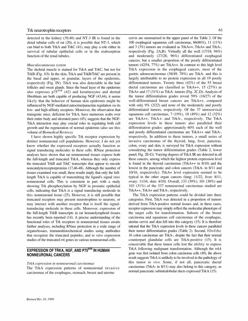
The TrkA expression patterns of nonneuronal invasive
carcinomas of the esophagus, stomach, breast and uterine
cervix are summarized in the upper panel of the Table 2. Of the
100 esophageal squamous cell carcinomas, 86(86%), 11 (11%)
and 3 (3%) tumors are evaluated as TrkA++, TrkA+ and TrkA-,
respectively (Fig. 2A,B). Virtually all the well (17/18, 94%)
and moderately (27/28, 96%) differentiated esophageal
cancers, but a smaller proportion of the poorly differentiated
tumors (42/54, 77%) are TrkA++. In contrast to this high level
TrkA expression in the esophageal cancers, most of the
gastric adenocarcinomas (38/49, 78%) are TrkA- and this is
largely attributable to no protein expression in all 19 poorly
differentiated tumors. Twenty three (42%) of the 55 breast
ductal carcinomas are classified as TrkA++, 15 (27%) as
TrkA+ and 17 (31%) as TrkA- tumors (Fig. 2C,D). Analysis of
the tumor differentiation grades reveal 59% (16/27) of the
well-differentiated breast cancers are TrkA++, compared
with only 9% (2/22) and none of the moderately and poorly
differentiated tumors, respectively. Of the 37 uterocervical
squamous cell carcinomas, 7 (19%), 18 (49%) and 12 (32%)
are TrkA++, TrkA-t- and TrkA-, respectively. The TrkA
expression levels in these tumors also paralleled their
differentiation grades: approximately 60% each of the well
and poorly differentiated carcinomas are TrkA++ and TrkA-,
respectively. In addition to these tumors, a small series of
invasive carcinomas of the thyroid, lung, liver, pancreas,
colon, ovary and skin, is surveyed for TrkA expression without
considering the tumor differentiation grades (Table 2, lower
panel; Fig. 2E-G). Varying degrees of TrkA IR are detected in all
these cancers, among which the highest protein expression level
is found in the thyroid carcinomas (TrkA++ in 8/10) and the
lowest in the pancreatic and colon cancers (TrkA- in 8/11 and
10/16, respectively). TrkA+ level expression seemed to be
typical in the other organ cancers (lung: 11/22, liver: 8/11,
ovary: 11/16, skin: 4/10). Overall, 133 (39%), 101 (30%) and
103 (31%) of the 337 nonneuronal carcinomas studied are
TrkA++, TrkA+ and TrkA-, respectively.
The TrkA expression patterns could be divided into three
categories. First, TrkA was detected in a proportion of tumors
derived from TrkA-positive normal tissues and, in these cases,
receptor expression may simply reflect the molecular phenotype of
the target cells for transformation. Subsets of the breast
carcinoma and squamous cell carcinomas of the esophagus,
uterine cervix and skin fell into this category (15). It is therefore
rational that the TrkA expression levels in these cancers paralleled
their tumor differentiation grades (Table 2). Second, lOofthe
16 colon carcinomas are TrkA-, despite the fact that their normal
counterpart glandular cells are TrkA-positive (15). It is
conceivable that these tumor cells lost the ability to express
TrkA following malignant transformation. Although the trkA
gene was first isolated from colon carcinoma cells (49), the above
result suggests TrkA is unlikely to be involved in the pathology of
this tumor in vivo. Some, if not all, pancreatic ductal
carcinomas (TrkA- in 8/11) may also belong to this category, as
normal pancreatic subintralobular ducts expressed TrkA (15).
61 Trk neurotrophin receptors
Biomed Rev 10, 1999

Biomed Rev 10, 1999
Koizumi 62

Third, TrkA++ or TrkA+ expression was detected in all the
thyroid carcinomas (10/10: 100%), which may result from the
reported trkA rearrangement in this tumor (50,51), and the
majority of the liver (8/11: 73%) and ovarian (14/16: 88%)
carcinomas. It is significant that we observed TrkA expression in
these tumors, but it was not detected in their counterpart
normal cells, i.e., thyroid follicular cells, hepatocytes, and ovarian
mesothelial cells (15). In such cases, receptor expression may
be related to the transformed phenotype or to increased
proliferative activity, not to the tissue derivation of the tumor
cells.
Expression of NGF and p75NTR in esophageal and breast
carcinomas
Immunostaining for NGF and p75NGFR was carried out in frozen
sections of 7 esophageal and 10 breast invasive carcinomas.
Normal esophageal tissues adjacent to the tumors showed NGF
IR in the epithelial spinocellular cells and submucosal
fibroblasts, whereas they exhibit p75NTR immunostaining
exclusively at the epithelial basal and parabasal layers. No NGF
immunostaining is seen in the luminal epithelium and
myoepithelium of normal mammary glands, although they stain
positively for p75NTR, and the stromal fibroblasts surrounding the
mammary glands show NGF immunoreactivity. These results
are consistent partly with previous reports (19, 52, 53) (Fig.
3C,D for p75NTR). NGF IR is present in the stromal fibroblasts, as
well as in various numbers of the cancer cells in tumor
portions of the esophageal and breast tissues, although none of
these tissues exhibit p75NTR immunostaining (Fig. 3A-D). These
findings are common to all the specimens tested. Altogether, I
suggest that paracrine regulation via the stromal NGF-tumoral
TrkA interaction might mediate certain effect, e.g., cell growth, in
these nonneuronal tumors. This concept has already been
suggested for prostate cancer. Djakiew et al (22,23,25,54)
localized Trk- and NGF-immunoreactive proteins in the epithelial
and stromal cells, respectively, in human prostate tissue, and
showed that NGF or its related stromal protein specifically
bound to the epithelial TrkA, thereby inducing tyrosine
phosphorylation of the receptor and tumor proliferation in a
prostate cancer cell line. Similar findings of NGF-
dependent clonal growth of lung cancer cells (26) and NGF
production by tissue fibroblasts (55,56) are reported recently.
Therefore, our findings extend the types of tumor that express
TrkA and stromal NGF and indicate that the paracrine regulation
model of tumor growth might be applicable to a variety of
nonneuronal human carcinomas. I suggest that the low-affinity
p75NTR is either not involved, or plays a minor role in this
paracrine process, as p75NTR immunoreactivity appears to have
been lost by most of the nonneuronal cancers that expressed
TrkA, such as those of the esophagus, prostate, thyroid, breast
and ovary (25,52,57) (Fig. 3C,D). Moreover, immunostaining of
NGF and TrkA in serial sections reveal that NGF is colocalized in
some, although not all, TrkA-expressing tumor cells (Fig.
3E,F). This result raises a possibility that autocrine regulation
via the tumoral NGF-tumoral TrkA interaction mechanism might
also involve in proliferation of some, if not all, nonneuronal
carcinomas.
The mechanism(s) underlying NGF-mediated nonneuronal
tumor growth hypothesized is(are) not well understood, although
the product of the NGF inducible-A (NGFI-A) gene in rat
prostate cells is found to prompt transcription of the Rb gene
(58), the protein product of which is crucial for Gl-S phase
transition of the cell cycle that eventually results in cell division
and proliferation (59). Finally, it is noteworthy that NGF-
stimulated expression of proteolytic enzymes, such as endo-f5-
glucuronidase heparanase, which are important for basement
membrane penetration, in TrkA-negative/p75NTR-positive
neuroectodermal melanoma cells (60,61), suggesting a further
role for NGF in tumor invasion and metastasis.
ACKNOWLEDGMENTS
I wish to thank Drs Eiichi Shibay ama, Mikita Morita, and Shiny a
Mikami for their contributions to the present studies. I am also
grateful to Professors Toshiyuki Uchikoshi and Mamoru -
Tadokoro for constant support and encouragement. The studies
carried out in our laboratory were supported in part by Grant-in-
Aid for Scientific Research from the Ministry of Education,
Science and Culture, Japan.
Table 2. (legend)
hnmunohistochemical detection ot TrkA in paraffin sections of human tumors. Counterpart normal cells of each tumor and their
status of TrkA expression are shown in parentheses following the organ names (Ref 15) (Y, yes; N, no), ep, epithelium. Well, well
differentiated type; moderate, moderately differentiated type; poor, poorly differentiated type; n, number of specimens. Staining
results were scored according to the following scale: -, no staining; +, low percentage (<50%) of positively stained tumor cells;
++, at least 50% of the tumor cells stained positive. *TrkA was present in the parietal cells but absent from the chief cells,
foveolar/neck cells and pyloric glands. 'TrkA was present in the intercalated and intralobular ducts but absent from the
interiobular and main pancreatic ducts.
Biomed Rev 10, 1999
Trk neurotrophin receptors 63

Koizumi 64
Biomed Rev 10, 1999
REFERENCES
1. Chao MV. Neurotrophin receptors: a window into neu-ronal differentiation. Neuron 1992; 9: 583-593.
2. Hide FF, Lowenstein DH, Reichardt LF. Neurotrophins and their receptors - current concepts and implications for neurologic disease. Ex/? JVewro/ 1993; 121:200-214.
3. Kaplan DR, Stephens RM. Neurotrophin signal transduc-tion by theTrkreceptor. JNeurobioll994;25:1404-1417.
4. ManessLM, KastinAJ,WeberJT, BanksWA,BeckmanBS, Zadina JE. The neurotrophins and their receptors: structure, function, and neuropathology. Neurosci Biobehav Rev 1994,18:143-159.
5. Kaplan DR, Martin-ZancaD, ParadaLF. Tyrosine phospho-rylation and tyrosine kinase activity of the trk proto-oncogene product induced by NGF. Nature 1991; 350:158-160.
6. Kaplan DR, Hempstead BL, Martin-Zanca D, Chao MV, Parada LF. The trk proto-oncogene product: a signal transducing receptor for nerve growth factor. Science 1991; 252: 554-558.
7. Hempstead B, Martin-ZancaD, Kaplan DR, ParadaLF, Chao MV. High-affinity NGF binding requires coexpression of the trk proto-oncogene and the low-affinity NGF receptor. Nature 1991; 350:678-683.
8. KleinR, NanduriV, Jing SQ, LamballeF, Tapley P, BryantS etal. The trkB tyrosine protein kinase is areceptor for brain-derived neurotrophic factor and neurotrophin-3. Cell 1991;
66:395-403
9. SoppetD, EscandonE, Maragos J, Middlemas DS, ReidSW, Blair J et al.The neurotrophic factors brain-derived neurotrophic factor and neurotrophin-3 are ligands for the trkB tyrosine kinase receptor. Cell 1991; 65: 895-903.
10. Klein R,LamballeF,Bryant S,BarbacidM. The trkB tyrosine protein kinase is areceptorforneurotrophin-4. Neuron 1992; 8:947-956.
11. IpNY,IbanezCF,NyeSH,McClainJ,JonesPF,GiesDRef al. Mammalian neurotrophin-4: structure, chromosomal localization, tissue distribution, and receptor specificity. Proc
NatlAcadSci USA 1992; 89:3060-3064. 12. BerkemeierLR, Winslow JW, Kaplan DR,Nikolics K, Goeddel
DV, Rosenthal A. Neurotrophin-5: a novel neurotrophic factor that activates trkA and trkB. Neuron 1991;7:857-866.
13. Lamballe F, Klein R, B arbacid M. trkC, a new member of the trk family of tyrosine protein kinases, is a receptor for neurotrophin-3. Celll99l; 66:967-979.
14. Cordon-Cardo C, Tapley P, Jing S, Nanduri V, O'RourkeE, Lamballe F et al. The trk tyrosine protein kinase mediates the mitogenic proterties of nerve growth factor and neurotrophin-3. Celll99l;66:173-183.
15. Shibayama E, Koizumi H. Cellular localization of the Trk neurotrophin receptor family in human non-neuronal tissues. AmJ Pathol 1996; 148:1807-1818.
16. LaurenziMA, Barbany G, TimmuskT, Lindgren JA, Persson H. Expression of mRNA encoding neurotrophins and neurotrophin receptors in rat thymus, spleen tissue and immunocompetent cells: regulation of neurotrophin-4 mRNA expression by mitogen and leukotriene B4. Eur JBiochem 1994; 223:733-741.
17. LomenHC, Shooter EM. Widespread neurotrophin receptor expression in the immune system and other non-neuronal rat tissues. JNeurochem 1995; 64:1780-1789.
18. Anand P, Terenghi G, Warner G, Kopelman P, Williams-Chestnut RE, Sinicropi DV. The role of endogenous nerve growthfactorinhumandiabeticneuropathy. NatMed\996;
2:703-707. 19. Jahansson O, Liang Y. Neurotrophins and their receptors in
the skin: a tribute to Rita Levi-Montalcini.fiw/weo?/̂ ev 1999; 10:15-24.
20. Zhou XF, Rush RA. Localization of neurotrophin-3-like immunoreactivity in peripheral tissues of the rat. Brain Res
1993;621:189-199. 21. Baker DL, MolenaarWM, Trojanowski JQ. Nerve growth
factor receptor expression in peripheral and central neuro-ectodermal tumors, other pediatric brain tumors, and during development of the adrenal gland. AmJPathol 1991; 139: 115-122.
22. Djakiew D, Delsite R, Pflug B, Wrathall J, LynchJH, Onoda M. Regulation of growth by a nerve growth factor-like protein which modulates paracrine interactions between a neoplastic epithelial cell line andstromal cells of the human prostate. Cancer Res 1991; 51:3304-3310.
23. Djakiew D. Role of nerve growth factor-like protein in the paracrine regulation of prostate growth. JAndroll992; 13: 476-487.
24. Lommatzsch M, Braun A, Mannsfeldt A, Botchkarev VA, Botchkareva NA, Paus R et al. Abundant production of brain-derived neurotrophic factor by adult visceral epithe-lia. Implications for paracrine and target-derived neurotrophic functions. AmJ Pathol 1999; 155:1183-1193.
25. Pflug BR, Dionne C, Kaplan DR, Lynch J, Dj akiew D. Expression of a Trk high affinity nerve growth factor receptor in the human prostate. Endocrinology 1995; 136:262-268. .
26. OelmannE,SreterL,SchullerI. Nerve growth factor stimulates clonal growth of human lung cancer cell lines and a human glioblastoma cell line expressing high-affinity nerve growth factor binding sites involving tyrosine kinase signaling. Cancer Res 1995; 55:2212-2219.
27. KoizumiH,MoritaM,Mikami, S, ShibayamaE,UchikoshiT. Immunohistochemical analysis of TrkA neurotrophin receptor expression inhuman non-neuronal carcinomas. Pathol
Int 1998;48,93-101. 28. Fawcett DW. Bloom and Fawcett, Textbook of Histology.
New York, Chapman and Hall, 1994, pp. 689-703. 29. Pulford K, Rigney E, Micklem K, Gatter K, Mason D. KP1: a
new monoclonal antibody that detects a monocyte/mac-
Koizumi 66
Biomed Rev 10, 1999

rophage associated antigen in routinely processed tissue
sections. JClinPathol 1989; 42: 414-421.
30. Pescarmona E, Pisacane A, Pignatelli E, Baroni CD.
Expression of epidermal and nerve growth factor receptors in
human thymus and thymomas. Histopathology 1993; 23: 39-
44.
31. Lobach DF, Haynes BF. Ontogeny of the human thymus
during fetal development. JClinlmmunol 1987;7: 81-97.
32. Zhou XF, Rush RA. Localization of neurotrophin-3-like
immunoreacti vity in peripheral tissues of the rat. Brain Res
1993;621:189-199.
33. Ojeda SR, Dissen GA, Junier MP. Neurotrophic factors and
female sexual development. Front Neuroendocrinol 1992;
13:120-162.
34. Timmusk T, Belluardo N, Metsis M, Persson H. Widespread
and developmentally regulated expression of neurotrophin-4
mRNA in rat brain and peripheral tissues. EurJNeurosci
1993;5:605-613.
35. MirandaRC, Sohrabji F, Toran AD. Interactions of estrogen
with the neurotrophins and their receptors during neural
development. HormBehav 1994;28:367-375.
36. Sohrabji F, Miranda RC, Toran AC. Estrogen differentially
regulates estrogen and nerve growth factor receptor mRN As
in adult sensory neurons. JNeurosci 1994; 14:459-471.
37. Sohrabj i F, Greene LA, Miranda RC, Toran AC. Reciprocal
regulation of estrogen and NGF receptors by their ligands
inPC12cells./AteMro&0/1994;25:974-988.
38. Durbeej M, SoderstromS,EbendalT,BirchmeierC,Ekblom
P. Differential expression of neurotrophin receptors during
renal development. Development 1993; 119:977-989.
39. Wheeler EF, B othwell M. Spatiotemporal patterns of expression
of NGF and the low-affinity NGF receptor in rat embryos
suggest functional roles in tissue morphogenesis and
myogenesis. JNeurosci 1992; 12:930-945.
40. Altar CA, Criden MR, Lindsay RM, DiStefano PS.
Characterization and topography of high-affinity I25I-
neurotrophin-3 binding to mammalian brain. JNeurosci 1993;
13:733-743.
41. Barbacid M. The Trk family of neurotrophin receptors. J
Neurobioll994;25:1386-1403.
42. Di Marco E, MathorM, B ondanza S, Cutuli N, Marchisio PC,
Cancedda R et al. Nerve growth factor binds to normal
human keratinocytes through high and low affinity receptors
and stimulates their growth by a novel autocrine loop.
JBiolChem 1993; 268:22838-22846.
43. Yaar M, filler MS, DiBenedetto P, Reenstra WR, Zhai S,
McQuaid Tetal. The trk family of receptors mediates nerve
growth factor and neurotrophin-3 effects in melanocytes. J
Clinlnvestl994;94:1550-1562.
44. YaarM, Grossman K, EllerM, Gilchrest BA. Evidence for
nerve growth factor-mediated paracrine effects in human
epidermis./Ce//Bioll99l; 115: 821-828
45. Smey ne RJ, Klein R, Schnapp A, Long LK, Bry ant S, Lewin A
et al. Severe sensory and sympathetic neuropathies in
mice carrying a disrupted Trk/NGF receptor gene. Nature
1994;368:246-249.
46. Klein R, Conway D, Parada LF, Barbacid M. The trkB
tyrosine protein kinase gene codes for a second neurogenic
receptor that lacks the catalytic kinase domain. Cell 1990;
61:647-656
47. ValenzuelaDM.MaisonpierrePC, GlassDJ,RojasE,Nunez L,
Kong Y et al. Alternative forms of rat TrkC with different
functional capabilities. Neuron 1993; 10:963-974.
48. Barker PA, Lomen-Hoerth C, GenschEM, Meakin SO, Glass
DJ, Shooter EM. Tissue-specific alternative splicing generates
two isoforms of the trkA receptor. J Biol Chem 1993;
268:15150-15157.
49. Mitra G, Martin-Zanca D, Barbacid M. Identification and
biochemical characterization of p70TRK, product of the
human TRK oncogene. ProcNatlAcadSci USA 7987; 84:
6707-6711.
50. Bongarzone I, Pierotti MA, Monzini N. High frequency of
activation of tyrosine kinase oncogenes in human papillary
thyroid carcinoma. Oncogene 1989;4:1457-1462.
51. Fugazzola L, Pilotti S, Pinchera A. Oncogenic
rearrangements of the RET proto-oncogene in papillary
thyroid carcinomas from children exposed to the
Chernobyl nuclear accident. CancerRes 1995; 55:5617-
5620.
52. ChesaPG, Rettig WJ, Thomson TM, OldLJ, MelamedMR.
Immunohistochemical analysis of nerve growth factor
receptor expression in normal and malignant human tissues.
JHistochemCytocheml9%8;36:3S3-389.
53. Mearow KM, Kril Y, Diamond J. Increased NGF mRNA
expressionindenervatedratskin. NeuroReport\993;4:351-
354.
54. DjakiewD,PflugB,OnodaM. Stromal-epithelialparacrine
interactions in the neoplastic rat and human prostate. 'Adv
ExpMedBioll993;330:185-202.
55. Yoshida K, Gage FH. Cooperative regulation of nerve
growth factor synthesis and secretion in fibroblasts and
astrocytes by fibroblast growth factor and other cytokines.
BrainResl992;569:14-25.
56. Cartwright M, Mikheev AM, Heinrich G. Expression of
neurotrophin genes in human fibroblasts: differential
regulation of the brain-derived neurotrophic factor gene.
Int J DevNeuroscil994; 12:685-693.
57. PflugBR,OnodaM,LynchJH,DjakiewD. Reduced
expression of the low affinity nerve growth factor
receptor in benign and malignant human prostate tissue
and loss of expression in four human metastatic prostate
tumor cell lines. CancerRes 1992; 52:5403-5406.
58. Day ML, Wu S, Easier JW. Prostatic nerve growth factor
inducible A gene binds a novel element in the retinoblas-
toma gene promoter. CancerRes 1993; 53:5597-5599.
59. Ewen ME. The cell cycle and the retinoblastoma protein
family. Cancer Metastasis Rev 1994; 13:45-66.
60. NicolsonGL,NakajimaM,HerrmannJL. Malignantmela-
67 Trk neurotrophin receptors
Biomed Rev 10, 1999

Koizumi
noma metastasis to brain: role of degradative enzymes and 61.Nicolson GL, Menter DG, Herrmann J. Tumor metastasis
responses to paracrine growth factors. / Neurooncol to brain: role of endothelial cells, neurotrophins, and
1993; 18:139-149. . paracrine growth factors. Crit Rev Oncog 1994; 5: 451-
Biomed Rev 10, 1999
68