Embed Size (px)
Citation preview
Facilitator: Caswell Mavimbela
CELL BIOLOGY
Introduction to

Module Overview
• Mineral elements and their roles
• Compounds required in human body and their biological importance.
• Cell structure and function
• Cellular organelles and their functions
• Reproduction in cells

What is cell biology?
• Branch of life science which deals with the study on cells in terms of structure, function and chemistry.
• Structure: how they are made from molecules• Function: how individual cells cooperate to make an
organism as complex as a human being• Chemistry: Interaction of different molecules in the
synthesis of complex molecules essential for cell function.
Before we can fully understand how a healthy human body functions, how it develops and ages, and what goes wrong with it in disease, we need to understand the cells of which it is made.
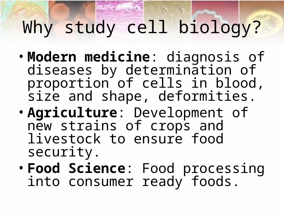
Why study cell biology?
• Modern medicine: diagnosis of diseases by determination of proportion of cells in blood, size and shape, deformities.
• Agriculture: Development of new strains of crops and livestock to ensure food security.
• Food Science: Food processing into consumer ready foods.
Historical perspective (task)
• Cell theory
• Antonio van Leeuwenhoek

Recent Advances in Cell Biology
• A possible cure of HIV with bone marrow transplant. Doctors announced in Berlin that a man who received a bone marrow transplant for leukemia was now also free of his HIV infection. He received bone marrow from a donor with CCR5 gene delta 32 gene believed to be resistant to HIV.
• Breakthrough windpipe transplant of Claudia Castillo - tissue grown from Ms Castillo’s own bone marrow stem cells, using them to fashion the new bronchus – a branch of the windpipe

Introduction
• Cell: basic building unit of from which all living organism are made and can function independently.
• Some organism are single celled (bacteria and protozoa) while others are multicellular (mammals and trees).
• Cells are precursors of tissues and then organs.• Cells can grow & divide; move; respond to
complicated instructions; measure time (they get old and die); can go wrong and make the living organisms sick etc

Chemistry of Life
• Living organisms rely on complex processes to perform and regulate the internal functions that are necessary to maintain life.
• The term to describe all the processes is homeostasis, the ability of a living organism to maintain its structure and function intact, separate from its environment.
• The maintenance of homeostasis requires a series of chemical reactions and each step in each reaction requires the presence of specific substances.
• Vitamins and mineral elements are among the necessary substances – not all are produced internally.

Chemistry of Life
• Require mineral elements to sustain life and perform their function
• Macroelements – required in quantities – used as bulding materials of organic compounds
• Microelements – required in small quantities – have great and essential contribution.

A lack of iodine in one’s diet can cause swelling of the thyroid gland resulting in a GOITER. The condition is reversible if iodine is taken. (Don’t worry, we iodize salt)
Iodine is used by thyroid cells to make hormones (chemicals released by one cell into the blood and bind to a receptor on another cell, which is one way cells talk to each other).
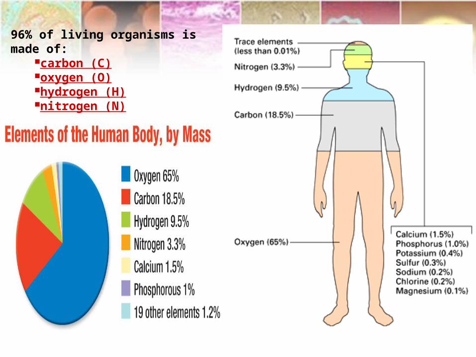
96% of living organisms is made of:
carbon (C)oxygen (O)hydrogen (H)nitrogen (N)

Macroelements
• Calcium (Ca+)– abundant in body – structural component of bones and function of nervous system.
• Phosphorus (P)– Ass. with Ca in bone – important in energy metabolism.
• Potassium (K+) – Milk – electrolyte – involved in fluid balance.
• Sodium (Na+)– Combine with K+- fluid balance• Chloride (Cl-) – Production of HCl for digestion.• Sulphur – involved in protein synthesis.• Magnesium (Mg)– Suppress certain hormones and
reduces stress. Metabolize Na, K, and Ca

Microelements• Iron (Fe) – essential in erythropoesis and oxygen transport.• Copper (Cu) – Enzyme component involved in energy
production, strengthening of connective tissue and brain neurotransmission
• Chromium (Cr) – insulin performance and cellular glucose uptake.
• Zinc (Zn) – Wound healing, antibody production and immune response.
• Manganese (Mn) – essential for enzyme for fertility and growth
• Cobalt (Co) – Part of Vitamin B12• Iodine (I) – Thyroid metabolism• Selenium (Se) – Combined with Vit E – essential in anti-
oxidant, support immune system by preventing disease and stress.

Inorganic to organic molecules• Carbon, hydrogen, oxygen and nitrogen (95% of body weight)
are common elements in livings things.• Carbon can bond with other elements (C,H,O,N) to create
different organic molecules due to possession of 4 electrons • Ability of C to bond with itself, produce carbon chain of
various lengths (50+ carbon atoms) and shapes.• Carbon chains makes up skeleton of organic molecules• Hydrocarbon chain composed of H & C are usually
hydrophobic and combination with functional group that ionizes them render them hydrophilic.
• Functional groups and isomers (molecules with identical molecular formula but different arrangement of atoms) bring diversity to organic molecules.
• Organic molecules (sugars, fatty acids, amino acids and nucleotides) are based on carbon chain backbone.
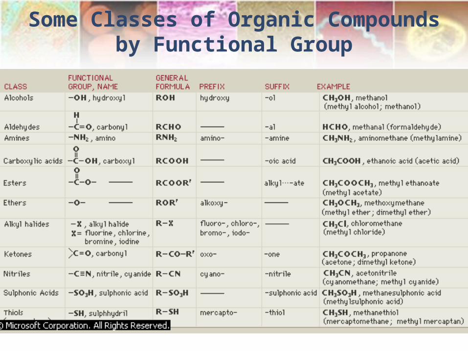
Some Classes of Organic Compounds by Functional Group

Organic molecules
• Can be small and large• Large organic molecules are called polymers and are
made up of small organic molecules called monomers.• Simple single sugars are called monosaccharides
(glucose or fructose), two of those would be disaccharide (sucrose) while many forming a long chain are called polysaccharides (starch or glycogen).
• Fatty acids and glycerol form fats• Amino acids join to form proteins• Nucleotides are the subunits of nucleic acids (DNA and
RNA)

1. Carbohydrates
• Def: any of a large group of compounds in which hydrogen and oxygen, in the proportions in which they exist in water, are combined with carbon.
• Most abundant organic molecules in nature, produced by autotrophs during photosynthesis.
• General formula to represent them is (CH2O)n.
• They are classified as mono-, di- and polysaccharides.

1.1. Monosaccharides (MBChB 24/02/2009)
• Monosaccharides – simple sugars with a carbon backbone (6 carbons)
• Example: glucose(6C - hexose), fructose (6C)and ribose (5C – pentose).
• Glucose and fructose are isomers but differ in structure and functional group. There is aldehyde and ketone respectiely.
• Characteristics: clear, sweet taste, crystalline, solid and water soluble.


TASK
Find out the structure of galactose

1.2. Disaccharides
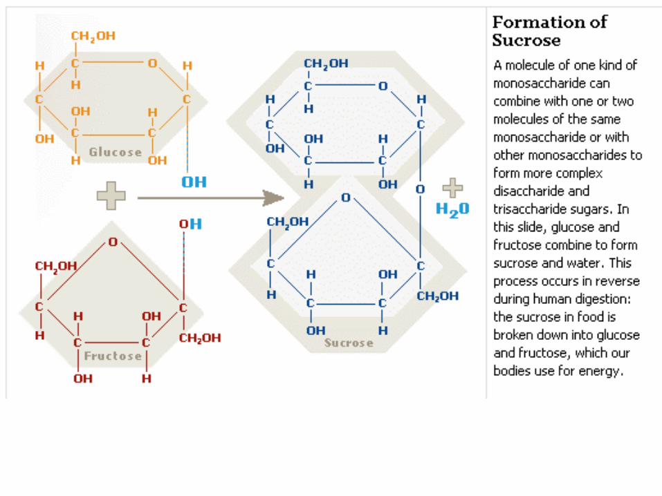
• Formed from two monosaccharides during dehydration synthesis reaction (condensation). The reverse is known as hydrolysis. It is the splitting of a large molecule into two or more parts by addition of water.
• Glucose + fructose = sucrose (sugar cane)• Glucose + galactose = lactose (milk)• Glucose + glucose = maltose (in stomach)
monosaccharides
disaccharides

Task
• Learn all the structures of monosaccharide and disaccharides and their respective chemical reactions.

1.3. Polysaccharides
• Most common include starch, glycogen and cellulose (contains chains of glucose molecule) and chitin (contain modified glucose).
• Starch and glycogen – energy storage in plants and animals respectively. Cellulose and chitin – structural purposes.
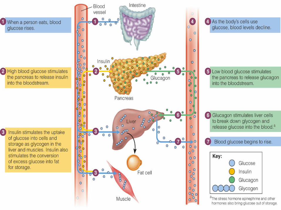
• In humans – glucose is converted to glycogen by liver and muscles. Between meals glycogen is broken down to glucose to keep stable blood glucose conc. Starch from plants is broken down to maltose and then glucose in stomach

1.3.1. Starch
• Composed of hundred of glucose molecules
• Made by plants to store energy for future use.
• Easily hydrolyzed into glucose molecules
• Humans and other animals (herbivores) utilizes this energy reserves of plants
1.3.2. Glycogen
• Made in animal cells by condensation or dehydration synthesis.
• Made for purpose of short-term energy storage.• Stored in liver and muscles• Diabetes mellitus: insulin dependence (type2) –
inadequate or ineffective pancreatic insulin production to metabolise glucose. Glucose become abundant in blood causing hyperglycemia.
presented by NM Mogale
1.3.3. Cellulose
• Structural compound found in cell walls of plant cells.
• Provide rigidity in stems, leaves and roots.
• Indigestible by humans
• Dietary cellulose also called fiber or roughage, useful in humans to prevents constipation.

• In plant cells each cellulose fibril contains several microfibrils. Each microfibril contains many polymers of glucose hydrogen – bonded together.
1.3.4. Chitin
• Has an amino group attached to each glucose molecule.
• Main component of arthropods exoskeleton
• Indigestible by humans.

BIOLOGICAL IMPORTANCE OF CARBOHYDRATES
• Carbohydrates serve both essential structure and energy-storage functions:
1. As structural components in cells eg chitin in the exoskeleton; cellulose in the cell walls of plants and many protists; in vertebrate animals, the cell coatings of connective tissue contain carbohydrates
2. As main source of energy: sugar (glucose) as short-term ernergy source; starch (plants) and glycogen (animals) as intermediate-term energy storage.

2. Lipids
• Insoluble in water – lacks polar group. Soluble in chloroform, benzene and ether.
• Composed of H, C and carboxyl functional group.
• Most important lipids are fats, oils, phospholipids and steroids.

2.1. Fats and OilsFats Oils
Made up fatty acids and glycerol Made up of fatty acids and glycerol
Animal origin Plant origin
Solid at room temp°C Liquid at room temp°C
Contains saturated fatty acids – c-chain, no double bond
Contains unsaturated fatty acids – c-chain, has double bond.
Less healthy More healthier
Functions: Insulation, cushioning and energy storage.
Prevents freezing.
• Glycerol contains 3 hydroxyl group – polar and water soluble.• Fat result in reaction between 3 fatty acid portions and 3 hydroxyl
group of glycerol. 3 water molecules are produced.• Glycerol fatty acids ratio = 1: 3. Fats and oils can be referred to
as triglycerides• Fats and oils are long-term energy storage in both plants and
animals and are richer than glucose and glycogen

Structure of fat (triglyceride)

2.2. Phospholipids
• Contains phosphate group and are derived from fatty acids, glycerol, phosphoric acid and nitrogen base.
• They are constructed much the same as neutral fats but instead of 3 fatty acids, one is replaced with a phosphate and nitrogen group (resulting in a polar head and non-polar tail)
• Its properties implies that they can form a separation between two solutions in terms of the interior and exterior. In the presence of water, they arrange themselves in a double layer.
• Function: form the lipid bilayer of plasma membrane

Structure of a phospholipid/nitrogen

Steroids
• A type of lipid with 4 fused carbon ring backbone.• Varies according to the functional group attached to the
rings.• Steroids include sterols (e.g. cholesterol), bile acids and
important hormones.• Cholesterol is a precursor of several other steroids (bile
acids and hormones)• Component of cell membrane• Used in the production of bile• Necessary for Vitamin D synthesis• Forms sheaths of some neurons.• Excess of cholesterol is linked to atherosclerosis
(hardening of arteries) – leading to possible coronary heart disease.

Steroids and SterolsFigure 1 shows the basic ring system of a steroid, a carbon skeleton with four rings. The addition of one or more alcohol groups (-OH), creates a sterol, in Figure 2, a cholesterol molecule, and in Figure 3, oestradiol or estradiol, an important female sex hormone.

Proteins• Large organic compounds made up of amino acids.• There are about 20 amino acids commonly found in cells e.g. alanine,
valine, serine, cysteine and lysine to name a few.• Amino acids differ in the nature of the R group they contain. This R group
range from a single H to complex ring compound, with some R group being polar and others not.
• All amino acids contains 2 important functional groups ( carboxyl - COOH and amino group – NH2). This makes them hydrophilic at normal body pH.
• Amino acids combine to form peptides and polypeptides. The bond between the amino acids is covalent bond called peptide.
• Structural proteins - important in maintaining shape of cells or organisms. Example: Collagen (make up bone, skim, tendon, and cartilage), Keratin (hair, skin ,nails and feathers), Fibrinogen (blood plasma protein responsible for blood clotting) and Actin and myosin (muscle contraction).
• Globular or regulator proteins – responsible for cellular and body functions. Example: Enzymes (catalysts), hormones (chemical messengers e.g. insulin secreted by pancreas to regulate blood sugar conc.), antibodies (immune response) and microtubules (has structural function and can conduct substance from one side of the cell to the other.
• Protein denature in response of pH and Temp.

Chemical structure of amino acids

Nucleic acids• Monomers of nucleic acids are nucleotides.• Each nucleotide is composed of 3 molecular components i.e. phosphate, pentose
sugar and nitrogen containing base.• Nucleic acids are huge polymers of nucleotides with specific func in cells e.g. DNA
and RNA • DNA is a genetic material that stores info on its own replication and order amino acids
follows to make proteins.• RNA is an intermediary in protein synthesis and conveys info from DNA regarding
amino acids sequence in protein synthesis.• DNA has a sugar deoxyribose while RNA, ribose - differs by lack of oxygen in
deoxyribose.• There are types of nucleotides each having its own base.• Bases (purines: adenine and guanine) and pyrimidines (thymine and cytosil). In RNA
uracil replaces thymine• Nucleotides form linear molecule called 2 strand (looks like a ladder) with sides of
ladder made by phosphate and sugar unit and rungs made by complementary base pairs
• Bases appear in any order but (T) pairs with (A), (G) with (C) with hydrogen bonds= complementary base pairs -
• Overall structure of DNA form a bouble helix while RNA is a single strand

Structure of DNA molecule
DNA MoleculeThe structure of a DNA molecule resembles a ladder formed of sugars and phosphates, and four nucleotide bases: adenine (A), thymine (T), cytosine (C), and guanine (G). The coding information in the DNA is given by the order of the nucleotide bases, and each gene possesses a unique sequence of base pairs. Scientists use these base sequences to locate the position of genes on chromosomes and to construct a map of the entire human genome.
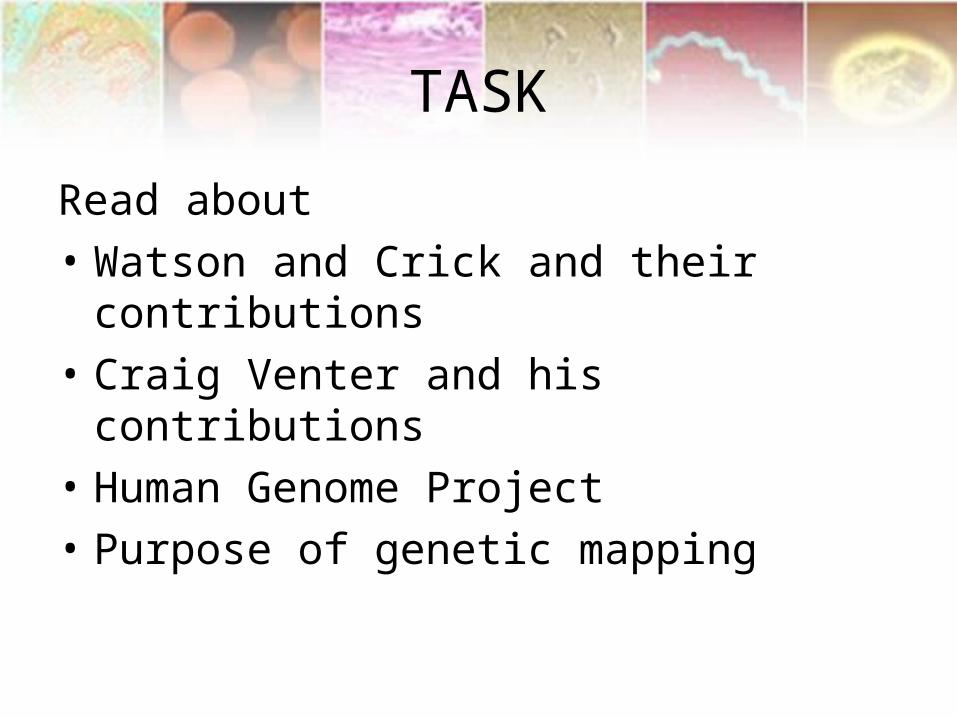
TASK
Read about
• Watson and Crick and their contributions
• Craig Venter and his contributions
• Human Genome Project
• Purpose of genetic mapping

Cell Structure and Functions
Chapter II
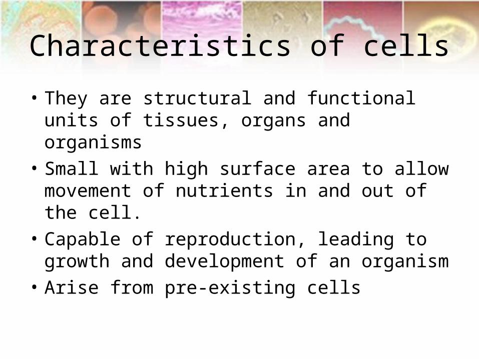
Characteristics of cells
• They are structural and functional units of tissues, organs and organisms
• Small with high surface area to allow movement of nutrients in and out of the cell.
• Capable of reproduction, leading to growth and development of an organism
• Arise from pre-existing cells

Cells
• Smallest living unit• Most are microscopic

Discovery of Cells
• Robert Hooke (mid-1600s)– Observed sliver of cork– Saw “row of empty boxes”– Coined the term cell
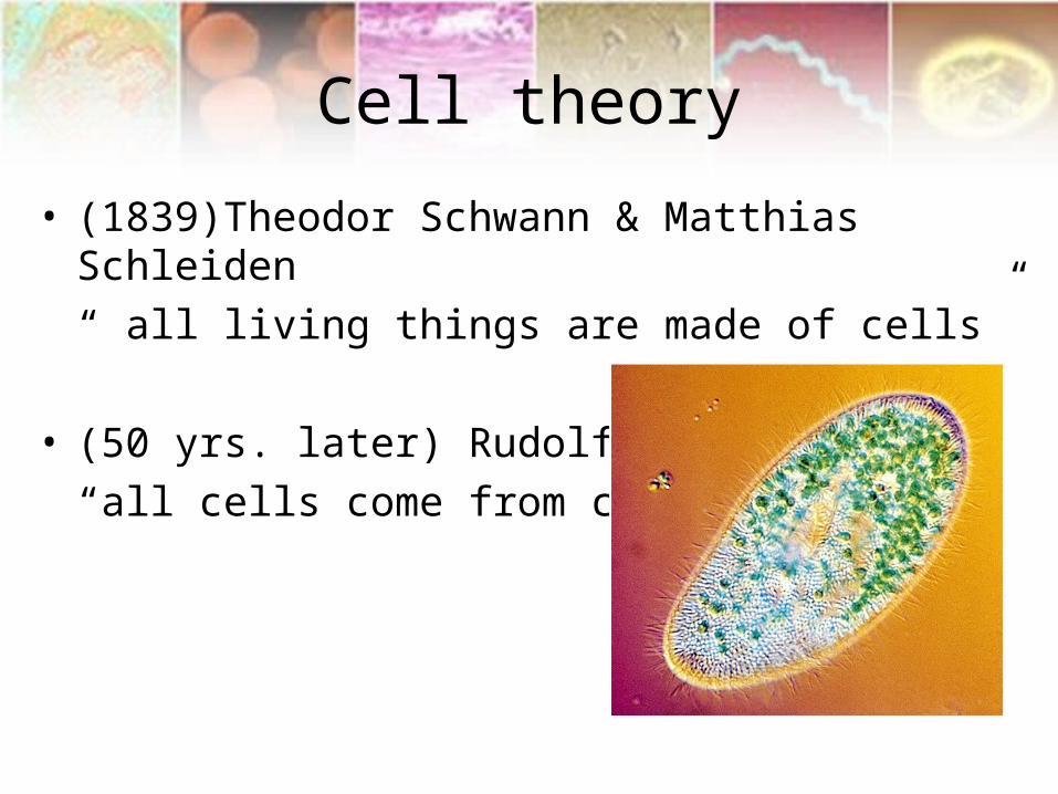
Cell theory
• (1839)Theodor Schwann & Matthias Schleiden
“ all living things are made of cells”
• (50 yrs. later) Rudolf Virchow
“all cells come from cells”

Principles of Cell Theory
• All living things are made of cells
• Smallest living unit of structure and function of all organisms is the cell
• All cells arise from preexisting cells
(this principle discarded the idea of
spontaneous generation)

Cell Size

Major types of cells
Major Cell Types
Prokaryote“Lacks true nucleus – nucleoid region –
not bound by membrane
Eukaryote“have true nucleus bound by membrane”
Animal cell Plant cellSingle celled organisme.g. bacteria
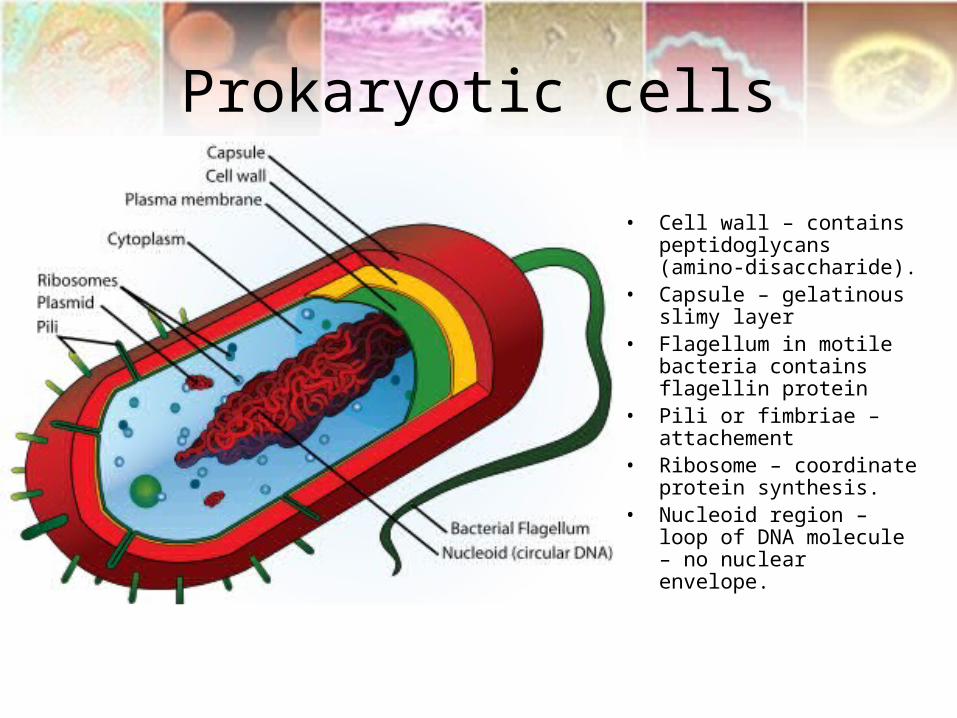
Prokaryotic cells
• Cell wall – contains peptidoglycans (amino-disaccharide).
• Capsule – gelatinous slimy layer
• Flagellum in motile bacteria contains flagellin protein
• Pili or fimbriae – attachement
• Ribosome – coordinate protein synthesis.
• Nucleoid region – loop of DNA molecule – no nuclear envelope.
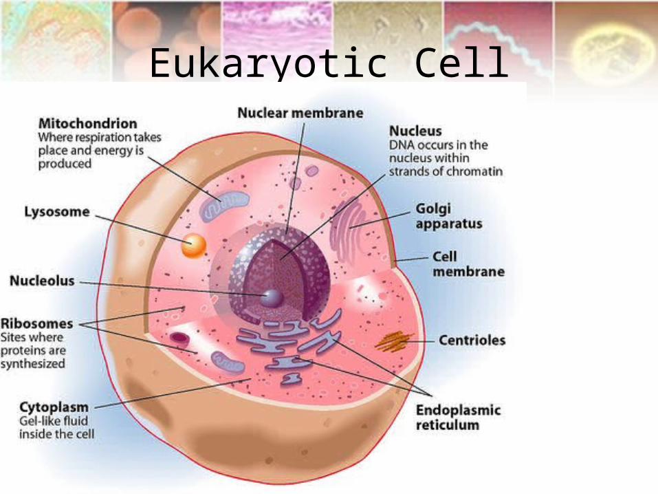
Eukaryotic Cell

Differences between Pro and Eukaryotic cells
Prokaryotic Eukaryotic
No true nucleus – nucleoid region. Has true nucleus surrounded by nuclear membrane
Smaller Larger
Has outer surrounding capsule and cell wall No capsule and only cell wall in plant cells
Less complex More complex
Cell wall contain peptidoglycans Plant cell wall contain cellulose. No wall in animal cell.
DNA forms a loop – single chromosome DNA housed in chromatin
No compartmentalization Highly organized (internally) due to compartmentalization by organelles
Lack organelles Have many organelles

Difference between animal and plant cells
Animal Plant
Lacks cell wall Contains cell wall – cellulose
Lack chlorophyll Have chlorophyll
Have centriole Lacks centriole
Have a small vacuole Have a large central vacuole

Cell structures and functions

Lecturer overview
• Cell membrane
• Cell organelle
• Mitosis
• Meiosis

Plasma Membrane• Fluid mosaic model• Proteins in plasma membrane• Cell to cell recognition• Crossing of plasma membrane
– Passive• Diffusion• Osmosis - tonicity• Facilitated transport
– and active transport• Active transport• Bulk transport
– Endocytosis– Exocytosis
» Phagocytosis and pinocytosis• Cellular junctions
– Adhesion – Tight– Gap– Desmosomes

Cell membrane
• Also called plasmalemma, plasma membrane or phospholipid bi-layer.
• Made up of phospholipids – forming a double layer
• It is selectively permeable• Very thin and measures about 7-10 micrometers• Phospholipids molecules are fluid to allow
stretching responding to stress or movement of cholesterol and proteins.
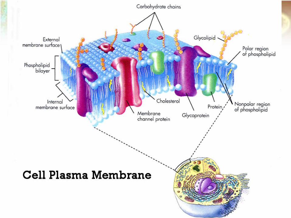
Phospholipid bilayer

Chromosomes

Cell Cycle Mitosis Meiosis
Genetics

Eukaryotic chromosomes contain DNA and protein
The chromosomes carry the genetic information


When a cell divides, chromatin fibers are very highly folded, and become visible in the light microscope as chromosomes.
During interphase (between divisions), chromatin is more extended, a form used for expression genetic information.

• DNA is organized into informational units called genes
• Chromosomes contain hundreds to thousands of genes

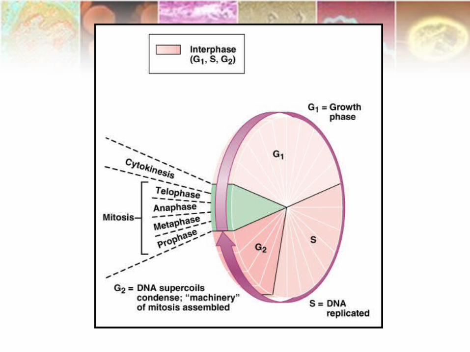
Cell Cycle
• The cell cycle is a sequence of cell growth and division.
• The cell cycle is the period from the beginning of one division to the beginning of the next.
• The time it takes to complete one cell cycle is the generation time.
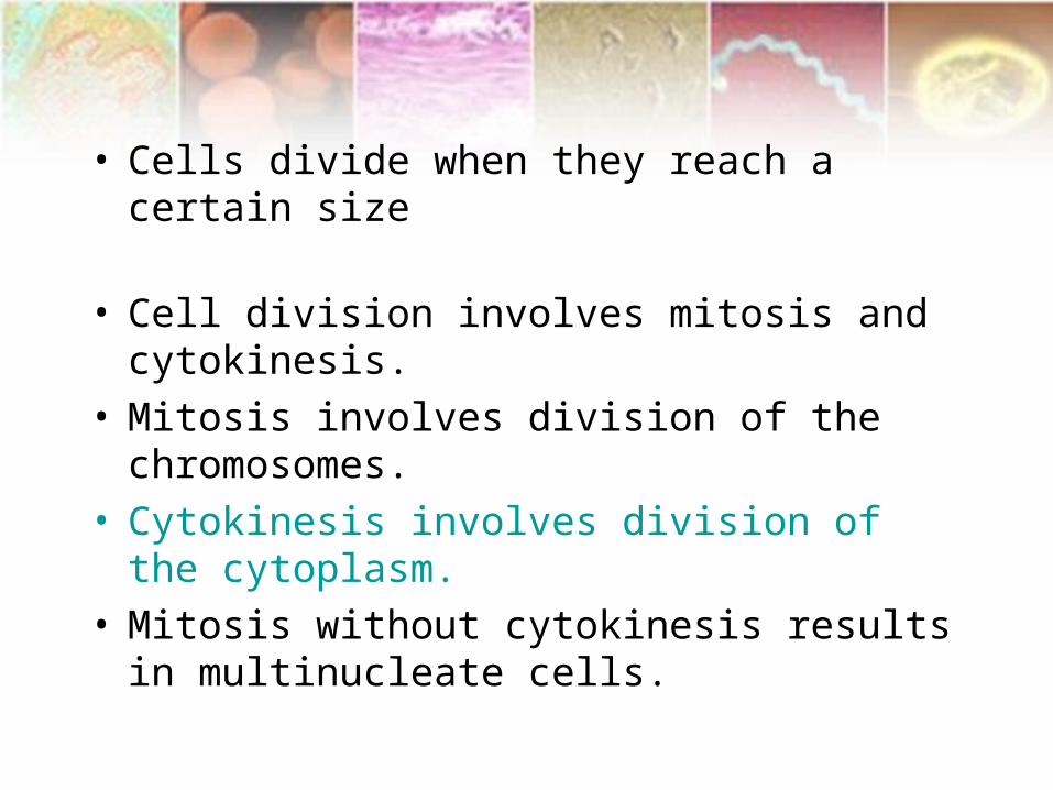
• Cells divide when they reach a certain size
• Cell division involves mitosis and cytokinesis.
• Mitosis involves division of the chromosomes.
• Cytokinesis involves division of the cytoplasm.
• Mitosis without cytokinesis results in multinucleate cells.

• Eukaryotic cell cycle– Beginning of one division to beginning of next– Stages in eukaryotic cell cycle– Interphase
• First gap phase
• Synthesis phase
• Second gap phase
– M phase• Mitosis
• Cytokinesis
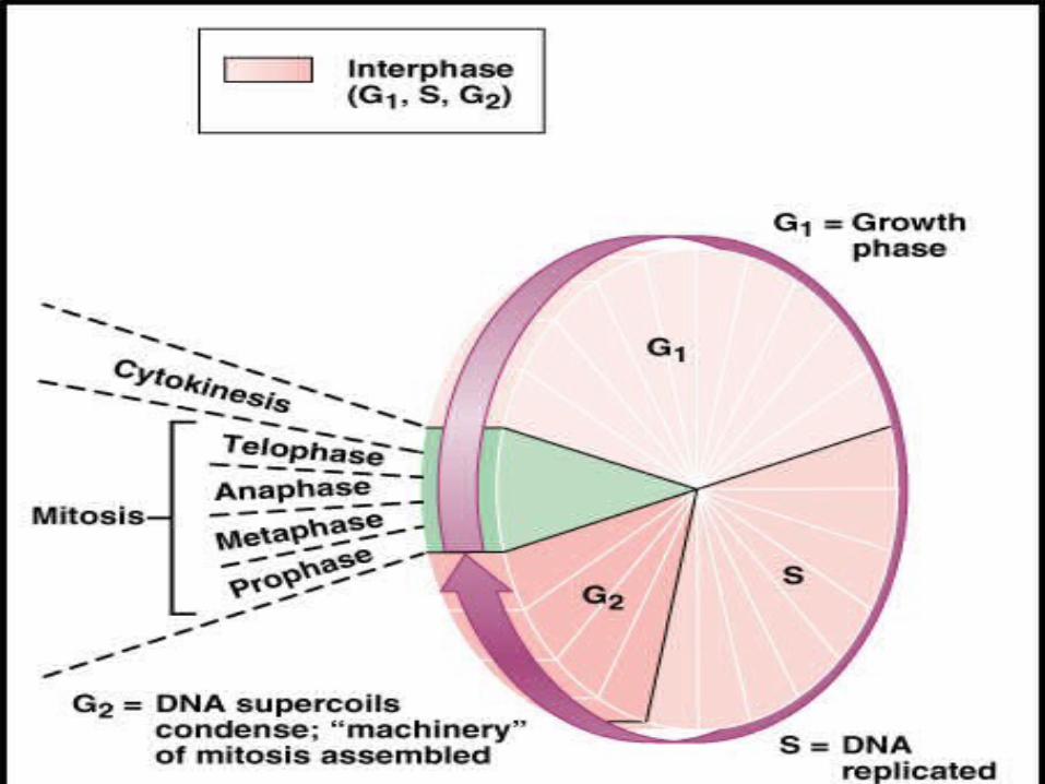

• Chromosomes become duplicated during interphase
• Cells are very active during interphase, synthesizing biological molecules and growing the G1 (gap) phase
• The S (synthesis) phase is marked by DNA replication
• The G2 (gap) phase occurs between the S phase and mitosis

• Despite differences between prokaryotes and eukaryotes, there are several common features in their cell division processes. – Replication of the DNA must occur.– Segregation of the "original" and its
"replica" follow. – Cytokinesis ends the cell division
process.• Whether the cell was eukaryotic or
prokaryotic, these basic events must occur.

Hereditary material is passed on to new cells by mitosis or meiosis
Cell division, growth, and reproduction
InterphaseMitosisCytokinesisMeiosis
Cell division
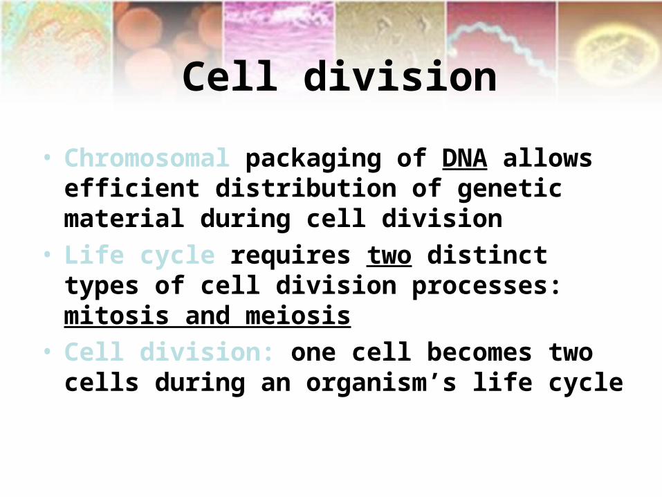
• Chromosomal packaging of DNA allows efficient distribution of genetic material during cell division
• Life cycle requires two distinct types of cell division processes: mitosis and meiosis
• Cell division: one cell becomes two cells during an organism’s life cycle

Mitosis
• Mitosis is nuclear division plus cytokinesis, and produces two identical daughter cells during the following steps:– Prophase– Metaphase– Anaphase– Telophase.
• Interphase is often included in discussions of mitosis, but interphase is technically not part of mitosis, but rather encompasses stages G1, S, and G2 of the cell cycle.

Interphase
The cell is engaged in metabolic activity and performing its prepare for mitosis (the next four phases that lead up to and include nuclear division). Chromosomes are not clearly discerned in the nucleus, although a dark spot called the nucleolus may be visible. The cell may contain a pair of centrioles (or microtubule organizing centers in plants) both of which are organizational sites for microtubules.

Prophase
Chromatin in the nucleus begins to condense and becomes visible in the light microscope as chromosomes.
The nucleolus disappears.
Centrioles begin moving to opposite ends of the cell and fibers extend from the centromeres.
Some fibers cross the cell to form the mitotic spindle.

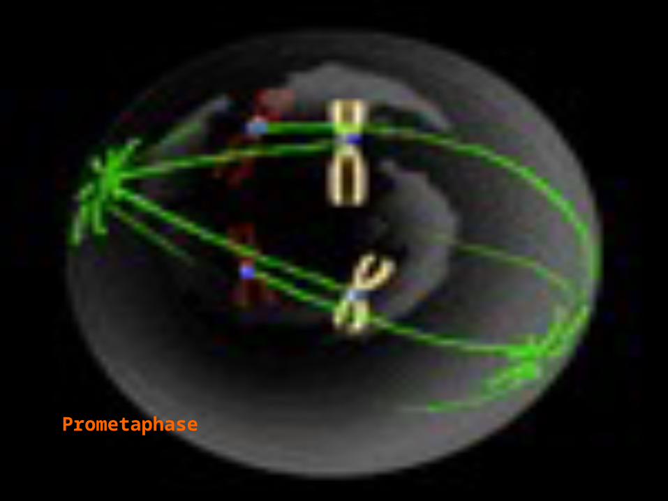
Prometaphase
The nuclear membrane dissolves, marking the beginning of prometaphase.
Proteins attach to the centromeres creating the kinetochores.
Microtubules attach at the kinetochores and the chromosomes begin moving.

Metaphase
Spindle fibers line the chromosomes along the middle of the cell nucleus. This line is referred to as the metaphase plate. Polar microtubules extend from the pole to the equator, and typically overlap Kinetochore microtubules extend from the pole to the kinetochores
This organization helps to ensure that in the next phase, when the chromosomes are separated, each new nucleus will receive one copy of each chromosome.
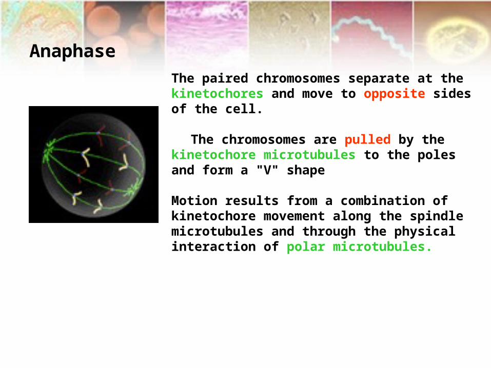
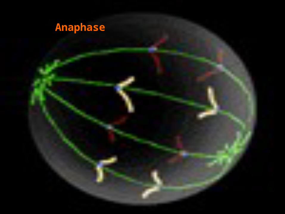
Anaphase
The paired chromosomes separate at the kinetochores and move to opposite sides of the cell.
The chromosomes are pulled by the kinetochore microtubules to the poles and form a "V" shape
Motion results from a combination of kinetochore movement along the spindle microtubules and through the physical interaction of polar microtubules.

Telophase
Chromatids arrive at opposite poles of cell, and new membranes form around the daughter nuclei.
The chromosomes disperse and are no longer visible under the light microscope.
The spindle fibers disperse, and cytokinesis will start.

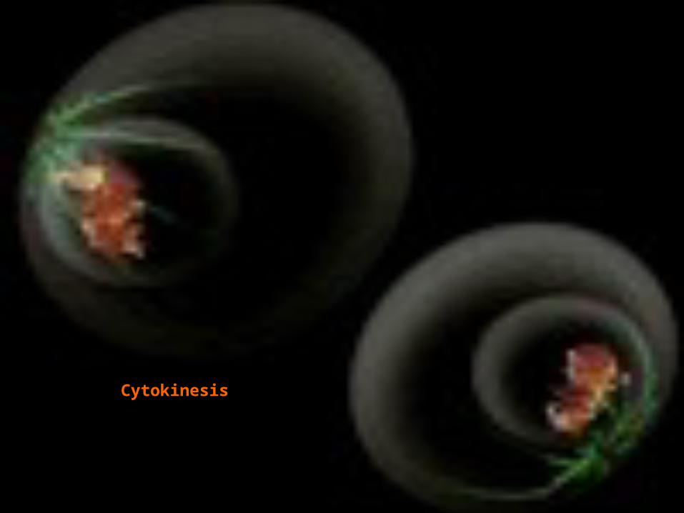
Cytokinesis
In animal cells, cytokinesis results when a fiber ring composed of a protein called actin around the center of the cell contracts pinching the cell into two daughter cells, each with one nucleus.
In plant cells, synthesis of new cell wall between two daughter cells rather than cleavage furrow in
cytoplasm

Interphase

Prophase
Prometaphase

Metaphase
Anaphase

Telophase
Cytokinesis


Animated GIF (203Kb)

Reproduction
• Asexual reproduction • Sexual reproduction

Asexual Reproduction
• A form of duplication using only mitosis.
• Example, a new plant grows out of the root or a shoot from an existing plant.
• Produces only genetically identical offspring since all divisions are by mitosis.

Sexual reproduction
• Formation of new individual by a combination of two haploid sex cells (gametes).
• Fertilization- combination of genetic information from two separate cells that have one half the original genetic information
• Gametes for fertilization usually come from separate parents

1. Female- produces an egg 2. Male produces sperm
• Both gametes are haploid, with a single set of chromosomes
• The new individual is called a zygote, with two sets of chromosomes (diploid).
• Meiosis is a process to convert a diploid cell to a haploid gamete, and cause a change in the genetic information to increase diversity in the offspring.

Chromosomes in a Diploid Cell
Summary of chromosome characteristics
• Diploid set for humans; 2n = 46 • Autosomes; homologous chromosomes, one
from each parent (humans = 22 sets of 2) • Sex chromosomes (humans have 1 set) 1.Female-sex chromosomes are homologous
(XX) 2.Male-sex chromosomes are non-
homologous (XY)

Number of sets of chromosomes in a cell
• Haploid (n)-- one set chromosomes • Diploid (2n)-- two sets
chromosomes • Most plant and animal adults are
diploid (2n) • Eggs and sperm are haploid (n)

• Most cells in the human body are produced by mitosis. These are the somatic (or vegetative) line cells.
• Cells that become gametes are referred to as germ line cells. The vast majority of cell divisions in the human body are mitotic, with meiosis being restricted to the gonads.

• Diploid cells – Characteristic number of
chromosome pairs per cell• Homologous chromosomes• Similar in length, shape, other features,
and carry similar attributes
• Haploid cells– Contain only one member of each
homologous chromosome pair

Meiosis• Diploid cells undergo meiosis to form
haploid cells
• Meiosis potentially produces four haploid cells
• Meiosis involves two separate divisions

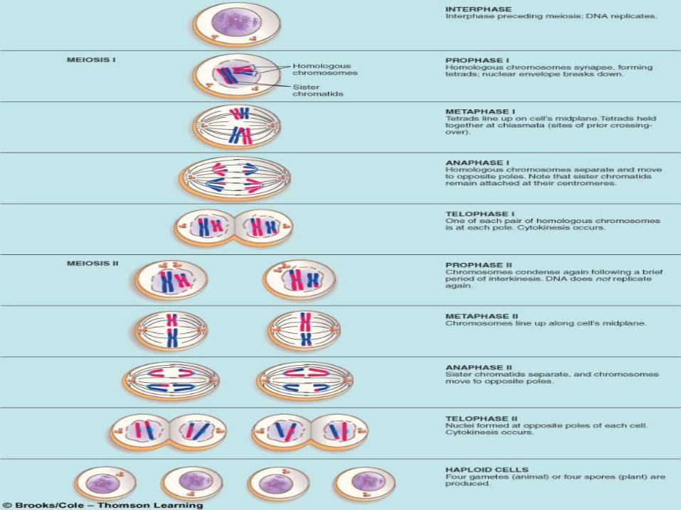
• Two successive nuclear divisions occur, Meiosis I (Reduction) and Meiosis II (Division).
• Meiosis I reduces the ploidy level from 2n to n (reduction) while Meiosis II divides the remaining set of chromosomes in a mitosis-like process (division).

• In meiosis, homologous chromosomes are separated into different daughter cells
• Meiosis I and meiosis II each include prophase, metaphase, anaphase, and telophase

The First Division Meiosis I
• Prophase I is one of the most important stages of meiosis.
During this stage, many crucial events occur.
In prophase I,
• The spindle appears.
• Nuclear envelopes disappear.
• The DNA of the chromosomes begin to twist and condense, making the DNA visible to the microscope.
• Each chromosome actively seeks out its homologous pair (which also has a sister chromatid).

• The two replicated homologous pairs find each other and form a synapse. The structure formed is referred to as a tetrad (four chromatids).
• The point at which the two non-sister chromatids intertwine is called a chiasma. Sometimes a process known as crossing over occurs at this point.
• This is where two non-sister chromatids exchange genetic material. This exchange does not become evident, however, until the two homologous pairs separate.


• Prophase I includes synapsis and crossing over
• Homologous chromosomes pair and undergo synapsis
• One member of a pair is the maternal homologue, the other is the paternal homologue
• Synapsis is the association of four chromatids (two from each homologue)

• In metaphase I, the tetrads line up along the equator.
• Anaphase I results in the separation of homologous pairs. Cells are haploid at this point.
• Telophase I results in a brief reappearance of nuclear envelopes, and the spindle disappears. The cell waits momentarily during interkinesis.
• Interkinesis separates meiosis I and II; no DNA synthesis occurs

The Second Division Meiosis II
• In prophase II, the spindle reappears, and the nuclear membrane fragments.
• In metaphase II, the chromosomes align at the equator.
• In anaphase II, sister chromatids separate. • In telophase II, the nuclear envelopes reappear,
and four haploid cells are the result.

Prophase IITelophase I
Anaphase IMetaphase I

Metaphase II Anaphase II
Telophase II
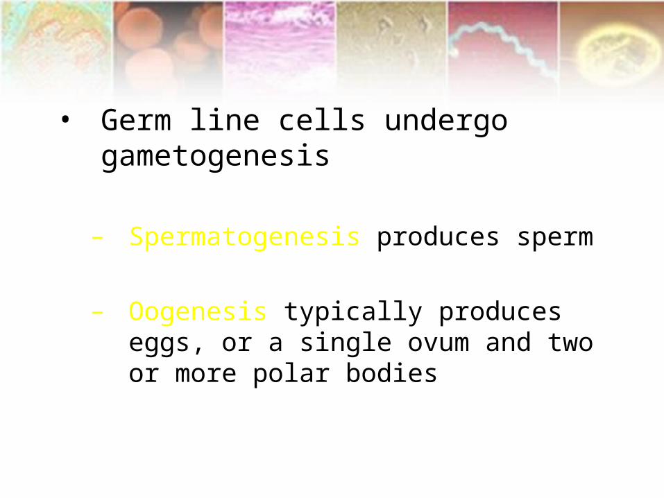
• Germ line cells undergo gametogenesis
– Spermatogenesis produces sperm
– Oogenesis typically produces eggs, or a single ovum and two or more polar bodies


![Tabletop Exercise Facilitator Handbook Template · Web viewFOR OFFICIAL USE ONLYAbout this Facilitator Guide FACILITATOR HANDBOOK [Exercise Name]Facilitator Handbook FACILITATOR HANDBOOK](https://img.pdfslide.net/doc/110x75/5ae2303b7f8b9a0d7d8bfd35/tabletop-exercise-facilitator-handbook-viewfor-official-use-onlyabout-this-facilitator.jpg)