Embed Size (px)
Citation preview
Feeding pattern and the visual light environment in
myctophid fish larvae
A. SABATES*, A. BOZZANO AND I. VALLVEY
Institut de Ciencies del Mar (CMIMA-CSIC), Passeig Marıtim de la Barceloneta37–49, 08003 Barcelona, Spain
(Received 29 January 2003, Accepted 10 September 2003)
The trophic spectrum and feeding pattern of two myctophid larvae, Benthosema glaciale and
Myctophum punctatum, were analysed in relation to changes in daily light intensity. The larvae
of both species live relatively deep, occurring in the first 100m, though distribution of
M. punctatum extends to 150m depth. The present study, carried out in the western Mediterranean
(Alboran Sea), indicated that the larvae of the two species exhibit different foraging strategies.
Both started to feed at dawn, but while feeding ofM. punctatum was high at dawn and dusk, the
feeding of B. glaciale remained high throughout the day. The light intensity profiles taken
during the day indicated that at the depths where the species dwelt, light intensity was enough
to provoke a feeding response. The larvae of these species, in contrast to the majority of fish
larvae, had an enhanced sensitivity due to their pure rod-like retina, an adaptation for foraging
at low light intensities. Both species showed an ontogenetic change in their diet: B. glaciale
preflexion larvae fed mainly on copepod eggs and nauplii, while postflexion larvae consumed
calanoid copepodites; M. punctatum larvae showed a more diversified diet, composed of larger
prey items. The stalked and elongated eye of M. punctatum larvae would enable the detection of
a greater range of prey in terms of shape and size. In addition, the retina of this species was
characterized by a higher summation ratio and longer photoreceptors, indicating a preference
for dimmer environments. This could explain the decreasing feeding activity of M. punctatum
during the high light intensity of the middle daylight hours. As a clear relationship existed
between feeding pattern and light intensity in these myctophid larvae, the visual characteristics
of each species could help to explain the different strategies of foraging behaviour, therefore
avoiding a possible overlap in their trophic niche. # 2003 The Fisheries Society of the British Isles
Key words: feeding; fish larvae; Mediterranean; Myctophidae; vision.
INTRODUCTION
A widespread consensus exists that the magnitude of recruitment in fishes isdetermined during the egg and larvae period, with larval feeding success being akey factor for growth and larval survival (Heath, 1992; Legget & DeBlois,1994). High growth and survival reduce the vulnerability of the larvae topredators that feed on small prey with limited mobility (Houde, 1987; Legget &DeBlois, 1994). Various factors contribute to feeding success, such as the
*Author to whom correspondence should be addressed. Tel.: þ34 932309500; fax: þ34 932309555;
email: [email protected]
Journal of Fish Biology (2003) 63, 1476–1490
doi:10.1046/j.1095-8649.2003.00259.x,availableonlineathttp://www.blackwell-synergy.com
1476# 2003TheFisheries Society of theBritish Isles
concentration and type of food (Hunter, 1981), turbulence (MacKenzie et al.,1994; Dower et al., 1997), temperature (Paul, 1983) and light conditions (Blaxter,1986; Miner & Stein, 1993).Although larvae of some fish species have non-visual senses for feeding (Blaxter,
1969; Batty & Hoyt, 1995), most fish larvae are visual predators (Blaxter, 1986;Batty, 1987; Davis & Olla, 1995; Porter & Theilacker, 1999) and environmentallight conditions represent a key factor in feeding success. The intensity and thespectral quality of light affect larval feeding capabilities by altering prey searchbehaviour, reactive distances and ultimately feeding success itself (Batty, 1987;Huse, 1994). The response of fish larvae to particular light characteristics is speciesspecific and varies with the age of the larva or development stage (Puvanendran &Brown, 1998). It has been reported that fish larval feeding often follows a distinctdiel pattern (Connaughton et al., 1996; Conway et al., 1998; MacKenzie et al.,1999; Hillbruger & Kloppmann, 2000) related to light intensity, with species-specific timing of peak feeding and duration of feeding periods. Even differentpopulations from the same species show different types of foraging, growth andsurvival in relation to the light intensity (Puvanendran & Brown, 1998). Thesestudies, however, have tended to concentrate on species that mainly inhabit surfacewaters during the whole of their life cycle. Consequently, the larvae of deep-seafishes have received less attention (Sabates & Saiz, 2000).Among the deep-sea fishes, the myctophids constitute an important compon-
ent of the oceanic ecosystems due to their high abundance and universaldistribution in all the oceans (Gjosater & Kawaguchi, 1980). Their larvae arevery abundant in plankton, achieving up to 50% of all the larvae collected insamples from the open sea (Moser & Ahlstrom, 1974) and, therefore, playing avery important role in the planktonic food chain. In addition to their highabundance, the myctophid larvae exhibit a high diversity of morphologicaltraits (Moser, 1981). The shape and proportions of the different parts of thebody, as well as the shape and size of the eye, vary markedly in the differentspecies and throughout larval development. This wide variety of morphologicalcharacters and larval adaptations causes interspecific variations in their loco-motive abilities and visual capabilities that might result in different feedingstrategies (Sabates & Saiz, 2000).The glacier lanternfish Benthosema glaciale (Reinhardt) and the spotted
lanternfish Myctophum punctatum Rafinesque are two species of Myctophidae,both included in the subfamily Myctophinae. The larvae of this subfamily arecharacterized by having elongated eyes, unlike the species of the subfamilyLampanyctinae that have rounded eyes (Moser & Ahlstrom, 1974). In addition,the larvae of M. punctatum have their eyes placed on short stalks (Moser &Ahlstrom, 1970). The vertical distribution of the larvae of both species isrelatively deep: B. glaciale occurs between depths of 40 and 110m (Ropke,1989), whereas M. punctatum larvae have a wider distribution range, between25 and 150m (John & Re, 1995; Olivar et al., 1998). The larvae of both speciesare visual predators since they feed during the day (Sabates & Saiz, 2000).The aim of this study was to determine the diet and daytime feeding
behaviour of B. glaciale and M. punctatum larvae, which have a partially over-lapping depth distribution, and to try to account for a visual feeding behaviourin an environment where light could be a limiting factor.
FEEDING PATTERN IN MYCTOPHID FISH LARVAE 1477
# 2003TheFisheries Society of theBritish Isles, Journal of FishBiology 2003, 63, 1476–1490
MATERIALS AND METHODS
FIELD SAMPLING
The research detailed here was part of a wider study on mesoscale features in planktondistribution in the western Alboran Sea (western Mediterranean) carried out from 14 to24 September 1999. The sampling was undertaken at three fixed stations located in atransect perpendicular to the coast (36�000–36�230 N; 4�140–4�160 W). A different stationwas sampled each day at dawn, noon and dusk, and each station was visited three timesduring the cruise. No night fishing was carried out because a preliminary study, under-taken on the same species, demonstrated that the larvae did not feed during the night(Sabates & Saiz, 2000). The fish larvae were collected with a bongo net with a mouthopening 60 cm in diameter and a mesh-size of 333mm. Fishing was carried out from amaximum depth of 200m to the surface, at a ship speed of 3�7 kmh�1 (2 knots). Planktonsamples were preserved in 5% buffered formalin.
Surface incident irradiance was continuously measured with a pyranometer connectedto a Li-Cor LI-1000 data logger. In addition, vertical profiles of photosynthetically activeradiation (PAR, 400–700 nm) were obtained with a Li-Cor spherical quantum sensorattached to a Seabird 25 CTD system. Light intensity profiles were measured from thesea surface down to a depth of 50m at 0900, 1030 and 1700 hours (GMT) on differentoccasions during the cruise. In the present study, only profiles corresponding to thehighest environmental light intensity were considered. The extinction coefficient K wascalculated using Beer’s Law: Iz¼ I0 � e�kz, where Iz is the intensity of light at depth z, I0 isthe intensity of light at the surface and k is the extinction coefficient. In order to obtainlight intensity profiles for the whole depth distribution range of the Myctophidae larvaestudied (to 150m), the extinction coefficient k was calculated for depths >50m using theregression curve obtained for the values measured with the Li-Cor sensor, and the resultwas applied to extrapolated light intensity down to 150m. The light intensity profile at0700 hours was obtained from the surface irradiance value (218�5mmol s�1m�2) meas-ured by the pyranometer connected to the Li-Cor LI-1000 data logger.
LABORATORY ANALYSIS
In the laboratory, all fish larvae were sorted from the bongo samples. The bulk of theselarvae corresponded to mesopelagic species from the family Myctophidae, with B. glacialeand M. punctatum being most abundant, since they represented 53% of the total fishlarvae collected.
Gut contents analysis was conducted on both species. Prior to dissection, the fishlarvae standard length (LS) was measured (to the nearest 0�1mm) from the tip of thesnout to the end of the notochord in preflexion and flexion larvae, and from the tip of thesnout to the posterior margin of the hypural plate in postflexion larvae. For the study ofthe gut contents, a maximum of 40 individuals per sample was analysed. The digestivetract, including the stomach and gut, were dissected with fine needles and any prey itemsfound were identified and counted.
Feeding incidence, the proportion of feeding larvae, and the mean number of fooditems per gut (using only feeding larvae) were calculated per each larval size range.Differences in mean number of prey between size groups were tested using one-factorANOVA. One-way ANOVA was also performed to analyse the effect of daytime onfeeding incidence. If significant differences were found, a Tukey–Kramer multiple com-parison test was used to identify group differences.
Histological analysis of the retinal structure was carried out on two postflexion larvaeof B. glaciale (7�2 and 7�3mm LS) and M. punctatum (9�7 and 10�3mm LS). The sizesrepresented a similar development stage in both species, since notochord flexion takesplace at c. 6mm in B. glaciale, while in M. punctatum it occurs at larger sizes, c. 7�8mm.The individuals, fixed on board, were stored in 70% alcohol. The dorso-ventraland rostro-temporal eye diameters were measured under light microscopy prior toembedding. No correction was applied for fixation shrinkage. The individuals were
1478 A. SABATES ET AL .
# 2003TheFisheries Society of the British Isles, Journal of FishBiology 2003, 63, 1476–1490
subsequently dehydrated in an ethanol series and embedded in Historesin (Leica).Transverse semi-thin sections (2 mm) were cut on a Reichert-Jung microtome and stainedwith methylene blue and basic fuchsin. For both species, the lenses were measured in thesections until they reached their maximum diameter, and in these sections (five for eachlarva) measurements of the retina were taken using an Optimas 6.0 image analyser.Photoreceptor cell type was examined under light microscopy. In each section obtainedfrom the dorso-temporal area of the retina, 15 measurements were taken for all theretinal layers: photoreceptor outer segment (OS), outer nuclear layer (ONL), innernuclear layer (INL), inner plaxiform layer (IPL) and ganglion cell layer (GCL). Anystatistical differences between intra- and interspecific retinal measurements were deter-mined by a t-test.In the four larvae examined, photoreceptors and ganglion cells were counted in the
dorso-temporal retina under light microscopy at �1000 magnification. In order to limitthe error due to the retinal curvature, counts were made in two linear transects of 50mmin three serial sections far from the optic disc. Cell density was calculated using Van derMeer & Anker (1986) equation: density¼ 106m [( tþ d� 2f ) w]�1 cells permm2, where mis the mean number of cells counted, t is the section thickness, d is the mean diameter ofthe cells, f is the thickness of the smallest cell fragments counted ( f¼ 0�1d ) and w is thewidth of the sampled strip.
RESULTS
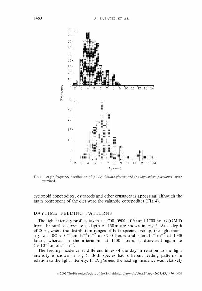
The stomach contents were examined from a total of 542 B. glaciale and 186M. punctatum larvae. The size frequency distributions for the dissected larvaeof both species are shown in Fig. 1. The range of sizes was between 2–10mm inB. glaciale and 3�5–14mm in M. punctatum.
FEEDING INCIDENCE AND DIET COMPOSITION
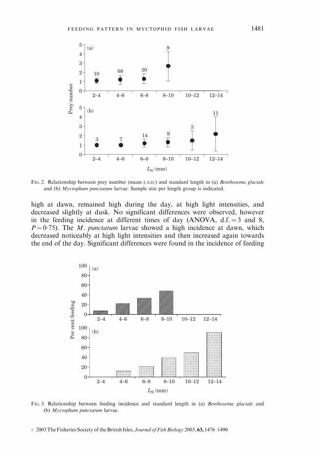
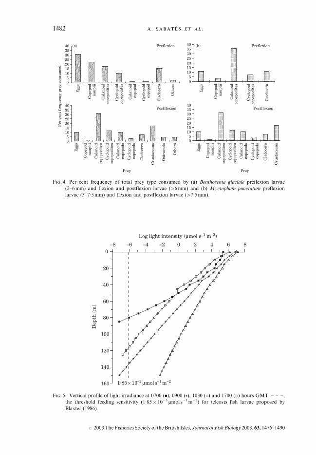
In general, the feeding incidence was relatively low during the samplingperiod. Nevertheless, in both species the incidence increased with larval growth(Fig. 2), and it was 100% in the largest sized M. punctatum larvae examined. InB. glaciale, the average number of prey items per larva was quite constant(mean� S.D. 1�2� 0�5) up to 8mm LS, and increased significantly (2�6� 1�6) inthe 8–10mm larvae (ANOVA, d.f.¼ 3 and 115, P< 0�001) (Fig. 3). InM. punctatum, the average number of prey items per larva increased progres-sively as development progressed (from 1�2� 0�4 to 2�2� 1�8). Nevertheless, nosignificant differences were observed among the size ranges considered(ANOVA, d.f.¼ 5 and 38, P¼ 0�35) (Fig. 3), probably due to the high vari-ability in the number of prey (one to seven per larva) detected in the largestlarvae examined.The diet composition analysis of both species showed that the preflexion
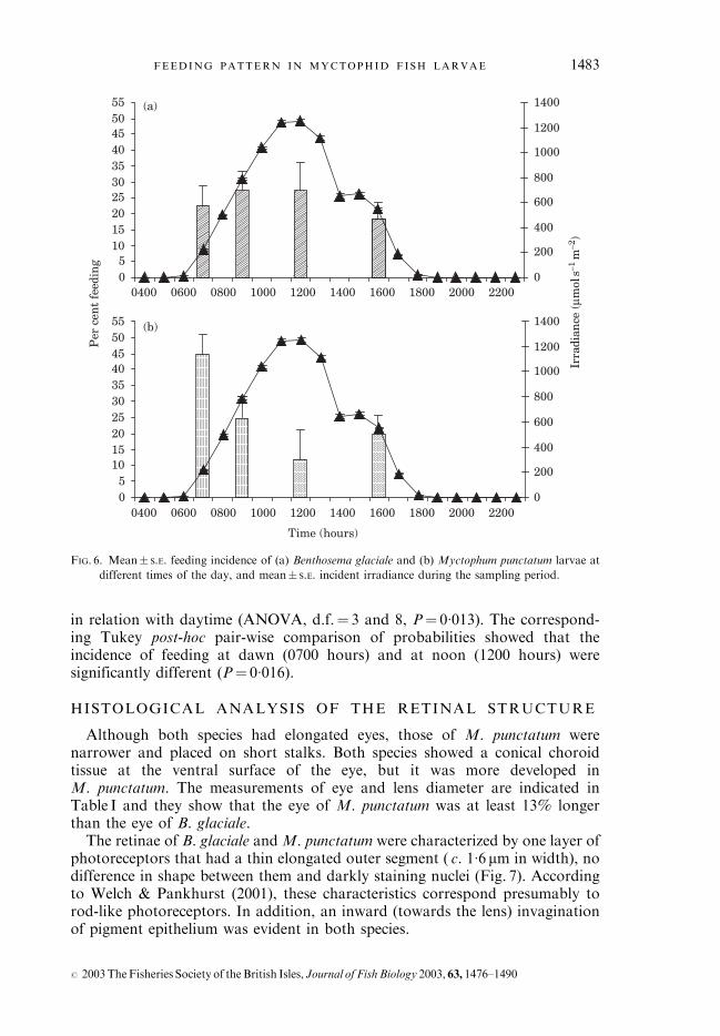
B. glaciale larvae were feeding mainly on small sized prey, such as eggs and naupliiof copepods (which represented >50% of the composition of the diet) (Fig. 4).Copepodites and cladocerans, however, also represented an important part ofthe diet. In the flexion and postflexion larvae, >6mm, the most abundant preywere calanoid copepodites and the larvae of other crustaceans. Adult copepodsand ostracods also appeared in the diet, while the proportion of nauplii andcladocerans decreased noticeably. In the preflexion M. punctatum larvae, thediet was not particularly varied and was dominated by calanoid copepodites. Inthe flexion and postflexion larvae, >7�5mm, the diet was more diversified, with
FEEDING PATTERN IN MYCTOPHID FISH LARVAE 1479
# 2003TheFisheries Society of theBritish Isles, Journal of FishBiology 2003, 63, 1476–1490
cyclopoid copepodites, ostracods and other crustaceans appearing, although themain component of the diet were the calanoid copepodites (Fig. 4).
DAYTIME FEEDING PATTERNS
The light intensity profiles taken at 0700, 0900, 1030 and 1700 hours (GMT)from the surface down to a depth of 150m are shown in Fig. 5. At a depthof 80m, where the distribution ranges of both species overlap, the light inten-sity was 0�2� 10�2mmol s�1m�2 at 0700 hours and 4 mmol s�1m�2 at 1030hours, whereas in the afternoon, at 1700 hours, it decreased again to5� 10�2 mmol s�1m�2.The feeding incidence at different times of the day in relation to the light
intensity is shown in Fig. 6. Both species had different feeding patterns inrelation to the light intensity. In B. glaciale, the feeding incidence was relatively
2 3 4 5 6 7 8 9 10 11 12 13 14
2 3 4 5 6 7 8 9 10 11 12 13 14
0
10
20
30
40
50
60
70
80
90
LS (mm)
0
5
10
15
20
25
30Fre
quen
cy
(a)
(b)
FIG. 1. Length frequency distribution of (a) Benthosema glaciale and (b) Myctophum punctatum larvae
examined.
1480 A. SABATES ET AL .
# 2003TheFisheries Society of the British Isles, Journal of FishBiology 2003, 63, 1476–1490
high at dawn, remained high during the day, at high light intensities, anddecreased slightly at dusk. No significant differences were observed, howeverin the feeding incidence at different times of day (ANOVA, d.f.¼ 3 and 8,P¼ 0�75). The M. punctatum larvae showed a high incidence at dawn, whichdecreased noticeably at high light intensities and then increased again towardsthe end of the day. Significant differences were found in the incidence of feeding
0
1
2
3
4
5
2–4 4–6 6–8 8–10 10–12 12–14
2–4 4–6 6–8 8–10 10–12 12–14
Pre
y n
umbe
r
0
1
2
3
4
5
LS (mm)
10
9
3068
11
5914
73
(a)
(b)
FIG. 2. Relationship between prey number (mean� S.D.) and standard length in (a) Benthosema glaciale
and (b) Myctophum punctatum larvae. Sample size per length group is indicated.
0
20
40
60
80
100
2–4 4–6 6–8 8–10 10–12 12–14
Per
cen
t fe
edin
g
0
20
40
60
80
100
2–4 4–6 6–8 8–10 10–12 12–14
LS (mm)
(a)
(b)
FIG. 3. Relationship between feeding incidence and standard length in (a) Benthosema glaciale and
(b) Myctophum punctatum larvae.
FEEDING PATTERN IN MYCTOPHID FISH LARVAE 1481
# 2003TheFisheries Society of theBritish Isles, Journal of FishBiology 2003, 63, 1476–1490
05
10152025303540
Egg
s
Cop
epod
nau
plii
Cal
anoi
dco
pepo
dite
s
Cyc
lopo
idco
pepo
dite
s
Cal
anoi
dco
pepo
d
Cyc
lopo
idco
pepo
d
Cla
doce
ra
Cla
doce
ra
Oth
ers
(a)
05
10152025303540
Egg
s
Cop
epod
nau
plii
Cal
anoi
dco
pepo
dite
s
Cyc
lopo
idco
pepo
dite
s
Cal
anoi
dco
pepo
ds
Cyc
lopo
idco
pepo
ds
Ost
raco
da
Oth
ers
Per
cen
t fr
eque
ncy
pre
y co
nsu
med
(b)
05
10152025303540
Postflexion
PreflexionPreflexion
Postflexion
05
10152025303540
Egg
s
Cop
epod
nau
plii
Cal
anoi
dco
pepo
dite
s
Cyc
lopo
idco
pepo
dite
s
Cal
anoi
dco
pepo
ds
Cyc
lopo
idco
pepo
ds
Cla
doce
ra
Egg
s
Cop
epod
nau
plii
Cal
anoi
dco
pepo
dite
s
Cyc
lopo
idco
pepo
dite
s
Cla
doce
ra
Cru
stac
ean
s
Cru
stac
ean
s
Prey Prey
FIG. 4. Per cent frequency of total prey type consumed by (a) Benthosema glaciale preflexion larvae
(2–6mm) and flexion and postflexion larvae (>6mm) and (b) Myctophum punctatum preflexion
larvae (3–7�5mm) and flexion and postflexion larvae (>7�5mm).
–8 –6 –4 –2 0 2 4 6 8
Log light intensity (µmol s–1 m–2)
160
140
120
100
80
60
40
20
0
Dep
th (
m)
1.85 × 10⁻³ µmol s–1 m–2
FIG. 5. Vertical profile of light irradiance at 0700 (&), 0900 ( ), 1030 (n) and 1700 (*) hours GMT. ,
the threshold feeding sensitivity (1�85� 10�3 mmol s�1m�2) for teleosts fish larvae proposed by
Blaxter (1986).
1482 A. SABATES ET AL .
# 2003TheFisheries Society of the British Isles, Journal of FishBiology 2003, 63, 1476–1490
in relation with daytime (ANOVA, d.f.¼ 3 and 8, P¼ 0�013). The correspond-ing Tukey post-hoc pair-wise comparison of probabilities showed that theincidence of feeding at dawn (0700 hours) and at noon (1200 hours) weresignificantly different (P¼ 0�016).
HISTOLOGICAL ANALYSIS OF THE RETINAL STRUCTURE
Although both species had elongated eyes, those of M. punctatum werenarrower and placed on short stalks. Both species showed a conical choroidtissue at the ventral surface of the eye, but it was more developed inM. punctatum. The measurements of eye and lens diameter are indicated inTable I and they show that the eye of M. punctatum was at least 13% longerthan the eye of B. glaciale.The retinae of B. glaciale andM. punctatum were characterized by one layer of
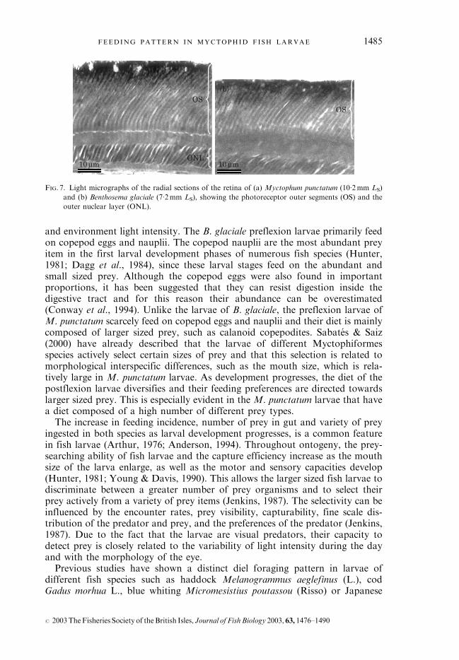
photoreceptors that had a thin elongated outer segment ( c. 1�6 mm in width), nodifference in shape between them and darkly staining nuclei (Fig. 7). Accordingto Welch & Pankhurst (2001), these characteristics correspond presumably torod-like photoreceptors. In addition, an inward (towards the lens) invaginationof pigment epithelium was evident in both species.
05
10152025303540455055
0400 0600 0800 1000 1200 1400 1600 1800 2000 2200
0400 0600 0800 1000 1200 1400 1600 1800 2000 2200
0
200
400
600
800
1000
1200
1400
Irra
dian
ce (
µmol
s–1
m–2
)
05
10152025303540455055
Time (hours)
0
200
400
600
800
1000
1200
1400
Per
cen
t fe
edin
g(a)
(b)
FIG. 6. Mean� S.E. feeding incidence of (a) Benthosema glaciale and (b) Myctophum punctatum larvae at
different times of the day, and mean� S.E. incident irradiance during the sampling period.
FEEDING PATTERN IN MYCTOPHID FISH LARVAE 1483
# 2003TheFisheries Society of theBritish Isles, Journal of FishBiology 2003, 63, 1476–1490
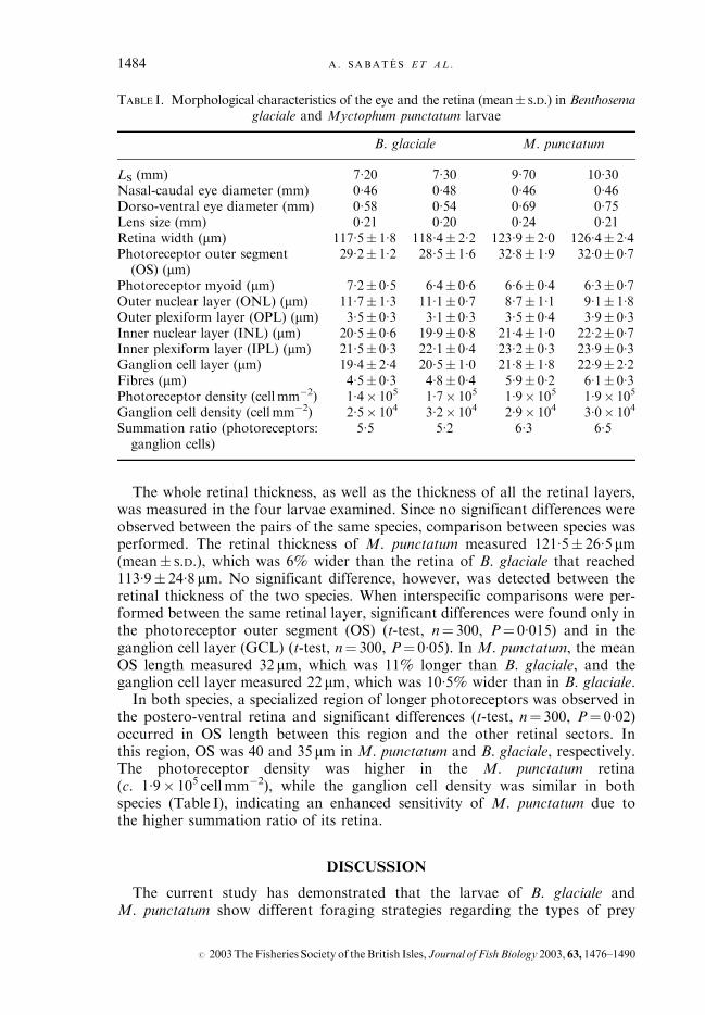
The whole retinal thickness, as well as the thickness of all the retinal layers,was measured in the four larvae examined. Since no significant differences wereobserved between the pairs of the same species, comparison between species wasperformed. The retinal thickness of M. punctatum measured 121�5� 26�5mm(mean� S.D.), which was 6% wider than the retina of B. glaciale that reached113�9� 24�8 mm. No significant difference, however, was detected between theretinal thickness of the two species. When interspecific comparisons were per-formed between the same retinal layer, significant differences were found only inthe photoreceptor outer segment (OS) (t-test, n¼ 300, P¼ 0�015) and in theganglion cell layer (GCL) (t-test, n¼ 300, P¼ 0�05). In M. punctatum, the meanOS length measured 32mm, which was 11% longer than B. glaciale, and theganglion cell layer measured 22mm, which was 10�5% wider than in B. glaciale.In both species, a specialized region of longer photoreceptors was observed in
the postero-ventral retina and significant differences (t-test, n¼ 300, P¼ 0�02)occurred in OS length between this region and the other retinal sectors. Inthis region, OS was 40 and 35mm in M. punctatum and B. glaciale, respectively.The photoreceptor density was higher in the M. punctatum retina(c. 1�9� 105 cellmm�2), while the ganglion cell density was similar in bothspecies (Table I), indicating an enhanced sensitivity of M. punctatum due tothe higher summation ratio of its retina.
DISCUSSION
The current study has demonstrated that the larvae of B. glaciale andM. punctatum show different foraging strategies regarding the types of prey
TABLE I. Morphological characteristics of the eye and the retina (mean� S.D.) in Benthosemaglaciale and Myctophum punctatum larvae
B. glaciale M. punctatum
LS (mm) 7�20 7�30 9�70 10�30Nasal-caudal eye diameter (mm) 0�46 0�48 0�46 0�46Dorso-ventral eye diameter (mm) 0�58 0�54 0�69 0�75Lens size (mm) 0�21 0�20 0�24 0�21Retina width (mm) 117�5� 1�8 118�4� 2�2 123�9� 2�0 126�4� 2�4Photoreceptor outer segment
(OS) (mm)29�2� 1�2 28�5� 1�6 32�8� 1�9 32�0� 0�7
Photoreceptor myoid (mm) 7�2� 0�5 6�4� 0�6 6�6� 0�4 6�3� 0�7Outer nuclear layer (ONL) (mm) 11�7� 1�3 11�1� 0�7 8�7� 1�1 9�1� 1�8Outer plexiform layer (OPL) (mm) 3�5� 0�3 3�1� 0�3 3�5� 0�4 3�9� 0�3Inner nuclear layer (INL) (mm) 20�5� 0�6 19�9� 0�8 21�4� 1�0 22�2� 0�7Inner plexiform layer (IPL) (mm) 21�5� 0�3 22�1� 0�4 23�2� 0�3 23�9� 0�3Ganglion cell layer (mm) 19�4� 2�4 20�5� 1�0 21�8� 1�8 22�9� 2�2Fibres (mm) 4�5� 0�3 4�8� 0�4 5�9� 0�2 6�1� 0�3Photoreceptor density (cellmm�2) 1�4� 105 1�7� 105 1�9� 105 1�9� 105
Ganglion cell density (cell mm�2) 2�5� 104 3�2� 104 2�9� 104 3�0� 104
Summation ratio (photoreceptors:ganglion cells)
5�5 5�2 6�3 6�5
1484 A. SABATES ET AL .
# 2003TheFisheries Society of the British Isles, Journal of FishBiology 2003, 63, 1476–1490
and environment light intensity. The B. glaciale preflexion larvae primarily feedon copepod eggs and nauplii. The copepod nauplii are the most abundant preyitem in the first larval development phases of numerous fish species (Hunter,1981; Dagg et al., 1984), since these larval stages feed on the abundant andsmall sized prey. Although the copepod eggs were also found in importantproportions, it has been suggested that they can resist digestion inside thedigestive tract and for this reason their abundance can be overestimated(Conway et al., 1994). Unlike the larvae of B. glaciale, the preflexion larvae ofM. punctatum scarcely feed on copepod eggs and nauplii and their diet is mainlycomposed of larger sized prey, such as calanoid copepodites. Sabates & Saiz(2000) have already described that the larvae of different Myctophiformesspecies actively select certain sizes of prey and that this selection is related tomorphological interspecific differences, such as the mouth size, which is rela-tively large in M. punctatum larvae. As development progresses, the diet of thepostflexion larvae diversifies and their feeding preferences are directed towardslarger sized prey. This is especially evident in the M. punctatum larvae that havea diet composed of a high number of different prey types.The increase in feeding incidence, number of prey in gut and variety of prey
ingested in both species as larval development progresses, is a common featurein fish larvae (Arthur, 1976; Anderson, 1994). Throughout ontogeny, the prey-searching ability of fish larvae and the capture efficiency increase as the mouthsize of the larva enlarge, as well as the motor and sensory capacities develop(Hunter, 1981; Young & Davis, 1990). This allows the larger sized fish larvae todiscriminate between a greater number of prey organisms and to select theirprey actively from a variety of prey items (Jenkins, 1987). The selectivity can beinfluenced by the encounter rates, prey visibility, capturability, fine scale dis-tribution of the predator and prey, and the preferences of the predator (Jenkins,1987). Due to the fact that the larvae are visual predators, their capacity todetect prey is closely related to the variability of light intensity during the dayand with the morphology of the eye.Previous studies have shown a distinct diel foraging pattern in larvae of
different fish species such as haddock Melanogrammus aeglefinus (L.), codGadus morhua L., blue whiting Micromesistius poutassou (Risso) or Japanese
(a)
(b)OS
OS
ONL10 µm 10 µm
FIG. 7. Light micrographs of the radial sections of the retina of (a) Myctophum punctatum (10�2mm LS)
and (b) Benthosema glaciale (7�2mm LS), showing the photoreceptor outer segments (OS) and the
outer nuclear layer (ONL).
FEEDING PATTERN IN MYCTOPHID FISH LARVAE 1485
# 2003TheFisheries Society of theBritish Isles, Journal of FishBiology 2003, 63, 1476–1490
Spanish mackerel Scomberomorus niphonius (Cuvier) in various geographicalareas (Kane, 1984; McLaren & Avendano, 1995; McLaren et al., 1997; MacKenzieet al., 1999; Shoji et al., 1999; Hillbruger & Kloppmann, 2000). These studiesconfirm that the highest incidences of feeding occur at or shortly after sunset.Active feeding at dusk would allow the fish larvae to store energy before theystop feeding during the night. Nevertheless, little is known about diel feedinghabits in larvae of mesopelagic species that are located at greater depths in thewater column. The present study has demonstrated that the fish larvae thatinhabit relatively deep waters are also subject to daily light cycles, showingdifferent diel periods for feeding incidence. In B. glaciale, the incidence offeeding increases at sunrise to reach a maximum at midday and then decreasesbefore sunset. On the other hand, the larvae of M. punctatum show a highincidence at dawn that decreases during midday, showing a new increasetowards the end of the day. Blaxter (1986) and Batty (1987) demonstratedthat for fish larvae whose feeding is visually mediated, a minimum light inten-sity in the water column is required to detect and capture prey organisms.Consequently, the vertical distribution of fish larvae will be determined by theexponential decrease in the light intensity by depth and the lower limit willbe determined by the minimum light intensity at which feeding is possible.This relationship will vary for every species. Blaxter (1986) indicated thatmost teleost larvae have a threshold feeding sensitivity at c. 0�1 lux(1�8� 10�3 mmol s�1m�2). Subsequent studies have indicated a feeding responseat a higher threshold light intensity level in larvae of striped trumpeter Latrislineata (Forster) (0�1–10mmol s�1m�2) (Cobcroft et al., 2001) and in larvae ofvarious tropical fish species (10 mmol s�1m�2) (Job & Bellwood, 2000). On theother hand, Huse (1994) observed low thresholds at 1�8� 10�3 mmol s�1m�2 inthe larvae of plaice Pleuronectes platessa L. and turbot Scophthalmus maximus(L.). All these studies, however, were carried out in controlled laboratoryconditions.In the present study, the levels of light observed in September between
depths of 40 and 80m, where the larvae of B. glaciale and M. punctatumare mainly located (Ropke, 1989; Olivar et al., 1998), are higher than1�8� 10�3 mmol s�1m�2 (0�1 lux) during the diurnal period. Values of1�8� 10�3 mmol s�1m�2 are reached at 0700 and 1700 hours for depths of 80and 120m, respectively. At midday, the intensity of available light at thesedepths is even higher and the value of 1�8� 10�3 mmol s�1m�2 is reached at235m. Consequently, it would be logical to assume that the larvae of the speciesstudied have a sufficient quantity of light to stimulate a visual response at thedepths they inhabit.In addition, the eyes of these fish larvae show a pure rod-like retina and a
invagination of pigment epithelium indicating, firstly, a possible specializationfor scotopic vision and secondly the need to control the amount of light reach-ing the retina. Pure rod-like retinas were also observed by Pankhurst (1987) inthe larvae of the lanternfish Lampanyctodes hectoris (Gunther) which mainlydwell in the upper levels of the water column (0–20m depth) (Olivar et al.,1992). These larvae have photoreceptors with >50% shorter OS than the OSmeasured in the present study in deeper larvae of M. punctatum and B. glaciale.In addition, the summation ratio of 5�2 calculated in L. hectoris is fairly similar
1486 A. SABATES ET AL .
# 2003TheFisheries Society of the British Isles, Journal of FishBiology 2003, 63, 1476–1490
to the summation ratio found in B. glaciale, but lower than the summation ratioof M. punctatum. This difference in scotopic sensitivity of M. punctatum couldallow this species to detect prey at higher depth or in dimmer light.In any case, pure rod retinae are very unusual in fish larvae, since the
majority of larvae have a pure cone retina in the first development phases(Blaxter & Staines, 1970). In clupeid species, such as the herring Clupeaharengus L. (Blaxter & Jones, 1967) and the South American pilchard Sardinopsmelanostictus (Jenyns) (Matsuoka, 1999), a pure cone retina up to 22 and 20mmLS, respectively has been reported. Pure rod retinae were found by Pankhurst(1984) in the larva of the European eel Anguilla anguilla (L.), and by Blaxter &Staines (1970) and Locket (1980) in some deep species, such as nine lepto-cephalus eels of 80mm, a deep-sea macruridae larvae of 17mm, and a 29mmindividual of the viperfish Chauliodus sloanei Bloch & Schneider. Therefore,presumably, a pure rod retina is a suitable adaptation for larvae that live in adim light environment. Deep-sea fishes, which remain at low levels of illumin-ation, have a pure rod retina in the adult phase and it might be assumed thatthe larvae of these species also have pure rod retina.Even if B. glaciale and M. punctatum do not show great differences in retinal
characteristics, specific features found in their visual system could explaincertain differences observed in their diet and feeding pattern. The stalked eyesof M. punctatum larvae permit them to scan a greater volume of water (Weihs &Moser, 1981) and its elongated form allows focused images to be projected onthe dorso-ventral parts of the retina. With these characteristics, therefore, thelarvae can detect a greater range of prey in terms of shape and size. In addition,the presence of longer photoreceptors and higher summation ratio in M. punctatummay indicate a preference for dimmer environments. It could explain thedecreasing feeding activity of this species in relation to a constraining higherlight intensity in the middle daylight hours. The decrease in feeding success athigh light intensities has been reported in other species, such as cod andhaddock (Huse, 1994; Downing & Litvak, 2001). According to Downing &Litvak (2001), high light intensities may create excessive reflectance that coulddiminish the contrast of prey in relation to the background.From the results of this study high sensitivity seems to be the main visual
characteristic of B. glaciale and M. punctatum in anticipation of migration todeeper waters where the adults of both species are found (Badcock & Merrett,1976). Both species show longer rod-like photoreceptor especially in the ventralsector of their retina. This retinal area receives light from above and longerphotoreceptors ensure a better visual sensitivity for an upward view. As Herring(1996) pointed out, the upward view is characteristic of fishes living in the upperfew hundred metres where light is highly directional. The ventral specializationof the retina could indicate that the main visual axis points in the dorsaldirection of the fishes, where these species can detect the contrast and themovement of their prey.In conclusion, there is a clear relationship between light intensity and feeding
behaviour in the two myctophid fish larvae studied. Taking into account thepartial overlap in their depth distribution, the visual capabilities could help toexplain the different strategies of foraging behaviour, therefore avoiding apossible overlap in their trophic niches.
FEEDING PATTERN IN MYCTOPHID FISH LARVAE 1487
# 2003TheFisheries Society of theBritish Isles, Journal of FishBiology 2003, 63, 1476–1490
The authors would like to thank C. Rodgers for kindly reviewing the final Englishversion. We thank two anonymous reviewers for their accurate and constructive criti-cisms on an earlier version of the manuscript. We thank M. Estrada for providingthe data on light intensity profiles. This work was supported by the European Unionin the framework of the MAST Program (MAS-CT96-0051) and by a Spanish grant(MAR 99-1202).
References
Anderson, J. T. (1994). Feeding ecology and condition of larval and pelagic juvenileredfish Sebastes spp. Marine Ecology Progress Series 104, 211–226.
Arthur, D. K. (1976). Food and feeding of larvae of three fishes occurring in theCalifornia Current, Sardinops sagax, Engraulis mordax, and Trachurus symmetricus.U.S. Fishery Bulletin 74, 517–530.
Badcock, K. & Merrett, N. R. (1976). Midwater fishes in the eastern North Atlantic. I.Vertical distribution and associated biology in 30� N 23� W with developmentnotes on some myctophids. Progress in Oceanography 7, 3–58.
Batty, R. S. (1987). Effect of light intensity on activity and food-searching of larvalherring, Clupea harengus: a laboratory study. Marine Biology 94, 323–327.
Batty, R. S. & Hoyt, R. D. (1995). The role of sense organs in the feeding bahaviour ofjuvenile sole and plaice. Journal of Fish Biology 47, 931–939.
Blaxter, J. H. S. (1969). Visual tresholds and spectral sensitivity of flatfish larvae. Journalof Experimental Biology 51, 221–230.
Blaxter, J. H. S. (1986). Development of sense organs and behavior of teleost larvae withspecial reference to feeding and predator avoidance. Transactions of the AmericanFisheries Society 115, 98–114.
Blaxter, J. H. S. & Jones, M. P. (1967). The development of the retina and retinomotorresponses in the herring. Journal of the Marine Biological Association of the UnitedKingdom 47, 677–697.
Blaxter, J. H. S. & Staines, M. (1970). Pure-cone retinae and retinomotor responses inlarval teleosts. Journal of the Marine Biological Association of the United Kingdom50, 449–460.
Connaughton, V. P., Epifanio, C. E. & Thomas, R. (1996). Effect of varying irradiance onfeeding in larval weakfish (Cynoscion regalis). Journal of Experimental MarineBiology and Ecology 180, 151–163.
Conway, D. V. P., McFadzen, I. R. B. & Tranter, P. R. G. (1994). Digestion of copepodeggs by larval turbot Scophthalmus maximus and egg viability following gutpassage. Marine Ecology Progress Series 106, 303–309.
Conway, D. V. P., Coombs, S. H. & Smith, C. (1998). Feeding of anchovy Engraulisencrasicolus larvae in the northwestern Adriatic Sea in response to changinghydrobiological conditions. Marine Ecology Progress Series 175, 35–49.
Cobcroft, J. M., Pankhurst, P. M., Hart, P. R. & Battaglene, S. C. (2001). The effects oflight intensity and algae-induced turbidity on feeding behaviour of larval stripedtrumpeter. Journal of Fish Biology 59, 1181–1197.
Dagg, M. J., Clarke, M. E., Nishiyama, T. & Smith, S. L. (1984). Production and standingstock of copepod nauplii, food items for larvae of the walleye pollock Theragra chalco-gramma in the southeastern Bering Sea. Marine Ecology Progress Series 19, 7–16.
Davis, M. W. & Olla, B. L. (1995). Formation and maintenance of aggregations in wall-eye pollock, Theragra chalcogramma, larvae under laboratory conditions: role ofvisual and chemical stimuli. Environmental Biology of Fishes 44, 385–392.
Dower, J. F., Miller, T. J. & Legget, W. C. (1997). The role of microscale turbulence in thefeeding ecology of larval fish. Advances in Marine Biology 31, 170–220.
Downing, G. & Litvak, M. K. (2001). The effect of light intensity and spectrum on theincidence of first feeding by larval haddock. Journal of Fish Biology 59, 1566–1578.doi: 10.1006/jfbi.2001.1792.
Gjosater, J. & Kawaguchi, K. (1980). A review of the world resources of mesopelagic fish.FAO Fisheries Technical Paper 193.
1488 A. SABATES ET AL .
# 2003TheFisheries Society of the British Isles, Journal of FishBiology 2003, 63, 1476–1490
Heath, M. R. (1992). Field investigations of the early life history stages of marine fish.Advances in Marine Biology 28, 2–174.
Herring, P. J. (1996). Light, colour, and vision in the ocean. In Oceanography, AnIllustrated Guide (Summerhayes, C. P. & Thorpe, S. A., eds), pp. 212–227.London: Manson Publishing.
Hillbruger, N. & Kloppmann, M. (2000). Vertical distribution and feeding of larval bluewhiting in turbulent waters above Porcupine Bank. Journal of Fish Biology 57,1290–1311. doi: 10.1006/jfbi.2000.1397.
Houde, E. D. (1987). Fish early life dynamics and recruitment variability. AmericanFisheries Society Symposium 2, 17–29.
Hunter, J. R. (1981). Feeding ecology and predation of marine fish larvae. InMarine FishLarvae: Morphology, Ecology and Relation to Fisheries (Lasker, R., ed.), pp. 34–77.Seattle, WA: University of Washington Press.
Huse, I. (1994). Feeding at different illumination levels in larvae of three marine teleostsspecies: cod, Gadus morhua L., plaice, Pleuronectes platessa L., and turbot,Scophthalmus maximus (L.). Aquaculture and Fisheries Management 25, 687–695.
Jenkins, G. P. (1987). Comparative diets, prey selection, and predatory impact ofco-occurring larvae of two flounder species. Journal of Experimental Marine Biologyand Ecology 110, 147–170.
Job, S. D. & Bellwood, D. R. (2000). Light sensitivity in larval fishes: implications forvertical zonation in the pelagic zone. Limnology and Oceanography 45, 362–371.
John, H.-C. & Re, P. (1995). Cross-shelf zonation, vertical distribution and drift of fishlarvae off northern Portugal during weak upwelling. Arquivos Museo Bocage, NovaSerie 2, 607–632.
Kane, J. (1984). The feeding habits of co-occurring cod and hadcock larvae from GeorgesBank. Marine Ecology Progress Series 16, 9–20.
Legget, W. & DeBlois, E. (1994). Recruitment in marine fishes: is it regulated bystarvation and predation in the egg and larval stages? Netherland Journal of SeaResearch 32, 119–134.
Locket, N. A. (1980). Variation of architecture with size in the multiple-bank retina of adeep-sea teleost, Chauliodus sloani. Proceedings of the Royal Society of London B208, 223–242.
MacKenzie, B. R., Miller, T. J., Cyr, S. & Legget, W. C. (1994). Evidence of a dome-shaped relationship between turbulence and larval fish ingestion rate. Limnologyand Oceanography 39, 1790–1799.
MacKenzie, B. R., Ueberschar, B., Basford, D., Heath, M. & Gallego, A. (1999). Dielvariability of feeding activity in haddock (Melanogrammus aeglefinus) larvae inthe East Sheltland area, North Sea. Marine Biology 135, 361–368.
Matsuoka, M. (1999). Histological characteristics and development of the retina in theJapanese sardine Sardinops melanostictus. Fisheries Science 65, 224–229.
McLaren, I. A. & Avendano, P. (1995). Prey field and diet of larval cod on Western Bank,Scotian Shelf. Canadian Journal of Fisheries and Aquatic Sciences 52, 448–463.
McLaren, I. A., Avendano, P., Taggart, C. T. & Lochmann, S. E. (1997). Feeding bylarval cod in different water masses on Western Bank, Scotian Shelf. FisheriesOceanography 6, 250–265.
Miner, J. G. & Stein, R. A. (1993). Interactive influence of turbidity and light on larvalbluegill (Lepomis machochirus) foraging. Canadian Journal of Fisheries and AquaticSciences 50, 781–788.
Moser, H. G. (1981). Morphological and functional aspects of marine fish larvae. InMarine Fish Larvae: Morphology, Ecology and Relation to Fisheries (Lasker, R.,ed.), pp. 89–31. Seattle, WA: University of Washington Press.
Moser, H. G. & Ahlstrom, E. H. (1970). Development of lanternfishes (family Mycto-phidae) in the California Current. Part I. Species with narrow-eyed larvae. Bulletinof the Los Angeles County Museum of Natural History Science 7.
Moser, H. G. & Ahlstrom, E. H. (1974). Role of larval stages in systematic investiga-tions of marine teleosts, the Myctophidae, a case study. U.S. Fishery Bulletin 72,391–413.
FEEDING PATTERN IN MYCTOPHID FISH LARVAE 1489
# 2003TheFisheries Society of theBritish Isles, Journal of FishBiology 2003, 63, 1476–1490
Olivar, M. P., Rubies, P. & Salat, J. (1992). Horizontal and vertical distribution patternsof ichthyoplankton under intense upwelling regimes off Namibia. In BenguelaTrophic Functioning (Payne, A. I. L., Brink, K. H., Mann, K. H. & Hilborn, R.,eds). South African Journal of Marine Science 12, 71–82.
Olivar, M. P., Sabates, A., Abello, P. & Garcıa, M. (1998). Transitory hydrographicstructures and distribution of fish larvae and neustonic crustaceans in the north-western Mediterranean. Oceanologica Acta 21, 95–104.
Pankhurst, N. W. (1984). Retinal development in larval and juvenile European eel,Anguilla anguilla (L.). Canadian Journal of Zoology 62, 335–343.
Pankhurst, N. W. (1987). Intra- and interspecific changes in retinal morphology amongmesopelagic and demersal teleosts from the slope water of New Zeland. Environ-mental Biology of Fishes 19, 269–280.
Paul, A. J. (1983). Light, temperature, nauplii concentrations, and prey capture by firstfeeding pollock larvae Theragra chalcogramma.Marine Ecology Progress Series 13,175–179.
Porter, S. M. & Theilacker, G. H. (1999). The development of the digestive tract and eyein larval walley pollock, Theragra chalcogramma. Fishery Bulletin 97, 722–729.
Puvanendran, V. & Brown, J. A. (1998). Effects of light on the foraging and growth ofAtlantic cod larvae: interpopulation differences. Marine Ecology Progress Series167, 207–214.
Ropke, A. (1989). Small-scale vertical distribution of ichthyoplankton in the Celtic Sea inApril 1986. Meeresforschung 32, 192–203.
Sabates, A. & Saiz, E. (2000). Intra- and interspecific variability in prey size and nichebreadth of mesopelagic fish larvae. Marine Ecology Progress Series 201, 261–271.
Shoji, J., Maehara, T. & Tanaka, M. (1999). Diel vertical movement and feeding rhythmof Japanese Spanish mackerel larvae in the central Seto Inland Sea. FisheriesScience 65, 726–730.
Van der Meer, H. J. & Anker, G. Ch. (1986). The influence of light deprivation onthe development of the eye and retina in the cichlid Sarotherodon mossambicus(Teleostei). Netherlands Journal of Zoology 36, 480–498.
Young, J. W. & Davis, T. L. O. (1990). Feeding ecology of larvae of southern bluefin,albacore and skipjack tunas (Pisces: Scombridae) in the eastern Indian Ocean.Marine Ecology Progress Series 61, 17–29.
Weihs, D. & Moser, H. G. (1981). Stalked eyes as an adaptation towards more efficientforaging in marine fish larvae. Bulletin of Marine Science 31, 31–36.
Welch, D. & Pankhurst, P. M. (2001). Visual morphology and feeding behaviour of thedaggertooth. Journal of Fish Biology 58, 1427–1437. doi: 10.1006/jfbi.2000.1552.
1490 A. SABATES ET AL .
# 2003TheFisheries Society of the British Isles, Journal of FishBiology 2003, 63, 1476–1490